POLYAMINE HOMEOSTASIS LOIKKANENjultika.oulu.fi/files/isbn951427668X.pdf · Loikkanen, Ildikó,...
82
POLYAMINE HOMEOSTASIS Cellular responses to perturbation of polyamine biosynthetic enzymes ILDIKÓ LOIKKANEN Faculty of Science, Department of Biochemistry, University of Oulu OULU 2005
Transcript of POLYAMINE HOMEOSTASIS LOIKKANENjultika.oulu.fi/files/isbn951427668X.pdf · Loikkanen, Ildikó,...

POLYAMINE HOMEOSTASISCellular responses to perturbation of polyamine biosynthetic enzymes
ILDIKÓLOIKKANEN
Faculty of Science,Department of Biochemistry,
University of Oulu
OULU 2005


ILDIKÓ LOIKKANEN
POLYAMINE HOMEOSTASISCellular responses to perturbation of polyamine biosynthetic enzymes
Academic Dissertation to be presented with the assent ofthe Faculty of Science, University of Oulu, for publicdiscussion in Raahensali (Auditorium L10), Linnanmaa, on June 3rd, 2005, at 12 noon
OULUN YLIOPISTO, OULU 2005

Copyright © 2005University of Oulu, 2005
Supervised byDocent Antti Pajunen
Reviewed byProfessor Martti ParvinenDocent Jarmo Wahlfors
ISBN 951-42-7667-1 (nid.)ISBN 951-42-7668-X (PDF) http://herkules.oulu.fi/isbn951427668X/
ISSN 0355-3191 http://herkules.oulu.fi/issn03553191/
OULU UNIVERSITY PRESSOULU 2005

Loikkanen, Ildikó, Polyamine homeostasis. Cellular responses to perturbation ofpolyamine biosynthetic enzymesFaculty of Science, Department of Biochemistry, University of Oulu, P.O.Box 3000, FIN-90014University of Oulu, Finland 2005Oulu, Finland
AbstractThe polyamines putrescine, spermidine and spermine are highly regulated polycations present invirtually all cells of higher eukaryotes. They are essential for proper cell growth and differentiationby participating in various physiological processes including DNA, RNA and protein synthesis,apoptosis and interactions with ion-channels. The complexity of polyamine metabolism and themultitude of compensatory mechanisms that are invoked to maintain polyamine homeostasis arguethat these molecules are critical for cell survival.
The primary aim of this study was to gain a better understanding of the mode of action ofpolyamines and the regulatory mechanisms in which they are involved. Transgenic miceoverexpressing the polyamine biosynthetic enzymes S-AdoMetDC and ODC were found to maintaintheir polyamine pools by acetylation of spermidine and spermine and an increased export of theseacetylated compounds. The expression of various genes was studied as a response to polyaminedeprivation in cell- and kidney organ culture. Among these genes acetyl-CoA synthetase andornithine decarboxylase were demonstrated to be developmentally regulated. Changes in geneexpression patterns, with most of the transcripts upregulated in the polyamine-depleted samples,indicated selective stabilization of mRNAs. Polyamines were shown to play an important role inkidney organogenesis as their depletion results in a reduction of ureteric branching and retardation oftubule formation. The selective changes of various genes in the ureteric bud and mesenchyme indicatethat polyamines might have a role in the regulation of epithelial-mesenchymal interactions duringmouse kidney development.
Keywords: epithelial-mesenchymal interactions, polyamine homeostasis, polyamines


"Ebben a dalban van négy akkord Ebben a dalban van két gitár
Ebben a dalban van egypár barát És egy kicsit benne vagyok én"
(Sztevanovity Dusán, Presser Gábor: Egészen egyszerű dal)


Acknowledgements
This work was carried out at the Department of Biochemistry, University of Oulu, during the years 1996-2005.
I wish to express sincere gratitude to my supervisor, Docent Antti Pajunen for introducing me to the world of molecular biology and giving me the possibility to work in his group. I also want to express my appreciation to Professor Seppo Vainio for his enthusiasm and patient guidance during the years of our collaboration. I owe my sincere thanks to the head of the Department of Biochemistry, Professor Kalervo Hiltunen, Professor Raili Myllylä and all the other professors and group leaders in the department for creating a positive atmosphere and excellent working conditions for all of us.
I am very grateful to Professor Matti Parvinen and Docent Jarmo Wahlfors for their valuable comments when reviewing the manuscript of this thesis. Dr. Deborah Kaska is warmly thanked for her fast and careful revision of the language.
I feel deep gratitude to all my co-authors for their expertise and knowledge that were essential for this study. I am thankful to all the former and present members of the PA- and SV-team, especially to Ritva Heljasvaara and Anitta Pulkka for their support and friendship during these years.
I thank Sirpa Salo for keeping me company in various activities, from Finnish language studies in the night-school Leskinen to troubleshooting histology staining, from my first days in Oulu. The continuously changing international team around me, especially Olga Beltcheva, Jocelyne Lietard and Verena Wolfram made my years in Finland more joyful in the laboratory and also outside. Thanks to all of you.
I wish to thank M.Sc. Seppo Kilpeläinen and Dr. Ari-Pekka Kvist for setting up excellent computer facilities and networks at the department. Virpi Hannus, Anneli Kaattari, Tuula Koret and Helena Heikura are warmly thanked for helping me out with many practical, secretarial, administrative and library matters.
I am grateful to everybody, especially to Terttu Keskitalo, who helped me on my long spiritual way to enter the mouse facility and touch a mouse. I also want to thank Eeva-Liisa Stefanius, Kaisa Penttilä, Johanna Kekolahti-Liias, Hannele Härkman, Soili Miettunen, Mervi Matero and Jaana Kujala for their skillful technical assistance and for the relaxed atmosphere they created in the lab. Maila Konttila and Pirjo Mustaniemi are acknowledged for taking care of the basic laboratory tasks thus saving time for all of us.

Jaakko Keskitalo and Kyösti Keränen are thanked for keeping things rolling and repairing everything that happened to break down in the lab.
My special thanks go to Eszter Vuojala for her friendship and for standing by me for all these years.
Finally, I would like to thank my family for their love and encouragement. I thank my dear husband, Mikko Loikkanen for his support and understanding. He never lets me give up. Our children, Joel, Leevi and the little coming one set up a strict time-table in this work and lighted-up my days. Thank you.
Végül, de nem utolsó sorban szüleimet köszöntöm. Köszönöm nektek, hogy felneveltetek, majd engedtétek, hogy a saját utamat járjam akkor is, ha épp nem arra mentem, amerre ti láttátok jónak. Remélem, hogy ez a kis könyv nektek is örömet szerez.
Oulu, Finland, February 2005

Abbreviations
ABC ATP binding cassette AceCS AMP-forming acetyl-CoA synthetase ANT adenine nucleotide translocator BCR B cell antigen receptor BrdU 5-bromo-2’-deoxyuridine cAMP cyclic adenosine monophosphate Cdk cyclin-dependent kinase CHENSpm N1-cycloheptylmethyl-N11-ethylnorspermine CHX cycloheximide CK2 casein kinase II CRP cAMP receptor protein CSRE carbon source-responsive element DAB 3,3’-diaminobenzidine DENSPM N1,N11-diethylnorspermine DFMO α-difluoromethylornithine eIF eukaryotic initiation factor ERK extracellular signal-regulated kinase EST expressed sequence tag GAPDH glyceraldehyde-3-phosphate dehydrogenase Gy gyro HSF heat-shock factor IAP inhibitors of apoptosis IPENSpm (S)-N1-(2-methyl-1-butyl)-N11-ethyl-4,8-diazaundecane IRES internal ribosome entry site JNK c-Jun kinase Kir channels inward rectifier K+ channels MAT methionine adenosyltransferase MGBG methylglyoxal bis(guanylhydrazone) NF nuclear factor NOHA Nω-hydroxy-L-arginine ODC ornithine decarboxylase OppA oligopeptide-binding protein

ORF open reading frame Orn ornithine PAO polyamine oxidase PHEX phosphate regulating gene PMF-1 polyamine-modulated factor-1 PMT plasma membrane transporter pRb retinoblastoma protein PRE polyamine-responsive element Put putrescine Sir2 silent information regulator protein SMO spermine oxidase Spd spermidine ROS reactive oxygen species S-AdoMet S-adenosylmethionine S-AdoMetDC S-adenosylmethionine decarboxylase SREBPs sterol regulatory element-binding proteins SSAT spermidine/spermine N1-acetyltransferase TNF-α tumor necrosis factor α TRAMP transgenic adenocarcinoma of mouse prostate UTR untranslated region VA vesicular polyamine-H+ antiporter V-ATPase vacuolar-ATPase WT1 Wilms’ tumor suppressor ΔΨm mitochondrial inner transmembrane potential

List of original articles
This thesis is based on the following articles, which are referred to in the text by their Roman numerals:
I Heljasvaara R, Veress I, Halmekytö M, Alhonen L, Jänne J, Laajala P & Pajunen A (1997) Transgenic mice overexpressing ornithine and S-adenosyl-methionine decarboxylases maintain a physiological polyamine homeostasis in their tissues. Biochem J 323 (2), 457-62.*
II Veress I, Haghighi S, Pulkka A & Pajunen A (2000) Changes in gene expression in response to polyamine depletion indicates selective stabilization of mRNAs. Biochem J 346, 185–191.*
III Loikkanen I, Haghighi S, Vainio S & Pajunen A (2002) Expression of cytosolic acetyl-CoA synthetase gene is developmentally regulated. Mech Dev 115:1-2 139-41. **
IV Loikkanen I, Lin Y, Railo A, Pajunen A & Vainio S (2005) Polyamines are involved in murine kidney development controlling expression of c-ret, Pax2/8 and E-cadherin genes. Submitted.
*Reproduced with permission from © the Biochemical Society. **Reprinted with permission from Elsevier


Contents
Abstract Acknowledgement Abbreviation List of original articles Contents 1. Introduction ..................................................................................................................13 2. Review of the literature ................................................................................................14
2.1 Polyamines .............................................................................................................14 2.1.1 Polyamine metabolism in mammals ................................................................15
2.1.1.1 Biosynthesis..............................................................................................15 2.1.1.2 Catabolism................................................................................................18 2.1.1.3 Transport of polyamines ...........................................................................19
2.1.2 Manipulation of the polyamine homeostasis ...................................................21 2.1.2.1 Polyamine inhibitors and analogs .............................................................21 2.1.2.2 Genetic engineering of polyamine metabolism ........................................22
2.1.3 Physiological roles of polyamines ...................................................................24 2.1.3.1 DNA-polyamine interactions....................................................................24 2.1.3.2 Cell growth ...............................................................................................24 2.1.3.3 Apoptosis ..................................................................................................26 2.1.3.4 DNA and protein synthesis .......................................................................30 2.1.3.5 Hypusine formation ..................................................................................30 2.1.3.6 Interactions with ion channels ..................................................................32 2.1.3.7 Embryonic development...........................................................................33 2.1.3.8 Polyamines in the urogenital organs .........................................................34
2.2 Acetyl-CoA synthetase ...........................................................................................35 2.2.1 Regulation of AMP-forming acetyl-CoA synthetase .......................................35 2.2.2 Posttranslational regulation of AceCS .............................................................36
3. Aims of the research .....................................................................................................37 4. Materials and methods..................................................................................................38
4.1 Mouse lines (I, II, III, IV) .......................................................................................38 4.2 Cell culture (I, II, IV)..............................................................................................38 4.3 Polyamine determination (I, II) ..............................................................................39

4.4 Northern blot analysis (I, II, III) .............................................................................39 4.5 Differential display (II)...........................................................................................40 4.6 Whole mount and section in situ hybridization (III, IV).........................................40 4.7 Sex typing (III) .......................................................................................................40 4.8 Organ culture (IV) ..................................................................................................41 4.9 Cell proliferation assay (IV) ...................................................................................41 4.10 Histology, β-galactosidase and antibody staining (IV) .........................................41 4.11 Other methods (I, II) .............................................................................................42
5. Results ..........................................................................................................................43 5.1 Overexpression of S-AdoMetDC and ODC in transgenic mice does not
dramatically affect tissue spermidine and spermine pools (I) .................................43 5.2 Transgenic mice compensate for increased S-AdoMetDC and ODC
activities by acetylating and excreting excessive polyamines (I)............................44 5.3 Changes in gene expression patterns in response to polyamine
depletion and activation of biosynthesis (II) ...........................................................44 5.4 Increased GAPDH and β-actin expression in polyamine-depleted cells (II) ..........47 5.5 Polyamine depletion results in selective stabilization of the mRNAs (II) ..............47 5.6 Cytosolic acetyl-CoA synthetase and polyamines ..................................................47 5.7 Expression of AceCS1 during mouse development (III) ........................................48 5.8 Expression of ODC is developmentally regulated (IV) ..........................................48 5.9 DFMO treatment reduces kidney size via reduction in cell proliferation (IV) .......49 5.10 DFMO causes reduced branching and a delay in renal tubule formation (IV) .....49 5.11 Changes in gene expression patterns in response to polyamine
depletion during kidney organogenesis (IV)...........................................................49 5.12 Effects of polyamine depletion on the Pax-2 promoter (IV).................................51
6. Discussion ....................................................................................................................52 6.1 Compensation mechanisms against increased S-AdoMetDC and ODC
activities in transgenic mice....................................................................................52 6.2 Genes responding to polyamine depletion and activation
of polyamine biosynthesis.......................................................................................53 6.2.1 The differential display approach ....................................................................53 6.2.2 The kidney culture approach ...........................................................................54 6.2.3 Common features of genes responding to polyamine depletion ......................55
7. Conclusions ..................................................................................................................57 References

1 Introduction
The polyamines putrescine spermidine and spermine are puzzling molecules. They are involved in various physiological processes and are crucial to the growth and proliferation of mammalian cells. Yet, despite extensive research during the last forty years, their exact function in these processes is still elusive. High polyamine concentrations are required for rapid cell growth and low polyamine content is typical for quiescent cells, suggesting that drugs that inhibit the synthesis of polyamines could prevent cancer and may be used for therapeutic purposes. However, the development of polyamine synthesis inhibitors has had disappointing results due to the unexpected compensatory mechanisms such as an increase in polyamine uptake from the circulation. Cells have a complex regulatory system, involving biosynthesis, catabolism and transport, to ensure tight control of intracellular polyamine pools to avoid cytotoxicity caused by high spermidine and spermine concentrations. A general approach to the study of the mechanisms in which polyamines are involved is to inhibit polyamine biosynthetic enzymes using specific inhibitors or overexpress these enzymes by generating genetically engineered animals.
The present study focused on cellular responses following perturbations in the function of polyamine biosynthetic enzymes using various models. Mechanisms by which transgenic mice overexpressing the polyamine biosynthetic enzymes S-AdoMetDC and ODC maintain their polyamine pools were clarified. Depletion of polyamines was shown to alter the expression of a panel of genes in cell- and also kidney organ culture, of which acetyl-CoA synthetase and ornithine decarboxylase were demonstrated to be developmentally regulated. Changes in the gene expression patterns indicated selective stabilization of mRNAs. Polyamines were shown to have an important role in kidney organogenesis being involved in inductive epithelial-mesenchymal tissue interactions.

2 Review of the literature
2.1 Polyamines
The history of polyamines is long; the first observation dating back to 1678, when Leeuwenhoek observed the crystallization of spermine phosphate in human semen (Cohen 1998). More than 300 years have passed, but still no one can definitively describe the function of spermine or the other natural polyamines. The natural polyamines (shown in figure 1) include, in addition to the tetraamine spermine, the triamine spermidine and its precursor, the diamine putrescine.
Fig. 1. Natural polyamines Spermidine and spermine are simple aliphatic amines containing two and three flexible carbon chains, respectively, separated by secondary amines with primary amines at either end (Usherwood 2000). Although putrescine is a primary diamine it is customary to treat it as a polyamine (Raina & Jänne 1975).
The amino groups are fully protonated at physiological pH, which allows polyamines to participate in many cellular processes through binding to RNA, DNA, nucleotide triphosphates and other acidic substances (Igarashi & Kashiwagi 2000).
NH3+
+H3N Putrescine
NH3+
+H3N N H2
+ Spermidine
H2+
N +H3N
N H2
+
N H3+
Spermine

15
2.1.1 Polyamine metabolism in mammals
Polyamines are involved in the regulation of cell growth. Their concentrations are high in actively proliferating cells, during periods of tissue growth and in mature tissues with highly active protein synthesis. Intracellular polyamine levels are strictly regulated by multiple metabolic processes including biosynthesis, catabolism and transport (reviewed in Pegg & McCann 1982, Wallace et al. 2003, Jänne et al. 2004, Seiler 2004) (Figure 2).
2.1.1.1 Biosynthesis
In eukaryotic cells polyamines are synthesized from two amino acids, L-arginine and L-methionine in a series of enzymatic reactions. L-ornithine is cleaved from L-arginine by arginase II (EC 3.5.3.1) or taken up in the diet. L-ornithine is decarboxylated by ornithine decarboxylase (ODC; EC 4.1.1.17) to form the diamine putrescine. Methionine adenosyltransferase (MAT; EC 2.5.1.6) converts L-methionine to S-adenosylmethionine (S-AdoMet), which is further decarboxylated by S-adenosylmethionine decarboxylase (S-AdoMetDC; EC 4.1.1.50). The resulting decarboxylated S-AdoMet serves as the aminopropyl donor for spermidine and spermine synthesis. The aminopropyl moiety of S-AdoMet is transferred to putrescine by spermidine synthase (EC 2.5.1.16) and to spermidine by spermine synthase (EC 2.5.1.22) to produce spermidine and spermine respectively. ODC and S-AdoMetDC are considered the rate-limiting enzymes in polyamine biosynthesis. Both have a fast turnover rate with a half-life less than 1 hour (Berntsson et al. 1999) and their translation is negatively regulated by polyamines (Kahana & Nathans 1985, Shantz et al. 1992, Shantz & Pegg 1999).
ODC is a homodimer with two active sites located at the interface between the subunits (Tobias & Kahana 1993, Coleman et al. 1994). Its regulation occurs at the levels of transcription, translation and degradation. ODC is a delayed early gene whose transcriptional activation requires ongoing protein synthesis in the stimulated cells (Tobias et al. 1995). Its promoter contains response elements for several trans-acting factors, including the Wilms’ tumor suppressor WT1 (Moshier et al. 1996), several Sp1 sites (Kumar et al. 1995) and a cAMP response element (Palvimo et al. 1991, Abrahamsen et al. 1992). ODC is a transcriptional target of the proto-oncogene c-Myc through c-Myc/Max heterodimers, in association with induction of cell cycle progression, cell proliferation and transformation (Bello-Fernandez et al. 1993, Pena et al. 1993, Auvinen et al. 2003).

16
Fig. 2. Pathways of polyamine metabolism. Enzymes are marked with bold font type. Abbreviations: MAT, methionine adenosyltransferase; S-AdoMetDC, S-adenosylmethionine decarboxylase; ODC, ornithine decarboxylase; PAO, polyamine oxidase; SSAT, spermidine/spermine N1-acetyltransferase; SMO, spermine oxidase The translation of ODC mRNA is regulated by its 5’-untranslated region (UTR). This GC-rich, long 5’UTR has the potential for stable hairpin formation that has inhibitory effect on ODC mRNA translation (Manzella & Blackshear 1990). Interaction of the
Antizyme
Antizyme
S-adenosylmethionine
Decarboxylated S-adenosylmethionine
Methionine
MAT
S-AdoMetDC
CO2
L-ornithine
Arginine
Arginase II
ODC
CO2
Putrescine
Spermidine synthase
Methylthioadenosine Spermidine
N1-acetylspermidine
PAO
SSAT
Methylthioadenosine Spermine
N1-acetylspermine
PAO
SSAT
Intracellular
Extracellular
Polyamine transport
SMO

17
5’UTR with the 3’UTR reverses this inhibition and enhances translation (Grens & Scheffler 1990, Lorenzini & Scheffler 1997). Within the 5’UTR of the ODC mRNA there is a small internal open reading frame (ORF). The sequence and length of the predicted protein differs between species and its role in translational regulation has not yet been established (Shantz & Pegg 1999). ODC mRNA can be translated by both cap-dependent and independent mechanisms (Hayashi et al. 2000, Pyronnet et al. 2000). The eukaryotic initiation factor (eIF) 4E has been reported to enhance the cap-dependent translation at the level of translation initiation (Rousseau et al. 1996, Shantz et al. 1996), whereas eIF-4G stimulates both cap-dependent and independent protein synthesis (Hayashi et al. 2000). Low levels of spermidine (0.2 mM) stimulate, whereas high levels (0.6-1mM) inhibit ODC translation via the 5’UTR at the level of initiation complex formation (Ito et al. 1990, Shimogori et al. 1996).
ODC is degraded by the 26S proteasome in an unusual ATP-dependent, but ubiquitin-independent, manner (Bercovich et al. 1989, Murakami et al. 1992). The degradation is accelerated by antizyme, the enzyme inhibitor of ODC. Antizyme binds to the ODC monomer (Mitchell & Chen 1990) and directs the proteasome to degrade the enzyme. Antizyme expression is regulated by a special +1 translational frameshift that is induced by polyamines in a concentration-dependent manner (Rom & Kahana 1994, Matsufuji et al. 1995). (For details see 2.1.3.4.) In addition to its effects on ODC, antizyme also negatively regulates polyamine transport into cells (Mitchell et al. 1994, Suzuki et al. 1994, Zhu et al. 1999). These processes together form a complete feedback loop: when polyamine levels rise, they induce antizyme production. Antizyme inhibits and degrades ODC which causes a decline in polyamine biosynthesis.
S-AdoMetDC is a pyruvoyl-dependent enzyme, synthesized as an inactive proenzyme that undergoes autocatalytic cleavage into two subunits. The mature enzyme consists of a dimer of these subunits (Pajunen et al. 1988). The autocatalytic cleavage reaction also forms the pyruvoyl cofactor from an internal serine residue at the amino terminus of the α subunit (Stanley et al. 1989, Tolbert et al. 2003). Regulation of the mammalian enzyme occurs at multiple levels; the cleavage reaction and the catalytic activity of the mature enzyme are both stimulated by putrescine (Kameji & Pegg 1987a, Stanley & Pegg 1991, Stanley et al. 1994), whereas the synthesis of the proenzyme is repressed by spermidine and spermine (Kameji & Pegg 1987b). The promoter region of the mammalian S-AdoMetDC gene contains DNA elements for the binding of transcription factors Sp1, AP-1, AP-2, CREB and multiple steroid receptors (Maric et al. 1992, Pulkka et al. 1993, Nishimura et al. 1998).
Like ODC, S-AdoMetDC mRNA also has a long 5’UTR with a short internal ORF (Shantz & Pegg 1999). The ORF in the S-AdoMetDC 5’UTR is a mediator of the polyamine-dependent regulation of the enzyme synthesis (Nishimura et al. 1999, Raney et al. 2000). This well conserved ORF, which encodes the peptide sequence MAGDIS (Shantz & Pegg 1999), suppresses the translation of the downstream cistron by stalling ribosomes close to the terminal codon of the ORF and thus limiting the number of scanning ribosomes that can reach the downstream start site. The ribosome stalling is affected by polyamine levels: high levels causing more effective stalling, and by the peptide sequence encoded by the upstream ORF (Hill & Morris 1993, Law et al. 2001, Raney et al. 2002).

18
The degradation of S-AdoMetDC is an important control mechanism maintaining polyamine levels. S-AdoMetDC activity can be lost by substrate-mediated transamination when an amine group from the substrate S-AdoMet is transferred to the pyruvoyl cofactor of the enzyme, converting it to alanine (Anton & Kutny 1987, Xiong et al. 1999). It was recently shown that S-AdoMetDC is degraded by the 26S proteasome in an ubiquitin-dependent manner and the degradation is accelerated by substrate-mediated transamination (Yerlikaya & Stanley 2004).
Spermidine and spermine synthases, stable enzymes and present in excess have not received much attention in polyamine research. Their activities are regulated mainly by the amount of enzyme protein and the availability of their substrate, decarboxylated S-AdoMet (Pegg 1986). Nevertheless the first human polyamine deficiency syndrome was recently characterized as a defect in the X-linked spermine synthase gene. The defect results from a splice mutation, and is associated with the Synder-Robinson syndrome, an X-linked mental retardation disorder (Cason et al. 2003).
2.1.1.2 Catabolism
Although the reactions catalyzed by spermidine and spermine synthase are practically irreversible, spermidine and spermine can be reconverted to putrescine in vivo. Cytosolic spermidine/spermine N1-acetyltransferase (SSAT; EC 2.3.1.57) is the first enzyme in the interconversion process, using acetyl-CoA to produce N1-acetylspermidine and N1-acetylspermine. These intermediates are either exported from the cells or they become substrates of the FAD-dependent peroxisomal polyamine oxidase (PAO; EC 1.5.3.11). As PAO prefers acetylated polyamines as substrates, acetylation is the rate-limiting step in this interconversion. Spermine can be directly converted to spermidine in the absence of SSAT by a recently discovered spermine oxidase (SMO) (Wang et al. 2001b).
SSAT is a highly regulated inducible enzyme. Its short biological half-life (less than one hour) is comparable to that of ODC and S-AdoMetDC (Seiler 2004). The active enzyme is either a homodimer (Ragione & Pegg 1982, Shinki & Suda 1989) or a tetramer (Casero & Pegg 1993, Fukuchi et al. 1994) with binding sites for acetyl-CoA and polyamines (Coleman et al. 1995, 1996, Lu et al. 1996).
The TATA-less promoter of the human SSAT gene contains binding sites for several transcription factors, including Sp1, GAGA factor, heat-shock factor (HSF), AP1, NF-κB and the polyamine-responsive element (PRE)-binding Nrf-2 (Tomitori et al. 2002). Nrf-2 has been shown to regulate polyamine-dependent SSAT expression by acting in cooperation with the polyamine-modulated factor-1 (PMF-1) (Wang et al. 1999, Wang et al. 2001a). In addition to transcriptional control, SSAT regulation includes stabilization of the mRNA (Fogel-Petrovic et al. 1993) and the protein (Fogel-Petrovic et al. 1997, Marverti et al. 2004), regulation of the mRNA translation (Parry et al. 1995) as well as protein degradation (Coleman & Pegg 1997).
SSAT is normally present in very small amounts in the cell and induced by a wide variety of factors, such as the polyamines themselves (Shappell et al. 1993), polyamine analogs (Casero et al. 1990, Fogel-Petrovic et al. 1996), hormones (Seidel & Snyder

19
1989), growth factors (Desiderio et al. 1998) and different toxic agents (Matsui & Pegg 1982). SSAT belongs to the heat-shock protein family because the enzyme is rapidly induced by heat-shock, an induction negatively regulated by polyamines (Fuller et al. 1990).
SSAT is degraded via the proteasomal/ubiquitin pathway. Proteasomes interact with the terminal MATEE motif at the carboxyl end of the ubiquitinated SSAT protein. Degradation is prevented in the presence of polyamines or polyamine analogs which cause conformational changes in the SSAT protein and decrease its ability to serve as a substrate for efficient ubiquitination (Coleman & Pegg 1997, 2001).
2.1.1.3 Transport of polyamines
The uptake and release of polyamines are integral parts of the regulatory machinery that adjusts polyamine levels according to the needs of cells. The uptake of a particular polyamine is often stimulated when its synthesis is inhibited. To date polyamine carriers on the plasma membrane have been molecularly identified only in prokaryotes, whereas in eukaryotic cells the genes encoding polyamine transporters have not yet been isolated (Igarashi & Kashiwagi 1999, Gugliucci 2004). Polyamine uptake is a saturable, carrier-mediated and energy-dependent process. In Escherichia coli three different polyamine transport systems have been characterized (Figure 3). Uptake is mainly catalyzed by two ABC (ATP binding cassette) transporters, one is spermidine-specific (Figure 3a) and the other prefers putrescine (Figure 3b). These two polyamine transporters consist of four different proteins, a substrate-binding protein (PotD or PotF), an ATPase (PotA or PotG) and two channel-forming proteins (PotB, PotC or PotH, PotI) (Furuchi et al. 1991, Pistocchi et al. 1993). The third uptake system (Figure 3c,d) involves the PotE protein, which can catalyze both the uptake and excretion of putrescine. Putrescine uptake (Figure 3d) is dependent on membrane potential whilst excretion (Figure 3c) is catalyzed by a putrescine/ornithine antiporter activity of PotE (Kashiwagi et al. 1992, Igarashi & Kashiwagi 1999).
Fig. 3. Polyamine transport systems in E. coli. Spd, spermidine; Put, putrescine; Orn, ornithine

20
Although the genes responsible for eukaryotic polyamine carriers are still unknown, the properties and physiological function of the polyamine transport system has been studied in different cell types. Polyamine uptake is subject to hormonal regulation (Lessard et al. 1995), pH- and membrane-potential-dependent, Na+-independent and requires extracellular divalent cations such as Ca2+ or Mg2+ (Poulin et al. 1995a). Polyamine transport is negatively regulated by antizyme, the enzyme inhibitor of ornithine-decarboxylase (Suzuki et al. 1994, Zhu et al. 1999). Antizyme not only inhibits polyamine uptake but also stimulates polyamine and acetylpolyamine excretion (Sakata et al. 2000). In addition to the natural polyamines polyamine transport systems are capable of transporting a significant range of polyamine analogs. Based on experiments with fluorescent polyamine conjugates (Cullis et al. 1999, Soulet et al. 2002) two models have been proposed concerning polyamine uptake mechanisms in mammalian cells: polyamines are internalized by a plasma membrane carrier and then sequestered into pre-existing vesicles (model A) or polyamines are directly captured by polyamine receptors and undergo endocytosis (model B). The results of recent studies support model A, indicating that polyamine transport occurs in two steps via a vesicular H+/polyamine carrier (Soulet et al. 2004).
Fig. 4. Model A for the intravesicular polyamine accumulation in eukaryotic cells. PA, polyamines; PMT, plasma membrane transporter; VA, vesicular polyamine-H+ antiporter; V-ATPase, vacuolar ATPase

21
2.1.2 Manipulation of the polyamine homeostasis
Proper polyamine homeostasis is essential for cell growth and replication. Failure to maintain individual polyamine concentrations leads to cell-cycle-arrest, cell death or transformation. Cancer cells show elevated ODC and S-AdoMetDC activities as well as increased polyamine levels. Agents inhibiting polyamine biosynthesis prevent, or at least limit cell growth. These facts made the polyamine pathway a promising target for the development of antiproliferative drugs. (Wallace et al. 2003.)
2.1.2.1 Polyamine inhibitors and analogs
A number of single enzyme inhibitors have been developed to block the polyamine pathway. The first effective, rationally designed drug that is still in use for polyamine depletion was α-difluoromethylornithine (DFMO) (Metcalf et al. 1978). DFMO is cleaved by ODC but the product is not released. It remains in the active site and irreversibly inactivates the enzyme (Wallace & Fraser 2004). In vitro DFMO prevents cell growth through depletion of putrescine and spermidine without affecting spermine levels (Meyskens & Gerner 1999). In experimental animal models it was especially effective in the treatment of carcinogen-induced epithelial cancers of many organs (Weeks et al. 1982, Nigro et al. 1987). Despite the promising in vitro and animal studies, DFMO as a single drug proved to be disappointing in human cancer therapy resulting in cytostatic rather than cytotoxic effects in vivo. This failure is mainly due to the compensatory increases in the uptake of polyamines from the circulation. However, due to its low toxicity, DFMO recently became a potential drug for cancer prevention (Meyskens et al. 1998, Wallace et al. 2003).
DFMO has been more successful in the treatment of African sleeping sickness caused by Trypanosoma brucei gambiense. Trypanosomes are more sensitive to the drug than human cells, possibly due to the slower turnover of ornithine decarboxylase in these organisms. DFMO treatment leads to the complete inhibition of ODC activity and depletion of polyamines making these parasites more vulnerable to the host immune system (Bacchi et al. 1980, Burri & Brun 2003, Docampo & Moreno 2003).
Another important polyamine biosynthetic enzyme inhibitor is methylglyoxal bis(guanylhydrazone) (MGBG), a potent competitive inhibitor of S-adenosylmethionine decarboxylase. It inhibits cell growth, lowers spermidine and spermine concentrations but causes a significant accumulation of putrescine. In addition to inhibition of S-AdoMetDC, MGBG also inhibits diamine oxidase, making it impossible to deplete polyamines in tissues with high diamine oxidase activity. Although it has been efficient in the treatment of several leukemias and lymphomas, the use of MGBG as an anticancer drug is limited by its extreme toxicity and profound antimitochondrial action.
Studies with single enzyme inhibitors indicate that the successful inhibition of tumor growth by the manipulation of the polyamine pathway requires the impairment of several reactions at the same time. (Jänne et al. 1991, Wallace & Fraser 2004.)

22
Polyamine analogs target more than one reaction in the polyamine pathway. They are designed to be sufficiently similar in structure to the parent compound to allow their recognition and uptake by the polyamine transporter and downregulate ODC and S-AdoMetDC, but dissimilar enough not to induce compensatory changes in metabolism (Wallace et al. 2003). Polyamine analogs can be divided into two main categories based on their ability to change the polyamine pool size. Antimetabolites activate catabolism and export of natural polyamines leading to cell growth arrest and cell death. The first generation of polyamine analogues, the symmetrical, terminally alkylated bis(ethyl) analogues of spermidine or spermine (Fukuchi et al. 1992) or the second generation, asymmetrically alkylated, spermine analog (S)-N1-(2-methyl-1-butyl)-N11-ethyl-4,8-diazaundecane (IPENSpm) (Fraser et al. 2002), belong to this group.
The polyamine mimetics, like the N1-cycloheptylmethyl-N11-ethylnorspermine (CHENSpm) (McCloskey et al. 2000), decrease cell growth but do not necessarily deplete polyamine pools.
Recently a third generation of polyamine analogs has been developed, including oligoamines, amines with inserted double bonds and amines with internal cyclic structures (Valasinas et al. 2001). These compounds have also shown some anti-proliferative effects in human tumor cells. Regarding the future it seems that the use of polyamine analogs as multisite inhibitors in cancer therapy is still a promising approach as it exploits the cell’s own regulatory mechanisms to deplete polyamines and induce growth arrest and apoptosis (Wallace et al. 2003).
2.1.2.2 Genetic engineering of polyamine metabolism
The genes for almost every reaction of polyamine metabolism have been genetically engineered in transgenic animals and embryonic stem cells. To better understand their physiological function in vivo, ODC, antizyme, S-AdoMetDC, SSAT, spermidine and spermine synthase have been generally or tissue-specifically overexpressed or disrupted (Jänne et al. 2004).
Polyamine biosynthesis has been activated in transgenic mice overexpressing the human ornithine decarboxylase gene under its own promoter (Halmekytö et al. 1991c). ODC activity and polyamine content showed great deviations between different tissues with the highest ODC activity and subsequent dramatically increased putrescine levels in the testis and brain associated with male infertility. However, with the exception of testis, spermidine and spermine levels were not markedly affected in any of the studied tissues of these animals, suggesting there is an effective compensatory mechanism to prevent the toxic accumulation of spermidine and spermine (Halmekytö et al. 1991a, Halmekytö et al. 1993).
ODC has been tissue-specifically overexpressed in the skin of transgenic mice driven by the bovine keratin promoter (Megosh et al. 1995, O'Brien et al. 1997) and in the heart using the α-myosin-heavy-chain promoter (Shantz et al. 2001). These animals showed elevated putrescine and spermidine levels in the target tissues. Elevated ODC activity caused clear phenotypic abnormalities such as the development of dermal follicular cysts,

23
skin wrinkling, hair loss, spontaneous tumor development (Megosh et al. 1995) and severe cardiac hypertrophy following β-adrenergic stimulation (Shantz et al. 2001), respectively.
Transgenic mice generally overexpressing spermidine (Kauppinen et al. 1993) and spermine synthase (Ikeguchi et al. 2004) have been generated as well. In the spermidine synthase overexpressing mice, the elevated enzyme activity did not affect the tissue polyamine pool sizes (Kauppinen et al. 1993), whereas in the mice overexpressing spermine synthase there was a significant decrease in spermidine and increase in spermine content. However, the latter changes in the tissue polyamine levels were smaller than expected on the basis of the elevated spermine synthase activity. Crossing of transgenic mice generally overexpressing spermine synthase with a mouse line with targeted S-AdoMetDC expression in the heart resulted in a lethal phenotype. This indicates that decarboxylated S-AdoMet is the limiting factor in spermine synthesis. (Ikeguchi et al. 2004.)
Polyamine catabolism has been activated by generally overexpressing its rate-limiting enzyme, SSAT. These mice accumulated N1-acetylspermidine, normally not present in mouse tissues, and putrescine, whereas spermidine and/or spermine pools decreased. The animals lost hair at the age of three to four weeks, their skin was heavily wrinkled and females were infertile (Pietilä et al. 1997). When SSAT and ODC overexpressing mouse lines were crossbred, the putrescine levels in the offspring increased further but the diminished spermidine and spermine pools were not replenished, suggesting that catabolism is the overriding control mechanism in polyamine metabolism (Suppola et al. 2001).
To further study the possible physiological roles of the polyamine biosynthetic enzymes, recently two knockout mouse lines have been generated by the disruption of the ODC (Pendeville et al. 2001) and the S-adenosylmethionine decarboxylase encoding Amd1 (Nishimura et al. 2002) genes respectively. ODC heterozygous mice did not exhibit visible pathology, at least by 1 year of age. Embryos that lacked ODC developed normally to the blastocyst stage and implanted at embryonic day E3.5 but died shortly thereafter, before the onset of gastrulation (Pendeville et al. 2001). Similarly, Amd1 heterozygous mice were normal and fertile, but homozygous embryos died early in embryonic development, between E3.5 and E6.5 (Nishimura et al. 2002).
The gyro (Gy) mouse line represents a model for X-linked dominant hypophosphatemic rickets. Gy mice had a contiguous gene deletion, containing part of the PHEX (phosphate regulating gene) and spermidine synthase genes. Gy male mice were infertile and showed a marked decrease in their hepatic and pancreatic spermine pools (Lorenz et al. 1998). Studies with cardiac myocytes derived from Gy mice indicate that spermidine and spermine are major controllers of inward rectification at potassium channels (Lopatin et al. 2000).

24
2.1.3 Physiological roles of polyamines
Polyamines have been implicated in a large number of cellular processes, including cellular growth, apoptosis, functioning of ion-channels, DNA replication and packaging, transcription and translation. Despite the intensive research, the exact molecular mechanisms describing their physiological functions in cells still remain to be clarified.
2.1.3.1 DNA-polyamine interactions
The positively charged polyamines interact readily with the anionic compounds of the cell such as DNA and RNA. Polyamines are associated with highly condensed mitotic chromosomes (Hougaard et al. 1987), inducing more stabilizing than regulating effects on the chromatin structure during the cell cycle (Morgan et al. 1987, Snyder 1989, Laitinen et al. 1998). The polyamine-mediated stabilization of highly condensed chromosomal fibers is inhibited by histone hyperacetylation, which confirms the hypothesis that polyamines act as transcriptional repressors via condensed chromatin stabilization (Pollard et al. 1999). Spermidine and spermine protect DNA from oxidative stress- (Muscari et al. 1995, Ha et al. 1998a, Ha et al. 1998b) or radiation-induced (Douki et al. 2000) strand breakage and subsequent mutations. Polyamines can promote DNA conformational changes such as conversion of the right-handed B-DNA form to left-handed Z-DNA (Thomas & Messner 1986). The positively charged polyamines readily generate ionic bonds with cellular phosphate anions and form aggregates. These polycationic aggregates are able to interact with genomic DNA and protect its integrity with much higher efficiency than single polyamines (D'Agostino & Di Luccia 2002). Several models for the interaction between polyamines and DNA have been proposed based on different biochemical, physical and chemical techniques. Recent studies with Fourier transform spectroscopy suggest that putrescine forms intrastrand complexes with DNA as well as interacts electrostatically with the exo-groove phosphate moiety. Spermidine binds inter- and intrastrand whereas spermine favors interstrand attachment to the DNA duplex. The ability of higher polyamines to form interstrand interactions with DNA could explain their higher protection against strand breaks. (Ruiz-Chica et al. 2001, Ouameur & Tajmir-Riahi 2004.)
2.1.3.2 Cell growth
Polyamines are necessary for normal cell growth. Investigations using polyamine biosynthetic inhibitors indicate that depletion of cellular polyamines blocks cell growth, and polyamine-depleted cells are stimulated to grow in the presence of exogenous polyamines (Luk et al. 1981, Herr et al. 1984, Pegg 1984, Kapyaho et al. 1985, Regenass et al. 1992, He et al. 1995).
25
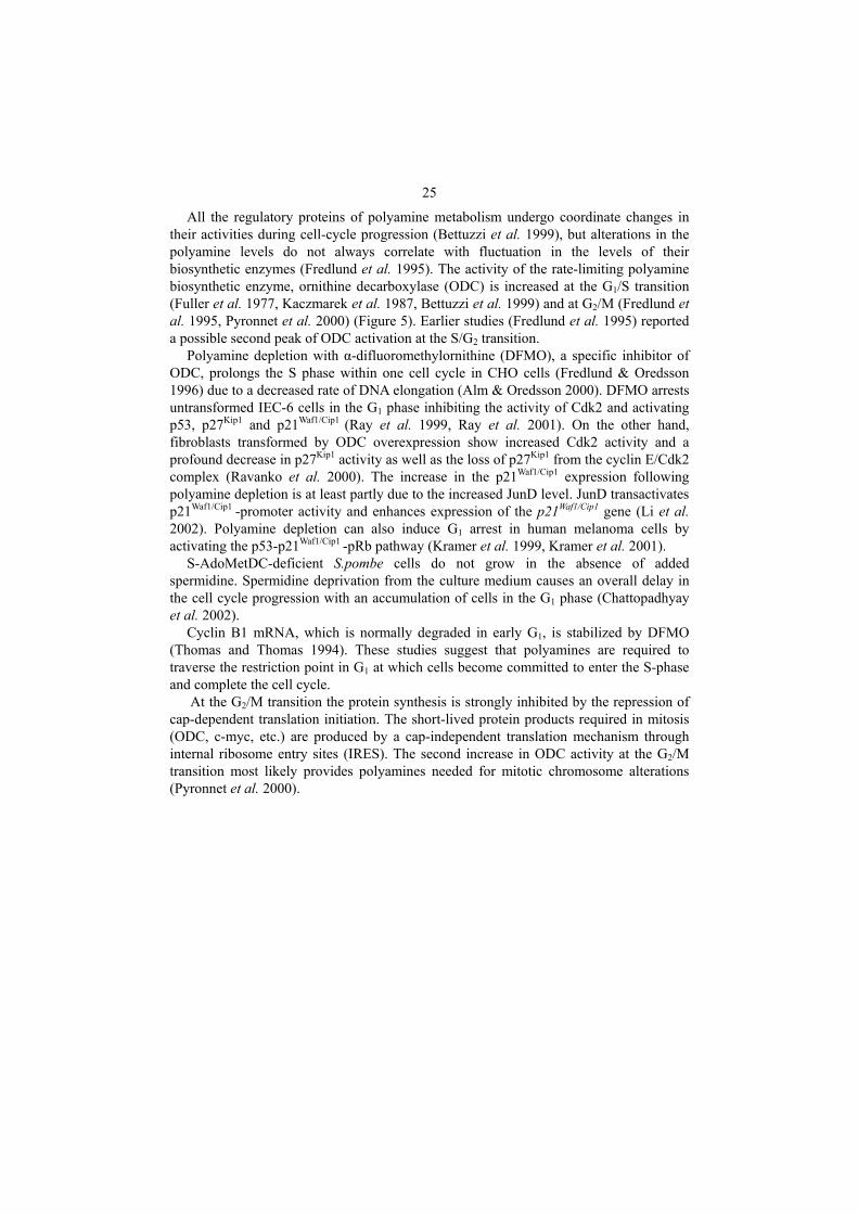
All the regulatory proteins of polyamine metabolism undergo coordinate changes in their activities during cell-cycle progression (Bettuzzi et al. 1999), but alterations in the polyamine levels do not always correlate with fluctuation in the levels of their biosynthetic enzymes (Fredlund et al. 1995). The activity of the rate-limiting polyamine biosynthetic enzyme, ornithine decarboxylase (ODC) is increased at the G1/S transition (Fuller et al. 1977, Kaczmarek et al. 1987, Bettuzzi et al. 1999) and at G2/M (Fredlund et al. 1995, Pyronnet et al. 2000) (Figure 5). Earlier studies (Fredlund et al. 1995) reported a possible second peak of ODC activation at the S/G2 transition.
Polyamine depletion with α-difluoromethylornithine (DFMO), a specific inhibitor of ODC, prolongs the S phase within one cell cycle in CHO cells (Fredlund & Oredsson 1996) due to a decreased rate of DNA elongation (Alm & Oredsson 2000). DFMO arrests untransformed IEC-6 cells in the G1 phase inhibiting the activity of Cdk2 and activating p53, p27Kip1 and p21Waf1/Cip1 (Ray et al. 1999, Ray et al. 2001). On the other hand, fibroblasts transformed by ODC overexpression show increased Cdk2 activity and a profound decrease in p27Kip1 activity as well as the loss of p27Kip1 from the cyclin E/Cdk2 complex (Ravanko et al. 2000). The increase in the p21Waf1/Cip1 expression following polyamine depletion is at least partly due to the increased JunD level. JunD transactivates p21Waf1/Cip1 -promoter activity and enhances expression of the p21Waf1/Cip1 gene (Li et al. 2002). Polyamine depletion can also induce G1 arrest in human melanoma cells by activating the p53-p21Waf1/Cip1 -pRb pathway (Kramer et al. 1999, Kramer et al. 2001).
S-AdoMetDC-deficient S.pombe cells do not grow in the absence of added spermidine. Spermidine deprivation from the culture medium causes an overall delay in the cell cycle progression with an accumulation of cells in the G1 phase (Chattopadhyay et al. 2002).
Cyclin B1 mRNA, which is normally degraded in early G1, is stabilized by DFMO (Thomas and Thomas 1994). These studies suggest that polyamines are required to traverse the restriction point in G1 at which cells become committed to enter the S-phase and complete the cell cycle.
At the G2/M transition the protein synthesis is strongly inhibited by the repression of cap-dependent translation initiation. The short-lived protein products required in mitosis (ODC, c-myc, etc.) are produced by a cap-independent translation mechanism through internal ribosome entry sites (IRES). The second increase in ODC activity at the G2/M transition most likely provides polyamines needed for mitotic chromosome alterations (Pyronnet et al. 2000).

26
Fig. 5. The cell cycle. DFMO induces G1 arrest inhibiting the activity of Cdk2 and activating p53, p27 and p21.
2.1.3.3 Apoptosis
Eukaryotic cells possess a genetic program to commit cellular suicide, a phenomenon also known as apoptosis. The characteristics of apoptosis include mitochondrial structural changes, activation of caspase proteases, chromatin condensation and DNA fragmentation. Failure of apoptosis can lead to auto-immune diseases or cancer. There is an emerging body of literature indicating the involvement of polyamines in this process by several mechanisms (Susin et al. 1998, Schipper et al. 2000) (Figure 6). Both a decrease (Desiderio et al. 1995, Grassilli et al. 1995, Penning et al. 1998) and an increase (Poulin et al. 1995c, Tobias & Kahana 1995, Tome et al. 1997, Xie et al. 1997) in polyamine levels have been reported to trigger apoptosis, and in several cases polyamines, particularly spermine, appear to protect cells against apoptotic cell death (Harada & Sugimoto 1997).
Ornithine decarboxylase is an effector of c-Myc-induced apoptosis. Under conditions in which ODC expression is normally down-regulated, elevated ODC enzyme activity is sufficient to induce apoptosis (Packham & Cleveland 1994). On the other hand, c-Myc, through ODC, has a role in protecting cells from death during periods of cellular stress.
G0 Resting
G2 Gap2
M Mitosis
G1 Gap1
S Synthesis
Cyclin E Cdk2
Increased ODC activity
Rb
DNA replication
p27
p21
p53
E2F

27
Many cellular insults that induce c-Myc expression also enhance ODC expression. The inductions of ODC and c-Myc seem to be mediated by reactive oxygen species (ROS) (Park et al. 2002). The protective role of ODC is most likely due to the production of spermidine and spermine which prevents the DNA strand breakage induced by ROS (Brune et al. 1991, Muscari et al. 1995, Ha et al. 1998b).
Spermine stabilizes DNA and chromatin structure (Penning et al. 1998) and interferes with different ion channels such as K+ channels or NMDA receptors (Harada & Sugimoto 1997), that can explain its protective action on programmed cell death.
Putrescine overaccumulation induces apoptosis (Tobias & Kahana 1995) by a mechanism involving suppression of hypusine formation in the eukaryotic initiation factor (eIF) 5A. The formation of hypusine, in which the butylamine moiety of spermidine is transferred to the lysine residue in eIF-5A, is an important part of the normal function of spermidine (For details see 2.1.3.5) Excess putrescine seems to act post-transcriptionally to reduce eIF-5A levels (Tome et al. 1997).
During B cell receptor (BCR)-mediated apoptosis intracellular polyamine levels decrease. The addition of exogenous spermine prevents mitochondrial inner transmembrane potential (ΔΨm) collapse but does not completely prevent apoptotic changes (Nitta et al. 2001).
Spermine and spermidine have been shown to trigger caspase activation in cell-free models of apoptosis (Stefanelli et al. 1999) and to cause a selective, rapid and saturable leakage of cytochrome c from isolated mitochondria (Stefanelli et al. 2000).
During apoptosis the Bax protein undergoes a transformational change at its NH2 terminus, translocates from the cytosol to the mitochondria, inserts itself into the outer mitochondrial membrane and induces the release of cytochrome c (Goping et al. 1998, Murphy et al. 2000). Polyamine depletion inhibits the translocation of Bax to the mitochondria and consequently prevents cytochrome c release (Yuan et al. 2002). Bid, a proapoptotic Bcl-2 family protein, is cleaved by caspase 8. The truncated Bid also translocates to the mitochondria and induces cytochrome c release (Li et al. 1998, Luo et al. 1998).
Exogenous L-ornithine antagonizes the apoptotic effect of Nω-hydroxy-L-arginine (NOHA) in releasing cytochrome c from the mitochondria and all subsequent steps downstream leading to cell death, but does not inhibit the activation of caspase-8 and cleavage of Bid (Singh et al. 2002). On the other hand polyamine depletion decreases the cleavage of Bid, that contributes, at least partially, to the decrease in release of cytochrome c from the mitochondria (Yuan et al. 2002).
Extracellular signal-regulated kinases (ERKs) 1 and 2 can act in signal transduction pathways leading to apoptosis. In etoposide-treated fibroblasts, polyamine depletion inhibits ERK 1/2 phosphorylation and the subsequent caspase activation (Stefanelli et al. 2002).
Polyamines are negative regulators of nuclear factor (NF)-κB activation. Depletion of cellular polyamines increases the basal level of NF-κB proteins, induces NF-κB nuclear translocation, and activates the sequence-specific DNA-binding activity (Li et al. 2001). NF-κB is an inducible transcription factor and is thought to be the central regulator of transcription of genes involved in apoptosis. It can play a proapoptotic or antiapoptotic role depending on the death stimulus and the cell type. In intestinal epithelial cells, decreased polyamines activate NF-κB and stimulate expression of the inhibitors of

28
apoptosis (IAP) genes, leading to the accumulation of IAP proteins. The increased IAPs inhibit the activation of caspases and protect cells from TNF-α/CHX induced apoptosis (Zou et al. 2004).
The serine-threonine kinase Akt is a multifunctional signaling intermediate in the regulation of cell cycle progression, apoptosis, and energy metabolism. Akt activation suppresses apoptosis induced by different death-stimuli in a variety of cell types. In normal intestinal cells polyamine depletion induces Akt activation mediating suppression of apoptosis at least partly through inhibition of caspase-3 (Zhang et al. 2004). In intestinal epithelial cells, polyamine depletion inhibits the activation of NH2-terminal c-Jun kinase (JNK), and subsequently prevents the activation of caspases-6, -8, -9 and -3 (Bhattacharya et al. 2003).
These results indicate that polyamines are involved in the transduction of the cell-death message.

29
Fig. 6. Involvement of polyamines in apoptosis. , activated protein; , phosphorylated protein, ROS, reactive oxygen species; eIF, eukaryotic initiation factor; ΔΨm, mitochondrial inner transmembrane potential; Bid’, truncated Bid; NOHA, Nω-hydroxy-L-arginine; ERK, extracellular signal-regulated kinase; NF, nuclear factor; IAP, inhibitors of apoptosis; JNK, c-Jun kinase
P
Cytochrome c release
Caspase activation
Apoptosis
Oxidative stress
Radiation
DNA damage
c-Myc
ODC
Spermidine Spermine
Putrescine
Modified eIF-5A
Hypusine
eIF-5A
ΔΨm collapse
Spermidine
Spermine
Bax
Polyamine depletion
NOHA
L-ornithine
Spermine
Bax
Bid
Caspase 8
Bid’
ROS
Polyamine depletion
ERK ERK P
IAP NF-κB
Caspase 3
JNK
JNK
Bid’
Akt Akt P

30
2.1.3.4 DNA and protein synthesis
Increased polyamine levels stimulate (Ginty et al. 1989, Tantini et al. 2001) and conversely the deprivation of polyamines inhibits DNA, RNA and protein synthesis (Oredsson et al. 1990). Polyamine deficient cells are not able to ligate Okazaki-fragments which causes cessation of cellular DNA synthesis (Pohjanpelto & Hölttä 1996). Polyamines might regulate the expression of many genes via DNA bending required for transcription initiation (Childs et al. 2003). Polyamines were shown to participate in protein synthesis in various ways, such as enhancing the synthesis and the activity of RNA polymerases (Jänne et al. 1975, Moruzzi et al. 1975, Jacob & Rose 1976, Yoshida et al. 2002), stimulating the synthesis of specific proteins (Atkins et al. 1975, Watanabe et al. 1981, Kashiwagi et al. 1990), decreasing misincorporation of amino acids during polypeptide synthesis (Jelenc & Kurland 1979, Ito & Igarashi 1986), stimulating the assembly of ribosomal subunits (Kakegawa et al. 1986), influencing tRNA methylation (Leboy 1971, Mach et al. 1982) and stimulating the formation of Ile-tRNA (Igarashi et al. 1978, Kusama-Eguchi et al. 1991).
The addition of polyamines to a cell-free protein-synthesis system results in a 3-to 5-fold stimulation of amino acid incorporation and increases the synthesis of high molecular weight proteins (Atkins et al. 1975). Polyamine regulation of protein synthesis depends on the size and base composition of the 5’-UTR of the mRNA. There is a tendency for polyamines to regulate the translation of mRNAs having long 5’-UTR in both prokaryotes and eukaryotes (Igarashi et al. 1997). Spermidine regulation (stimulation at low concentrations and inhibition at high concentrations) of protein synthesis directed by mRNA having a GC-rich 5’-UTR occurs at the level of initiation complex formation of Met-tRNAi, mRNA and ribosomes (Shimogori et al. 1996).
Polyamines also participate in a rare autoregulatory mechanism of protein synthesis, called translational frameshifting. Antizyme mRNA contains two overlapping open reading frames, ORF1 and ORF2. ORF1 contains sequences required for the initiation of translation but ORF2 encodes most of the protein. The functional protein can be translated only by starting at ORF1, having a frameshift one codon just before its stop codon, and continuing translation in the +1 frame until reaching the stop codon of ORF2. Polyamines have a stimulatory effect on this frameshifting process producing their negative regulator antizyme. (Matsufuji et al. 1996, Coffino 2001.)
2.1.3.5 Hypusine formation
Eukaryotic translation initiation factor 5A (eIF-5A) is a small, acidic protein, highly conserved from yeast to mammalian cells (Childs et al. 2003). The inactive eIF-5A precursor is post-translationally processed by the synthesis of an unusual amino acid, hypusine (Park et al. 1981). The biosynthesis of hypusine occurs via two consecutive enzymatic steps (Figure 7) (Murphey & Gerner 1987, Park & Wolff 1988). In the first step of this process, deoxyhypusine synthase (EC 2.5.1.46) catalyzes the NAD-dependent transfer of the butylamine moiety of the donor spermidine to the amino group of a

31
specific lysine residue of the acceptor eIF-5A precursor (Chen & Dou 1988, Wolff et al. 1995, Lee et al. 2001). This intermediate is then hydroxylated by deoxyhypusine hydroxylase (EC 1.14.99.29) to complete the synthesis of hypusine and the maturation of eIF-5A (Abbruzzese et al. 1986). Hypusine synthesis is one of the most specific post-translational modifications and the only biological function for a polyamine described at the molecular level to date. Mature eIF-5A has an essential role in eukaryotic cell proliferation. Gene disruption studies in the yeast, Saccharomyces cerevisiae, show that inactivation of the two eIF-5A genes (Schnier et al. 1991) or of the single-copy deoxyhypusine synthase gene (Sasaki et al. 1996, Park et al. 1998) results in a loss of cell viability. In addition, inhibitors of deoxyhypusine synthase (Park et al. 1994) or deoxyhypusine hydroxylase (Hanauske-Abel et al. 1994) suppress growth in various mammalian cells. However, eIF-5A is not required for general protein synthesis, as eIF-5A-depleted yeast cells are still able to synthesize the major part of their proteins (Kang & Hershey 1994). Although the exact function of eIF-5A is still unclear, its highly conserved structure in eukaryotes and unique post-translational modification machinery suggest a critical biological role.
Fig. 7. Biosynthesis of eukaryotic initiation factor 5A (eIF-5A)
eIF-5A precursor
NH2
(CH2)4
-NH-CH-COO-
+ Lysine
NH2
(CH2)3
NH2
(CH2)2
CH2
CH2
NH
NH2
(CH2)2
CH2
CH2
NH
(CH2)4
-NH-CH-COO-
Spermidine
1.
NH2
(CH2)2
CH
CH2
NH
(CH2)4
-NH-CH-COO-
OH 2.
eIF-5A
1. deoxyhypusine synthase 2. deoxyhypusine hydroxylase
Hypusine Deoxyhypusine

32
2.1.3.6 Interactions with ion channels
Spermidine and spermine play a role in blocking and modulating various ion channels, such as the inward rectifier K+ channels (Kir channels) and different glutamate receptors (Williams 1997a). In the case of K+ channels, inward rectification means that the inward flow of K+ ions at negative membrane potentials is always greater than the outward flow for the opposite driving force. Strong inward rectification is essential to maintain the resting potential of cells and permit prolonged depolarization, a feature of the cardiac action potential (Nichols & Lopatin 1997). Inward rectification may result from two independent mechanisms, an instantaneous block by Mg2+ and a phenomenon called intrinsic gating caused by intracellular spermidine and spermine (Ficker et al. 1994, Lopatin et al. 1994). Intrinsic gating involves a slow decrease in the current (50-100 ms) at depolarized membrane potentials. Spermine was shown to enter a Kir channel and bind electrostatically to its negatively charged residues at various depths, thus blocking the channel pore and the outward K+ flow in a voltage dependent manner (Kubo & Murata 2001, Xie et al. 2002).
Glutamate receptors mediate fast synaptic transmission in the central nervous system. They are classified on the basis of their selectively activating agonists into three groups: NMDA-, AMPA- and kainate receptors (Williams 1997a). NMDA- (Traynelis et al. 1995, Masuko et al. 1999) and kainate receptors (Mott et al. 2003) are regulated by protons at ambient pH (pH 7.3). Spermine is able to relieve proton inhibition and stimulate these receptors possibly via binding to their LIVBP-like domain (Mott et al. 2003).
Intracellular polyamines control the rectification of the Ca2+-permeable AMPA and kainate receptors as well (Donevan & Rogawski 1995, Williams 1997b). Bowie et al. (1998) have suggested that polyamines modulate these glutamate receptors dynamically via both open- and closed-channel blocking mechanisms.
Activation of NMDA receptors requires binding of glutamate and glycine at separate sites on the receptor. Extracellular spermidine and spermine have multiple effects on NMDA receptors. They facilitate the binding of glycine to the receptor complex (glycine-dependent stimulation) (Sacaan & Johnson 1989, McGurk et al. 1990, Ransom & Deschenes 1990) as well as potentiate NMDA currents via increase in frequency of channel opening (glycine-independent stimulation) (Rock & MacDonald 1992, Araneda et al. 1993).
Polyamines also block responses of the NMDA receptors by reducing conductance in a voltage-dependent manner (Araneda et al. 1993). Spermine reduces NMDA single-channel currents, and blocks and permeates NMDA receptor channels from both the extracellular and intracellular sides (Araneda et al. 1999). Recently it was shown (Turecek et al. 2004) that intracellular spermine modulates the activity of NMDA receptors by a direct mechanism involving a decrease in the probability that receptor channels were open.

33
2.1.3.7 Embryonic development
The role of polyamines in invertebrate development was studied by using the polychete Ophryotrocha labronica (Heby & Emanuelsson 1981) and the nematode (or round worm) Caenorhabditis elegans (MacRae et al. 1998) as model systems. DFMO treatment of fertilized Ophryotrocha labronica eggs prevents putrescine accumulation at the beginning of gastrulation. This polyamine limitation results in a developmental arrest at gastrulation, prevents nucleolar formation and suppresses ribosomal activity (Heby & Emanuelsson 1981). C.elegans odc-1 null mutant worms with no detectable ODC activity show two different phenotypes in polyamine-free medium, depending on the developmental stage at which it was imposed. Polyamine depletion at the early L1 stage results in animals that are morphologically adult but do not contain or lay eggs. If mutant larvae are transferred to polyamine-deficient medium at the L4 stage, they develop and lay eggs normally, but the embryos fail to hatch and arrest at about the 550-cell stage (MacRae et al. 1998).
In polyamine research the most widely used model systems for vertebrate development are Xenopus (Shiokawa et al. 2000), chick (Goyns 1979, Lowkvist et al. 1980, Heby & Emanuelsson 1981), rat (Russel & McVicker 1972) and mouse (Fozard et al. 1980a, Jotova et al. 1999, Pendeville et al. 2001, Nishimura et al. 2002). S-AdoMetDC mRNA injection into Xenopus fertilized eggs activates a maternally preset apoptotic program at the early gastrula stage. S-AdoMetDC overexpression induces an S-adenosylmethionine deficient state, that in turn induces the inhibition of protein synthesis. The fact, that various other agents which damage DNA, RNA and protein synthesis switch on the apoptotic program at the same developmental step (at the early gastrula stage, shortly after the midblastula transition), indicates the existence of an important developmental check-point at this stage (Shiokawa et al. 2000).
Administration of DFMO into fertilized chick eggs also blocks embryonic development at gastrulation (Lowkvist et al. 1980, Heby & Emanuelsson 1981). ODC inhibition interferes with nucleolar formation with a reduction of the fibrillar component of the nucleolus and segregation of the nucleolar material (Lowkvist et al. 1983). DFMO treatment in mice at early pregnancy abolishes increases in uterine ODC activity and in putrescine and spermidine concentrations seen during normal gestation, but markedly increases S-AdoMetDC activity. As a consequence, decidualization takes place normally after implantation, but embryonic development is arrested at a stage typical of days 6 to 7 of normal gestation that corresponds to the onset of gastrulation. Rats and rabbits show similar responses to DFMO (Fozard et al. 1980b).
The ODC and the S-adenosylmethionine decarboxylase knock-out mouse lines (Pendeville et al. 2001, Nishimura et al. 2002) were discussed more in detail in 2.1.2.2. Briefly, homozygous embryos were not viable, dying before the onset of gastrulation, between E3.5 and E6.5. These knockout studies confirm the generally accepted view that polyamine homeostasis plays an essential role in development and cell growth. However, the generation of conditional knockout mouse lines is needed to further clarify the role of polyamines and their biosynthetic enzymes during vertebrate embryogenesis.

34
2.1.3.8 Polyamines in the urogenital organs
Polyamines have a critical role in germ cell development. Putrescine, spermidine and spermine concentrations show continuous and cell type specific changes during testicular maturation that are consistent with fluctuations of their biosynthetic enzymes (Shubhada et al. 1989b). In rodent testes, ornithine decarboxylase (ODC) mRNA levels increase during late meiosis and early spermiogenesis and decrease to background levels in later stages of spermatid development and in spermatozoa suggesting that polyamines are needed during haploid gene expression (Alcivar et al. 1989, Shubhada et al. 1989a, Kaipia et al. 1990). Transgenic mice overexpressing ODC exhibit male infertility and dramatic morphological changes in testicular tissue with grossly impaired spermatogenesis. These mice have more than a 20 fold higher putrescine concentration in their testes when compaired with their nontransgenic littermates (Halmekytö et al. 1991a, Halmekytö et al. 1991c). Putrescine has a selective mode of action during mitotic and meiotic cell cycles in spermatogenesis, strongly stimulating the last mitosis before the onset of meiosis in type B spermatogonia and inhibiting meiotic DNA synthesis. Aberrantly high putrescine levels may lead to decreased fertility in the ODC-overexpressing transgenic mice (Hakovirta et al. 1993). During sperm development, ODC expression is regulated by antizyme 3, an ODC antizyme expressed only in the haploid germ cells in the testis between early spermiogenesis and the late spermatid phase (Ivanov et al. 2000, Tosaka et al. 2000). The expression pattern of antizyme 3 suggests that it provides spatial and temporal regulation of ODC during spermatogenesis sharply limiting polyamine accumulation in cells that have completed meiotic reduction and are about to be remodeled into mature spermatozoa (Coffino 2000).
In the developing chick ovary ODC has two activity peaks: one in the early developmental stages correlated with early morphological development and germ cell proliferation and another related to the maturation of a large population of follicular cells. Polyamine levels follow the changes in ODC activity (Teng & Teng 1980). Interestingly, transgenic mice overexpressing spermidine/spermine N1-acetyltransferase (SSAT) are characterized by permanent hair loss and female infertility. In transgenic animals, primary and small secondary follicles are present but larger developing follicles and corpus luteum formation are absent suggesting a role for polyamines in follicular development (Pietilä et al. 1997).
In the adult murine kidney, ODC is expressed mainly in the proximal tubules (Levillain & Hus-Citharel 1998) and its expression is regulated by androgens at multiple levels, including transcription, posttranscription and translation (Seely et al. 1982, Isomaa et al. 1983, Berger et al. 1986, Watson & Paigen 1988). Testosterone enhances ODC expression causing a significant difference in renal ODC activity and consequently in putrescine levels between sexes, being high in males and low in females (Goldstone et al. 1982). This renal sexual dimorphism is manifested after weaning, simultaneously with an increase in plasma testosterone concentration (Sanchez-Capelo et al. 1994, Sanchez-Capelo et al. 1999). Despite the low ODC level in female kidneys, antizyme 1 expression levels are similar to those of males suggesting a continuous degradation process of the ODC protein in females (Murakami et al. 1988, Levillain et al. 2003).

35
2.2 Acetyl-CoA synthetase
Acetyl-CoA synthetase activates acetate to acetyl-CoA, an essential molecule utilized in various metabolic pathways including the fatty acid and cholesterol synthesis as well as the tricarboxylic acid cycle. There are two different forms of acetyl-CoA synthetase, the ADP-forming and the AMP-forming enzymes, which are encoded by two independent, nonhomologous genes (Starai & Escalante-Semerena 2004). The ADP-forming acetyl-CoA synthetase (EC 6.2.1.13) catalyzes the reversible reaction of acetate+ ATP ADP + Pi + acetyl-CoA. This type of enzyme is restricted to lower organisms, such as anaerobic protists and some archeal halophytes and thermophiles. AMP-forming acetyl-CoA synthetase (AceCS, EC 6.2.1.1) synthesizes acetyl-CoA in two steps via an acyl-AMP intermediate. The simplified reaction is acetate+ ATP (acetyl-AMP) acetyl-CoA + PPi + AMP. AceCS has a broad distribution from prokaryotes to human (Karan et al. 2001, Starai & Escalante-Semerena 2004). The characteristics of this latter enzyme will be discussed more in detail below.
2.2.1 Regulation of AMP-forming acetyl-CoA synthetase
The coding regions of the AMP-forming acetyl-CoA synthetase (AceCS) are well conserved among different species (Karan et al. 2001). AceCS gene expression is controlled by complex regulatory mechanisms as a function of carbon flux including a posttranslational NAD+/sirtuin-dependent protein acetylation/deacetylation system.
In E.coli AceCS is the promoter-proximal gene of an operon involved in acetate metabolism. The AceCS gene is cotranscribed with the acetate transporter coding actP gene, and with the yjcH gene whose function is unknown to date (Gimenez et al. 2003). AceCS is induced through the actions of different transcription factors such as the carbon regulator cAMP receptor protein (CRP), the oxygen regulator FNR, the glyoxylate shunt repressor IclR and its activator FadR, (Kumari et al. 2000a). AceCS transcription occurs as a function of sigma factor σ70 activity (Kumari et al. 2000b).
Eukaryotes possess two isoforms of AceCS, one in the mitochondria, participating in the energy generation processes and a second one in the cytosol supplying acetyl-CoA for lipid biosynthesis. In S. cerevisiae ACS1 is the mitochondrial enzyme, only expressed during respiratory and respirofermentative growth, whereas ACS2 is expressed in the cytosol during anaerobic growth on glucose (Starai & Escalante-Semerena 2004). Transcriptional regulation of the two genes differs strongly.
ACS1 is repressed by high concentrations of glucose and other fermentable carbon sources (de Jong-Gubbels et al. 1997). Induction of ACS1 on non-fermentable ethanol or acetate is positively controlled by a carbon source-responsive element (CSRE) and an alcohol dehydrogenase regulator, Adr1p in the promoter region whereas binding of the Ume6p protein to the URS1 motifs exerts negative control on the ACS1 promoter (Kratzer & Schuller 1995, 1997).
ACS2 is coregulated with structural genes of lipid biosynthesis via binding of the heterodimeric activator Ino2p/Ino4p protein to the ICRE regulatory motif as well as

36
binding of the pleiotropic transcription factor Abf1p to the ACS2 promoter region (Hiesinger et al. 1997).
Mammalian acetyl-CoA synthetase also has two isoforms with an unfortunate nomenclature. Mammalian cytoplasmic AceCS is called AceCS1 and the mitochondrial isoform is AceCS2, which is the opposite and confusing numbering compared to yeast.
Cytosolic acetyl-CoA synthetase is involved in generating acetyl-CoA for lipid biosynthesis. AceCS1 transcription is negatively regulated by sterols (Luong et al. 2000) and induced via binding of the sterol regulatory element-binding proteins (SREBPs) and Sp1 or Sp3 to the promoter region (Ikeda et al. 2001). AceCS1 is also regulated by insulin and the diabetic status of the individual. It also responds to dietary changes possibly under the control of SREBP-1 (Sone et al. 2002).
AceCS2 is a mitochondrial enzyme and its acetyl-CoA product is mainly used for oxidation by the citric acid cycle. AceCS2 transcripts are abundant in the heart and skeletal muscle of mouse. Under ketogenic conditions such as starvation and diabetes AceCS2 is induced via unknown mechanisms (Fujino et al. 2001).
2.2.2 Posttranslational regulation of AceCS
AceCS expression is further regulated by a sirtuin-dependent posttranslational control mechanism. Sirtuin is the collective name of the silent information regulator protein (Sir2) and its homologs involved in gene silencing and chromosome stability. Sir2 protein has a genetically conserved NAD+-dependent protein deacetylase activity (Smith et al. 2000) and one of its substrates is AceCS. Deacetylation by Sir2 activates AceCS whereas a single acetylation is enough to block enzyme activity. The site of acetylation is a lysine residue in a well conserved motif of the AMP-forming enzymes suggesting a common regulatory mechanism in pro- and eukaryotes (Starai et al. 2002, Starai et al. 2004, Takasaki et al. 2004).

3 Aims of the research
To date, over 60.000 articles have been published involving polyamines, but it is still not possible to define exactly how polyamines function in cells. While it is difficult to find a single cellular function in which they would not be involved, polyamines were studied mostly because of their role in malignant cell growth. Polyamines accumulate in cancerous tissues and their concentration is elevated in the body fluids of cancer patients. However, cells have a sophisticated regulatory machinery to maintain their polyamine pools that makes polyamine research very complicated. To date, inhibitors of virtually all of their biosynthetic enzymes are available and transgenic and knock-out animals have been generated for both their biosynthetic and catabolic enzymes to study their cellular functions and interactions with cellular components. In this work we have used a variety of approaches from cell culture to transgenic animals with tools of molecular and developmental biology to elucidate the function of polyamines and specify mechanisms in which they are involved.
The specific aims of the present work were:
1. to clarify mechanisms by which transgenic mice overexpressing polyamine biosynthetic enzymes maintain their polyamine pools;
2. to identify genes responsive to changes in polyamine synthesis; 3. to characterize mammalian cytosolic acetyl-CoA synthetase (AceCS1) 4. to gain a better understanding of the regulatory functions of polyamines using the
embryonic kidney as a model organ

4 Materials and methods
Detailed description of the materials and methods are presented in the original publications I-IV.
4.1 Mouse lines (I, II, III, IV)
A transgenic mouse line overexpressing S-AdoMetDC (UKU99) was obtained by the standard pronuclear microinjection technique (Hogan et al. 1986). A 19.5 kb fragment consisting of the entire rat S-AdoMetDC gene with 3kb of 5’- and 0.9 kb of 3’- flanking sequences was injected into fertilized mouse oocytes. A hybrid transgenic mouse line was generated by mating the UKU99 mice with UKU2, a transgenic mouse line which overexpresses the human ODC gene (Halmekytö et al. 1991a) (I, II).
For whole-mount (III, IV) and organ culture (IV) experiments whole embryos or embryonic tissues were isolated from the CD-1mouse line, with day 0.5 being the day of detection of the vaginal plug.
The effect of DFMO on Pax-2 epithelial expression was studied by using a Pax-2/lacZ mouse line, which expresses a β-galactosidase gene under the control of a 8.5 kb-long Pax-2 promoter (Kuschert et al. 2001) (IV).
4.2 Cell culture (I, II, IV)
To study mechanisms by which hybrid mice overexpressing ODC and S-AdoMetDC maintain their polyamine pools (I), and for the differential display analysis (II) early-passage mouse embryonic transgenic and non-transgenic fibroblasts from UKU2/UKU99 cross mated mice hybrids were grown in DMEM with 20% (v/v) fetal bovine serum in 5% CO2/95% air at 37°C. In the pulse labeling experiments (I) subconfluent cells were incubated for 2 hours with 0.4 μCi/ml of L-[U-14C] ornithine. After labeling, the cells

39
were grown for 4-24 hours, and the concentrations and radioactivity of the polyamines were determined.
For differential display analysis (II), Rat-2 cells (ATCC, Rockville, MD, USA) were grown in DMEM supplemented with 2 mM glutamine, 100 units/ml penicillin, 100 µg/ml streptomycin and 5% (v/v) fetal bovine serum in 5% CO2/95% air at 37°C. Cells were treated with 5 mM DFMO for 90 min or 48 h prior to differential display and for 48 hours before exposure to 4 μM actinomycin D for the mRNA half-life assays. To study the effect of DFMO on the turnover of Tex261 mRNA (II), subconfluent cells were transfected with a pBK-CMV-Tex261 construct using a standard calcium-phosphate transfection protocol (Sambrook et al. 1989) and grown for 48 hours in a medium containing 5 mM DFMO and/or 5μM MGBG.
For the analysis of the minimal Pax-2 promoter (IV), HEK 293 cells (ATCC) were grown in Eagle minimal essential medium supplemented with 2mM glutamine, 1mM sodium-pyruvate and 10% (v/v) heat-inactivated horse serum in 5% CO2/95% air at 37°C. Subconfluent cells were transfected with luciferase reporter plasmids using Fugene 6 transfection reagent (Roche Molecular Biochemicals) according to the manufacturer’s instructions and grown for 48 hours in the presence or absence of 10 mM DFMO before harvesting for the luciferase assay.
4.3 Polyamine determination (I, II)
Polyamine concentrations were determined essentially as described earlier (Gilbert et al. 1991). Briefly, samples were passed over using a weakly acidic cation exchange resin, Biorex-70. After concentration (freeze-drying), samples were derivatized with dansyl-chloride and analyzed by high performance liquid chromatography (HPLC) on a reversed phase Ultrasphere ODS column using a methanol-acetonitrile-water gradient. The detector was a Waters 470 fluorescent scanning detector at an excitation wavelength of 330 nm and an emission wavelength of 510 nm. To standardize the different cell samples, DNA was quantified by the method described by Giles & Myers (1965).
4.4 Northern blot analysis (I, II, III)
Total RNA (Chomczynski & Sacchi 1987) was fractionated (15-20 µg/lane) on a 1% agarose gel containing formaldehyde (Meinkoth & Wahl 1984), blotted onto nitrocellulose and hybridized with cDNA fragments labeled with [α-32P]dCTP using the random-priming method. The amount and the quality of total RNA loaded in the gels were checked by staining the transferred membrane with methylene blue (Herrin & Schmidt 1988).
40
4.5 Differential display (II)
Differential display was performed essentially as described previously (Liang & Pardee 1992, Liang et al. 1993). Briefly, total RNA was used in a reverse-transcription reaction using each of the four 1-bp-anchored 3´ oligo(dT) primers (Liang et al. 1994). The reverse-transcribed cDNA was used for each PCR reaction, performed using the same 1-bp-anchored 3´ oligo(dT) primer and four 5´ arbitrary oligonucleotides of 10 nucleotides in length. The parameters for PCR were 40 cycles of denaturation at 94 °C for 30 s, annealing at 40 °C for 2 min, and extension at 72 °C for 30 s. The PCR reaction mixture was loaded onto an 8% sequencing gel, and the differentially amplified PCR fragments were visualized by exposing the dried sequencing gel to X-ray film. Candidate PCR products were excised from the gel and DNA eluted from the gel slices by boiling in TE buffer for 10 min. The eluted DNA fragments were re-amplified by using the same primer pair and subsequently cloned into a TA cloning vector. Inserts were sequenced by cycle sequencing on an ABI sequencer and DNA sequences analyzed using the FASTA and BLAST computer programs. The isolated cDNA fragments were used as probes to confirm differential expression by northern-blot analyses.
4.6 Whole mount and section in situ hybridization (III, IV)
Whole-mount and section in situ hybridization was performed based on standard procedures (Wilkinson 1992). Digoxigenin labeled AceCS1 antisense and sense probes were prepared from the mouse clone AF216873 in the pBluescript SK- vector (III). Full-length cDNAs from Pax-2 (Dressler et al. 1990), Pax-8 (Plachov et al. 1990), Wnt-11, c-ret, GDNF (Majumdar et al. 2003), E-cadherin (Ringwald et al. 1987), and BF-2 (Hatini et al. 1996) were also labeled with digoxigenin. The antisense and sense ODC probe was synthesized from the 749 bp HindIII-digest of the full-length mouse cDNA inserted into pBluescript SK- vector (IV).
4.7 Sex typing (III)
The sex of E11.5 and E12.5 embryos was determined by a PCR method. A 353-bp region from the last intron of the mouse Zfy gene was amplified as described previously (Chang et al. 1994).

41
4.8 Organ culture (IV)
Kidney rudiments were isolated from mouse embryos at E11.0 and placed on small pieces of Nucleopore filter supported by stainless steel grids in a medium consisting of DMEM with Glutamax-I supplemented with 100 units/ml penicillin, 100 µg/ml streptomycin and 10% (v/v) fetal bovine serum (Lin et al. 2001). Cultured kidneys were grown in 5% CO2/95% air at 37 °C for 1, 2, 3, 4, 6 days, in the presence or absence of DFMO or together with DFMO and putrescine, the medium being changed every second day.
4.9 Cell proliferation assay (IV)
The cell proliferation assay was performed using cell proliferation kit (Amersham) following the manufacturer’s instructions. E11.0 mouse kidneys were grown in culture for 3 days. After 1 hour of BrdU-labeling, kidney specimens were paraffin-embedded and cut into 5 µm sections. BrdU was incorporated into replicating DNA of proliferating cells and subsequently localized using specific monoclonal antibody. Detection of bound antibody was achieved using peroxidase conjugated antibody to mouse immunoglobulin and DAB as peroxidase substrate, giving blue-black staining at sites of BrdU incorporation.
4.10 Histology, β-galactosidase and antibody staining (IV)
Hematoxylin-eosin and β-galactosidase staining was performed using standard protocols (Hogan et al. 1994). For hematoxylin-eosin staining, kidney specimens were fixed in 4% paraformaldehyde, washed in PBS, dehydrated and embedded in paraffin. 5μm sections were cut and stained with hematoxylin and eosin.
For β-galactosidase staining, kidney specimens were fixed for 30 min at 4 ˚C in a solution containing 1% formaldehyde and 0.2% glutaraldehyde, washed at room temperature in PBS and stained overnight at 37˚C in PBS containing 5mM K3Fe(CN)6, 5mM K4Fe(CN)6, 2mM MgCl2, 0.01% NaDeoxycholate, 0.02% NP-40 and 1mg/ml X-gal. After staining, specimens were washed in PBS, post-fixed in 4% paraformaldehyde and stored in 80% glycerol.
Methanol fixed organ culture samples were double-stained with Troma-1 against cytokeratin Endo A (Developmental Studies Hybridoma Bank, USA) and brushborder (BB) (Ekblom et al. 1980) antibodies. The secondary antibodies used were FITC-conjugated donkey anti-rat IgG (Jackson Immuno Research Laboratories, USA) and TRITC-conjugated donkey anti-rabbit IgG (Rockland Immunochemicals, USA) respectively.

42
4.11 Other methods (I, II)
The activity of S-AdoMetDC and ODC was measured as described earlier (Jänne and Williams-Ashman 1971b, 1971a) (I).
RT-PCR was used for the identification of the rat-specific S-AdoMetDC mRNA (I), the differential display analysis (II) and the determination of the half-life of S-AdoMetDC mRNA (II). In brief, total RNA (Chomczynski & Sacchi 1987) was reverse-transcribed with a sequence specific sense primer using an AMV (I) or a M-MLV (II) reverse transcriptase (Promega). After the first strand synthesis, antisense primer and a thermostable DNA polymerase were added, and PCR amplification was performed as described in the articles referenced above.

5 Results
5.1 Overexpression of S-AdoMetDC and ODC in transgenic mice does not dramatically affect tissue spermidine
and spermine pools (I)
Cells have an efficient compensation machinery to correct disturbances in polyamine homeostasis. To study these mechanisms we have generated a mouse line overexpressing S-AdoMetDC. The UKU99 S-AdoMetDC-overexpressing transgenic mice have a 2-4 fold increase in S-AdoMetDC activity in all tissues tested. This increase in enzyme activity is not dramatic, but the expression of the transgene is presumably controlled by tissue-specific control elements located in the 3-kb-long 5’-flanking region of the transgene construct. Mice from another transgenic line, UKU2, generated by our collaborators (Halmekytö et al. 1991a) overexpress ODC 10-100-fold in their tissues, most markedly in the testis and brain. These mice have been generated with a shorter, 800bp, 5’-flanking region in the transgene construct, probably lacking the tissue-specific control elements (Halmekytö et al. 1991b).
In the S-AdoMetDC-overexpressing mice, tissue putrescine levels decreased but spermidine and spermine pools were practically unaltered. Nevertheless, the molar ratio of spermidine to spermine was lower in the transgenic animals, indicating a faster conversion of putrescine and spermidine into spermine in the presence of excess decarboxylated S-AdoMet. To study whether the decreased putrescine level is responsible for the unchanged spermidine and spermine pools, a hybrid UKU2/UKU99 transgenic mouse line was generated. In the hybrid mice overexpressing both ODC and S-AdoMetDC, the putrescine pool tended to normalize compared to the aberrantly high putrescine levels seen in UKU2 mice, whereas spermidine and spermine concentrations remained unchanged.

44
5.2 Transgenic mice compensate for increased S-AdoMetDC and ODC activities by acetylating and excreting
excessive polyamines (I)
To study the mechanisms by which UKU2/UKU99 hybrid mice overexpressing both ODC and S-AdoMetDC maintain their polyamine homeostasis, we performed pulse-labeling of polyamine-flux in transgenic and non-transgenic early-passage embryonic fibroblasts. Pulse-labeling was carried-out using 14C-labeled L-ornithine, the substrate of the rate limiting polyamine biosynthetic enzyme, ODC. Polyamine synthesis and secretion was monitored by measuring intracellular and extracellular concentrations of polyamines and their acetylated derivatives, as well as by following the incorporation of radioactivity into acetylated and non-acetylated polyamines.
We found a time-dependent polyamine concentration shift between hybrid and control fibroblasts. Intracellular putrescine levels were elevated in the transgenic fibroblasts 4 hours after pulse-labeling, but normalized after 24 hours. On the other hand, 24 hours after pulse-labeling, extracellular putrescine and N1-acetylspermidine accumulated in the medium of the transgenic fibroblasts. By following the fate of radioactive ornithine, we have shown that polyamine biosynthesis is accelerated and polyamine acetylation is increased in the UKU2/UKU99 hybrid fibroblasts. These results indicate that a compensation mechanism, including acetylation and increased excretion, prevents the toxic accumulation of higher polyamines in cells.
5.3 Changes in gene expression patterns in response to polyamine depletion and activation of biosynthesis (II)
To further clarify the regulatory mechanisms involving polyamines, we applied a differential display approach for identifying mRNAs responsive to changes in polyamine levels. Rat-2 cells were treated for short-term (90 min) and for long-term (48 hours) with DFMO to block the function of ODC and consequently deplete cellular polyamines. The two different time periods served as a control to be able to distinguish between direct effects caused by polyamine depletion and secondary effects due to the cessation of cell growth. The concentration of higher polyamines, spermidine and spermine did not change after 90 min of DFMO treatment, but putrescine level decreased by about 30%. Long-term DFMO treatment almost completely depleted cellular putrescine, spermidine concentration decreased to 10% of the control level, whereas the spermine level remained unchanged.
Differential display resulted in 15 cDNA bands after 90 min and 44 cDNA bands after 48 hours with significant intensity differences. The majority of these cDNA bands (43) were more intense in the polyamine-depleted samples and only 16 of them indicated down-regulation after DFMO treatment. A repeated differential display analysis confirmed 35 cDNA bands of the 59 and these were subcloned, sequenced and 11 of them

45
subjected to Northern blot analysis to further confirm differential display results. Six clones, mitochondrial adenine nucleotide translocator, CarG-box binding factor, ribosomal proteins L3 and 3Sa, acetoacetyl-CoA thiolase (EC 2.3.1.9) as well as the mouse EST (MM97319), later characterized as cytosolic acetyl-CoA synthetase (EC 6.2.1.1) (see in chapters 5.6-5.7), showed results similar to those obtained from differential display analysis, the first two transcripts being present both after short-term and long-term DFMO treatment. Five of the six clones were upregulated and only one down-regulated in polyamine-depleted cells.
To study the compensation mechanisms under conditions where polyamine synthesis is enhanced, we performed differential display analysis with kidneys from UKU2/UKU99 hybrid mice overexpressing both ODC and S-AdoMetDC (I) and from non-transgenic mice. Following the confirmation procedure described previously, we have found only two cDNA clones having different gene-expression pattern in transgenic and non-transgenic kidneys, the ribosomal protein L3 and Tex261, both downregulated in transgenic animals. Table 1 shows the general features of the confirmed genes from both the cell culture and the transgenic approach.

Table 1. Gene-expression differences in cell culture in response to polyamine level alterations, function of genes and properties of their promoters
Gene Effect of DFMO treatment
Status in transgenic mice
Function Mammalian promoter properties
Cytosolic acetyl-CoA synthetase
down Supplies acetyl-CoA for lipid biosynthesis
Lacks TATA box, CCAAT box, SRE, E-box, GC-box binding Sp1 and/or Sp3 (Ikeda et al. 2001)
Mitochondrial adenine nucleotide translocator
up Mitochondrial ATP/ADP exchanger, part of the mitochondrial permeability transition pore complex, involved in apoptosis
TATA box, CCAT box, intronic Sp1 binding sites (Li et al. 1989)
CArG-box binding factor
up Transcription regulator, enhances myogenic differentiation
Not published
Ribosomal protein L3 (rpL3)
up down Involved in the formation of the peptidyltransferase center
Not published
Ribosomal protein 3Sa
up 40S ribosomal subunit protein; involved in induction of apoptosis and malignant transformation
Lacks TATA and CAAT boxes, CpG-islands with potential Sp1 binding site, CREB/ATF binding sites (Nolte et al. 1996)
Mitochondrial acetoacetyl-CoA thiolase
up Catalyses the conversion of acetyl-CoA to acetoacetyl-CoA in the cholesterol biosynthetic pathway
Lacks TATA box, CAAT boxes, Sp1, AP-2 and GTI binding sites (Kano et al. 1991)
Tex261 down Potential transmembrane protein
High rate of CpG-islands (Lopez-Fernandez et al. 1998)

47
5.4 Increased GAPDH and β-actin expression in polyamine-depleted cells (II)
Glyceraldehyde-3-phosphate dehydrogenase (GAPDH) and β-actin probes are widely used as loading standards in Northern-blot analysis. Earlier studies have shown that β-actin levels increase upon DFMO treatment (Celano et al. 1988) and that was the case also under our experimental conditions. GAPDH responded similarly to polyamine depletion, with an increase in the intensity of the transcripts, whereas 28S and 18S rRNA levels remained unchanged. Hence we propose that hybridization or staining of 28S and 18S rRNA in Northern-blot analysis is a more reliable loading control than the use of β-actin or GAPDH.
5.5 Polyamine depletion results in selective stabilization of the mRNAs (II)
To clarify the mechanisms leading to an increase in mRNA levels in the majority of our polyamine-depleted samples, we studied whether mRNA accumulation is due to the stabilization of the message. Turnover of S-AdoMetDC, GAPDH and Tex261 was measured after actinomycin D treatment. All of the three transcripts were clearly stabilized after DFMO treatment with a constant mRNA level up to 6 hours for S-AdoMetDC, the same transcript having a half-life of 3 hours in control cells. These results indicate that polyamine depletion results in selective changes in the expression of a panel of genes, mostly by upregulation of transcripts, via stabilization of the message.
5.6 Cytosolic acetyl-CoA synthetase and polyamines
Cytosolic acetyl-CoA synthetase (AceCS1) was the only downregulated gene identified in polyamine-depleted cells using differential display analysis (II). Its response to polyamine concentration changes was similar to that of SSAT, the latter using acetyl-CoA to produce N1-acetylspermidine and N1-acetylspermine. Our hypothesis was that AceCS1might provide acetyl-CoA for polyamine acetylation by SSAT. We isolated the mouse cDNA clone (AF216873), used it for transfection of Rat-2 cells and measured polyamine concentrations. Although AceCS1 activity was two-three times higher in the transfected cells, there was no difference in the level of polyamines or acetylated polyamines relative to controls (unpublished results). At the same time we studied the tissue distribution of AceCS1 in adult mice and during embryonic development, to better understand the possible functions of this enzyme. Meanwhile Luong and co-workers (2000) published their results concerning the characterization of the homologous human AceCS1 gene, linking it to lipid biosynthesis. As by that date we did not have any

48
promising link between AceCS1 and polyamine catabolism, we decided to focus on the tissue distribution studies.
5.7 Expression of AceCS1 during mouse development (III)
Tissue distribution of the AceCS1 mRNA in adult mice was determined with Northern-blot hybridization. A 3-kb-long single transcript was found in all the tissues studied with the highest intensity in the ovaries, testes and kidney.
To study AceCS1 expression during embryonic development, we used whole-mount and tissue section in situ hybridization techniques. In the early embryonic stages (E9.5-E11.5) AceCS1 transcripts were found in the cephalic region, with the strongest intensity in the forebrain. From E10.5 AceCS1 was expressed also in the spinal cord and in the dorsal root ganglia and at E11.5 expression expanded to the liver as well. In later embryonic stages AceCS1 transcripts were detected in the testes (from E12.5) and in the ovaries (from E13.5). Ovarian expression decreased gradually from E15.5, whereas in kidneys there was no detectable whole-mount staining at any of these stages. Section in situ hybridization of urogenital blocks from E14.5-E16.5 embryos and newborn mice revealed the distribution of AceCS1 mRNA inside different tissues. In testes AceCS1 transcripts were localized to the interstitial Leydig cells at all stages studied, while ovaries showed a transient expression, peaking at E14.5. In the kidneys AceCS1 signals appeared at E16.5 in a subset of renal tubules and expression remained similar in newborns. These results indicate that expression of AceCS1 is developmentally regulated, suggesting a specific role during mouse embryogenesis in addition to the generally accepted function of providing acetyl-CoA for lipid biosynthesis.
5.8 Expression of ODC is developmentally regulated (IV)
For our studies on polyamine homeostasis, we applied a new approach in polyamine research, taking advantage of the various tools of developmental biology. We studied changes in morphogenesis and the expression of the developmental control genes in the embryonic mouse kidney in response to polyamine depletion, using kidney organ culture. Gritli-Linde and coworkers (2001) have previously systematically analyzed ODC expression during fetal development in the mouse, finding its expression mainly in the epithelium of the developing tooth, testis, kidney, gut and hair follicles, all of which are developmentally dependent on proper epithelial-mesenchymal interactions. We used section in situ hybridization to study the expression pattern of ODC in early renal organogenesis. We found that although at E14.5 and E18.5 ODC is expressed mainly in the epithelial tissues such as the ureteric bud and tubules (Gritli-Linde et al. 2001), at earlier developmental stages (at E11.0 and E12.5) it is present also in the surrounding condensing mesenchyme, suggesting that ODC plays an active role in both epithelia and mesenchyme during kidney organogenesis.

49
5.9 DFMO treatment reduces kidney size via reduction in cell proliferation (IV)
To explore the potential pathways through which polyamines act during kidney organogenesis, we adopted an organ culture approach. Kidney rudiments were isolated from mouse embryos at E11.0 and treated with DFMO to inhibit ODC and consequently reduce the polyamine pools. DFMO treatment caused a clear phenotype in the polyamine-depleted specimens including smaller size and reduction in the size and number of tubules. The morphological changes were at least partly reversed by the addition of putrescine. A cell proliferation assay indicated a 50% decrease in the number of actively proliferating cells in the DFMO-treated kidneys, with only a few of the tubular cells proliferating. These results show that polyamine homeostasis is essential for kidney development and that polyamines are involved in the control of cell proliferation.
5.10 DFMO causes reduced branching and a delay in renal tubule formation (IV)
Morphological changes caused by polyamine depletion were studied using Troma-I and brushborder (BB) double immunostaining. Troma-I stains the branching ureter and BB visualizes the tubules. Kidney rudiments from mouse embryos at E11.0 were cultured for different time periods and following immunostaining, the branches were counted. From the second day onwards the branching was continuously reduced in the DFMO-treated kidneys, reaching 56% of the controls after 3 days in culture. The addition of 10 µM putrescine to the growth medium partly reversed the reduction in branching to 70% of the control level. Tubule formation was delayed by one day in DFMO-treated kidneys, appearing only on day 4. Hence polyamine depletion leads to inhibition of ureteric branching and tubule formation.
5.11 Changes in gene expression patterns in response to polyamine depletion during kidney organogenesis (IV)
To explore the molecular mechanisms that could be responsible for the developmental dysmorphology that follows polyamine depletion, we studied gene expression patterns using whole-mount in situ hybridization. E11.0 mouse kidneys were grown for 2 days in culture in the presence or absence of DFMO. Table 2 shows the general features of the differently expressed genes from the kidney organ culture approach.

50
Table 2. Gene-expression differences in kidney organ culture in response to polyamine depletion, function of genes and properties of their promoters. *: unchanged general but upregulated epithelial expression
Gene Effect of DFMO treatment
Function Mammalian promoter properties
ODC up Decarboxylates L-ornithine to form putrescine
TATA box, CAAT box, Sp1, a cell-specific modulatory element and WT1 binding sites (Moshier et al. 1996, Palvimo et al. 1996)
C-ret up protooncogene, transmembrane receptor tyrosine kinase, crucial for neural and kidney development
Lacks TATA box, Sp1-binding sites (Sukumaran et al. 2001)
E-cadherin (uvomorulin)
up Ca2+-dependent cell adhesion molecule
Lacks TATA box, CCAAT box, Sp1, AP2, CP1, glucocorticoid receptor and progesterone receptor binding sites (Ringwald et al. 1991)
Wnt-11 unchanged Secreted glycoprotein regulating ureteric branching
Not published
Pax-8 down Transcription factor controlling mesenchymal-epithelial conversion throughout kidney development together with Pax-2
TATA box, CCAAT box, AP2, Sp1, PEA3, zeste and NF-κB binding sites (Okladnova et al. 1997)
Pax-2 unchanged/ up* Transcription factor controlling mesenchymal-epithelial conversion throughout kidney development together with Pax-8
Lacks TATA and CCAAT boxes, WT1, NF-IL6, EGR-1, Sp1 and PEA3 binding sites (Stayner et al. 1998)
GDNF unchanged Neurotrophic factor signaling via c-ret
TATA box, CpG-islands, Sp1 binding sites (Tanaka et al. 2001)
BF-2 (Foxd1) unchanged Transcription factor, regulating the expression of the stroma-cell-derived signal in kidney
Not published
Polyamine depletion increased ODC mRNA expression and transcripts were found to accumulate in the ureter buds. Epithelial c-ret and E-cadherin expression increased as well, while the Wnt-11 transcripts did not show any significant intensity changes but did indicate a decrease in tip numbers. From the mesenchymal genes tested, only Pax-8 responded to polyamine depletion with a marked decline in mRNA level, whereas the GDNF, BF-2 and Pax-2 transcripts did not show significant differences between the controls and DFMO-treated samples. These results suggest that polyamine depletion has a selective regulatory effect on epithelial and mesenchymal gene expression during kidney organogenesis.

51
5.12 Effects of polyamine depletion on the Pax-2 promoter (IV)
Under normal conditions Pax-2 is expressed in the ductal and mesenchymal components of the developing urogenital system, including the Wolffian duct, the ureter and the condensing mesenchyme. Whole-mount immunostaining visualizes both epithelial and mesenchymal Pax-2 expression. Epithelial expression of Pax-2 was studied in kidneys isolated from a Pax-2/lacZ mouse line (Kuschert et al. 2001), in which an 8.5kb Pax-2 promoter drives β-galactosidase expression only in the epithelium of the ureter and the Wolffian duct, whereas the mesenchymal cells are not stained. Dissected E11.0 kidneys, cultured for 48 hours in the presence of DFMO, showed a strongly elevated epithelial Pax-2 expression, whereas the addition of spermidine to the growth medium reduced this increase to close to the control level.
To study the mechanisms by which polyamine depletion results in increased Pax-2 expression in the Pax-2/lacZ mice, we transiently transfected HEK 293 cells with a vector construct containing the minimal 4.1 kb Pax-2 promoter fused to the luciferase gene (Patel & Dressler 2004). Polyamine depletion with DFMO caused a slight but significant increase in transcription from the Pax-2 promoter. Thus we can conclude that polyamines participate in regulation of the Pax-2 gene at the transcription level.

6 Discussion
6.1 Compensation mechanisms against increased S-AdoMetDC and ODC activities in transgenic mice
Polyamines are present in millimolar concentrations in cells and their homeostasis is tightly regulated by complex machinery including biosynthesis, catabolism and transport. One way to study this regulatory machinery is the generation of transgenic animals with enhanced polyamine biosynthesis. In this work a transgenic mouse line moderately overexpressing S-AdoMetDC was generated. Although in S-AdoMetDC-overexpressing cells (Kramer et al. 1995, Manni et al. 1995) polyamine profiles changed markedly, showing an increase in spermine and decrease in putrescine and spermidine levels, transgenic mice did not show any phenotypic alterations. A slight decrease in their tissue putrescine concentrations was observed, but spermidine and spermine levels remained practically invariable despite the two to four fold higher S-AdoMetDC activity. The fact that these animals endeavor to balance their polyamine pools is not surprising, as accumulation of spermidine and spermine is toxic for the cells (Poulin et al. 1995b). A transgenic mouse line generated earlier moderately overexpressing spermidine synthase (Kauppinen et al. 1993) showed similarly stable polyamine distribution, suggesting that accumulation of spermidine and spermine is possibly controlled by a more complex mechanism. In order to provide more putrescine as substrate for S-AdoMetDC, a hybrid mouse line overexpressing S-AdoMetDC and ODC was generated. In these hybrid mice the putrescine level was between those of the S-AdoMetDC-overexpressing and the ODC-overexpressing mice, still without an increase in spermidine and spermine tissue concentrations. Pulse-labeling studies with primary fetal fibroblasts revealed that in hybrid mice the polyamine flow was faster than in the nontransgenic ones and that accumulation of the higher polyamines is at least partly prevented by their acetylation and increased export from the cells. The fact that SSAT activity did not show any marked increase in these animals predicts new compensatory pathways and enzymes to be discovered in the regulation of polyamine homeostasis. A possible approach for future work could be the generation of a triple transgenic mouse line, overexpressing S-AdoMetDC, spermidine synthase and ODC together, possibly with higher S-AdoMetDC and spermidine synthase activities.

53
6.2 Genes responding to polyamine depletion and activation of polyamine biosynthesis
6.2.1 The differential display approach
We aimed to identify novel genes responsive to changes in polyamine synthesis. Differential display analysis resulted in seven candidate genes, of which only one was downregulated after polyamine deprivation (Table I). Among these candidate genes there were genes coding for ribosomal and mitochondrial proteins, proteins involved in lipid biosynthesis and apoptosis, a transmembrane protein and a transcription regulator. However, these genes did not share any obvious common functions and it was difficult to find a link between them and the various physiological processes in which polyamines are involved.
Polyamine depletion causes suppression of cell growth and inhibits DNA, RNA and protein synthesis (Oredsson et al. 1990). DFMO treatment resulted in both up- and downregulated genes indicating that these changes are not solely due to the generally decreased transcription rate during growth arrest. However most of the transcripts were upregulated suggesting a general phenomenon. In addition to polyamine depletion, similar prolongation in the half-lives of mRNAs has been observed by using protein synthesis inhibitors (Stimac et al. 1984, Altus & Nagamine 1991, Ichikawa et al. 2003). If polyamine depletion stabilizes mRNAs via inhibition of protein synthesis, a possible link between them could be the eukaryotic initiation factor 5A (eIF-5A). eIF-5A is a polyamine-dependent protein, necessary for cell proliferation, processed post-translationally by the synthesis of hypusine from spermidine and a lysine residue (Park et al. 1981). Spermidine depletion reduces the modified eIF-5A pool and suppresses cell growth (Tome & Gerner 1996). eIF-5A likely plays a role in mRNA turnover, since mRNAs are stabilized in yeast in which the TIF51A gene, encoding eIF-5A, is inactivated (Zuk & Jacobson 1998). Polyamine depletion was also reported to stabilize certain mRNAs containing an eIF-5A consensus binding sequence in their 3’-UTR (Parker & Gerner 2002). Stabilization of mRNAs contributes to strong and rapid induction of genes. mRNA stabilization with inhibition of translation might reflect a physiological process that occurs during mitosis (Ross 1997). Ross hypothesized that translational repression during mitosis inactivates a critical component of the mRNA degradation machinery to preserve the capacity of cells to synthesize essential proteins needed in the G1 phase. If labile mRNAs were not stabilized during mitosis they would be depleted as the cell entered G1. mRNA stabilization might serve a similar purpose during polyamine starvation or any other stress negatively effecting translation.
The only downregulated gene in polyamine-depleted cells was acetyl-CoA synthetase (AceCS1). AceCS1 was characterized as an enzyme regulated by sterol regulatory element-binding proteins (SREBPs) (Luong et al. 2000). SREBPs are transcription factors that coordinate the regulation of genes involved in the synthesis, transport and metabolism of lipids such as cholesterol and fatty acids. Lipogenic gene expression is upregulated by androgens, accompanied by an increase in the levels of transcriptionally

54
active SREBPs (Heemers et al. 2003), that could explain our finding of strong AceCS1 expression in the testosterone-secreting Leydig cells in the developing mouse testis.
Similarly to AceCS1, polyamine depletion also reduces the mRNA level of SSAT, the rate-limiting enzyme in the degradation and interconversion of polyamines (Shappell et al. 1993). Protein synthesis inhibitors induce the accumulation the SSAT mRNA (Fogel-Petrovic et al. 1996) indicating two separate pathways. SSAT uses acetyl-CoA to produce N1-acetylspermidine and N1-acetylspermine. A recent study with TRAMP (transgenic adenocarcinoma of mouse prostate)/SSAT hybrid mice reported a markedly decreased acetyl-CoA pool in the bigenic animals relative to TRAMP mice (Kee et al. 2004). AceCS1 might provide acetyl-CoA to SSAT for polyamine acetylation, a reaction downregulated in polyamine-depleted cells, but we were not able to show it experimentally.
The fact that the expression of AceCS1 is developmentally regulated suggests that its function might be more complex than providing acetyl-CoA for general lipid biosynthesis.
6.2.2 The kidney culture approach
The expression of ODC is developmentally regulated in the mouse. During early kidney organogenesis it is expressed in the ureteric bud and the surrounding mesenchyme, suggesting for it an active role in epithelial-mesenchymal interactions. Polyamine-depleted kidneys in organ culture have a severe phenotype, such as smaller size, reduced branching and retarded tubule formation, resulting at least partly from the decreased rate of cell proliferation.
Polyamine depletion results in a change in the expression of a panel of genes (Table 2). In addition to ODC, the epithelial markers E-cadherin and c-ret are upregulated, mesenchymal Pax-8 downregulated, whereas GDNF, Wnt-11, BF-2 and Pax-2 transcripts remain unchanged. One possible reason for the increased OCD, E-cadherin and c-ret levels in the polyamine-depleted specimens might be mRNA accumulation as a result of the selective stabilization of the messages described earlier.
The Ret/GDNF signaling pathway is of great importance in the coordination of ureteric branching. GDNF released from the mesenchyme activates Ret in the adjacent epithelium and induces proliferation of epithelial cells, ureter growth and development (Durbec et al. 1996, Srinivas et al. 1999). Wnt-11 was reported to regulate mesenchymal GDNF expression, while ureteric Wnt-11 expression is reciprocally dependent upon Ret/GDNF signaling (Majumdar et al. 2003). Under our experimental conditions c-ret (the gene coding for Ret) mRNA levels increased but GDNF and Wnt-11 expression did not show any changes after polyamine depletion. Srinivas (1999) reported that increased expression of c-ret in the ureteric bud results in the retardation of kidney development. Kidneys of the c-ret overexpressing mice had phenotype similar to the polyamine-depleted kidneys presented in this work, suggesting that polyamines might act directly on c-ret expression to induce cell proliferation without affecting GDNF or Wnt-11 levels. The selective changes of various genes in the ureteric bud and

55
mesenchyme indicate a possible role for polyamines in the regulation of epithelial-mesenchymal interactions during mouse kidney development.
6.2.3 Common features of genes responding to polyamine depletion
Spermidine was shown to regulate protein synthesis by mRNAs containing a GC-rich 5’-untranslated region (Shimogori et al. 1996), with stimulation at low and inhibition at high spermidine levels. The common feature in all of the genes we found to be upregulated after polyamine depletion was a similar GC-rich 5’ structure with one or more Sp1 binding sites (Tables 1 and 2). Transfection studies with the minimal Pax-2 promoter revealed enhanced transcription in polyamine-depleted cells, although the detected difference in the Pax-2 mRNA level was much higher than expected from the promoter studies, suggesting a more complex mechanism. The lack of classical TATA or CCAAT boxes, an increased GC content with functional Sp1 site(s) in the proximal promoter region, and a CpG island close to the transcriptional initiation site are features typical of housekeeping genes (Rundlof et al. 2001), but a subclass of polymerase II promoters including developmental genes like WT1 and Pax-2 shows similar characteristics (Stayner et al. 1998). However, the ODC gene does have a TATA box, indicating that elevated transcription is not dependent on the existence of this core promoter element. Sp1 is considered to be a constitutively expressed transcription factor and has been implicated in the regulation of a wide variety of housekeeping genes, tissue-specific genes, and genes involved in the regulation of growth (Song et al. 2001). Sp1 can interact with other cellular transcription factors and activate expression of various genes with or without TATA boxes (Mitchell & Tjian 1989). Glycosylation and phosphorylation can affect the stability or activity of the Sp1 protein: a variety of cellular kinases are able to phosphorylate it (Leggett et al. 1995, Armstrong et al. 1997, Roos et al. 1997). One of the Sp1-phosphorylating kinases is casein kinase II (CK2), present in the cytoplasm and nucleus of eukaryotic organisms. Polyamines were shown to bind CK2, inducing conformational changes in the holoenzyme and stimulate CK2-mediated phosphorylation (Leroy et al. 1997). CK2-mediated phosphorylation decreases the DNA binding activity of Sp1, suggesting that CK2 may play a role in the regulation of this zinc finger transcription factor (Armstrong et al. 1997). In a different study (Chen et al. 1997) polyamines were shown to inhibit the binding of Sp1 to its cognate binding motif. One can hypothesize that depletion of polyamines might diminish Sp1 phosphorylation via CK2, and as a consequence Sp1 would be able to bind to the promoter and induce transcription of various genes (Figure 8).

56
Fig. 8. Hypothesis of the effect of polyamines impeding transcription activation of Sp1. Detailed description in the text. Interestingly, cell-type differential transcription was proposed to be regulated by Sp1 and Sp3, epithelial cells having higher Sp1 levels than fibroblasts (Apt et al. 1996). Based on the expression pattern of ODC, Gritli-Linde et al. (2001) have proposed that during early development of organs involving epithelial-mesenchymal interactions, polyamines are produced by the epithelia, secreted and then taken up by mesenchymal cells. ODC inhibitors were also the most effective in the inhibition of tumor formation in epithelial tissues (Meyskens & Gerner 1999) indicating cell-type specific mechanisms. Taken together with our results when epithelial E-cadherin, c-ret and Pax-2 were upregulated after polyamine depletion, we propose that in addition to the selective stabilization of the mRNAs, polyamines might influence gene expression by regulation of transcription via the transcription factor Sp1. Naturally, regulation of a gene is not the function of a single parameter, as Sp1-binding GC-boxes also play a role in the gene expression of nonepithelial cells, and genes containing Sp1 binding sites in their promoters might respond differently to polyamine depletion, confirming that expression of any given gene depends on the simultaneous interaction of a specific combination of proteins.
GC-box
Sp1
Sp1
DNA
Promoter region
P

7 Conclusions
In this work different approaches were used to elucidate the function of polyamines and specify mechanisms in which they are involved. Transgenic animals, overexpressing polyamine biosynthetic enzymes S-AdoMetDC and ODC were found to have an effective compensation mechanism, including acetylation and increased excretion, which prevents the toxic accumulation of higher polyamines in cells.
Polyamine depletion results in selective changes in the expression of a panel of genes, mostly by the upregulation of transcripts and by increasing the half-life of the mRNAs. The stabilization of mRNAs presumably occurs via inhibition of protein synthesis, resulting from the diminished pool of the eukaryotic initiation factor 5A (eIF-5A). Based on the promoter sequences of genes responding to changes in polyamine levels, it is possible that in addition to the selective stabilization of the mRNAs, polyamines might influence gene expression by regulation of transcription via the transcription factor Sp1. The only downregulated gene in polyamine-depleted cells, acetyl-CoA synthetase (AceCS1) is developmentally regulated. AceCS1 is involved in lipid biosynthesis, having among others a strong expression in the testosterone-secreting Leydig cells in the developing mouse testis.
The expression of ODC is also developmentally regulated, its transcripts being present in the ureteric bud and the surrounding mesenchyme during early renal organogenesis. Polyamine depletion results in clear alterations in kidney organogenesis in the mouse: ureteric branching is reduced and tubule formation retarded in DFMO-treated kidney specimens via a reduction in cell proliferation. Polyamine depletion results in a change in the expression pattern of epithelial and mesenchymal genes, indicating a possible role for polyamines in the regulation of mesenchymal-epithelial interactions during kidney development. Polyamines were shown to have an important role in proper kidney organogenesis and the kidney seems to serve as a suitable model for studying the detailed molecular mechanisms by which these cationic molecules are involved in inductive epithelial-mesenchymal tissue interactions.

References
Abbruzzese, A, Park, MH & Folk, JE (1986) Deoxyhypusine hydroxylase from rat testis. Partial purification and characterization. J Biol Chem 261(7): 3085-9.
Abrahamsen, MS, Li, RS, Dietrich-Goetz, W & Morris, DR (1992) Multiple DNA elements responsible for transcriptional regulation of the ornithine decarboxylase gene by protein kinase A. J Biol Chem 267(26): 18866-73.
Alcivar, AA, Hake, LE, Mali, P, Kaipia, A, Parvinen, M & Hecht, NB (1989) Developmental and differential expression of the ornithine decarboxylase gene in rodent testis. Biol Reprod 41(6): 1133-42.
Alm, K & Oredsson, SM (2000) The organization of replicon clusters is not affected by polyamine depletion. J Struct Biol 131(1): 1-9.
Altus, MS & Nagamine, Y (1991) Protein synthesis inhibition stabilizes urokinase-type plasminogen activator mRNA. Studies in vivo and in cell-free decay reactions. J Biol Chem 266(31): 21190-6.
Anton, DL & Kutny, R (1987) Mechanism of substrate inactivation of Escherichia coli S-adenosylmethionine decarboxylase. Biochemistry 26(20): 6444-7.
Apt, D, Watts, RM, Suske, G & Bernard, HU (1996) High Sp1/Sp3 ratios in epithelial cells during epithelial differentiation and cellular transformation correlate with the activation of the HPV-16 promoter. Virology 224(1): 281-91.
Araneda, RC, Lan, JY, Zheng, X, Zukin, RS & Bennett, MV (1999) Spermine and arcaine block and permeate N-methyl-D-aspartate receptor channels. Biophys J 76(6): 2899-911.
Araneda, RC, Zukin, RS & Bennett, MV (1993) Effects of polyamines on NMDA-induced currents in rat hippocampal neurons: a whole-cell and single-channel study. Neurosci Lett 152(1-2): 107-12.
Armstrong, SA, Barry, DA, Leggett, RW & Mueller, CR (1997) Casein Kinase II-mediated Phosphorylation of the C Terminus of Sp1 Decreases Its DNA Binding Activity. J. Biol. Chem. 272(21): 13489-13495.
Atkins, JF, Lewis, JB, Anderson, CW & Gesteland, RF (1975) Enhanced differential synthesis of proteins in a mammalian cell-free system by addition of polyamines. J Biol Chem 250(14): 5688-95.
Auvinen, M, Järvinen, K, Hotti, A, Okkeri, J, Laitinen, J, Jänne, OA, Coffino, P, Bergman, M, Andersson, LC, Alitalo, K & Hölttä, E (2003) Transcriptional regulation of the ornithine
59
decarboxylase gene by c-Myc/Max/Mad network and retinoblastoma protein interacting with c-Myc. Int J Biochem Cell Biol 35(4): 496-521.
Bacchi, CJ, Nathan, HC, Hutner, SH, McCann, PP & Sjoerdsma, A (1980) Polyamine metabolism: a potential therapeutic target in trypanosomes. Science 210(4467): 332-4.
Bello-Fernandez, C, Packham, G & Cleveland, JL (1993) The ornithine decarboxylase gene is a transcriptional target of c-Myc. Proc Natl Acad Sci U S A 90(16): 7804-8.
Bercovich, Z, Rosenberg-Hasson, Y, Ciechanover, A & Kahana, C (1989) Degradation of ornithine decarboxylase in reticulocyte lysate is ATP-dependent but ubiquitin-independent. J Biol Chem 264(27): 15949-52.
Berger, FG, Loose, D, Meisner, H & Watson, G (1986) Androgen induction of messenger RNA concentrations in mouse kidney is posttranscriptional. Biochemistry 25(5): 1170-5.
Berntsson, PSH, Alm, K & Oredsson, SM (1999) Half-Lives of Ornithine Decarboxylase and S-Adenosylmethionine Decarboxylase Activities during the Cell Cycle of Chinese Hamster Ovary Cells. Biochem Biophys Res Commun 263(1): 13-16.
Bettuzzi, S, Davalli, P, Astancolle, S, Pinna, C, Roncaglia, R, Boraldi, F, Tiozzo, R, Sharrard, M & Corti, A (1999) Coordinate changes of polyamine metabolism regulatory proteins during the cell cycle of normal human dermal fibroblasts. FEBS Lett 446(1): 18-22.
Bhattacharya, S, Ray, RM, Viar, MJ & Johnson, LR (2003) Polyamines are required for activation of c-Jun NH2-terminal kinase and apoptosis in response to TNF-{alpha} in IEC-6 cells. Am J Physiol Gastrointest Liver Physiol 285(5): G980-991.
Brune, B, Hartzell, P, Nicotera, P & Orrenius, S (1991) Spermine prevents endonuclease activation and apoptosis in thymocytes. Exp Cell Res 195(2): 323-9.
Burri, C & Brun, R (2003) Eflornithine for the treatment of human African trypanosomiasis. Parasitol Res 90 Supp 1: S49-52.
Casero, RA, Jr., Celano, P, Ervin, SJ, Wiest, L & Pegg, AE (1990) High specific induction of spermidine/spermine N1-acetyltransferase in a human large cell lung carcinoma. Biochem J 270(3): 615-20.
Casero, RA, Jr. & Pegg, AE (1993) Spermidine/spermine N1-acetyltransferase--the turning point in polyamine metabolism. FASEB J 7(8): 653-61.
Cason, AL, Ikeguchi, Y, Skinner, C, Wood, TC, Holden, KR, Lubs, HA, Martinez, F, Simensen, RJ, Stevenson, RE, Pegg, AE & Schwartz, CE (2003) X-linked spermine synthase gene (SMS) defect: the first polyamine deficiency syndrome. Eur J Hum Genet 11(12): 937-44.
Celano, P, Baylin, SB, Giardiello, FM, Nelkin, BD & Casero, RA, Jr. (1988) Effect of polyamine depletion on c-myc expression in human colon carcinoma cells. J Biol Chem 263(12): 5491-4.
Chang, BH, Shimmin, LC, Shyue, SK, Hewett-Emmett, D & Li, WH (1994) Weak male-driven molecular evolution in rodents. Proc Natl Acad Sci U S A 91(2): 827-31.
Chattopadhyay, MK, Tabor, CW & Tabor, H (2002) Absolute requirement of spermidine for growth and cell cycle progression of fission yeast (Schizosaccharomycespombe). PNAS 99(16): 10330-10334.
Chen, KY & Dou, QP (1988) NAD+ stimulated the spermidine-dependent hypusine formation on the 18 kDa protein in cytosolic lysates derived from NB-15 mouse neuroblastoma cells. FEBS Lett 229(2): 325-8.
Chen, TT, Wu, RL, Castro-Munozledo, F & Sun, TT (1997) Regulation of K3 keratin gene transcription by Sp1 and AP-2 in differentiating rabbit corneal epithelial cells. Mol Cell Biol 17(6): 3056-64.

60
Childs, AC, Mehta, DJ & Gerner, EW (2003) Polyamine-dependent gene expression. Cell Mol Life Sci 60(7): 1394-406.
Chomczynski, P & Sacchi, N (1987) Single-step method of RNA isolation by acid guanidinium thiocyanate-phenol-chloroform extraction. Anal Biochem 162(1): 156-9.
Coffino, P (2000) Polyamines in spermiogenesis: not now, darling. Proc Natl Acad Sci U S A 97(9): 4421-3.
Coffino, P (2001) Regulation of cellular polyamines by antizyme. Nat Rev Mol Cell Biol 2(3): 188-94.
Cohen, SS (1998) A guide to the polyamines. Oxford University Press. Oxford, UK. Coleman, CS, Huang, H & Pegg, AE (1995) Role of the carboxyl terminal MATEE sequence of
spermidine/spermine N1-acetyltransferase in the activity and stabilization by the polyamine analog N1,N12-bis(ethyl)spermine. Biochemistry 34(41): 13423-30.
Coleman, CS, Huang, H & Pegg, AE (1996) Structure and critical residues at the active site of spermidine/spermine-N1-acetyltransferase. Biochem J 316 ( Pt 3): 697-701.
Coleman, CS & Pegg, AE (1997) Proteasomal degradation of spermidine/spermine N1-acetyltransferase requires the carboxyl-terminal glutamic acid residues. J Biol Chem 272(18): 12164-9.
Coleman, CS & Pegg, AE (2001) Polyamine analogues inhibit the ubiquitination of spermidine/spermine N1-acetyltransferase and prevent its targeting to the proteasome for degradation. Biochem J 358(Pt 1): 137-45.
Coleman, CS, Stanley, BA, Viswanath, R & Pegg, AE (1994) Rapid exchange of subunits of mammalian ornithine decarboxylase. J Biol Chem 269(5): 3155-8.
Cullis, PM, Green, RE, Merson-Davies, L & Travis, N (1999) Probing the mechanism of transport and compartmentalisation of polyamines in mammalian cells. Chem Biol 6(10): 717-29.
D'Agostino, L & Di Luccia, A (2002) Polyamines interact with DNA as molecular aggregates. Eur J Biochem 269(17): 4317-25.
de Jong-Gubbels, P, van den Berg, MA, Steensma, HY, van Dijken, JP & Pronk, JT (1997) The Saccharomyces cerevisiae acetyl-coenzyme A synthetase encoded by the ACS1 gene, but not the ACS2-encoded enzyme, is subject to glucose catabolite inactivation. FEMS Microbiol Lett 153(1): 75-81.
Desiderio, MA, Grassilli, E, Bellesia, E, Salomoni, P & Franceschi, C (1995) Involvement of ornithine decarboxylase and polyamines in glucocorticoid-induced apoptosis of rat thymocytes. Cell Growth Differ 6(5): 505-13.
Desiderio, MA, Pogliaghi, G & Dansi, P (1998) Regulation of spermidine/spermine N1-acetyltransferase expression by cytokines and polyamines in human hepatocarcinoma cells (HepG2). J Cell Physiol 174(1): 125-34.
Docampo, R & Moreno, SN (2003) Current chemotherapy of human African trypanosomiasis. Parasitol Res 90 Supp 1: S10-3.
Donevan, SD & Rogawski, MA (1995) Intracellular polyamines mediate inward rectification of Ca(2+)-permeable alpha-amino-3-hydroxy-5-methyl-4-isoxazolepropionic acid receptors. Proc Natl Acad Sci U S A 92(20): 9298-302.
Douki, T, Bretonniere, Y & Cadet, J (2000) Protection against radiation-induced degradation of DNA bases by polyamines. Radiat Res 153(1): 29-35.
Dressler, GR, Deutsch, U, Chowdhury, K, Nornes, HO & Gruss, P (1990) Pax2, a new murine paired-box-containing gene and its expression in the developing excretory system. Development 109(4): 787-95.

61
Durbec, P, Marcos-Gutierrez, CV, Kilkenny, C, Grigoriou, M, Wartiowaara, K, Suvanto, P, Smith, D, Ponder, B, Costantini, F & Saarma, M (1996) GDNF signalling through the Ret receptor tyrosine kinase. Nature 381(6585): 789-93.
Ekblom, P, Miettinen, A & Saxen, L (1980) Induction of brush border antigens of the proximal tubule in the developing kidney. Dev Biol 74(2): 263-74.
Ficker, E, Taglialatela, M, Wible, BA, Henley, CM & Brown, AM (1994) Spermine and spermidine as gating molecules for inward rectifier K+ channels. Science 266(5187): 1068-72.
Fogel-Petrovic, M, Kramer, DL, Vujcic, S, Miller, J, McManis, JS, Bergeron, RJ & Porter, CW (1997) Structural basis for differential induction of spermidine/spermine N1-acetyltransferase activity by novel spermine analogs. Mol Pharmacol 52(1): 69-74.
Fogel-Petrovic, M, Shappell, NW, Bergeron, RJ & Porter, CW (1993) Polyamine and polyamine analog regulation of spermidine/spermine N1-acetyltransferase in MALME-3M human melanoma cells. J Biol Chem 268(25): 19118-25.
Fogel-Petrovic, M, Vujcic, S, Brown, PJ, Haddox, MK & Porter, CW (1996) Effects of polyamines, polyamine analogs, and inhibitors of protein synthesis on spermidine-spermine N1-acetyltransferase gene expression. Biochemistry 35(45): 14436-44.
Fozard, JR, Part, ML, Prakash, NJ & Grove, J (1980a) Inhibition of murine embryonic development by alpha-difluoromethylornithine, an irreversible inhibitor of ornithine decarboxylase. Eur J Pharmacol 65(4): 379-91.
Fozard, JR, Part, ML, Prakash, NJ, Grove, J, Schechter, PJ, Sjoerdsma, A & Koch-Weser, J (1980b) L-Ornithine decarboxylase:an essential role in early mammalian embryogenesis. Science 208(4443): 505-8.
Fraser, AV, Woster, PM & Wallace, HM (2002) Induction of apoptosis in human leukaemic cells by IPENSpm, a novel polyamine analogue and anti-metabolite. Biochem J 367(Pt 1): 307-12.
Fredlund, JO, Johansson, MC, Dahlberg, E & Oredsson, SM (1995) Ornithine decarboxylase and S-adenosylmethionine decarboxylase expression during the cell cycle of Chinese hamster ovary cells. Exp Cell Res 216(1): 86-92.
Fredlund, JO & Oredsson, SM (1996) Normal G1/S transition and prolonged S phase within one cell cycle after seeding cells in the presence of an ornithine decarboxylase inhibitor. Cell Prolif 29(8): 457-66.
Fujino, T, Kondo, J, Ishikawa, M, Morikawa, K & Yamamoto, TT (2001) Acetyl-CoA synthetase 2, a mitochondrial matrix enzyme involved in the oxidation of acetate. J Biol Chem 276(14): 11420-6.
Fukuchi, J, Kashiwagi, K, Kusama-Eguchi, K, Terao, K, Shirahata, A & Igarashi, K (1992) Mechanism of the inhibition of cell growth by N1,N12-bis(ethyl)spermine. Eur J Biochem 209(2): 689-96.
Fukuchi, J, Kashiwagi, K, Takio, K & Igarashi, K (1994) Properties and structure of spermidine acetyltransferase in Escherichia coli. J Biol Chem 269(36): 22581-5.
Fuller, DJ, Carper, SW, Clay, L, Chen, JR & Gerner, EW (1990) Polyamine regulation of heat-shock-induced spermidine N1-acetyltransferase activity. Biochem J 267(3): 601-5.
Fuller, DJ, Gerner, EW & Russell, DH (1977) Polyamine biosynthesis and accumulation during the G1 to S phase transition. J Cell Physiol 93(1): 81-8.
Furuchi, T, Kashiwagi, K, Kobayashi, H & Igarashi, K (1991) Characteristics of the gene for a spermidine and putrescine transport system that maps at 15 min on the Escherichia coli chromosome. J Biol Chem 266(31): 20928-33.

62
Gilbert, RS, Gonzalez, GG, Hawel, L & Byus, CV (1991) An ion-exchange chromatography procedure for the isolation and concentration of basic amino acids and polyamines from complex biological samples prior to high-performance liquid chromatography. Anal Biochem 199(1): 86-92.
Giles, KW & Myers, A (1965) An improved diphenylamine method for the estimation of deoxyribonucleic acid. Nature 206: 93.
Gimenez, R, Nunez, MF, Badia, J, Aguilar, J & Baldoma, L (2003) The gene yjcG, cotranscribed with the gene acs, encodes an acetate permease in Escherichia coli. J Bacteriol 185(21): 6448-55.
Ginty, DD, Osborne, DL & Seidel, ER (1989) Putrescine stimulates DNA synthesis in intestinal epithelial cells. Am J Physiol 257(1 Pt 1): G145-50.
Goldstone, A, Koenig, H & Lu, C (1982) Testosterone-dependent sexual dimorphism of the mouse kidney is mediated by polyamines. Biochem Biophys Res Commun 104(1): 165-72.
Goping, IS, Gross, A, Lavoie, JN, Nguyen, M, Jemmerson, R, Roth, K, Korsmeyer, SJ & Shore, GC (1998) Regulated Targeting of BAX to Mitochondria. J. Cell Biol. 143(1): 207-215.
Goyns, MH (1979) Changes in the nuclear polyamine content of chick erythrocytes during embryonic development. Biochem J 184(3): 607-12.
Grassilli, E, Desiderio, MA, Bellesia, E, Salomoni, P, Benatti, F & Franceschi, C (1995) Is polyamine decrease a common feature of apoptosis? Evidence from gamma rays- and heat shock-induced cell death. Biochem Biophys Res Commun 216(2): 708-14.
Grens, A & Scheffler, IE (1990) The 5'- and 3'-untranslated regions of ornithine decarboxylase mRNA affect the translational efficiency. J Biol Chem 265(20): 11810-6.
Gritli-Linde, A, Nilsson, J, Bohlooly, YM, Heby, O & Linde, A (2001) Nuclear translocation of antizyme and expression of ornithine decarboxylase and antizyme are developmentally regulated. Dev Dyn 220(3): 259-75.
Gugliucci, A (2004) Polyamines as clinical laboratory tools. Clinica Chimica Acta 344(1-2): 23-35. Ha, HC, Sirisoma, NS, Kuppusamy, P, Zweier, JL, Woster, PM & Casero, RA, Jr. (1998a) The
natural polyamine spermine functions directly as a free radical scavenger. Proc Natl Acad Sci U S A 95(19): 11140-5.
Ha, HC, Yager, JD, Woster, PA & Casero, RA (1998b) Structural specificity of polyamines and polyamine analogues in the protection of DNA from strand breaks induced by reactive oxygen species. Biochem Biophys Res Commun 244(1): 298-303.
Hakovirta, H, Keiski, A, Toppari, J, Halmekytö, M, Alhonen, L, Jänne, J & Parvinen, M (1993) Polyamines and regulation of spermatogenesis: selective stimulation of late spermatogonia in transgenic mice overexpressing the human ornithine decarboxylase gene. Mol Endocrinol 7(11): 1430-6.
Halmekytö, M, Alhonen, L, Alakuijala, L & Jänne, J (1993) Transgenic mice over-producing putrescine in their tissues do not convert the diamine into higher polyamines. Biochem J 291 ( Pt 2): 505-8.
Halmekytö, M, Alhonen, L, Wahlfors, J, Sinervirta, R, Eloranta, T & Jänne, J (1991a) Characterization of a transgenic mouse line over-expressing the human ornithine decarboxylase gene. Biochem J 278 ( Pt 3): 895-8.
Halmekytö, M, Alhonen, L, Wahlfors, J, Sinervirta, R, Jänne, OA & Jänne, J (1991b) Position-independent, aberrant expression of the human ornithine decarboxylase gene in transgenic mice. Biochem Biophys Res Commun 180(1): 262-7.

63
Halmekytö, M, Hyttinen, JM, Sinervirta, R, Utriainen, M, Myohanen, S, Voipio, HM, Wahlfors, J, Syrjanen, S, Syrjanen, K & Alhonen, L (1991c) Transgenic mice aberrantly expressing human ornithine decarboxylase gene. J Biol Chem 266(29): 19746-51.
Hanauske-Abel, HM, Park, MH, Hanauske, AR, Popowicz, AM, Lalande, M & Folk, JE (1994) Inhibition of the G1-S transition of the cell cycle by inhibitors of deoxyhypusine hydroxylation. Biochim Biophys Acta 1221(2): 115-24.
Harada, J & Sugimoto, M (1997) Polyamines prevent apoptotic cell death in cultured cerebellar granule neurons. Brain Res 753(2): 251-9.
Hatini, V, Huh, SO, Herzlinger, D, Soares, VC & Lai, E (1996) Essential role of stromal mesenchyme in kidney morphogenesis revealed by targeted disruption of Winged Helix transcription factor BF-2. Genes Dev 10(12): 1467-78.
Hayashi, S, Nishimura, K, Fukuchi-Shimogori, T, Kashiwagi, K & Igarashi, K (2000) Increase in cap- and IRES-dependent protein synthesis by overproduction of translation initiation factor eIF4G. Biochem Biophys Res Commun 277(1): 117-23.
He, Y, Shimogori, T, Kashiwagi, K, Shirahata, A & Igarashi, K (1995) Inhibition of cell growth by combination of alpha-difluoromethylornithine and an inhibitor of spermine synthase. 117(4): 824-9.
Heby, O & Emanuelsson, H (1981) Role of the polyamines in germ cell differentiation and in early embryonic development. Med Biol 59(5-6): 417-22.
Heemers, H, Vanderhoydonc, F, Roskams, T, Shechter, I, Heyns, W, Verhoeven, G & Swinnen, JV (2003) Androgens stimulate coordinated lipogenic gene expression in normal target tissues in vivo. Mol Cell Endocrinol 205(1-2): 21-31.
Herr, HW, Kleinert, EL, Conti, PS, Burchenal, JH & Whitmore, WF (1984) Effects of alpha-difluoromethylornithine and methylglyoxal bis(guanylhydrazone) on the growth of experimental renal adenocarcinoma in mice. Cancer Res 44(10): 4382-5.
Herrin, DL & Schmidt, GW (1988) Rapid, reversible staining of northern blots prior to hybridization. Biotechniques 6(3): 196-7, 199-200.
Hiesinger, M, Wagner, C & Schuller, HJ (1997) The acetyl-CoA synthetase gene ACS2 of the yeast Saccharomyces cerevisiae is coregulated with structural genes of fatty acid biosynthesis by the transcriptional activators Ino2p and Ino4p. FEBS Lett 415(1): 16-20.
Hill, JR & Morris, DR (1993) Cell-specific translational regulation of S-adenosylmethionine decarboxylase mRNA. Dependence on translation and coding capacity of the cis-acting upstream open reading frame. J Biol Chem 268(1): 726-31.
Hogan, B, Beddington, R, Constantini, F & Lacy, E (1994) Manipulating the Mouse Embryo: A Laboratory Manual 2nd edn. Cold Spring Harbor Press, New York.
Hogan, B, Constantini, F & Lacy, E (1986) Manipulating the Mouse Embryo. Cold Spring Harbor Laboratory Press, Cold Spring Harbor, NY.
Hougaard, DM, Bolund, L, Fujiwara, K & Larsson, LI (1987) Endogenous polyamines are intimately associated with highly condensed chromatin in vivo. A fluorescence cytochemical and immunocytochemical study of spermine and spermidine during the cell cycle and in reactivated nuclei. Eur J Cell Biol 44(1): 151-5.
Ichikawa, H, Fujimoto, T, Taira, E & Miki, N (2003) The accumulation of arc (an immediate early gene) mRNA by the inhibition of protein synthesis. J Pharmacol Sci 91(3): 247-54.
Igarashi, K, Eguchi, K, Tanaka, M & Hirose, S (1978) Mechanism of spermine stimulation of rat-liver isoleucyl-tRNA formation. Eur J Biochem 90(1): 13-9.

64
Igarashi, K & Kashiwagi, K (1999) Polyamine transport in bacteria and yeast. Biochem J 344 Pt 3: 633-42.
Igarashi, K & Kashiwagi, K (2000) Polyamines: mysterious modulators of cellular functions. Biochem Biophys Res Commun 271(3): 559-64.
Igarashi, K, Saisho, T, Yuguchi, M & Kashiwagi, K (1997) Molecular mechanism of polyamine stimulation of the synthesis of oligopeptide-binding protein. J Biol Chem 272(7): 4058-64.
Ikeda, Y, Yamamoto, J, Okamura, M, Fujino, T, Takahashi, S, Takeuchi, K, Osborne, TF, Yamamoto, TT, Ito, S & Sakai, J (2001) Transcriptional regulation of the murine acetyl-CoA synthetase 1 gene through multiple clustered binding sites for sterol regulatory element-binding proteins and a single neighboring site for Sp1. J Biol Chem 276(36): 34259-69.
Ikeguchi, Y, Wang, X, McCloskey, DE, Coleman, CS, Nelson, P, Hu, G, Shantz, LM & Pegg, AE (2004) Characterization of transgenic mice with widespread overexpression of spermine synthase. Biochem J 381(Pt 3): 701-7.
Isomaa, VV, Pajunen, AE, Bardin, CW & Jänne, OA (1983) Ornithine decarboxylase in mouse kidney. Purification, characterization, and radioimmunological determination of the enzyme protein. J Biol Chem 258(11): 6735-40.
Ito, K & Igarashi, K (1986) The increase by spermidine of fidelity of protamine synthesis in a wheat-germ cell-free system. Eur J Biochem 156(3): 505-10.
Ito, K, Kashiwagi, K, Watanabe, S, Kameji, T, Hayashi, S & Igarashi, K (1990) Influence of the 5'-untranslated region of ornithine decarboxylase mRNA and spermidine on ornithine decarboxylase synthesis. J Biol Chem 265(22): 13036-41.
Ivanov, IP, Rohrwasser, A, Terreros, DA, Gesteland, RF & Atkins, JF (2000) Discovery of a spermatogenesis stage-specific ornithine decarboxylase antizyme: antizyme 3. Proc Natl Acad Sci U S A 97(9): 4808-13.
Jacob, ST & Rose, KM (1976) Stimulation of RNA polymerases I, II and III from rat liver by spermine, and specific inhibition of RNA polymerase I by higher spermine concentrations. Biochim Biophys Acta 425(1): 125-8.
Jänne, J, Alhonen, L & Leinonen, P (1991) Polyamines: from molecular biology to clinical applications. Ann Med 23(3): 241-59.
Jänne, J, Alhonen, L, Pietilä, M & Keinanen, TA (2004) Genetic approaches to the cellular functions of polyamines in mammals. Eur J Biochem 271(5): 877-94.
Jänne, J & Williams-Ashman, HG (1971a) Dissociation of putrescine-activated decarboxylation of S-adenosyl-L-methionine from the enzymic synthesis of spermidine and spermine by purified prostatic enzyme preparations. Biochem Biophys Res Commun 42(2): 222-9.
Jänne, J & Williams-Ashman, HG (1971b) On the purification of L-ornithine decarboxylase from rat prostate and effects of thiol compounds on the enzyme. J Biol Chem 246(6): 1725-32.
Jänne, O, Bardin, CW & Jacob, ST (1975) DNA-dependent RNA polymerases I and II from kidney. Effect of polyamines on the in vitro transcription of DNA and chromatin. Biochemistry 14(16): 3589-97.
Jelenc, PC & Kurland, CG (1979) Nucleoside triphosphate regeneration decreases the frequency of translation errors. Proc Natl Acad Sci U S A 76(7): 3174-8.
Jotova, I, Pavlov, V, Dimitrov, O & Bachrach, U (1999) Developmental aspects of polyamine-oxidizing enzyme activities in the mouse kidney. Effects of testosterone. Amino Acids 17(3): 267-76.
Kaczmarek, L, Calabretta, B, Ferrari, S & de_Riel, JK (1987) Cell-cycle-dependent expression of human ornithine decarboxylase. J Cell Physiol 132(3): 545-51.

65
Kahana, C & Nathans, D (1985) Translational regulation of mammalian ornithine decarboxylase by polyamines. J Biol Chem 260(29): 15390-3.
Kaipia, A, Toppari, J, Mali, P, Kangasniemi, M, Alcivar, AA, Hecht, NB & Parvinen, M (1990) Stage- and cell-specific expression of the ornithine decarboxylase gene during rat and mouse spermatogenesis. Mol Cell Endocrinol 73(1): 45-52.
Kakegawa, T, Hirose, S, Kashiwagi, K & Igarashi, K (1986) Effect of polyamines on in vitro reconstitution of ribosomal subunits. Eur J Biochem 158(2): 265-9.
Kameji, T & Pegg, AE (1987a) Effect of putrescine on the synthesis of S-adenosylmethionine decarboxylase. Biochem J 243(1): 285-8.
Kameji, T & Pegg, AE (1987b) Inhibition of translation of mRNAs for ornithine decarboxylase and S-adenosylmethionine decarboxylase by polyamines. J Biol Chem 262(6): 2427-30.
Kang, HA & Hershey, JW (1994) Effect of initiation factor eIF-5A depletion on protein synthesis and proliferation of Saccharomyces cerevisiae. J Biol Chem 269(6): 3934-40.
Kano, M, Fukao, T, Yamaguchi, S, Orii, T, Osumi, T & Hashimoto, T (1991) Structure and expression of the human mitochondrial acetoacetyl-CoA thiolase-encoding gene. Gene 109(2): 285-90.
Kapyaho, K, Sinervirta, R & Jänne, J (1985) Effects of inhibitors of polyamine biosynthesis on the growth and melanogenesis of murine melanoma cells. Cancer Res 45(4): 1444-8.
Karan, D, David, JR & Capy, P (2001) Molecular evolution of the AMP-forming Acetyl-CoA synthetase. Gene 265(1-2): 95-101.
Kashiwagi, K, Miyamoto, S, Suzuki, F, Kobayashi, H & Igarashi, K (1992) Excretion of putrescine by the putrescine-ornithine antiporter encoded by the potE gene of Escherichia coli. Proc Natl Acad Sci U S A 89(10): 4529-33.
Kashiwagi, K, Yamaguchi, Y, Sakai, Y, Kobayashi, H & Igarashi, K (1990) Identification of the polyamine-induced protein as a periplasmic oligopeptide binding protein. J Biol Chem 265(15): 8387-91.
Kauppinen, L, Myohanen, S, Halmekytö, M, Alhonen, L & Jänne, J (1993) Transgenic mice over-expressing the human spermidine synthase gene. Biochem J 293 ( Pt 2): 513-6.
Kee, K, Foster, BA, Merali, S, Kramer, DL, Hensen, ML, Diegelman, P, Kisiel, N, Vujcic, S, Mazurchuk, RV & Porter, CW (2004) Activated polyamine catabolism depletes acetyl-CoA pools and suppresses prostate tumor growth in TRAMP mice. J Biol Chem 279(38): 40076-83.
Kramer, D, Mett, H, Evans, A, Regenass, U, Diegelman, P & Porter, CW (1995) Stable amplification of the S-adenosylmethionine decarboxylase gene in Chinese hamster ovary cells. J Biol Chem 270(5): 2124-32.
Kramer, DL, Chang, BD, Chen, Y, Diegelman, P, Alm, K, Black, AR, Roninson, IB & Porter, CW (2001) Polyamine depletion in human melanoma cells leads to G1 arrest associated with induction of p21WAF1/CIP1/SDI1, changes in the expression of p21-regulated genes, and a senescence-like phenotype. Cancer Res 61(21): 7754-62.
Kramer, DL, Vujcic, S, Diegelman, P, Alderfer, J, Miller, JT, Black, JD, Bergeron, RJ & Porter, CW (1999) Polyamine Analogue Induction of the p53-p21WAF1/CIP1-Rb Pathway and G1 Arrest in Human Melanoma Cells. Cancer Res 59(6): 1278-1286.
Kratzer, S & Schuller, HJ (1995) Carbon source-dependent regulation of the acetyl-coenzyme A synthetase-encoding gene ACS1 from Saccharomyces cerevisiae. Gene 161(1): 75-9.
Kratzer, S & Schuller, HJ (1997) Transcriptional control of the yeast acetyl-CoA synthetase gene, ACS1, by the positive regulators CAT8 and ADR1 and the pleiotropic repressor UME6. Mol Microbiol 26(4): 631-41.

66
Kubo, Y & Murata, Y (2001) Control of rectification and permeation by two distinct sites after the second transmembrane region in Kir2.1 K+ channel. J Physiol 531(Pt 3): 645-60.
Kumar, AP, Mar, PK, Zhao, B, Montgomery, RL, Kang, D-C & Butler, AP (1995) Regulation of Rat Ornithine Decarboxylase Promoter Activity by Binding of Transcription Factor Sp1. J. Biol. Chem. 270(9): 4341-4348.
Kumari, S, Beatty, CM, Browning, DF, Busby, SJ, Simel, EJ, Hovel-Miner, G & Wolfe, AJ (2000a) Regulation of acetyl coenzyme A synthetase in Escherichia coli. J Bacteriol 182(15): 4173-9.
Kumari, S, Simel, EJ & Wolfe, AJ (2000b) sigma(70) is the principal sigma factor responsible for transcription of acs, which encodes acetyl coenzyme A synthetase in Escherichia coli. J Bacteriol 182(2): 551-4.
Kusama-Eguchi, K, Watanabe, S, Irisawa, M, Watanabe, K & Igarashi, K (1991) Correlation between spermine stimulation of rat liver Ile-tRNA formation and structural change of the acceptor stem by spermine. Biochem Biophys Res Commun 177(2): 745-50.
Kuschert, S, Rowitch, DH, Haenig, B, McMahon, AP & Kispert, A (2001) Characterization of Pax-2 regulatory sequences that direct transgene expression in the Wolffian duct and its derivatives. Dev Biol 229(1): 128-40.
Laitinen, J, Stenius, K, Eloranta, TO & Hölttä, E (1998) Polyamines may regulate S-phase progression but not the dynamic changes of chromatin during the cell cycle. J Cell Biochem 68(2): 200-12.
Law, GL, Raney, A, Heusner, C & Morris, DR (2001) Polyamine regulation of ribosome pausing at the upstream open reading frame of S-adenosylmethionine decarboxylase. J Biol Chem 276(41): 38036-43.
Leboy, PS (1971) Influence of polyamines and salts on changing patterns of tRNA methylation. FEBS Lett 16(2): 117-120.
Lee, CH, Um, PY & Park, MH (2001) Structure-function studies of human deoxyhypusine synthase: identification of amino acid residues critical for the binding of spermidine and NAD. Biochem J 355(Pt 3): 841-9.
Leggett, RW, Armstrong, SA, Barry, D & Mueller, CR (1995) Sp1 is phosphorylated and its DNA binding activity down-regulated upon terminal differentiation of the liver. J Biol Chem 270(43): 25879-84.
Leroy, D, Heriche, J-K, Filhol, O, Chambaz, EM & Cochet, C (1997) Binding of Polyamines to an Autonomous Domain of the Regulatory Subunit of Protein Kinase CK2 Induces a Conformational Change in the Holoenzyme. A PROPOSED ROLE FOR THE KINASE STIMULATION. J. Biol. Chem. 272(33): 20820-20827.
Lessard, M, Zhao, C, Singh, SM & Poulin, R (1995) Hormonal and feedback regulation of putrescine and spermidine transport in human breast cancer cells. J Biol Chem 270(4): 1685-94.
Levillain, O, Greco, A, Diaz, JJ, Augier, R, Didier, A, Kindbeiter, K, Catez, F & Cayre, M (2003) Influence of testosterone on regulation of ODC, antizyme, and N1-SSAT gene expression in mouse kidney. Am J Physiol Renal Physiol 285(3): F498-506.
Levillain, O & Hus-Citharel, A (1998) Ornithine decarboxylase along the mouse and rat nephron. Am J Physiol 274(6 Pt 2): F1020-8.
Li, H, Zhu, H, Xu, CJ & Yuan, J (1998) Cleavage of BID by caspase 8 mediates the mitochondrial damage in the Fas pathway of apoptosis. Cell 94(4): 491-501.
Li, K, Warner, CK, Hodge, JA, Minoshima, S, Kudoh, J, Fukuyama, R, Maekawa, M, Shimizu, Y, Shimizu, N & Wallace, DC (1989) A human muscle adenine nucleotide translocator gene has

67
four exons, is located on chromosome 4, and is differentially expressed. J Biol Chem 264(24): 13998-4004.
Li, L, Liu, L, Rao, JN, Esmaili, A, Strauch, ED, Bass, BL & Wang, JY (2002) JunD stabilization results in inhibition of normal intestinal epithelial cell growth through P21 after polyamine depletion. Gastroenterology 123(3): 764-79.
Li, L, Rao, JN, Bass, BL & Wang, JY (2001) NF-kappaB activation and susceptibility to apoptosis after polyamine depletion in intestinal epithelial cells. Am J Physiol Gastrointest Liver Physiol 280(5): G992-G1004.
Liang, P, Averboukh, L & Pardee, AB (1993) Distribution and cloning of eukaryotic mRNAs by means of differential display: refinements and optimization. Nucleic Acids Res 21(14): 3269-75.
Liang, P & Pardee, AB (1992) Differential display of eukaryotic messenger RNA by means of the polymerase chain reaction. Science 257(5072): 967-71.
Liang, P, Zhu, W, Zhang, X, Guo, Z, O_Connell, RP, Averboukh, L, Wang, F & Pardee, AB (1994) Differential display using one-base anchored oligo-dT primers. Nucleic Acids Res 22(25): 5763-4.
Lin, Y, Liu, A, Zhang, S, Ruusunen, T, Kreidberg, JA, Peltoketo, H, Drummond, I & Vainio, S (2001) Induction of ureter branching as a response to Wnt-2b signaling during early kidney organogenesis. Dev Dyn 222(1): 26-39.
Lopatin, AN, Makhina, EN & Nichols, CG (1994) Potassium channel block by cytoplasmic polyamines as the mechanism of intrinsic rectification. Nature 372(6504): 366-9.
Lopatin, AN, Shantz, LM, Mackintosh, CA, Nichols, CG & Pegg, AE (2000) Modulation of potassium channels in the hearts of transgenic and mutant mice with altered polyamine biosynthesis. J Mol Cell Cardiol 32(11): 2007-24.
Lopez-Fernandez, LA, Parraga, M & del Mazo, J (1998) Tex261, a novel gene presumably related but distinct from steroidogenic acute regulatory (StAR) gene, is regulated during the development of germ cells. Biochem Biophys Res Commun 242(3): 565-9.
Lorenz, B, Francis, F, Gempel, K, Boddrich, A, Josten, M, Schmahl, W, Schmidt, J, Lehrach, H, Meitinger, T & Strom, TM (1998) Spermine deficiency in Gy mice caused by deletion of the spermine synthase gene. Hum Mol Genet 7(3): 541-7.
Lorenzini, EC & Scheffler, IE (1997) Co-operation of the 5' and 3' untranslated regions of ornithine decarboxylase mRNA and inhibitory role of its 3' untranslated region in regulating the translational efficiency of hybrid RNA species via cellular factor. Biochem J 326 ( Pt 2): 361-7.
Lowkvist, B, Emanuelsson, H & Heby, O (1983) Effects of polyamine limitation on nucleolar development and morphology in early chick embryos. Cell Differ 12(1): 19-26.
Lowkvist, B, Heby, O & Emanuelsson, H (1980) Essential role of the polyamines in early chick embryo development. J Embryol Exp Morphol 60: 83-92.
Lu, L, Berkey, KA & Casero, RA, Jr. (1996) RGFGIGS is an amino acid sequence required for acetyl coenzyme A binding and activity of human spermidine/spermine N1acetyltransferase. J Biol Chem 271(31): 18920-4.
Luk, GD, Goodwin, G, Marton, LJ & Baylin, SB (1981) Polyamines are necessary for the survival of human small-cell lung carcinoma in culture. Proc Natl Acad Sci U S A 78(4): 2355-8.
Luo, X, Budihardjo, I, Zou, H, Slaughter, C & Wang, X (1998) Bid, a Bcl2 interacting protein, mediates cytochrome c release from mitochondria in response to activation of cell surface death receptors. Cell 94(4): 481-90.

68
Luong, A, Hannah, VC, Brown, MS & Goldstein, JL (2000) Molecular characterization of human acetyl-CoA synthetase, an enzyme regulated by sterol regulatory element-binding proteins. The Journal of Biological Chemistry 275(34): 26458-66.
Mach, M, Kersten, H & Kersten, W (1982) Regulation of tRNA methyltransferase activities by spermidine and putrescine. Inhibition of polyamine synthesis and tRNA methylation by alpha-methylornithine or 1,3-diaminopropan-2-ol in Dictyostelium. Biochem J 202(1): 153-62.
MacRae, M, Kramer, DL & Coffino, P (1998) Developmental effect of polyamine depletion in Caenorhabditis elegans. Biochem J 333 ( Pt 2): 309-15.
Majumdar, A, Vainio, S, Kispert, A, McMahon, J & McMahon, AP (2003) Wnt11 and Ret/Gdnf pathways cooperate in regulating ureteric branching during metanephric kidney development. Development 130(14): 3175-85.
Manni, A, Badger, B, Grove, R, Kunselman, S & Demers, L (1995) Isolation and characterization of human breast cancer cells overexpressing S-adenosylmethionine decarboxylase. Cancer Lett 95(1-2): 23-8.
Manzella, JM & Blackshear, PJ (1990) Regulation of rat ornithine decarboxylase mRNA translation by its 5'-untranslated region. J Biol Chem 265(20): 11817-22.
Maric, SC, Crozat, A & Jänne, OA (1992) Structure and organization of the human S-adenosylmethionine decarboxylase gene. J Biol Chem 267(26): 18915-23.
Marverti, G, Monti, MG, Bettuzzi, S, Caporali, A, Astancolle, S & Moruzzi, MS (2004) Cisplatin-resistance modulates the effect of protein synthesis inhibitors on spermidine/spermine N(1)-acetyltransferase expression. Int J Biochem Cell Biol 36(1): 123-37.
Masuko, T, Kashiwagi, K, Kuno, T, Nguyen, ND, Pahk, AJ, Fukuchi, J, Igarashi, K & Williams, K (1999) A regulatory domain (R1-R2) in the amino terminus of the N-methyl-D-aspartate receptor: effects of spermine, protons, and ifenprodil, and structural similarity to bacterial leucine/isoleucine/valine binding protein. Mol Pharmacol 55(6): 957-69.
Matsufuji, S, Matsufuji, T, Miyazaki, Y, Murakami, Y, Atkins, JF, Gesteland, RF & Hayashi, S (1995) Autoregulatory frameshifting in decoding mammalian ornithine decarboxylase antizyme. Cell 80(1): 51-60.
Matsufuji, S, Matsufuji, T, Wills, N, Gesteland, R & Atkins, J (1996) Reading two bases twice: mammalian antizyme frameshifting in yeast. EMBO J. 15(6): 1360-1370.
Matsui, I & Pegg, AE (1982) Induction of spermidine N1-acetyltransferase by dialkylnitrosamines. Cancer Res 42(8): 2990-5.
McCloskey, DE, Woster, PM, Casero, RA, Jr. & Davidson, NE (2000) Effects of the polyamine analogues N1-ethyl-N11-((cyclopropyl)methyl)-4,8-diazaundecane and N1-ethylN-11-((cycloheptyl)methyl)-4,8-diazaundecane in human prostate cancer cells. Clin Cancer Res 6(1): 17-23.
McGurk, JF, Bennett, MV & Zukin, RS (1990) Polyamines potentiate responses of N-methyl-D-aspartate receptors expressed in xenopus oocytes. Proc Natl Acad Sci U S A 87(24): 9971-4.
Megosh, L, Gilmour, SK, Rosson, D, Soler, AP, Blessing, M, Sawicki, JA & O'Brien, TG (1995) Increased frequency of spontaneous skin tumors in transgenic mice which overexpress ornithine decarboxylase. Cancer Res 55(19): 4205-9.
Meinkoth, J & Wahl, G (1984) Hybridization of nucleic acids immobilized on solid supports. Anal Biochem 138(2): 267-84.
Metcalf, BW, Bey, P, Danzin, C, Jung, MJ, Casara, P & Vevert, JP (1978) Catalytic irreversible inhibition of mammalian ornithine decarboxylase (E.C. 4.1.1.17) by substrate and product analogs. J Am Chem Soc 100(8): 2551-2553.

69
Meyskens, F, Jr, Gerner, E, Emerson, S, Pelot, D, Durbin, T, Doyle, K & Lagerberg, W (1998) Effect of alpha-difluoromethylornithine on rectal mucosal levels of polyamines in a randomized, double-blinded trial for colon cancer prevention. J Natl Cancer Inst 90(16): 1212-1218.
Meyskens, FL, Jr. & Gerner, EW (1999) Development of Difluoromethylornithine (DFMO) as a Chemoprevention Agent. Clin Cancer Res 5(5): 945-951.
Mitchell, JL & Chen, HJ (1990) Conformational changes in ornithine decarboxylase enable recognition by antizyme. Biochim Biophys Acta 1037(1): 115-21.
Mitchell, JL, Judd, GG, Bareyal-Leyser, A & Ling, SY (1994) Feedback repression of polyamine transport is mediated by antizyme in mammalian tissue-culture cells. Biochem J 299 ( Pt 1): 19-22.
Mitchell, PJ & Tjian, R (1989) Transcriptional regulation in mammalian cells by sequence-specific DNA binding proteins. Science 245(4916): 371-8.
Morgan, JE, Blankenship, JW & Matthews, HR (1987) Polyamines and acetylpolyamines increase the stability and alter the conformation of nucleosome core particles. Biochemistry 26(12): 3643-9.
Moruzzi, G, Barbiroli, B, Moruzzi, MS & Tadolini, B (1975) The effect of spermine on transcription of mammalian chromatin by mammalian deoxyribonucleic acid-dependent ribonucleic acid polymerase. Biochem J 146(3): 697-703.
Moshier, J, Skunca, M, Wu, W, Boppana, S, Rauscher, F, 3rd & Dosescu, J (1996) Regulation of ornithine decarboxylase gene expression by the Wilms' tumor suppressor WT1. Nucl. Acids. Res. 24(6): 1149-1157.
Mott, DD, Washburn, MS, Zhang, S & Dingledine, RJ (2003) Subunit-dependent modulation of kainate receptors by extracellular protons and polyamines. J Neurosci 23(4): 1179-88.
Murakami, Y, Marumo, M & Hayashi, S (1988) Ornithine decarboxylase antizyme in kidneys of male and female mice. Biochem J 254(2): 367-72.
Murakami, Y, Matsufuji, S, Kameji, T, Hayashi, S, Igarashi, K, Tamura, T, Tanaka, K & Ichihara, A (1992) Ornithine decarboxylase is degraded by the 26S proteasome without ubiquitination. Nature 360(6404): 597-9.
Murphey, RJ & Gerner, EW (1987) Hypusine formation in protein by a two-step process in cell lysates. J Biol Chem 262(31): 15033-6.
Murphy, KM, Streips, UN & Lock, RB (2000) Bcl-2 Inhibits a Fas-induced Conformational Change in the Bax N Terminus and Bax Mitochondrial Translocation. J. Biol. Chem. 275(23): 17225-17228.
Muscari, C, Guarnieri, C, Stefanelli, C, Giaccari, A & Caldarera, CM (1995) Protective effect of spermine on DNA exposed to oxidative stress. Mol Cell Biochem 144(2): 125-9.
Nichols, CG & Lopatin, AN (1997) Inward rectifier potassium channels. Annu Rev Physiol 59: 171-91.
Nigro, ND, Bull, AW & Boyd, ME (1987) Importance of the duration of inhibition on intestinal carcinogenesis by difluoromethylornithine in rats. Cancer Lett 35(2): 153-8.
Nishimura, K, Kashiwagi, K, Matsuda, Y, Jänne, OA & Igarashi, K (1999) Gene structure and chromosomal localization of mouse S-adenosylmethionine decarboxylase. Gene 238(2): 343-50.
Nishimura, K, Liisanantti, M, Muta, Y, Kashiwagi, K, Shirahata, A, Janne, M, Kankare, K, Jänne, OA & Igarashi, K (1998) Structure and activity of mouse S-adenosylmethionine decarboxylase gene promoters and properties of the encoded proteins. Biochem J 332 ( Pt 3): 651-9.

70
Nishimura, K, Nakatsu, F, Kashiwagi, K, Ohno, H, Saito, T & Igarashi, K (2002) Essential role of S-adenosylmethionine decarboxylase in mouse embryonic development. Genes Cells 7(1): 41-7.
Nitta, T, Igarashi, K, Yamashita, A, Yamamoto, M & Yamamoto, N (2001) Involvement of polyamines in B cell receptor-mediated apoptosis: spermine functions as a negative modulator. Exp Cell Res 265(1): 174-83.
Nolte, D, Taimor, G, Kalff-Suske, M & Seifart, KH (1996) The human S3a ribosomal protein: sequence, location and cell-free transcription of the functional gene. Gene 169(2): 179-85.
O'Brien, TG, Megosh, LC, Gilliard, G & Soler, AP (1997) Ornithine decarboxylase overexpression is a sufficient condition for tumor promotion in mouse skin. Cancer Res 57(13): 2630-7.
Okladnova, O, Poleev, A, Fantes, J, Lee, M, Plachov, D & Horst, J (1997) The genomic organization of the murine Pax 8 gene and characterization of its basal promoter. Genomics 42(3): 452-61.
Oredsson, SM, Nicander, B & Heby, O (1990) Implications for a reduced DNA-elongation rate in polyamine-depleted cells. Eur J Biochem 190(3): 483-9.
Ouameur, AA & Tajmir-Riahi, HA (2004) Structural analysis of DNA interactions with biogenic polyamines and cobalt(III)hexamine studied by Fourier transform infrared and capillary electrophoresis. J Biol Chem 279(40): 42041-54.
Packham, G & Cleveland, JL (1994) Ornithine decarboxylase is a mediator of c-Myc-induced apoptosis. Mol Cell Biol 14(9): 5741-7.
Pajunen, A, Crozat, A, Jänne, OA, Ihalainen, R, Laitinen, PH, Stanley, B, Madhubala, R & Pegg, AE (1988) Structure and regulation of mammalian S-adenosylmethionine decarboxylase. J Biol Chem 263(32): 17040-9.
Palvimo, JJ, Eisenberg, LM & Jänne, OA (1991) Protein-DNA interactions in the cAMP responsive promoter region of the murine ornithine decarboxylase gene. Nucleic Acids Res 19(14): 3921-7.
Palvimo, JJ, Partanen, M & Jänne, OA (1996) Characterization of cell-specific modulatory element in the murine ornithine decarboxylase promoter. Biochem J 316 ( Pt 3): 993-8.
Park, JK, Chung, YM, Kang, S, Kim, JU, Kim, YT, Kim, HJ, Kim, YH, Kim, JS & Yoo, YD (2002) c-Myc Exerts a Protective Function through Ornithine Decarboxylase against Cellular Insults. Mol Pharmacol 62(6): 1400-8.
Park, MH, Cooper, HL & Folk, JE (1981) Identification of hypusine, an unusual amino acid, in a protein from human lymphocytes and of spermidine as its biosynthetic precursor. Proc Natl Acad Sci U S A 78(5): 2869-73.
Park, MH, Joe, YA & Kang, KR (1998) Deoxyhypusine synthase activity is essential for cell viability in the yeast Saccharomyces cerevisiae. J Biol Chem 273(3): 1677-83.
Park, MH & Wolff, EC (1988) Cell-free synthesis of deoxyhypusine. Separation of protein substrate and enzyme and identification of 1,3-diaminopropane as a product of spermidine cleavage. J Biol Chem 263(30): 15264-9.
Park, MH, Wolff, EC, Lee, YB & Folk, JE (1994) Antiproliferative effects of inhibitors of deoxyhypusine synthase. Inhibition of growth of Chinese hamster ovary cells by guanyl diamines. J Biol Chem 269(45): 27827-32.
Parker, MT & Gerner, EW (2002) Polyamine-mediated post-transcriptional regulation of COX-2. Biochimie 84(8): 815-9.

71
Parry, L, Balana Fouce, R & Pegg, AE (1995) Post-transcriptional regulation of the content of spermidine/spermine N1-acetyltransferase by N1N12-bis(ethyl)spermine. Biochem J 305 ( Pt 2): 451-8.
Patel, SR & Dressler, GR (2004) Expression of Pax2 in the intermediate mesoderm is regulated by YY1. Dev Biol 267(2): 505-16.
Pegg, AE (1984) The role of polyamine depletion and accumulation of decarboxylated S-adenosylmethionine in the inhibition of growth of SV-3T3 cells treated with alpha-difluoromethylornithine. Biochem J 224(1): 29-38.
Pegg, AE (1986) Recent advances in the biochemistry of polyamines in eukaryotes. Biochem J 234(2): 249-62.
Pegg, AE & McCann, PP (1982) Polyamine metabolism and function. Am J Physiol 243(5): C212-21.
Pena, A, Reddy, CD, Wu, S, Hickok, NJ, Reddy, EP, Yumet, G, Soprano, DR & Soprano, KJ (1993) Regulation of human ornithine decarboxylase expression by the c-Myc.Max protein complex. J Biol Chem 268(36): 27277-85.
Pendeville, H, Carpino, N, Marine, JC, Takahashi, Y, Muller, M, Martial, JA & Cleveland, JL (2001) The ornithine decarboxylase gene is essential for cell survival during early murine development. Mol Cell Biol 21(19): 6549-58.
Penning, LC, Schipper, RG, Vercammen, D, Verhofstad, AA, Denecker, T, Beyaert, R & Vandenabeele, P (1998) Sensitization of tnf-induced apoptosis with polyamine synthesis inhibitors in different human and murine tumour cell lines. Cytokine 10(6): 423-31.
Pietilä, M, Alhonen, L, Halmekytö, M, Kanter, P, Jänne, J & Porter, CW (1997) Activation of polyamine catabolism profoundly alters tissue polyamine pools and affects hair growth and female fertility in transgenic mice overexpressing spermidine/spermine N1-acetyltransferase. J Biol Chem 272(30): 18746-51.
Pistocchi, R, Kashiwagi, K, Miyamoto, S, Nukui, E, Sadakata, Y, Kobayashi, H & Igarashi, K (1993) Characteristics of the operon for a putrescine transport system that maps at 19 minutes on the Escherichia coli chromosome. J Biol Chem 268(1): 146-52.
Plachov, D, Chowdhury, K, Walther, C, Simon, D, Guenet, JL & Gruss, P (1990) Pax8, a murine paired box gene expressed in the developing excretory system and thyroid gland. Development 110(2): 643-51.
Pohjanpelto, P & Hölttä, E (1996) Phosphorylation of Okazaki-like DNA fragments in mammalian cells and role of polyamines in the processing of this DNA. EMBO J 15(5): 1193-200.
Pollard, KJ, Samuels, ML, Crowley, KA, Hansen, JC & Peterson, CL (1999) Functional interaction between GCN5 and polyamines: a new role for core histone acetylation. EMBO J 18(20): 5622-33.
Poulin, R, Lessard, M & Zhao, C (1995a) Inorganic cation dependence of putrescine and spermidine transport in human breast cancer cells. J Biol Chem 270(4): 1695-704.
Poulin, R, Pelletier, G & Pegg, AE (1995b) Induction of apoptosis by excessive polyamine accumulation in ornithine decarboxylase-overproducing L1210 cells. Biochem J 311 ( Pt 3): 723-7.
Poulin, R, Pelletier, G & Pegg, AE (1995c) Induction of apoptosis by excessive polyamine accumulation in ornithine decarboxylase-overproducing L1210 cells. The Biochemical Journal 311 ( Pt 3): 723-7.

72
Pulkka, A, Ihalainen, R, Suorsa, A, Riviere, M, Szpirer, J & Pajunen, A (1993) Structures and chromosomal localizations of two rat genes encoding S-adenosylmethionine decarboxylase. Genomics 16(2): 342-9.
Pyronnet, S, Pradayrol, L & Sonenberg, N (2000) A cell cycle-dependent internal ribosome entry site. Mol Cell 5(4): 607-16.
Ragione, FD & Pegg, AE (1982) Purification and characterization of spermidine/spermine N1-acetyltransferase from rat liver. Biochemistry 21(24): 6152-8.
Raina, A & Jänne, J (1975) Physiology of the natural polyamines putrescine, spermidine and spermine. Med Biol 53(3): 121-47.
Raney, A, Baron, AC, Mize, GJ, Law, GL & Morris, DR (2000) In vitro translation of the upstream open reading frame in the mammalian mRNA encoding S-adenosylmethionine decarboxylase. J Biol Chem 275(32): 24444-50.
Raney, A, Law, GL, Mize, GJ & Morris, DR (2002) Regulated translation termination at the upstream open reading frame in s-adenosylmethionine decarboxylase mRNA. J Biol Chem 277(8): 5988-94.
Ransom, RW & Deschenes, NL (1990) Polyamines regulate glycine interaction with the N-methyl-D-aspartate receptor. Synapse 5(4): 294-8.
Ravanko, K, Järvinen, K, Paasinen-Sohns, A & Hölttä, E (2000) Loss of p27Kip1 from cyclin E/cyclin-dependent kinase (CDK) 2 but not from cyclin D1/CDK4 complexes in cells transformed by polyamine biosynthetic enzymes. Cancer Res 60(18): 5244-53.
Ray, RM, McCormack, SA & Johnson, LR (2001) Polyamine depletion arrests growth of IEC-6 and Caco-2 cells by different mechanisms. Am J Physiol Gastrointest Liver Physiol 281(1): G37-43.
Ray, RM, Zimmerman, BJ, McCormack, SA, Patel, TB & Johnson, LR (1999) Polyamine depletion arrests cell cycle and induces inhibitors p21(Waf1/Cip1), p27(Kip1), and p53 in IEC-6 cells. Am J Physiol 276(3 Pt 1): C684-91.
Regenass, U, Caravatti, G, Mett, H, Stanek, J, Schneider, P, Muller, M, Matter, A, Vertino, P & Porter, CW (1992) New S-adenosylmethionine decarboxylase inhibitors with potent antitumor activity. Cancer Res 52(17): 4712-8.
Ringwald, M, Baribault, H, Schmidt, C & Kemler, R (1991) The structure of the gene coding for the mouse cell adhesion molecule uvomorulin. Nucleic Acids Res 19(23): 6533-9.
Ringwald, M, Schuh, R, Vestweber, D, Eistetter, H, Lottspeich, F, Engel, J, Dolz, R, Jahnig, F, Epplen, J, Mayer, S & et al. (1987) The structure of cell adhesion molecule uvomorulin. Insights into the molecular mechanism of Ca2+-dependent cell adhesion. EMBO J 6(12): 3647-53.
Rock, DM & MacDonald, RL (1992) Spermine and related polyamines produce a voltage-dependent reduction of N-methyl-D-aspartate receptor single-channel conductance. Mol Pharmacol 42(1): 157-64.
Rom, E & Kahana, C (1994) Polyamines regulate the expression of ornithine decarboxylase antizyme in vitro by inducing ribosomal frame-shifting. Proc Natl Acad Sci U S A 91(9): 3959-63.
Roos, MD, Su, K, Baker, JR & Kudlow, JE (1997) O glycosylation of an Sp1-derived peptide blocks known Sp1 protein interactions. Mol Cell Biol 17(11): 6472-80.
Ross, J (1997) A hypothesis to explain why translation inhibitors stabilize mRNAs in mammalian cells: mRNA stability and mitosis. Bioessays 19(6): 527-9.

73
Rousseau, D, Kaspar, R, Rosenwald, I, Gehrke, L & Sonenberg, N (1996) Translation initiation of ornithine decarboxylase and nucleocytoplasmic transport of cyclin D1 mRNA are increased in cells overexpressing eukaryotic initiation factor 4E. Proc Natl Acad Sci U S A 93(3): 1065-70.
Ruiz-Chica, J, Medina, MA, Sanchez-Jimenez, F & Ramirez, FJ (2001) Fourier transform Raman study of the structural specificities on the interaction between DNA and biogenic polyamines. Biophys J 80(1): 443-54.
Rundlof, AK, Carlsten, M & Arner, ES (2001) The core promoter of human thioredoxin reductase 1: cloning, transcriptional activity, and Oct-1, Sp1, and Sp3 binding reveal a housekeeping-type promoter for the AU-rich element-regulated gene. J Biol Chem 276(32): 30542-51.
Russel, DH & McVicker, TA (1972) Polyamines in the developing rat and in supportive tissues. Biochim Biophys Acta 259(2): 247-58.
Sacaan, AI & Johnson, KM (1989) Spermine enhances binding to the glycine site associated with the N-methyl-D-aspartate receptor complex. Mol Pharmacol 36(6): 836-9.
Sakata, K, Kashiwagi, K & Igarashi, K (2000) Properties of a polyamine transporter regulated by antizyme. Biochem J 347 Pt 1: 297-303.
Sambrook, J, Fritsch, EF & Maniatis, T (1989) Molecular cloning: A Laboratory Manual 2nd edn. Cold Spring Harbor Press. Cold Spring Harbor.
Sanchez-Capelo, A, Penafiel, R, Tovar, A, Galindo, JD & Cremades, A (1994) Postnatal development of ornithine decarboxylase and polyamines in the mouse kidney: influence of testosterone. Biol Neonate 66(2-3): 119-27.
Sanchez-Capelo, A, Tejada, F, Ruzafa, C, Cremades, A & Penafiel, R (1999) Postnatal exposure to androgens alters renal ornithine decarboxylase ontogeny and abolishes renal sexual dimorphism in mice. Biol Neonate 76(2): 72-83.
Sasaki, K, Abid, MR & Miyazaki, M (1996) Deoxyhypusine synthase gene is essential for cell viability in the yeast Saccharomyces cerevisiae. FEBS Lett 384(2): 151-4.
Schipper, RG, Penning, LC & Verhofstad, AA (2000) Involvement of polyamines in apoptosis. Facts and controversies: effectors or protectors? Semin Cancer Biol 10(1): 55-68.
Schnier, J, Schwelberger, HG, Smit-McBride, Z, Kang, HA & Hershey, JW (1991) Translation initiation factor 5A and its hypusine modification are essential for cell viability in the yeast Saccharomyces cerevisiae. Mol Cell Biol 11(6): 3105-14.
Seely, JE, Pösö, H & Pegg, AE (1982) Effect of androgens on turnover of ornithine decarboxylase in mouse kidney. Studies using labeling of the enzyme by reaction with [14C] alpha-difluoromethylornithine. J Biol Chem 257(13): 7549-53.
Seidel, ER & Snyder, RG (1989) Pentagastrin induction of spermine/spermidine N1-acetyltransferase and mucosal polyamines. Am J Physiol 256(1 Pt 1): G16-21.
Seiler, N (2004) Catabolism of polyamines. Amino Acids 26(3): 217-33. Shantz, LM, Feith, DJ & Pegg, AE (2001) Targeted overexpression of ornithine decarboxylase
enhances beta-adrenergic agonist-induced cardiac hypertrophy. Biochem J 358(Pt 1): 25-32. Shantz, LM, Holm, I, Jänne, OA & Pegg, AE (1992) Regulation of S-adenosylmethionine
decarboxylase activity by alterations in the intracellular polyamine content. Biochem J 288 ( Pt 2): 511-8.
Shantz, LM, Hu, RH & Pegg, AE (1996) Regulation of ornithine decarboxylase in a transformed cell line that overexpresses translation initiation factor eIF-4E. Cancer Res 56(14): 3265-9.
Shantz, LM & Pegg, AE (1999) Translational regulation of ornithine decarboxylase and other enzymes of the polyamine pathway. Int J Biochem Cell Biol 31(1): 107-22.

74
Shappell, NW, Fogel-Petrovic, MF & Porter, CW (1993) Regulation of spermidine/spermine N1-acetyltransferase by intracellular polyamine pools. Evidence for a functional role in polyamine homeostasis. FEBS Lett 321(2-3): 179-83.
Shimogori, T, Kashiwagi, K & Igarashi, K (1996) Spermidine regulation of protein synthesis at the level of initiation complex formation of Met-tRNAi, mRNA and ribosomes. Biochem Biophys Res Commun 223(3): 544-8.
Shinki, T & Suda, T (1989) Purification and characterization of spermidine N1-acetyltransferase from chick duodenum. Eur J Biochem 183(2): 285-90.
Shiokawa, K, Kai, M, Higo, T, Kaito, C, Yokoska, J, Yasuhiko, Y, Kajita, E, Nagano, M, Yamada, Y, Shibata, M, Muto, T, Shinga, J, Hara, H, Takayama, E, Fukamachi, H, Yaoita, Y & Igarashi, K (2000) Maternal program of apoptosis activated shortly after midblastula transition by overexpression of S-adenosylmethionine decarboxylase in Xenopus early embryos. Comp Biochem Physiol B Biochem Mol Biol 126(2): 149-55.
Shubhada, S, Daver, R & Tsai, YH (1989a) The changing profiles of L-ornithine decarboxylase and S-adenosyl-L-methionine decarboxylase activities in testicular cell types during sexual maturation of male rats. J Androl 10(2): 152-8.
Shubhada, S, Lin, SN, Qian, ZY, Steinberger, A & Tsai, YH (1989b) Polyamine profiles in rat testis, germ cells and Sertoli cells during testicular maturation. J Androl 10(2): 145-51.
Singh, R, Pervin, S & Chaudhuri, G (2002) Caspase-8-mediated BID Cleavage and Release of Mitochondrial Cytochrome c during Nomega -Hydroxy-L-arginine-induced Apoptosis in MDA-MB-468 Cells. ANTAGONISTIC EFFECTS OF L-ORNITHINE. J Biol Chem 277(40): 37630-6.
Smith, JS, Brachmann, CB, Celic, I, Kenna, MA, Muhammad, S, Starai, VJ, Avalos, JL, Escalante-Semerena, JC, Grubmeyer, C, Wolberger, C & Boeke, JD (2000) A phylogenetically conserved NAD+-dependent protein deacetylase activity in the Sir2 protein family. Proc Natl Acad Sci U S A 97(12): 6658-63.
Snyder, RD (1989) Polyamine depletion is associated with altered chromatin structure in HeLa cells. Biochem J 260(3): 697-704.
Sone, H, Shimano, H, Sakakura, Y, Inoue, N, Amemiya-Kudo, M, Yahagi, N, Osawa, M, Suzuki, H, Yokoo, T, Takahashi, A, Iida, K, Toyoshima, H, Iwama, A & Yamada, N (2002) Acetyl-coenzyme A synthetase is a lipogenic enzyme controlled by SREBP-1 and energy status. Am J Physiol Endocrinol Metab 282(1): E222-30.
Song, J, Ugai, H, Kanazawa, I, Sun, K & Yokoyama, KK (2001) Independent repression of a GC-rich housekeeping gene by Sp1 and MAZ involves the same cis-elements. J Biol Chem 276(23): 19897-904.
Soulet, D, Covassin, L, Kaouass, M, Charest-Gaudreault, R, Audette, M & Poulin, R (2002) Role of endocytosis in the internalization of spermidine-C(2)-BODIPY, a highly fluorescent probe of polyamine transport. Biochem J 367(Pt 2): 347-57.
Soulet, D, Gagnon, B, Rivest, S, Audette, M & Poulin, R (2004) A Fluorescent Probe of Polyamine Transport Accumulates into Intracellular Acidic Vesicles via a Two-step Mechanism. J Biol Chem 279(47): 49355-66.
Srinivas, S, Wu, Z, Chen, CM, D'Agati, V & Costantini, F (1999) Dominant effects of RET receptor misexpression and ligand-independent RET signaling on ureteric bud development. Development 126(7): 1375-86.

75
Stanley, BA & Pegg, AE (1991) Amino acid residues necessary for putrescine stimulation of human S-adenosylmethionine decarboxylase proenzyme processing and catalytic activity. J Biol Chem 266(28): 18502-6.
Stanley, BA, Pegg, AE & Holm, I (1989) Site of pyruvate formation and processing of mammalian S-adenosylmethionine decarboxylase proenzyme. J Biol Chem 264(35): 21073-9.
Stanley, BA, Shantz, LM & Pegg, AE (1994) Expression of mammalian S-adenosylmethionine decarboxylase in Escherichia coli. Determination of sites for putrescine activation of activity and processing. J Biol Chem 269(11): 7901-7.
Starai, VJ, Celic, I, Cole, RN, Boeke, JD & Escalante-Semerena, JC (2002) Sir2-dependent activation of acetyl-CoA synthetase by deacetylation of active lysine. Science 298(5602): 2390-2.
Starai, VJ & Escalante-Semerena, JC (2004) Acetyl-coenzyme A synthetase (AMP forming) Cell Mol Life Sci 61(16): 2020-30.
Starai, VJ, Takahashi, H, Boeke, JD & Escalante-Semerena, JC (2004) A link between transcription and intermediary metabolism: a role for Sir2 in the control of acetyl-coenzyme A synthetase. Current Opinion in Microbiology 7(2): 115-119.
Stayner, CK, Cunliffe, HE, Ward, TA & Eccles, MR (1998) Cloning and characterization of the human PAX2 promoter. J Biol Chem 273(39): 25472-9.
Stefanelli, C, Bonavita, F, Stanic', I, Pignatti, C, Flamigni, F, Guarnieri, C & Caldarera, CM (1999) Spermine triggers the activation of caspase-3 in a cell-free model of apoptosis. FEBS Lett 451(2): 95-98.
Stefanelli, C, Stanic_, I, Zini, M, Bonavita, F, Flamigni, F, Zambonin, L, Landi, L, Pignatti, C, Guarnieri, C & Caldarera, CM (2000) Polyamines directly induce release of cytochrome c from heart mitochondria. Biochem J 347 Pt 3: 875-80.
Stefanelli, C, Tantini, B, Fattori, M, Stanic, I, Pignatti, C, Clo, C, Guarnieri, C, Caldarera, CM, Mackintosh, CA, Pegg, AE & Flamigni, F (2002) Caspase activation in etoposide-treated fibroblasts is correlated to ERK phosphorylation and both events are blocked by polyamine depletion. FEBS Lett 527(1-3): 223-28.
Stimac, E, Groppi, VE, Jr. & Coffino, P (1984) Inhibition of protein synthesis stabilizes histone mRNA. Mol Cell Biol 4(10): 2082-90.
Sukumaran, M, Waxman, SG, Wood, JN & Pachnis, V (2001) Flanking regulatory sequences of the locus encoding the murine GDNF receptor, c-ret, directs lac Z (beta-galactosidase) expression in developing somatosensory system. Dev Dyn 222(3): 389-402.
Suppola, S, Heikkinen, S, Parkkinen, JJ, Uusi-Oukari, M, Korhonen, VP, Keinanen, T, Alhonen, L & Jänne, J (2001) Concurrent overexpression of ornithine decarboxylase and spermidine/spermine N(1)-acetyltransferase further accelerates the catabolism of hepatic polyamines in transgenic mice. Biochem J 358(Pt 2): 343-8.
Susin, SA, Zamzami, N & Kroemer, G (1998) Mitochondria as regulators of apoptosis: doubt no more. Biochim Biophys Acta 1366(1-2): 151-65.
Suzuki, T, He, Y, Kashiwagi, K, Murakami, Y, Hayashi, S & Igarashi, K (1994) Antizyme protects against abnormal accumulation and toxicity of polyamines in ornithine decarboxylase-overproducing cells. Proc Natl Acad Sci U S A 91(19): 8930-4.
Takasaki, K, Shoun, H, Yamaguchi, M, Takeo, K, Nakamura, A, Hoshino, T & Takaya, N (2004) Fungal ammonia fermentation, a novel metabolic mechanism that couples the dissimilatory and assimilatory pathways of both nitrate and ethanol. Role of acetyl CoA synthetase in anaerobic ATP synthesis. J Biol Chem 279(13): 12414-20.

76
Tanaka, M, Ito, S & Kiuchi, K (2001) The 5'-untranslated region of the mouse glial cell line-derived neurotrophic factor gene regulates expression at both the transcriptional and translational levels. Molecular Brain Research 91(1-2): 81-95.
Tantini, B, Flamigni, F, Pignatti, C, Stefanelli, C, Fattori, M, Facchini, A, Giordano, E, Clo, C & Caldarera, CM (2001) Polyamines, NO and cGMP mediate stimulation of DNA synthesis by tumor necrosis factor and lipopolysaccharide in chick embryo cardiomyocytes. Cardiovasc Res 49(2): 408-16.
Teng, CS & Teng, CT (1980) Studies on sex organ development. Effect of hormones on ornithine decarboxylase activity in the developing chick ovary. Biochem J 188(2): 313-9.
Thomas, T & Thomas, TJ (1994) Regulation of cyclin B1 by estradiol and polyamines in MCF-7 breast cancer cells. Cancer Res 54(4): 1077-84.
Thomas, TJ & Messner, RP (1986) A left-handed (Z) conformation of poly(dA-dC).poly(dG-dT) induced by polyamines. Nucleic Acids Res 14(16): 6721-33.
Tobias, KE & Kahana, C (1993) Intersubunit location of the active site of mammalian ornithine decarboxylase as determined by hybridization of site-directed mutants. Biochemistry 32(22): 5842-7.
Tobias, KE & Kahana, C (1995) Exposure to ornithine results in excessive accumulation of putrescine and apoptotic cell death in ornithine decarboxylase overproducing mouse myeloma cells. Cell Growth Differ 6(10): 1279-85.
Tobias, KE, Shor, J & Kahana, C (1995) c-Myc and Max transregulate the mouse ornithine decarboxylase promoter through interaction with two downstream CACGTG motifs. Oncogene 11(9): 1721-7.
Tolbert, WD, Zhang, Y, Cottet, SE, Bennett, EM, Ekstrom, JL, Pegg, AE & Ealick, SE (2003) Mechanism of human S-adenosylmethionine decarboxylase proenzyme processing as revealed by the structure of the S68A mutant. Biochemistry 42(8): 2386-95.
Tome, ME, Fiser, SM, Payne, CM & Gerner, EW (1997) Excess putrescine accumulation inhibits the formation of modified eukaryotic initiation factor 5A (eIF-5A) and induces apoptosis. Biochem J 328 ( Pt 3): 847-54.
Tome, ME & Gerner, EW (1996) Hypusine modification in eukaryotic initiation factor 5A in rodent cells selected for resistance to growth inhibition by ornithine decarboxylase-inhibiting drugs. Biochem J 320 ( Pt 1): 55-60.
Tomitori, H, Nenoi, M, Mita, K, Daino, K, Igarashi, K & Ichimura, S (2002) Functional characterization of the human spermidine/spermine N(1)-acetyltransferase gene promoter. Biochim Biophys Acta 1579(2-3): 180-4.
Tosaka, Y, Tanaka, H, Yano, Y, Masai, K, Nozaki, M, Yomogida, K, Otani, S, Nojima, H & Nishimune, Y (2000) Identification and characterization of testis specific ornithine decarboxylase antizyme (OAZ-t) gene: expression in haploid germ cells and polyamine-induced frameshifting. Genes Cells 5(4): 265-76.
Traynelis, SF, Hartley, M & Heinemann, SF (1995) Control of proton sensitivity of the NMDA receptor by RNA splicing and polyamines. Science 268(5212): 873-6.
Turecek, R, Vlcek, K, Petrovic, M, Horak, M, Vlachova, V & Vyklicky, L, Jr. (2004) Intracellular spermine decreases open probability of N-methyl-D-aspartate receptor channels. Neuroscience 125(4): 879-87.
Usherwood, PN (2000) Natural and synthetic polyamines: modulators of signalling proteins. Farmaco 55(3): 202-5.

77
Valasinas, A, Sarkar, A, Reddy, VK, Marton, LJ, Basu, HS & Frydman, B (2001) Conformationally restricted analogues of 1N,14N-bisethylhomospermine (BE-4-4-4): synthesis and growth inhibitory effects on human prostate cancer cells. J Med Chem 44(3): 390-403.
Wallace, HM & Fraser, AV (2004) Inhibitors of polyamine metabolism: Review article. Amino Acids 26(4): 353-65.
Wallace, HM, Fraser, AV & Hughes, A (2003) A perspective of polyamine metabolism. Biochem J 376(Pt 1): 1-14.
Wang, Y, Devereux, W, Stewart, TM & Casero, RA (2001a) Characterization of the interaction between the transcription factors human polyamine modulated factor (PMF-1) and NF-E2-related factor 2 (Nrf-2) in the transcriptional regulation of the spermidine/spermine N1-acetyltransferase (SSAT) gene. Biochem J 355(Pt 1): 45-9.
Wang, Y, Devereux, W, Stewart, TM & Casero, RA, Jr. (1999) Cloning and characterization of human polyamine-modulated factor-1, a transcriptional cofactor that regulates the transcription of the spermidine/spermine N(1)-acetyltransferase gene. J Biol Chem 274(31): 22095-101.
Wang, Y, Devereux, W, Woster, PM, Stewart, TM, Hacker, A & Casero, RA, Jr. (2001b) Cloning and characterization of a human polyamine oxidase that is inducible by polyamine analogue exposure. Cancer Res 61(14): 5370-3.
Watanabe, Y, Igarashi, K & Hirose, S (1981) Differential stimulation by polyamines of phage RNA-directed synthesis of proteins. Biochim Biophys Acta 656(2): 134-9.
Watson, G & Paigen, K (1988) mRNA synthesis rates in vivo for androgen-inducible sequences in mouse kidney. Mol Cell Biol 8(5): 2117-24.
Weeks, CE, Herrmann, AL, Nelson, FR & Slaga, TJ (1982) alpha-Difluoromethylornithine, an irreversible inhibitor of ornithine decarboxylase, inhibits tumor promoter-induced polyamine accumulation and carcinogenesis in mouse skin. Proc Natl Acad Sci U S A 79(19): 6028-32.
Wilkinson, DG (1992) Whole-mount in situ hybridization of vertebrate embryos. Wilkinson, DG (Ed.) In Situ Hybridization: A Practical Approach. Oxford University Press, Oxford.
Williams, K (1997a) Interactions of polyamines with ion channels. Biochem J 325 ( Pt 2): 289-97. Williams, K (1997b) Modulation and block of ion channels: a new biology of polyamines. Cell
Signal 9(1): 1-13. Wolff, EC, Lee, YB, Chung, SI, Folk, JE & Park, MH (1995) Deoxyhypusine synthase from rat
testis: purification and characterization. J Biol Chem 270(15): 8660-6. Xie, LH, John, SA & Weiss, JN (2002) Spermine block of the strong inward rectifier potassium
channel Kir2.1: dual roles of surface charge screening and pore block. J Gen Physiol 120(1): 53-66.
Xie, X, Tome, ME & Gerner, EW (1997) Loss of intracellular putrescine pool-size regulation induces apoptosis. Exp Cell Res 230(2): 386-92.
Xiong, H, Stanley, BA & Pegg, AE (1999) Role of cysteine-82 in the catalytic mechanism of human S-adenosylmethionine decarboxylase. Biochemistry 38(8): 2462-70.
Yerlikaya, A & Stanley, BA (2004) S-adenosylmethionine decarboxylase degradation by the 26 S proteasome is accelerated by substrate-mediated transamination. J Biol Chem 279(13): 12469-78.
Yoshida, M, Kashiwagi, K, Kawai, G, Ishihama, A & Igarashi, K (2002) Polyamines enhance synthesis of the RNA polymerase sigma 38 subunit by suppression of an amber termination codon in the open reading frame. J Biol Chem 277(40): 37139-46.
78
Yuan, Q, Ray, RM & Johnson, LR (2002) Polyamine depletion prevents camptothecin-induced apoptosis by inhibiting the release of cytochrome c. Am J Physiol Cell Physiol 282(6): C1290-7.
Zhang, HM, Rao, JN, Guo, X, Liu, L, Zou, T, Turner, DJ & Wang, JY (2004) Akt kinase activation blocks apoptosis in intestinal epithelial cells by inhibiting caspase-3 after polyamine depletion. J Biol Chem 279(21): 22539-47.
Zhu, C, Lang, DW & Coffino, P (1999) Antizyme2 is a negative regulator of ornithine decarboxylase and polyamine transport. J Biol Chem 274(37): 26425-30.
Zou, T, Rao, JN, Guo, X, Liu, L, Zhang, HM, Strauch, ED, Bass, BL & Wang, JY (2004) NF-kappaB-mediated IAP expression induces resistance of intestinal epithelial cells to apoptosis after polyamine depletion. Am J Physiol Cell Physiol 286(5): C1009-18.
Zuk, D & Jacobson, A (1998) A single amino acid substitution in yeast eIF-5A results in mRNA stabilization. EMBO J 17(10): 2914-25.