POLY (VINYL ALCOHOL) (PVA) NANOFIBERS EMBEDDED … · 2018-07-08 · perak. Saiz yang lebih kecil...
24
PERPUSTAKAAN UMP 111111111111 1111 0000073654 POLY (VINYL ALCOHOL) (PVA) NANOFIBERS EMBEDDED WITH SILVER NANOPARTICLES FOR ANTIBACTERIAL STUDIES-BY ELECTROSP1NNIING METHOD INTAN SHAFINAZ BINTI ABD MANAF Report submitted in partial fuilfilment of the requirements for the award of Bachelor of Applied Science (Honours) in Industrial Chemistry i-I Faculty of Industrial Sciences & Technology UNIVERSITI MALAYSIA PAHANG 2012
Transcript of POLY (VINYL ALCOHOL) (PVA) NANOFIBERS EMBEDDED … · 2018-07-08 · perak. Saiz yang lebih kecil...

PERPUSTAKAAN UMP
111111111111 1111 0000073654
POLY (VINYL ALCOHOL) (PVA) NANOFIBERS EMBEDDED WITH SILVER NANOPARTICLES FOR ANTIBACTERIAL STUDIES-BY ELECTROSP1NNIING
METHOD
INTAN SHAFINAZ BINTI ABD MANAF
Report submitted in partial fuilfilment of the requirements for the award of Bachelor of Applied Science (Honours) in Industrial Chemistry
i-I
Faculty of Industrial Sciences & Technology UNIVERSITI MALAYSIA PAHANG
2012

ABSTRACT
In this research antibacterial study has been done by using the polymer nanofibers made from polyvinyl alcohol (PVA) embedded with silver nanoparticles. The smaller size of polymer, the surface area will increase. Simple preparation steps had been done for this study. PVA was selected as the matrix because of it special properties such as is water soluble and has excellent fiber forming ability, biocompatibility, chemical resistance and biodegradability. It is one of the good stabilizing agents. An electrospinning technique was used for the fabrication of polymer nanofibers. Recently, many researches have been done in the fabrication of nanofibers by using electrospinning method. This technique has been chosen by many researchers because of it special properties which gives nanofibers with dimensions ranging from micrometer to a few nanometers.SEM and UV-visible spectroscopy analysis has been done for the characterization of morphology of nanofibers. E. Co/i has been used as gram negative while S.Aureus has been used as gram positive. In conclusion, we state that this polymer nanofibers act as a very good antimicrobial, environmental friendly and also known as "green" nanofibers. In antibacterial studies, when the fibers were heated at higher temperatures, it did not show any antibacterial activity. When it was heated at low temperature, it showed excellent antibacterial activity.

VI
M.
Dalam kajian i, kajian antibakteria telah dilakukan dengan menggunakan nanofiber polimer yang dibuat daripada polyvinyl alkohol (PVA) yang tertanam dengan partikel perak. Saiz yang lebih kecil polimer, kawasan permukaan akan meningkat. Langkah -langkah persediaan yang ringkas telah dilakukan untuk kajian mi. PVA telah dipilih sebagai matriks kerana sifat-sifat khas seperti larut dalam air dan mempunyai fiber yang sangat baik membentuk keupayaan, Biocompatibiliti, kimia rintangan dan biodegradability. la merupakan salah satu agen penstabilan yang baik. Seseorang teknik electrospinning yang telah digunakan untuk fabrikasi nanofibers polimer. Baru-baru mi, banyak penyelidikan yang telah dilakukan dalam fabrikasi nanofibers dengan menggunakan kaedah electrospinning. Teknik mi telah dipilih oleh ramai penyelidik kerana ia sifat-sifat istimewa yang memberikan nanofibers dengan dimensi yang terdiri dari mikrometer untuk nanometers.SEM beberapa dan analisis spektroskopi UV yang dapat dilihat oleh telah dilakukan bagi pencirian morfologi nanofibers. E. Coli telah digunakan sebagai gram negatif manakala S.Aureus telah digunakan sebagai gram positif. Kesimpulannya, kita menyatakan bahawa mi polimer nanofibers bertindak sebagai antimikrobial yang sangat baik, mesra alam dan juga dikenali sebagai "hijau" nanofibers. Dalam kajian antibakteria, apabila gentian telah dipanaskan pada suhu yang lebih tinggi, ia tidak menunjukkan apa-apa aktiviti antibaktenia. Apabila ia telah dipanaskan pada suhu yang rendah, ia menunjukkan aktiviti anti-bakteria yang sangat baik.

TABLE OF CONTENTS
Page
SUPERVISOR'S DECLARATION ii
STUDENT'S DECLARATION jj
ACKNOWLEDGEMENTS iv
ABSTRACT
ABSTRAK vi
TABLE OF CONTENTS vu
LIST OF TABLES x
LIST OF FIGURES xi
LIST OF SYMBOLS xiii
LIST OF ABBREVIATIONS xiv
CHAPTER 1 INTRODUCTION
1.1 Introduction 1
1.2 Problem Statement 3
1.3 Research Objectives 3
1.4 Scope of Study 4
1.5 Significance of Study 4
CHAPTER 2 LITERATURE REVIEW
2.1 Overview of Nanoparticles 5
2.1 .1 Silver Nanoparticles (AgNP) 6
2.1.2 Nanoeffects of Silver Particles 7
2.1.3 AgNP in Term of Biocompatibility 8
2.1.4 Characterization of AgNP 9 2.2 Characterization of Polyvinyl (Alcohol) (PVA) 10
2.2.1 Classification of PVA. 11
VII

2.3 Characterization of Polymer Nanofibers 13
2.3.1 Morphology Characterization 13
2.3.2 Mechanical, Chemical and Thermal Characterization 13
2.4 Electrospinning 14
2.4.1 Electrospinning Processing 16
2.4.2 Electrospinning Process Parameters 17
2.5 Application of Polymer Nanofibers in Antibacterial Studies 28
2.6 Synthesis of Polymer Nanofibers 20
2.6.1 Ion Reduction Process 21
CHAPTER 3 RESEARCH METHODOLOGY
3.1 Materials and Apparatus 22
3.2 Preparation of Silver Nanoparticles and Polyvinyl Alcohol 22
Solution
3.3 Fabrication of PVA!AgNP Nanofibers 23
3.4 Characterization of Nanofibers 23
3.4.1 SEM Techniques 23
3.4.2 UV Spectroscopy Analysis 26
3.5 Antibacterial Studies 26
3.5.1 Day 27
3.5.2 Day 28
3.5.3 Day 3 28
3.6 Zone of Inhibition 29
VIII

ix
CHAPTER 4 RESULTS AND DISCUSSIONS
4.1 Preparation and Characterization of AgNP 30
4.2 Mechanism of PVA/ AgNP as Polymer Fiber Formation Via 30
electrospirming
4.3 Fabrication and Morphological Studies 31
4.3.2 SEM Analysis 32
4.3.3 UV- Visible Spectroscopy Analysis 32
4.4 Antibacterial Studies 34
4.5 Zone of Inhibition Area 34
CHAPTER 5 CONCLUSIONS AND RECOMMENDATIONS
5.1 Introduction 37
5.2 Conclusions 38
5.3 Recommendations 39
REFERENCES 40
APPENDICES 46

x
LIST OF TABLES
Table No. Page
2.1 General chemical identity and physical properties. of Polyvinyl 11
Alcohol
2.2 Classification of PVA 12
2.3 Different polymers used in electrospinning, characterization 21
methods and their applications
4.1 The set up during the analysis 33
4.2 Wavelength and absorbance value from UV analysis 34

LIST OF FIGURES
Figure No. Page
2.1 Face centered cubic unit cell of silver 8
2.2 UV-Vis spectrum of as-prepared Ag NPs synthesized by 10
chemical reduction of Ag+ by dextrose
2.3 Effect of molecular weight and hydrolysis level on the physical 13
properties of PVA
2.4 Electrospinning machine 15
2.5 Electrospinning set up, tube-less Spinneret 16
2.6 Nanofibers mat 16
2.7 Electrospinning process for polymer production 17
2.8 This scanning-electron micrograph shows a transparent 18
conducting film made up of silver nanowires (apparent as
lines), titanium nanoparticles and a conductive polymer.
2.9 a) Escherichia Coil 20
b) Staphylococcus Aureus
2.10 Silver nitrate structural formula 22
3.1 Working flow of SEM 26
3.2 The incident beam 26
3.3 Quartz cuvette 27
3.4 Broth and agar solution 28
3.5 Experimental flow chart for the fabrication and antibacterial 30
studies of PVA and AgNP.
4.1 SEM images of 5 % AgNO 3 embedded with PVA nanofibers. 32
4.2 UV-visible absorption spectrum of silver nanoparticles 34
embedded with PVA
A
XII
4.3 Inhibition area for E.Coli (lower temperature) 36
4.4 Inhibition area for S.Aureus (lower temperature) 36
4.5 Inhibition area for E.Coli (higher temperature) 37
4.6 Inhibition area for S.Aureus (higher temperature) 37
LIST OF SYMBOLS
Micro
oc Degree Celsius
% Percentage
XIII

LIST OF ABBREAVIATIONS
AFM Atomic Force Microscope
Ag Silver
Ag+ Silver Ion
AgNO3 Silver Nitrate
AgNP Silver Nanoparticles
DMF N,N-Dimethylformamide
DNA Deoxyribonucleic Acid
E. coli Escherichia coil
EDS Energy-Dispersive X-Ray Spectroscopy
g Gram
h Surface Plasmon Resonance
Ml Milliliter
Mbc Minimum Bactericidal Concentration
Mic Minimum Inhibitory Concentration
nm Nanometer
mw Molecular Weight
PAN Poly(Acrylonitrile)
PU Polyurethane
PVA Polyvinyl Alcohol
PVP Poly(N-vinylpyrrolidone)
S. Aureus Staphylococcus Aureus
SEM Scanning Electron Microscope
SPR Surface Plasmon Resonance
TEM Transmission Electron Microscope
UV—Vis Ultraviolet Visible
xiv

CHAPTER 1
INTRODUCTION
1.1 INTRODUCTION
Metal nanostructures are of special rich due to their amazing properties
incomparison to the bulk metal and are widely used in magnetic, optical receptors,
sensing devices, electronic and catalytic devices (Rotello, V.M. 2004; Hu, J et al., 1999;
Cui, Y. etal., 2001; Rai, M. etal., 2009 and El-Saved, M.A. 2001). Due to its extremely
smaller size, large surface area and shape-dependent optical properties, silver
nanoparticles are used as antibacterial (Cao, G. 2004 and Vigo, T.L. 2001). In
biomedical field in term of curing and prevention disease, thousand antimicrobial agents
have been developed (Lu, L. et al., 2005; Krishnan, S. et al., 2006; Kurt, P. et al., 2007;
Fuchs, A. 2006; Rosemary, M.J. et al., 2006 and Dias, H.V.R. et al., 2006). The greatest
actions as biocidal ability and non-toxicity to human cells are the main reasons for silver
ions and silver nanoparticles to become excellent antimicrobial agents (Balogh, L. et al.,
2001; Ramstedt, M. et al., 2007; Sainbhv, V. et al., 2006; Shi, Z. et al., 2004 and
Wadhera, M. et al., 2005).
2
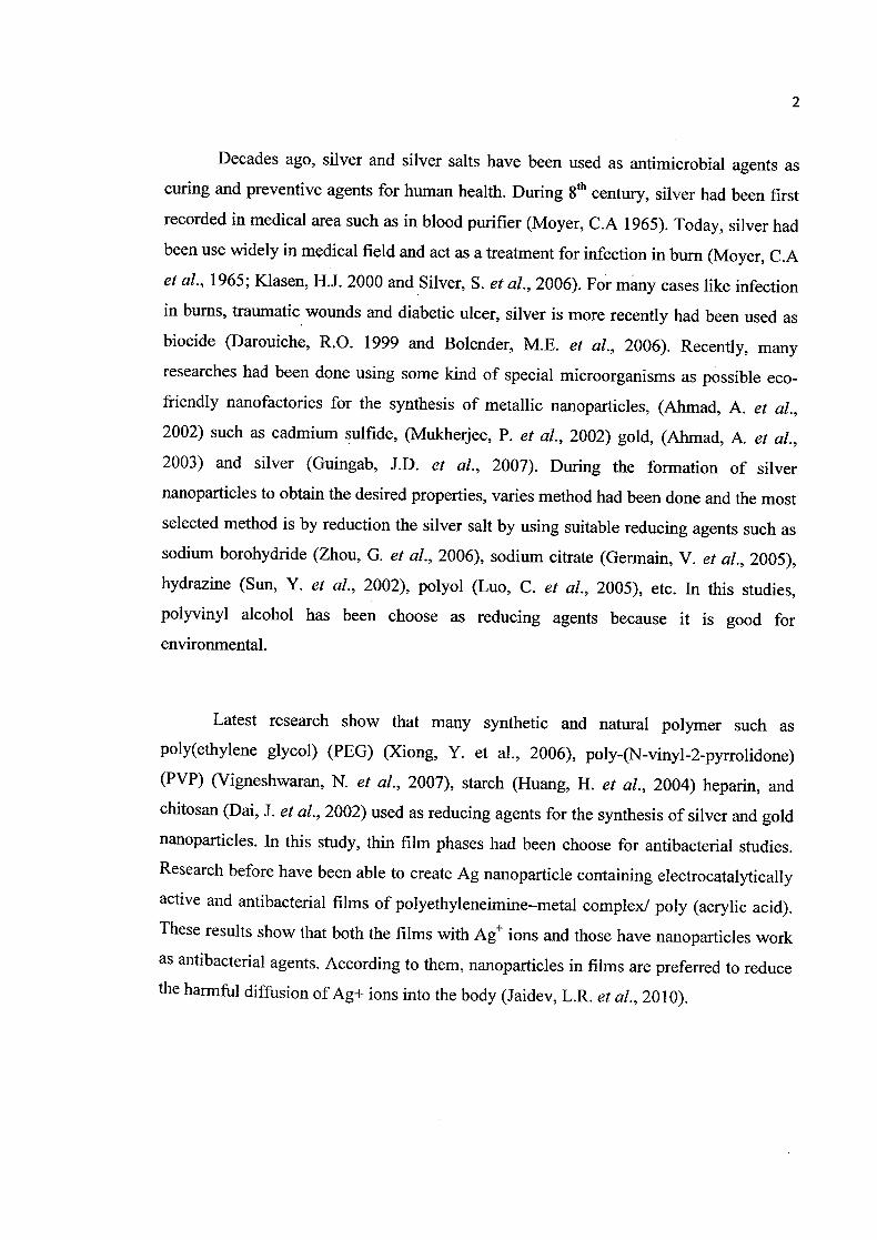
Decades ago, silver and silver salts have been used as antimicrobial agents as
curing and preventive agents for human health. During 8th century, silver had been first
recorded in medical area such as in blood purifier (Moyer, C.A 1965). Today, silver had
been use widely in medical field and act as a treatment for infection in burn (Moyer, C.A
et al., 1965; Kiasen, H. J. 2000 and Silver, S. et al., 2006). For many cases like infection
in bums, traumatic wounds and diabetic ulcer, silver is more recently had been used as
biocide (Darouiche, R.O. 1999 and Bolender, M.E. et al., 2006). Recently, many
researches had been done using some kind of special microorganisms as possible eco-
friendly nanofactories for the synthesis of metallic nanoparticles, (Ahmad, A. et al.,
2002) such as cadmium sulfide, (Mukherjee, P. et al., 2002) gold, (Ahmad, A. et al.,
2003) and silver (Guingab, J.D. et al., 2007). During the formation of silver
nanoparticles to obtain the desired properties, varies method had been done and the most
selected method is by reduction the silver salt by using suitable reducing agents such as
sodium borohydride (Zhou, G. et al., 2006), sodium citrate (Germain, V. et al., 2005),
hydrazine (Sun, Y. et al., 2002), polyol (Luo, C. et al., 2005), etc. In this studies,
polyvinyl alcohol has been choose as reducing agents because it is good for
environmental.
Latest research show that many synthetic and natural polymer such as
poly(ethylene glycol) (PEG) (Xiong, Y. et al., 2006), poly-(N-vinyl-2-pyrrolidone)
(PVP) (Vigneshwaran, N. et al., 2007), starch (Huang, H. et al., 2004) heparin, and
chitosan (Dai, J. et al., 2002) used as reducing agents for the synthesis of silver and gold
nanoparticles. In this study, thin film phases had been choose for antibacterial studies.
Research before have been able to create Ag nanoparticle containing electrocatalytically
active and antibacterial films of polyethyleneimine—metal complex! poly (acrylic acid).
These results show that both the films with Ag ions and those have nanoparticles work
as antibacterial agents. According to them, nanoparticles in films are preferred to reduce
the harmful diffusion of Ag+ ions into the body (Jaidev, L.R. etal., 2010).

3
Various method had been used for characterization the nanoparticles such as
Ultraviolet spectroscopy analysis, thermal electron microscope analysis and X-ray
diffraction analysis (Huang, Z.M. et al., 2003). Spin coating is used for the formation of
thin film. By using electrospinning, various type of polymer had been converted into
fibers (Li, X. et al., 2004). Some research has been done using hydroxyl cellulose as a
stearic stabilizer such as in the synthesis of nanocrystalline ceramic oxide powders
(Shukia, S. et al., 2002&2003).
1.2 PROBLEM STATEMENT
Most of the research in nanoparticles involves toxic solvents which are harmful to
the environment. In this study, the research approach no toxic solvent, green technology
and environmental friendly.
1.3 RESEARCH OBJECTIVES
The main objectives of this research are:
a) To study the efficiency of polymer nanofibers of PVAISilver nanoparticles
in antibacterial studies.
b) To obtain the optimum concentration of PVA and silver nitrate in killing the
bacteria.
C) To identify the effect of heating the polymer nanofibers in antibacterial studies.

4
1.4 SCOPE OF STUDY
In this research, the effectiveness of silver nanoparticles (Ag NP) using PVA
solution in antibacterial studies have been focused. The optimum concentration of silver
nanoparticles in curing is focused. These are the things that are very important because
during the experimental work, the exact amount of concentration used have to be recorded
to show the effectiveness. Other than that, we want to identify the effect of heating
towards polymer nanofibers in the antibacterial studies.
1.5 SIGNIFICANCE OF STUDY
Nanofibers of polyvinyl alcohol embedded with silver nanoparticles for
antibacterial studies will give many benefits and advantage in many fields especially in
medicine fields. Chemistry has a very strong bond in every single life even though
peoples do not notice that. Significance from this study, even though we use a very small
amount of curing but the result will be show an amazing curing. Some creative touch in
nanoparticles preparation and play with many concentrations will discovered a lot of
mystery. This study will show the effectiveness of silver nanoparticles embedded with
polyvinyl alcohol in antibacterial studies by using optimum concentration and also the
effect of heating towards antibacterial studies.

CHAPTER 2
LITERATURE REVIEW
2.1 OVERVIEW OF NANOPARTICLES
The term 'nanoparticles' is used to describe particles with the size in the range of
1 to 100 nm at least in one of the three dimensions. In this range, the physical, chemical
and biological properties are change in fundamental ways from the properties
corresponding to the bulk material (Revathi, J. et al., 2009). Generally, there are
designed with surface modification tailored to meet the need of specific applications that
they are to be used for. The large specific surface area of nanoparticles is the origin of a
number of their unique applications. High surface areas give strong interactions between
nanoparticles and the solid matrix in which they may be incorporated. Nanoparticles
can be synthesized by a variety of methods using solid, gas and liquid phase processes.
(Ramanathan, N. et al., 2008). Discoveries in the past decade have demonstrated that the
electromagnetic optical and catalytic properties of noble-metal nanoparticles such as
gold, silver and platinum, are strongly influenced by shape and size. (Daniel, M.C. et al., 2004).

6
2.1.1 Silver Nanoparticles (AgNP)
Among all metal, silver is more interesting due to its application and properties.
Silver acts as the stabilizer and reducing agent. Silver, and silver-based compounds, is
highly antimicrobial thanks to its antiseptic properties to several species of bacteria,
including the common kitchen microbe, E. coil. Silver nanoparticles interact with the
outer membrane of bacteria, causing structural changes that lead to degradation and
eventually death of the microbe. Silver nanoparticles are one of the most commonly
utilized nanomaterials due to their anti-microbial properties, high electrical conductivity,
and unique optical properties. Silver nanoparticles have so many applications like
electronic field, catalysis and wound dressing, but the most important one is antibacterial
studies (Resham, B. et al., 2008).
Silver has such advantages as broad spectrum antibacterial studies activity, non-
toxicity to human cells and long lasting effect (Yunarova, T. et al., 2003). Silver
nanoparticles used in this study were prepared by the reduction of silver nitrate and
characterized using UV—Visible spectroscopy (UV—Vis) and transmission electron
microscopy (TEM). Transmission electron microscopy (TEM) allows to directly image
the lattice structure of nanoparticles in the order of a few nanometers (Yamamuro, S. et
al., 2002) as well as to obtain diffraction data, amplitudes and phases of nanoparticle
structures. Studies by TEM can be used to determine the behavior and self-assembly of
nanoparticles under external influences such as magnetic fields (Ahniyaz, A. et al., 2007).
Furthermore, elemental analysis of nanoparticles can be made using energy
dispersive X-ray spectroscopy (EDS), and modern transmission electron microscopes
are equipped with tools to perform elemental mapping and analysis using incident probe
sizes in the order of a few nanometers in diameter (Fadeel, B. et al., 2010) . In order to
study the conversion of AgNO3 to Ag in the PVP nanofibres during the heat treatment,
UV—visible absorbance spectroscopy was used; specifically, this can be used to track the
formation of silver nanostructures of various dimensions, which exhibit surface plasmon

7
resonance (SPR) bands at different frequencies (Hernandez, E.A. et al., 2005). Silver
nanoparticles also can be prepared by a UV-irradiation photo reduction technique
(Ershov, B.G. & Henglein, A. 1993).
2.1.2 Nanoeffects of Silver Particles
About 20 - 50 000 silver atoms had been found in silver nanoparticles. The size
of atoms usually smaller than 100 nm(Chen, X. et al., 2008). Basically, total surface
energy is lower than single crystal because lower energy faces at the expense of an
internal strain. For silver structures, more { 111 } facets with the lowest surface energy
and planes surfaces are preferred when the size of particles decreased to nanosize. This
figure shows the highest atomic density. The morphology of silver nanoparticles is
favored with high atom density facets such as { 111 }. Silver nanoparticles with this type
of facets will interact with bacteria's thiol group which are contains sulfur hydrogen
bond. Elechiguerra et al had done a study and reported that only special size of silver
particles can bound to human within the range 1-10 nm. The strongest bacterial activity
occurs at basal planes compare to spherical or rod shaped. The direct interaction
between silver particles and bacteria only occur at the diameter of about 1-10 nm.
Figure 2.1: Face centered cubic unit cell of silver
Source: Cao, H. and Liu, X. (2010)

8
2.1.3 AgNP in Term of Biocompatibiity
Biocompatibility means the ability to coexist with living organism without
harming them. In 1996 studied (Ratner et al., 1996), biocompatibility is "the ability of a
material to perform with an appropriate host response in a specific application." Silver is
very great and excellent bactericidal metal because it is non-toxic to human and animal
cells but highly toxic to bacteria such as E.coli and S. Aureus (Klueh, U. et al., 2000 and Zhou, G. et al., 1998). This silver nanoparticles act as biocidal agents. (Shan-hui, H. et
al., 2010), Silver (Ag) is regarded as one of the noble metals with high biocompatibility.
Both nano Ag and nano Au were reported to modify the microphase separation on the
surface of H 12MDI-based PU and enhance the biostability and biocompatibility in vitro
and in vivo, with nano Ag being more effective (Chou, C.W. et al., 2008).
It is believed that the silver nanoparticles having phosphorylcholine groups to
enhance their biocompatibility and intracellular uptake and having rhodamine dye on its
surface as a fluorescent probe could be a promising biomedical material (Yi-Chang, C.
et al., 2007) search approaches green technology and environmental friendly because
water-base system is used during the formation of silver nanoparticles. Water is non-
toxicity and does not harmful to the environment and human health. Kulshreshtha, S.N
(1998), water is widely used in chemical reactions as a solvent or reactant and less
commonly as a solute or catalyst. In inorganic reactions, water is a common solvent,
dissolving many ionic compounds. In organic reactions, it is not usually used as a
reaction solvent, because it does not dissolve the reactants well and is amphoteric (acidic
and basic) and nucleophilic.
9
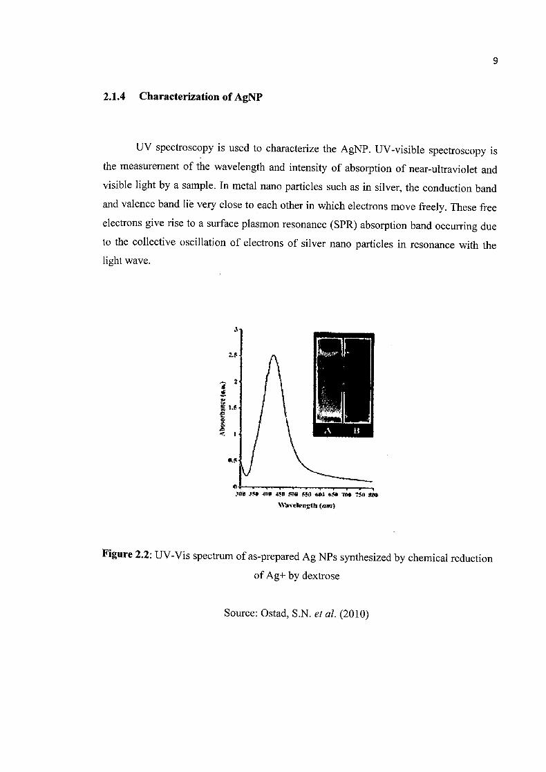
2.1.4 Characterization of AgNP
UV spectroscopy is used to characterize the AgNP. UV-visible spectroscopy is
the measurement of the wavelength and intensity of absorption of near-ultraviolet and
visible light by a sample. In metal nano particles such as in silver, the conduction band
and valence band Be very close to each other in which electrons move freely. These free
electrons give rise to a surface plasmon resonance (SPR) absorption band occurring due
to the collective oscillation of electrons of silver nano particles in resonance with the
light wave.
7t! S1 Rm
(flft;a)
Figure 2.2: UV-Vis spectrum of as-prepared Ag NPs synthesized by chemical reduction
of Ag+ by dextrose
Source: Ostad, S.N. et al. (2010)

10
2.2 CHARACTERIZATION OF POLYVINYL (ALCOHOL) (PVA)
PVA is a poly hydroxyl polymer, which is water soluble and has excellent fiber
forming ability, biocompatibility, chemical resistance and biodegradability (Lin, W.C. et
A, 2006 and Krevelen, D.W.V. et al., 1975). It is also known as a very good stabilizer
for some metals particles (Longenberger, L. et a!, 1995). In present works, PVA have an
ability act as nature reducing agents. In chemical synthesis of nanoparticles, polyvinyl
alcohol had been used as a stabilizer. Polyvinyl alcohol is a very good water soluble
polymer and suitable for nanofibers preparations (Koski, A. et al, 2004).
PVA also have been known as potential biodegradable polymeric materials for
environmental application and also for biomedical (Chen et al., 1997). It also exhibits
good mechanical properties; chemical resistance, water soluble and highly crystalline
(Fussell, G. et al., 1998). Table 2.1 generally shows about the chemical identity and
physical properties of PVA.
Table 2.1: General chemical identity and physical properties of polyvinyl alcohol
CAS No, 9002.89.5 USP1NF
Molecular weight* 30,000200000 hHandhoth Pharm. Excip. Structural formula* ciapan. Iharm. Excip, Dir. Empirical formula* CAO)nC4H60. Japan. Pharm. Excip. Dir. Physical appearance odorless, white to cream-colored granular powder Handbook Pharm. Excip. Specific gm',ity 1,19-131 Handbook ITharm. Excip. Solubility Insoluble in aliphatic and aromatic hydrocarbons, esters, ketones, and oils; water soluble. Handbook Pharm. F_cip
USPjNF, 2000, United States Pha opoda (24) and National Formulary (19). pp. 1352-1353. U.S. Pharmacopeial Convention, Rockville, MD.
Handbook of Pharmaceutical Excipients, 1994. second od. A. Wade, P.J. Weller (F.ds). pp. 383-384. American Pharmaecutieul Aseiation, Washington, DC.
The Japanese Pharmaceutical Excipients Directory, 1996. Monograph on Polyvinyl Alcohol, p.355. * \'ariablc based on PVA grade.
Source: DeMerlis, C.C. et al. (2003)
11
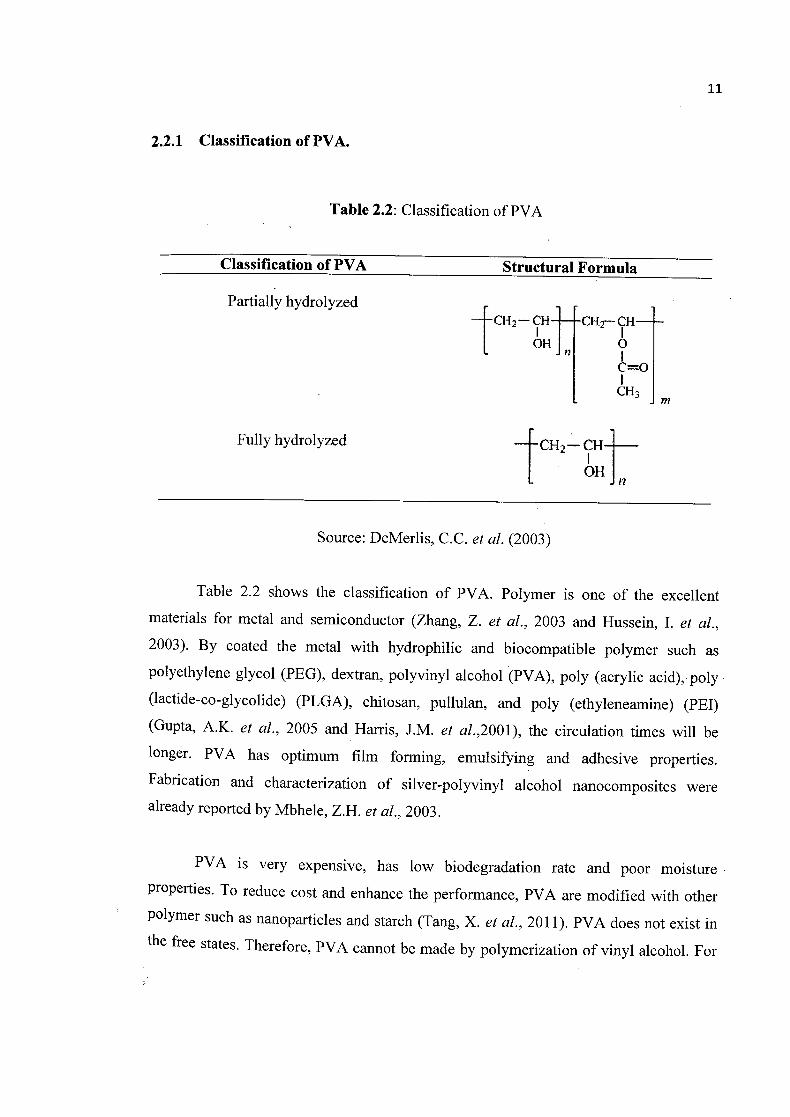
2.2.1 Classification of PVA.
Table 2.2: Classification of PVA
Classification of PVA Structural Formula
OH 4)
Partially hydrolyzedCM1 CHr- CH—L-
=OI
CU3 j
Fully hydrolyzed CII
n [ OH
Source: DeMerlis, C.C. et al. (2003)
Table 2.2 shows the classification of PVA. Polymer is one of the excellent
materials for metal and semiconductor (Zhang, Z. et al., 2003 and Hussein, I. et al., 2003). By coated the metal with hydrophilic and biocompatible polymer such as
polyethylene glycol (PEG), dextran, polyvinyl alcohol (PVA), poly (acrylic acid), poly
(lactide-co-glycolide) (PLGA), chitosan, pullulan, and poly (ethyleneamine) (PEI)
(Gupta, A.K. et al., 2005 and Harris, J.M. et al.,2001), the circulation times will be
longer. PVA has optimum film forming, emulsifying and adhesive properties.
Fabrication and characterization of silver-polyvinyl alcohol nanocomposites were
already reported by Mbhele, Z.H. ci' al., 2003.
PVA is very expensive, has low biodegradation rate and poor moisture
properties. To reduce cost and enhance the performance, PVA are modified with other
Polymer such as nanoparticles and starch (Tang, X. ci' al., 2011). PVA does not exist in
the free states. Therefore PVA cannot be made by polymerization of vinyl alcohol. For

12
preparation, they used partial or complete hydrolysis of polyvinyl acetate to remove the
acetate group. Since 1930's, PVA is known as biodegradable synthetic polymer. The
problem here is the higher cost compared to other polymer such as polypropylene and
polyethylene. The application for PVA is very broad. Usually this polymer had been use
for water soluble packaging fills, paper adhesives, textiles and paper coating (Chang, J.
et al., 2003 and Ibrahim, M. et al., 2010). This is because of the excellent film forming,
emulsifying and adhesive properties of PVA itself. High energy cost of evaporating
water needs for this polymer because of it water soluble properties. Table 2.3 show the
effect of molecular weight and hydrolysis level on the physical properties of PVA.
Increased viscosity Increased solubility Increased tensile strength Increased flexibtlity Increased water resistance Increased water sensitivity Increased solvent resistance Increased ease of solvation Increased adhesive strength
MJLECULAR
deaearn WEIGHTg increasing
HYDROLYSIS %
Increased solubility Increased flexibility Increased water sensitivity Increased adhesionto hydrophobic surfaces
Increased water resistance Increased tense strength Increased solvent resistance Increased adhesion to hydrophilic surfaces
Figure 2.3: Effect of molecular weight and hydrolysis level on the physical properties of
PVA
Source: Sekisui Specialty Chemicals America, U/C, (2010)

13
2.3 CHARACTERIZATION OF POLYMER NANOFIBERS
The principle of electrospinning method is very simple. The fiber form when the
electrostatic field stretches the polymer solution into fiber. But, the process is very
difficult to control. Usually, the polymer nanofibers can be characterized based on
morphology, mechanical, thermal and chemical properties.
2.3.1 Morphology Characterization
The quality of the fibers is typically inconsistent, for example, the fiber
deposition may be uneven or the distribution of fiber diameter. Scanning electron
microscope and transmission electron microscope is the instrument that can be used to
measure the diameter. But, both of these instruments are not too precise compare to
atomic force microscope (AFM) (Srinivasan, G. et al., 1995 and Li, W.J. et al., 2002).
For this instrument, a very sharp probe moves over the surfaces. The tip geometry in the
AFM make the fiber looks larger than the actual (Jaeger, R. et al., 1996).
2.3.2 Mechanical, Chemical and Thermal Characterization
While decreasing the diameter, the tensile strength will increase. The contact
area between filler and polymer increase due to the increase in fiber surface area/volume
ratio. Therefore, the flexibility of fiber will increase (Paul, D.R. et al., 2008). AFM is
also used to measure the mechanical properties of polymer nanofibers. To increase the
mechanical properties of polymer nanofibers, inorganic fiber such as glass and carbon
fibers and also aromatic organic fibers had been used in previous study.