Poly-ion Complex of Chondroitin Sulfate and Spermine and ... · PDF filePoly-ion Complex of...
9
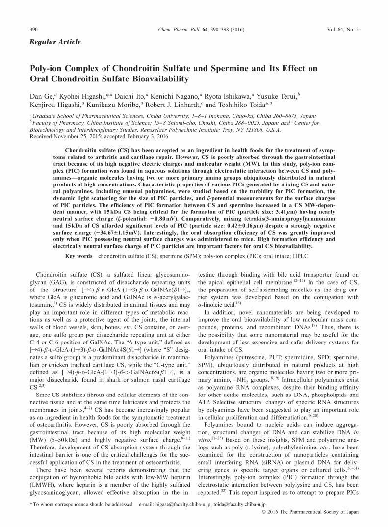
390 Vol. 64, No. 5 © 2016 The Pharmaceutical Society of Japan Chem. Pharm. Bull. 64, 390–398 (2016) Regular Article Poly-ion Complex of Chondroitin Sulfate and Spermine and Its Effect on Oral Chondroitin Sulfate Bioavailability Dan Ge, a Kyohei Higashi,* ,a Daichi Ito, a Kenichi Nagano, a Ryota Ishikawa, a Yusuke Terui, b Kenjirou Higashi, a Kunikazu Moribe, a Robert J. Linhardt, c and Toshihiko Toida* ,a a Graduate School of Pharmaceutical Sciences, Chiba University; 1–8–1 Inohana, Chuo-ku, Chiba 260–8675, Japan: b Faculty of Pharmacy, Chiba Institute of Science; 15–8 Shiomi-cho, Choshi, Chiba 288–0025, Japan: and c Center for Biotechnology and Interdisciplinary Studies, Rensselaer Polytechnic Institute; Troy, NY 121806, U. S.A. Received November 25, 2015; accepted February 3, 2016 Chondroitin sulfate (CS) has been accepted as an ingredient in health foods for the treatment of symp- toms related to arthritis and cartilage repair. However, CS is poorly absorbed through the gastrointestinal tract because of its high negative electric charges and molecular weight (MW). In this study, poly-ion com- plex (PIC) formation was found in aqueous solutions through electrostatic interaction between CS and poly- amines—organic molecules having two or more primary amino groups ubiquitously distributed in natural products at high concentrations. Characteristic properties of various PICs generated by mixing CS and natu- ral polyamines, including unusual polyamines, were studied based on the turbidity for PIC formation, the dynamic light scattering for the size of PIC particles, and ζ-potential measurements for the surface charges of PIC particles. The efficiency of PIC formation between CS and spermine increased in a CS MW-depen- dent manner, with 15 kDa CS being critical for the formation of PIC (particle size: 3.41 μm) having nearly neutral surface charge (ζ-potential: −0.80 mV). Comparatively, mixing tetrakis(3-aminopropyl)ammonium and 15 kDa of CS afforded significant levels of PIC (particle size: 0.42±0.16 μm) despite a strongly negative surface charge ( −34.67±1.15 mV). Interestingly, the oral absorption efficiency of CS was greatly improved only when PIC possessing neutral surface charges was administered to mice. High formation efficiency and electrically neutral surface charge of PIC particles are important factors for oral CS bioavailability. Key words chondroitin sulfate (CS); spermine (SPM); poly-ion complex (PIC); oral intake; HPLC Chondroitin sulfate (CS), a sulfated linear glycosamino- glycan (GAG), is constructed of disaccharide repeating units of the structure [ →4)-β-D-GlcA-(1 →3)-β-D-GalNAc( β1 →] n , where GlcA is glucuronic acid and GalNAc is N-acetylgalac- tosamine. 1) CS is widely distributed in animal tissues and may play an important role in different types of metabolic reac- tions as well as a protective agent of the joints, the internal walls of blood vessels, skin, bones, etc. CS contains, on aver- age, one sulfo group per disaccharide repeating unit at either C-4 or C-6 position of GalNAc. The “A-type unit,” defined as [ →4)-β-D-GlcA-(1 →3)-β-D-GalNAc4S( β1 →] (where “S” desig- nates a sulfo group) is a predominant disaccharide in mamma- lian or chicken tracheal cartilage CS, while the “C-type unit,” defined as [ →4)-β-D-GlcA-(1 →3)-β-D-GalNAc6S( β1 →], is a major disaccharide found in shark or salmon nasal cartilage CS. 2,3) Since CS stabilizes fibrous and cellular elements of the con- nective tissue and at the same time lubricates and protects the membranes in joints, 4–7) CS has become increasingly popular as an ingredient in health foods for the symptomatic treatment of osteoarthritis. However, CS is poorly absorbed through the gastrointestinal tract because of its high molecular weight (MW) (5–50 kDa) and highly negative surface charge. 8–11) Therefore, development of CS absorption system through the intestinal barrier is one of the critical challenges for the suc- cessful application of CS in the treatment of osteoarthritis. There have been several reports demonstrating that the conjugation of hydrophobic bile acids with low-MW heparin (LMWH), where heparin is a member of the highly sulfated glycosaminoglycan, allowed effective absorption in the in- testine through binding with bile acid transporter found on the apical epithelial cell membrane. 12–15) In the case of CS, the preparation of self-assembling micelles as the drug car- rier system was developed based on the conjugation with α-linoleic acid. 16) In addition, novel nanomaterials are being developed to improve the oral bioavailability of low molecular mass com- pounds, proteins, and recombinant DNAs. 17) Thus, there is the possibility that some nanomaterial may be useful for the development of less expensive and safer delivery systems for oral intake of CS. Polyamines (putrescine, PUT; spermidine, SPD; spermine, SPM), ubiquitously distributed in natural products at high concentrations, are organic molecules having two or more pri- mary amino, –NH 2 groups. 18,19) Intracellular polyamines exist as polyamine–RNA complexes, despite their binding affinity for other acidic molecules, such as DNA, phospholipids and ATP. Selective structural changes of specific RNA structures by polyamines have been suggested to play an important role in cellular proliferation and differentiation. 18,20) Polyamines bound to nucleic acids can induce aggrega- tion, structural changes of DNA and can stabilize DNA in vitro. 21–25) Based on these insights, SPM and polyamine ana- logs such as poly ( L-lysine), polyethylenimine, etc. , have been examined for the construction of nanoparticles containing small interfering RNA (siRNA) or plasmid DNA for deliv- ering genes to specific target organs or cultured cells. 26–31) Interestingly, poly-ion complex (PIC) formation through the electrostatic interaction between polylysine and CS, has been reported. 32) This report inspired us to attempt to prepare PICs * To whom correspondence should be addressed. e-mail: [email protected]; [email protected]
Transcript of Poly-ion Complex of Chondroitin Sulfate and Spermine and ... · PDF filePoly-ion Complex of...
390 Vol. 64, No. 5
© 2016 The Pharmaceutical Society of Japan
Chem. Pharm. Bull. 64, 390–398 (2016)
Regular Article
Poly-ion Complex of Chondroitin Sulfate and Spermine and Its Effect on Oral Chondroitin Sulfate Bioavailability
Dan Ge,a Kyohei Higashi,*,a Daichi Ito,a Kenichi Nagano,a Ryota Ishikawa,a Yusuke Terui,b Kenjirou Higashi,a Kunikazu Moribe,a Robert J. Linhardt,c and Toshihiko Toida*,a
a Graduate School of Pharmaceutical Sciences, Chiba University; 1–8–1 Inohana, Chuo-ku, Chiba 260–8675, Japan: b Faculty of Pharmacy, Chiba Institute of Science; 15–8 Shiomi-cho, Choshi, Chiba 288–0025, Japan: and c Center for Biotechnology and Interdisciplinary Studies, Rensselaer Polytechnic Institute; Troy, NY 121806, U.S.A.Received November 25, 2015; accepted February 3, 2016
Chondroitin sulfate (CS) has been accepted as an ingredient in health foods for the treatment of symp-toms related to arthritis and cartilage repair. However, CS is poorly absorbed through the gastrointestinal tract because of its high negative electric charges and molecular weight (MW). In this study, poly-ion com-plex (PIC) formation was found in aqueous solutions through electrostatic interaction between CS and poly-amines—organic molecules having two or more primary amino groups ubiquitously distributed in natural products at high concentrations. Characteristic properties of various PICs generated by mixing CS and natu-ral polyamines, including unusual polyamines, were studied based on the turbidity for PIC formation, the dynamic light scattering for the size of PIC particles, and ζ-potential measurements for the surface charges of PIC particles. The efficiency of PIC formation between CS and spermine increased in a CS MW-depen-dent manner, with 15 kDa CS being critical for the formation of PIC (particle size: 3.41 µm) having nearly neutral surface charge (ζ-potential: −0.80 mV). Comparatively, mixing tetrakis(3-aminopropyl)ammonium and 15 kDa of CS afforded significant levels of PIC (particle size: 0.42±0.16 µm) despite a strongly negative surface charge (−34.67±1.15 mV). Interestingly, the oral absorption efficiency of CS was greatly improved only when PIC possessing neutral surface charges was administered to mice. High formation efficiency and electrically neutral surface charge of PIC particles are important factors for oral CS bioavailability.
Key words chondroitin sulfate (CS); spermine (SPM); poly-ion complex (PIC); oral intake; HPLC
Chondroitin sulfate (CS), a sulfated linear glycosamino-glycan (GAG), is constructed of disaccharide repeating units of the structure [→4)-β-D-GlcA-(1→3)-β-D-GalNAc(β1→]n, where GlcA is glucuronic acid and GalNAc is N-acetylgalac-tosamine.1) CS is widely distributed in animal tissues and may play an important role in different types of metabolic reac-tions as well as a protective agent of the joints, the internal walls of blood vessels, skin, bones, etc. CS contains, on aver-age, one sulfo group per disaccharide repeating unit at either C-4 or C-6 position of GalNAc. The “A-type unit,” defined as [→4)-β-D-GlcA-(1→3)-β-D-GalNAc4S(β1→] (where “S” desig-nates a sulfo group) is a predominant disaccharide in mamma-lian or chicken tracheal cartilage CS, while the “C-type unit,” defined as [→4)-β-D-GlcA-(1→3)-β-D-GalNAc6S(β1→], is a major disaccharide found in shark or salmon nasal cartilage CS.2,3)
Since CS stabilizes fibrous and cellular elements of the con-nective tissue and at the same time lubricates and protects the membranes in joints,4–7) CS has become increasingly popular as an ingredient in health foods for the symptomatic treatment of osteoarthritis. However, CS is poorly absorbed through the gastrointestinal tract because of its high molecular weight (MW) (5–50 kDa) and highly negative surface charge.8–11) Therefore, development of CS absorption system through the intestinal barrier is one of the critical challenges for the suc-cessful application of CS in the treatment of osteoarthritis.
There have been several reports demonstrating that the conjugation of hydrophobic bile acids with low-MW heparin (LMWH), where heparin is a member of the highly sulfated glycosaminoglycan, allowed effective absorption in the in-
testine through binding with bile acid transporter found on the apical epithelial cell membrane.12–15) In the case of CS, the preparation of self-assembling micelles as the drug car-rier system was developed based on the conjugation with α-linoleic acid.16)
In addition, novel nanomaterials are being developed to improve the oral bioavailability of low molecular mass com-pounds, proteins, and recombinant DNAs.17) Thus, there is the possibility that some nanomaterial may be useful for the development of less expensive and safer delivery systems for oral intake of CS.
Polyamines (putrescine, PUT; spermidine, SPD; spermine, SPM), ubiquitously distributed in natural products at high concentrations, are organic molecules having two or more pri-mary amino, –NH2 groups.18,19) Intracellular polyamines exist as polyamine–RNA complexes, despite their binding affinity for other acidic molecules, such as DNA, phospholipids and ATP. Selective structural changes of specific RNA structures by polyamines have been suggested to play an important role in cellular proliferation and differentiation.18,20)
Polyamines bound to nucleic acids can induce aggrega-tion, structural changes of DNA and can stabilize DNA in vitro.21–25) Based on these insights, SPM and polyamine ana-logs such as poly (L-lysine), polyethylenimine, etc., have been examined for the construction of nanoparticles containing small interfering RNA (siRNA) or plasmid DNA for deliv-ering genes to specific target organs or cultured cells.26–31) Interestingly, poly-ion complex (PIC) formation through the electrostatic interaction between polylysine and CS, has been reported.32) This report inspired us to attempt to prepare PICs
* To whom correspondence should be addressed. e-mail: [email protected]; [email protected]
Vol. 64, No. 5 (2016) 391Chem. Pharm. Bull.
by mixing CS and natural polyamines in aqueous solutions and investigate their effect on CS oral absorption. Since poly-amines are found at high concentrations in natural foods, they are considered to have extremely low toxicity.
In the present study, the efficient formation of PICs from CS and polyamines has been accomplished. The dependence on the mixing ratio between CS and polyamines, MW of CS, nature of polyamines, and the pH for PIC formation have been investigated. The preparation of PICs, possessing nearly neu-tral surface charge, an important factor for the oral absorption of CS, was established from 15 kDa of CS and SPM. These results suggest that PIC generated by the interaction of CS and polyamines might represent a promising core structure for the development of CS delivery system in gastrointestinal tract.
ExperimentalChemicals and Materials Chondroitin sulfate-A (average
MW: 15 kDa, 14.0% of ΔDi-0S, 49.8% of ΔDi-4S, 36.1% of ΔDi-6S) from bovine tracheal cartilage was purchased from Shin-Nippon Yakugyo Co., Ltd. (Tokyo, Japan). Chondroitin sulfate-C (average MW: 46 kDa, 1.77% of ΔDi-0S, 13.7% of ΔDi-4S, 70.8% of ΔDi-6S, 2.89% of ∆Di-diSE, 10.8% of ∆Di-diSD) from shark cartilage, chondroitinase ABC (Proteus vulgaris EC 4.2.2.4), the unsaturated disaccharides (∆Di-0S, ∆Di-4S, ∆Di-6S, ∆Di-diSE, ∆Di-diSB, ∆Di-diSD, and ∆Di-triS) were purchased from Seikagaku Corp. (Tokyo, Japan). Fractioned CS standard samples (MW 6 kDa, 13 kDa, 31 kDa, 44 kDa) for MW determination were prepared as described previously.33) Spermine tetrahydrochloride (SPM·4HCl) was obtained from Nacalai Tesque Inc. (Kyoto, Japan). Titanium dioxide (anatase type, average particle size, 50 µm) was ob-tained from Wako Pure Chemical Industries, Ltd. (Osaka, Japan). All other chemicals were of analytical reagent grade.
MethodsPIC FormationCS and SPM were separately dissolved in double-distilled
water, and then, equivalent volumes of CS solution and SPM solution were fully mixed with one another to generate PIC. PIC formation efficiency was evaluated by turbidimetric mea-surement at 600 nm wavelength, namely the determination of % transmittance (T), using Thermo Scientific SPECTONIC 20D+ digital spectrophotometer.
The Effect of pH Value on the PIC Formation EfficiencyA 100 µL aliquot of 50 mM SPM aqueous solution was fully
mixed with the equal volume of 40 mg/mL of CS, and then the suspensions containing PIC were separately adjusted to pH value 1–9 using 1 M HCl or 1 M NaOH. PIC fraction, obtained by the centrifugation at 6000×g for 5 min, was suspended and precipitated twice, using 1 mL of pH conditioned H2O to remove the free SPM and CS that did not form PIC. Since PIC formation was unstable under basic conditions (pH value above 9), 400 µL aliquot of 0.2 M NaOH was added to the PIC pellets to dissociate CS and SPM from each other. Free SPM was separated from CS using Amicon® Ultra 10 K spin columns by centrifugation at 6000×g for 30 min at 4°C, and then the separated CS and SPM samples were quantitatively analyzed by HPLC.
HPLCThe HPLC system for CS quantitative analysis comprised
two DP-8020 Pumps (Tosoh Corporation, Tokyo, Japan) as the gradient eluent pumps and an NP-FX(II)-1 MINICHEMI
Pump (Nihon Seimitsu Kagaku Co., Ltd., Tokyo, Japan) as the post-column reaction solution pump. An AS-8020 (Tosoh Corporation, Tokyo, Japan) Auto Sampler, a CO631A (GL Sciences Inc., Tokyo, Japan) Column Oven, a DB-5 (Shi-mamura Instrument Co., Tokyo, Japan) Dry Reaction Bath, an FP-1520S (Jasco Corporation, Tokyo, Japan) Intelligent Fluorescence Detector and a DGU-12A (Shimadzu Corpora-tion, Kyoto, Japan) Degasser were also applied to this HPLC analysis system. Here, an Asahipak NH2P-50 4E column (4.6 mm i.d.×250 mm, 5 µm, Showa Denko K.K., Tokyo, Japan) was used along with a guard column Asahipak NH2P-50G 4A (4.6 mm i.d.×10 mm, 5 µm, Showa Denko K.K.). The mobile phases: (A) 0.1 M sodium carbonate buffer (pH 10.0) containing 0.1 M NaCl and (B) 0.1 M sodium carbonate buffer (pH 10.0) containing 1.0 M NaCl were eluted according to the following gradient program: 0–20 min (0–100% B), 20–25 min (100% B), 25–27 min (100–0% B), 27–35 min (0% B) (for ini-tialization). And the flow rate was constantly maintained at 0.5 mL/min. The post-column reaction reagents were 50 mM guanidine and 1.0 M NaOH and the flow rates were both main-tained at 0.25 mL/min. The column was kept at the room tem-perature and the post-column reaction temperature was set at 110°C. The reaction mixture passed the reaction coil (0.5 mm i.d.×10 m), fully heated in the reactor to accelerate the reac-tion, and then was cooled down in the cooling coil (0.25 mm i.d.×5 m). The detection wavelength was set at 425 nm with 320 nm of excitation. The 0.05, 0.1, 0.5, 1, 2 and 4 µg of CS (MW: 15 kDa) were used as the standard samples to plot the calibration curve for quantitatively determining CS contents in PIC.
Determination of SPM contents was performed according to the method of Igarashi et al.34)
Preparation of LMWCSLMWCS was prepared by photochemical depolymerization
with titanium dioxide (TiO2) as described previously with minor modifications.35,36) The photochemical reaction device (Sen Lights Corporation, Osaka, Japan) consisted of a VG1500 reaction tank with 5 inlets, a light source (high pressure mer-cury lamp HL100 CH-4, 100W), a power source (HB100P-1) and water-cooling jacket JW-1G. The apparatus was connected with a water circulating system to cool the lamp. Briefly, 1 g of CS-A (MW: 15 kDa) was dissolved in 100 mL of H2O with 100 mg of TiO2 in the VG1500 reaction tank. After being exposed to the light for specified time, MW of CS was monitored by gel permeation chromatography (GPC)-HPLC to obtain 8 kDa and 5 kDa of LMWCS.33) After the reaction, the sample was centrifuged at 1500×g for 5 min at 20°C and the supernatant was filtered through 0.45-µm disposable syringe filter unit (Dismic-13HP; Advantec, Tokyo, Japan) to eliminate all of the TiO2 particles. Then the product solution was neu-tralized with NaOH, followed by dialysis against water using Spectra/Por® Dialysis Membrane MWCO: 3500 and ultimately the obtained solution was lyophilized to prepare the LMWCS powder sample. LMWCS with MW of 8 and 5 kDa were gained at the reaction time of 7 and 14 h, respectively (data not shown). Structural verification of LMWCS by 1H-NMR spec-troscopy was performed as described previously.37)
The GPC-HPLC system for MW determination consisted of an L-6000 Pump (Hitachi High-Technologies Corporation, Tokyo, Japan) equipped with a Rheodyne Model-7725 Injector with 20 µL Sample Loop (Rheodyne Inc., Cotati, CA, U.S.A.),
392 Vol. 64, No. 5 (2016)Chem. Pharm. Bull.
an SPD-6A UV Spectrophotometric Detector (Shimadzu Cor-poration) set at 204 nm, and a D-2500 Chromato Integrator (Hitachi High-Technologies Corporation). The MW of CS was determined using gel permeation chromatography with Asa-hipak GF-510 HQ column (7.5 mm i.d.×300 mm, 5 µm, Showa Denko K.K.). Isocratic elution with 10 mM NH4HCO3 was used at a flow rate of 0.3 mL/min. The commercial CS standard samples (average MW: 6, 13, 31 and 44 kDa) were applied to construct the calibration curve using the retention time plotted against the antilogarithm of MW. Thereby, the average MW of the photodegraded CS was calculated from this calibration curve.
Particle Size and ζ-Potential DeterminationThe mean particle sizes of various kinds of PIC samples
were determined by dynamic light scattering (DLS) method on the MICROTRAC 9340 UPA-UT151 (Nikkiso Co., Ltd., Tokyo, Japan) equipped with a semi-conductor laser at the wavelength of 780 nm. The ζ-potential determinations were performed for each PIC samples on the ELS-Z1 (Otsuka Electronics Co., Ltd., Osaka, Japan) at the 60 V of voltage. Double-distilled water was used as the dispersant and the measurements were carried out at 25°C. The average of three measurements was taken as the final characteristic parameter.
Surface Structure AnalysisPIC samples generated from CS and polyamines were char-
acterized by Olympus BX51 upright microscope with Olym-pus DP-70 color camera (Olympus Optical Co., Ltd., Tokyo, Japan) for the particle morphology. Ten microliters of aque-ous PIC sample was poured on a microscope slide to form a sample film for observation.
Absorption of Orally Administered PIC in MiceAll animal experiments were approved by the Institutional
Animal Care and Use Committee of Chiba University and carried out according to the guidelines for Animal Research of Chiba University. The ddY mice (female, weighing 28–30 g, Japan SLC, Inc., Shizuoka, Japan) were housed at 25°C and 55% RH. A 12 h dark/light cycle was maintained throughout. Mice had free access to standard pellet diet (MF; Oriental Yeast Co., Ltd., Tokyo, Japan) and tap water, and were fasted overnight before the experiment. Mice were randomly divided into six groups comprising 6–9 animals in each and respec-tively administered with the following preparations: (i) 40 mg/mL of 15 kDa CS-A alone, (ii) PIC formed by 40 mg/mL of 15 kDa CS-A +50 mM of SPM, (iii) PIC formed by 20 mg/mL of 8 kDa CS-A +50 mM of SPM, (iv) PIC formed by 20 mg/mL of 5 kDa CS-A +20 mM of SPM, (v) PIC formed by 80 mg/mL of 46 kDa CS-C +100 mM of SPM, (vi) PIC formed by 40 mg/mL of 15 kDa CS-A +20 mM of Taa. Each PIC sus-pension was prepared by well mixing CS and polyamines in water solutions utilizing vortex mixer and was immediately orally administered to each group at a single dose of 500 mg CS/kg body weight (ca. 15 mg/each animal). CS alone dosed group (control) was similarly treated with 500 mg CS/kg body weight through the oral route. Blood samples were collected from the tail vein at time intervals of 0, 0.5, 1, 2, 4, 6, 8, 10 h after oral administration and were anticoagulated with 3.2% trisodium citrate solution at the ratio of blood : 3.2% triso-dium citrate=9 : 1. Each collected sample was immediately centrifuged at 800×g for 15 min to obtain the plasma, which was transferred to a new Eppendorf tube and stored in a deep freezer until used. Additionally, inflammation and wound
damage at the digestive system of animals were checked by surgical operation after oral dose of samples.
Determination of CS Contents in Mouse PlasmaMouse plasma (25 µL) was treated with 1.3 µL of 100% tri-
chloroacetic acid (TCA) (final 5% TCA) and incubated at 4°C for 30 min to obtain free CS. After centrifugation at 20000×g for 10 min, 15 µL of supernatant was neutralized with 1.5 µL of 2 M NaOH and diluted with 200 µL of H2O. The buffer of sam-ple solution was firstly exchanged with 0.2 M Tris–acetate (pH 8.0) using Amicon® Ultra 3K spin columns (EMD Millipore Co., MA, U.S.A.), followed by buffer exchange with H2O. The pure sample was collected and lyophilized. The chromato-graphic analyses for the unsaturated disaccharide composition of CS catalyzed by chondroitinase ABC were performed as described previously.37)
Preparation of Unusual PolyaminesTris(3-aminopropyl) amine was purchased from Tokyo
Chemical Industry Co. (Tokyo, Japan). Tetrakis(3-amino-propyl) ammonium, caldopentamine and caldohexamine were chemically synthesized as reported previously.38) The chemical structures of unusual polyamines were illustrated in Fig. 4a and all polyamines used in the study were in the form of their hydrochloride salts. When the unusual polyamine solutions were mixed with 15 kDa of CS to form PICs, the procedures were similar to those described above.
Results and DiscussionPreparation of PIC At first, we examined whether poly-
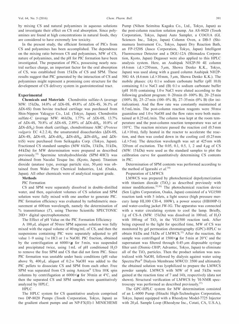
amine spermine (SPM) could aggregate with commercially available CS-A (MW: 15 kDa) prepared from bovine tracheal cartilage. As shown in Fig. 1a, the aqueous solution contain-ing CS (40 mg/mL) or SPM (50 mM) was transparent, how-ever, a turbid white suspension of PIC was generated when an equivalent volume of CS and SPM solution were fully mixed with one another. Furthermore, after approximately 20 min of settling, the turbid white suspension separated into two lay-ers. But after being thoroughly mixed again using a vortex mixer, the deposited PIC sample could be restored back to the original PIC suspension. Morphological characterization of the PIC sample showed that it was of spherical shape and in a mi-crometer size range, correlating closely with the particle size (3.41±0.64 µm) measured by dynamic light scattering (Fig. 1b, Table 1). However, if the PIC sample was settled for relatively longer time under microscope, the PIC particles would easily merge with the adjacent ones to form larger particles and show heterogeneous size, even though the sample was well mixed using vortex mixer previously.
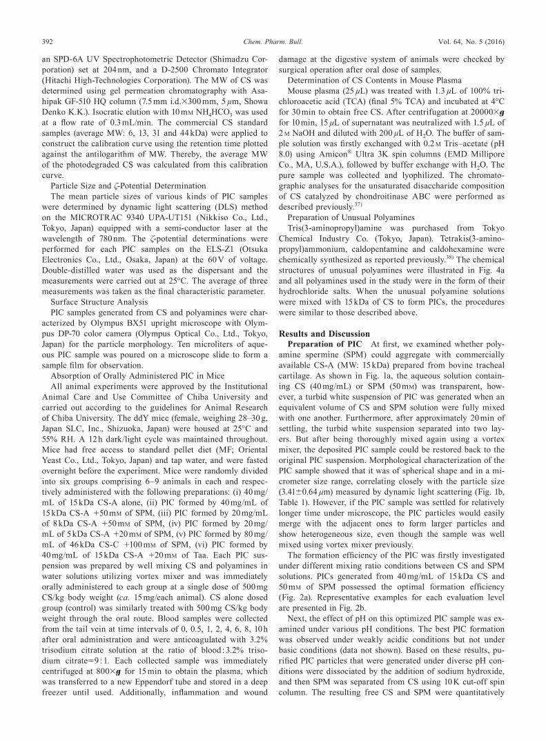
The formation efficiency of the PIC was firstly investigated under different mixing ratio conditions between CS and SPM solutions. PICs generated from 40 mg/mL of 15 kDa CS and 50 mM of SPM possessed the optimal formation efficiency (Fig. 2a). Representative examples for each evaluation level are presented in Fig. 2b.
Next, the effect of pH on this optimized PIC sample was ex-amined under various pH conditions. The best PIC formation was observed under weakly acidic conditions but not under basic conditions (data not shown). Based on these results, pu-rified PIC particles that were generated under diverse pH con-ditions were dissociated by the addition of sodium hydroxide, and then SPM was separated from CS using 10 K cut-off spin column. The resulting free CS and SPM were quantitatively
Vol. 64, No. 5 (2016) 393Chem. Pharm. Bull.
determined by HPLC (Fig. 3). It was observed that the PIC formation efficiency was the highest at pH 6.0. If the MW of CS is uniform, we are able to calculate that approximately 10 molecules of SPM (correspond to 40 positive charges) bound
to one molecule of 15 kDa (dp30) of CS (correspond to 60 negative charges). The observation that weakly acidic condi-tions are most suitable for the PIC formation can be explained by the combination ratio between 40 positive charges from
Fig. 2. Effects of the Molecular Weight of CS and the Mixing Concentration Ratio between CS and SPM on the PIC Formation Efficiency(a) PIC formation efficiency under different condition of mixing ratio between CS (5–46 kDa) and SPM. Aqueous solutions of SPM and CS with different molecular
weights were prepared at different concentrations and well mixed with each other by the equivalent volume to generate PIC. The PIC formation efficiencies were evalu-ated by turbidimetric assay at 600 nm. (b) The representative instances for each level of evaluation. PIC formation was evaluated by measuring the % transmittance (T): −, PIC formation could not be observed (≧90%T); +, PIC formation was poor (30–89%T); ++, PIC formation was good (10–29%T); +++, PIC formation was very good (1–9%T) and after 24 h still exhibited strongly turbid white suspension; ++++, PIC formation was the best of all conditions (<1%T) but PIC precipitated very quickly (within several seconds). The transmittances for the representative examples were 99.2%T (−), 30.6%T (+), 11.8%T (++), 3.2%T (+++) and 0.6%T (++++), respectively.
Table 1. The Sizes and Surface Charges of PIC Particles Generated under the Optimal Mixing Ratio Conditions between CS and Polyamines
PIC formation MV (µm) ζ (mV)
40 mg/mL of 15 kDa CS +50 mM of SPM 3.41±0.64 −0.80±0.2520 mg/mL of 8 kDa CS +50 mM of SPM 2.95±2.52 −9.94±0.5220 mg/mL of 5 kDa CS +20 mM of SPM 1.64±1.61 −1.96±1.3580 mg/mL of 46 kDa CS +100 mM of SPM 4.17±2.92 −11.82±0.8640 mg/mL of 15 kDa CS +20 mM of Cdp 1.65±0.53 −25.19±1.0380 mg/mL of 15 kDa CS +20 mM of Cdh 2.00±1.26 −32.93±0.4740 mg/mL of 15 kDa CS +20 mM of Taa 0.42±0.16 −34.67±1.15
Mean±S.D., n=3.
Fig. 1. Sample Appearances of SPM, CS and PIC(a) Photographs of (I) 40 mg/mL of CS, (II) 50 mM of SPM, (III) PIC, (IV) PIC that separated into two layers after standing for about 20 min. (b) Microscopic image of
PIC generated from 15 kDa of CS and SPM. The scale-bar indicates 20.0 µm.

394 Vol. 64, No. 5 (2016)Chem. Pharm. Bull.
amino groups of SPM (4 amino groups/SPM×10 mol), and 60 negative charges from sulfo and carboxyl groups of CS (2 negative charges/disaccharide unit×dp30). Moreover, this was also consistent with the analysis for the characteristic proper-ties of PIC that the ζ-potential was −0.80±0.25 mV (Table 1).
Effect of the Molecular Size of CS on PIC Formation Efficiency The effect of MW of CS on PIC formation was investigated using 46 kDa, 8 kDa and 5 kDa CS. These CS samples were dissolved in water and the conditions to gener-ate a maximum PIC with SPM were explored. As a result, the optimal conditions for PIC formation were different among the various chain lengths of CSs (Fig. 2a). Moreover, efficiency of PIC formation behaved in a CS MW-dependent manner. Similar to PIC generated from 15 kDa CS, the spherical PIC particles generated from LMWCS were also observed when PIC suspensions were adequately mixed. The size and shape of PIC could not be maintained for long time because adja-cent PIC particles were extremely prone to merging with one another (data not shown). Interestingly, the surface charges of PIC coming from 8 or 46 kDa of CS were significantly differ-ent from those of PIC generated by 15 or 5 kDa of CS despite an almost identical particle size of PIC (Table 1). In contrast with the ζ-potentials of the PICs, coming from 15 and 5 kDa of CS, being near 0 mV, the PICs formed using 8 and 46 kDa of CS showed ζ-potentials around −10 mV.
Effect of Unusual Polyamines on PIC Formation Ef-ficiency It has been known that thermophilic bacteria and archaea are a rich source of a wide variety of unusual polyamines. In particular, an extreme thermophile (eubac-terium), Thermus thermophilus, produces various kinds of unusual polyamines such as caldopentamine (Cdp), caldohex-amine (Cdh), tris(3-aminopropyl) amine (Mitsubishine) and tetrakis(3-aminopropyl) ammonium (Taa)25) (Fig. 4a). Since unusual polyamines, especially Taa, stabilize single-stranded
DNA (ssDNA) and tRNA more effectively than SPM, the effect of the unusual polyamines on PIC formation with 15 kDa of CS was examined. The optimal conditions of PIC formations were different among the unusual polyamines (Fig. 4b). Particularly, Mitsubishine was unable to form PIC with 15 kDa of CS, in contrast to the strong PIC formation ob-served for well mixed by the equivalent volumes of 40 mg/mL of 15 kDa CS and 20 mM Taa (Fig. 4b). Although amount of the resulting PIC particles coming from Taa was almost equal to that from SPM after the PIC suspensions separated into layers, the configuration and the color of the PIC generated by Taa was obviously different (Fig. 4c). Furthermore, it was observed that PIC coming from Taa maintained its particle size for a relatively long time (more than 1 h) and the particles did not quickly merge into one another (Fig. 4d). The particle size of the PIC formed by Taa was the smallest (0.42±0.16 µm) among all the PICs examined, however, Taa afforded the low-est ζ-potential value (−34.67±1.15 mV) (Table 1). The reason for this may be because Taa can bind to CS more effectively than any of the other polyamines.25)
It is thought that the maintenance of PIC particle size is an important factor to improve the absorption efficiency of the PIC in gastrointestinal tract. In general, CS chains may exist as a linear form in neutral pH and the electrostatic repulsion of each sulfonate/carboxyl group of CS chain must affect their coagulation. However, once some parts of negatively charged acidic groups of CS were electronically neutralized by amines, it might be allowed to change their forms in solution as a complex. As mentioned above, PIC formations by the com-bination of CS (5, 8, 15, 46 kDa) and linear polyamines such as SPM, Cdp and Cdh were successfully conducted, however their particles were extremely prone to merging with one an-other. It is conceivable that PIC particles obtained by the electrical attachment of linear polyamines to linear CS in the
Fig. 3. The Contents of SPM and 15 kDa of CS in PIC at Diverse pH ValuesPIC formation under various pH conditions and the determination of CS and SPM in PIC were performed as described under “Experimental.” These experiments were
repeated twice with reproducible results.

Vol. 64, No. 5 (2016) 395Chem. Pharm. Bull.
parallel positions are unable to keep their size. In the case of branched polyamines, Taa and Mitsubishine showed different PIC formation with linear CS. Especially, Taa exhibited the generation of different type of PIC compared with any of the other polyamines, suggesting that Taa but not Mitsubishine maybe caused the structural changes of CS (or bridge con-nection between CSs) and consequently maintained the PIC particle size. In the light of these observations, we speculate that maintenance of the size of PIC generated by CS and poly-amines requires structural changes of CS based on the binding with branched polyamines possessing numerous positively charged amino groups. Experiments are in progress to develop the more stable PIC generation using branched polyamines.
The Effect of PIC Formation on the Oral Intake of CS in Animal Models There have been several experiments evaluating the nanoparticles that have permeating activity into the epithelial cell layer.12–15) However, there are two issues for evaluation of PIC generated in this study. One is the toxic-ity of extracellular SPM because amine oxidase in fetal calf serum produces aminodialdehyde-generating acrolein.39) The other is the stability of PIC particles formed in this study, be-cause adjacent PIC particles quite tended to come together and make one larger particle, which caused the phase separation and might influence the oral intake efficiency of PIC. For this
reason, the effect of PIC formation on the oral intake of CS was directly evaluated in vivo.
Under our experimental conditions, orally absorbed CS was observed in the plasma of only one mouse in the total 7 mice (14.3% of mice) without any injury caused by scratching in the 15 kDa CS (500 mg CS/kg body weight) dosed group (Fig. 5a). In the case of colitis model mouse, induced by dextran sul-fate,40) a significant level of 15 kDa of CS was absorbed by the inflamed colon (data not shown), while we did not find any in-flammation in intestine or colon of mice in this study by sur-gical operation after the experiments. Subsequently, PIC was orally administered to mice at a single dose of 500 mg CS/kg body weight and the plasma concentration–time profiles of CS were plotted (Fig. 5b). CS concentration in plasma increased in 4 mice in the total 9 mice (44.4% of mice) when PICs, generated from 15 kDa of CS and SPM, were treated to mice. Generally, a lower MWCS is regarded as better absorbed than a greater CS, however, the PICs generated from 8 and 5 kDa CS almost did not help increase the CS oral absorption effi-ciency (Figs. 5c, d). Moreover, the effective absorption of PIC formed by 46 kDa CS and SPM or PIC generated by 15 kDa CS and Taa was hardly observed (Figs. 5e, f). To better under-stand the absorption of PIC, the ζ-potentials of various kinds of PIC particles were subsequently determined under their
Fig. 4. Effect of Unusual Polyamines on the PIC Formation Efficiency(a) The chemical structures of unusual polyamines. (b) Aqueous solutions of unusual polyamines and 15 kDa of CS were prepared at different concentrations and well
mixed with each other by the equivalent volume to generate PIC. The PIC formation efficiencies were evaluated by turbidimetric assay as described in Fig. 2. (c) Photo-graphs of the PIC (15 kDa CS-A+SPM) and PIC (15 kDa CS-A+Taa). (d) Microscope image of PIC generated from 15 kDa of CS and Taa. The scale-bar indicates 20.0 µm.

396 Vol. 64, No. 5 (2016)Chem. Pharm. Bull.
corresponding mixing ratio conditions for the best PIC forma-tion efficiency. As a result, the PIC generated by 15 kDa of CS and SPM possessed nearly electrically neutral particle surface charges, while other PIC particles (except for the mixing ratio condition of 5 kDa CS and SPM) were negatively surface charged (Table 1).
If PIC having neutral surface charge is critical for the absorption in gastrointestinal tract, it is important to better understand the condition of PIC derived from various length of CS. In the case of 15 kDa CS (dp30), we determined that approximately 10 molecules of SPM (correspond to 40 positive charges) bound to one molecule of 15 kDa CS (correspond to 60 negative charges) when CS (40 mg/mL) and SPM (50 mM) were mixed (Fig. 3). If the mixing ratio between the number of anionic groups in CS (5, 8 or 46 kDa) and that of cat-ionic groups in SPM also obeys this ratio between 15 kDa CS (40 mg/mL) and SPM (50 mM), similar PICs having nearly neutral surface charges were obtained. The ζ-potentials of PIC particles generated by 40 mg/mL of various lengths of CSs and 50 mM SPM were −3.12±8.96 mV (5 kDa CS), −2.96±2.35 mV (8 kDa CS) and −3.62±0.54 mV (46 kDa CS), respectively.
However, it should be noted that PIC amounts obtained under these conditions were less, compared with the optimal mixing ratio conditions (Fig. 2a). In the case of 5 kDa CS, the mixing condition of 20 mg/mL of 5 kDa CS and 20 mM SPM also af-forded the PIC having nearly neutral surface charge (Table 1), however, substantial absorption was not observed (Fig. 5d), which was probably due to the lower PIC formation efficiency. These results indicate that the high formation efficiency and electrically neutral surface charge of PIC particles are both important factors for their effect on oral CS bioavailability.
Unfortunately, it was yet unclear why the detected amounts of CS, which was absorbed in the oral PIC form, were so uneven in plasma samples of individual mice (Fig. 5b). We assume that under the physiological conditions in the intestine (pH 7–8), the PIC particles probably will be dissociated ac-cording to the results from Fig. 3, which may be one reason that inhibits the PIC oral absorption. Furthermore, the PIC particles formed by CS and SPM are unstable and easily merge with one another, possibly also influencing the oral in-take efficiency of PIC.
In light of these observations, the more stable PIC particles
Fig. 5. Effect of PIC Formation on the Oral Intake of CS in MiceCS contents in the plasma of mice orally administrated with CS alone or PIC were determined following the “Experimental.” Each profile represents one mouse. (a)
15 kDa CS only dosed group (n=7), orally absorbed CS was observed in only 14.3% of mice, with a relative absorption percentage of 0.0150% of the dosage (○). (b) 15 kDa CS+SPM PIC dosed group (n=9), CS level increased in 44.4% of mice with the relative absorption percentage of 0.0489% (□), 0.0471% (○), 0.0046% (△) and 0.0003% (◇) respectively. (c) 8 kDa CS+SPM PIC dosed group (n=7). (d) 5 kDa CS+SPM PIC dosed group (n=6). (e) 46 kDa CS+SPM PIC dosed group (n=6). (f) 15 kDa CS+Taa PIC dosed group (n=6). The absorption percentage was acquired using area under concentration–time curve (AUC). The Eppendorf tube photographs added at the right side of each plasma concentration–time profile graph displayed the orally administered PIC suspensions to mice in each group. The PIC suspensions (※) were immediately treated to mice after being well mixed by vortex mixer.
Vol. 64, No. 5 (2016) 397Chem. Pharm. Bull.
simultaneously possessing high formation efficiency and neu-tral surface charges might represent a promising core struc-ture for further development of oral CS delivery system.
It has been reported that N-trimethyl chitosan coating of polylactide-co-glycoside (ζ-potential: around −10 mV) afforded the positive surface charges on the resulting nanoparticles.41) Jain et al. also reported the preparation of cationic-coated liposomes using polyallyl amine hydrochloride.42) Based on these studies and the observation that Taa and 15 kDa of CS achieved more stable PICs with negative surface charges, the development of a cationic coating reagent, of PICs generated through the interaction of CS and Taa, is underway.
With regard to the exact mechanism of oral intake for PIC delivery system, there are lots of possibilities such as pinocytosis, nonspecific or receptor-mediated endocytosis or phagocytosis.17,43) It is preliminarily conjectured to be M cell-mediated transport of drug-loaded particulate systems in Peyer’s patches that microparticles are localized in the epithe-lial lining of the tissue, and exclusively uptaken by the Peyer’s patch.44) Further investigations are required to verify the con-jecture’s veracity.
ConclusionThe formation of PIC was found through the electrostatic
interaction between CS and polyamines in water solutions. Thus, the physicochemical properties of PICs were investi-gated in the aqueous phase. It was demonstrated that the effi-ciency of PIC formation was influenced by various factors in-cluding the mixing concentration ratio of CS and polyamines, the MW of CS, the nature of polyamines and pH value. The optimal PIC sample generated from 15 kDa of CS and SPM was the most stable at pH 6.0 and combining molar ratio of CS ∶ SPM was 1 ∶ 10. This PIC formation afforded a particle size of 3.41±0.64 µm and a ζ-potential of −0.80±0.25 mV. Furthermore, based on the in vivo experiments, the PIC gen-erated from SPM and 15 kDa of CS had the best absorption by oral administration when compared to CS alone or other PICs. PICs generated by 15 kDa of CS and Taa were the most stable PICs with particle size of 0.42±0.16 µm, despite their undesirable negative ζ-potential. In conclusion, PIC utilizing polyamines appears to be a promising core structure for the development of CS delivery system. The high formation effi-ciency and electrically neutral surface charge of PIC particles are both important factors for their effect on oral CS bioavail-ability. Deeper studies are in progress to furtherly improve the oral CS bioavailability by developing the cationic-coated PIC generated through the interaction of CS and polyamines.
Acknowledgments This research was supported by a Grant-in-Aid for Scientific Research from the Ministry of Education, Culture, Sports, Science and Technology (MEXT) of Japan (24590046 and 25870126).
Conflict of Interest The authors declare no conflict of interest.
References 1) Rodén L., “The Biochemistry of Glycoproteins and Proteoglycans,”
Chap. 7, ed. by Lennarz W.J., Plenum Press, New York, 1980, pp. 267–371.
2) Higashi K., Okamoto Y., Mano T., Wada T., Toida T., JJFCS, 21,
187–194 (2014). 3) Volpi N., J. Pharm. Pharmacol., 61, 1271–1280 (2009). 4) Lippiello L., Woodward J., Karpman R., Hammad T. A., Clin. Or-
thop. Relat. Res., 381, 229–240 (2000). 5) Nasonov E. L., Alekseeva L. I., Ter. Arkh., 73, 87–89 (2001). 6) Saito T., Takeuchi R., Mitsuhashi S., Uesugi M., Yoshida T., Ko-
shino T., Arthritis Rheum., 46, 1813–1819 (2002). 7) Wang J. Y., Roehrl M. H., Proc. Natl. Acad. Sci. U.S.A., 99, 14362–
14367 (2002). 8) Jackson C. G., Plaas A. H., Sandy J. D., Hua C., Kim-Rolands S.,
Barnhill J. G., Harris C. L., Clegg D. O., Osteoarthritis Cartilage, 18, 297–302 (2010).
9) Clegg D. O., Reda D. J., Harris C. L., Klein M. A., O’Dell J. R., Hooper M. M., Bradley J. D., Bingham C. O. 3rd, Weisman M. H., Jackson C. G., Lane N. E., Cush J. J., Moreland L. W., Schu-macher H. R. Jr., Oddis C. V., Wolfe F., Molitor J. A., Yocum D. E., Schnitzer T. J., Furst D. E., Sawitzke A. D., Shi H., Brandt K. D., Moskowitz R. W., Williams H. J., N. Engl. J. Med., 354, 795–808 (2006).
10) Baici A., Hörler D., Moser B., Hofer H. O., Fehr K., Wagenhäuser F. J., Rheumatol. Int., 12, 81–88 (1992).
11) Kubo M., Ando K., Mimura T., Matsusue Y., Mori K., Life Sci., 85, 477–483 (2009).
12) Alam F., Al-Hilal T. A., Chung S. W., Seo D., Mahmud F., Kim H. S., Kim S. Y., Byun Y., Biomaterials, 35, 6543–6552 (2014).
13) Khatun Z., Nurunnabi M., Cho K. J., Byun Y., Bae Y. H., Lee Y. K., J. Control. Release, 177, 64–73 (2014).
14) Al-Hilal T. A., Alam F., Park J. W., Kim K., Kwon I. C., Ryu G. H., Byun Y., J. Control. Release, 195, 155–161 (2014).
15) Al-Hilal T. A., Park J., Alam F., Chung S. W., Park J. W., Kim K., Kwon I. C., Kim I. S., Kim S. Y., Byun Y., J. Control. Release, 175, 17–24 (2014).
16) Xiao Y., Li P., Cheng Y., Zhang X., Sheng J., Wang D., Li J., Zhang Q., Zhong C., Cao R., Wang F., Int. J. Pharm., 465, 143–158 (2014).
17) Sahay G., Alakhova D. Y., Kabanov A. V., J. Control. Release, 145, 182–195 (2010).
18) Igarashi K., Kashiwagi K., IUBMB Life, 67, 160–169 (2015).19) Nishimura K., Shiina R., Kashiwagi K., Igarashi K., J. Biochem.,
139, 81–90 (2006).20) Higashi K., Terui Y., Suganami A., Tamura Y., Nishimura K.,
Kashiwagi K., Igarashi K., J. Biol. Chem., 283, 32989–32994 (2008).
21) Basu H. S., Marton L. J., Biochem. J., 244, 243–246 (1987).22) Vijayanathan V., Thomas T., Shirahata A., Thomas T. J., Biochemis-
try, 40, 13644–13651 (2001).23) Minyat E. E., Ivanov V. I., Kritzyn A. M., Minchenkova L. E.,
Schyolkina A. K., J. Mol. Biol., 128, 397–409 (1979).24) Hou M.-H., Lin S.-B., Yuann J.-M. P., Lin W.-C., Wang A. H.-J.,
Kan L. S., Nucleic Acids Res., 29, 5121–5128 (2001).25) Terui Y., Ohnuma M., Hiraga K., Kawashima E., Oshima T., Bio-
chem. J., 388, 427–433 (2005).26) Ruan G. X., Zhang T. Y., Li L. M., Zhang X. G., Shen Y. Q., Tabata
Y., Gao J. Q., Mol. Pharm., 11, 3322–3329 (2014).27) Vijayanathan V., Agostinelli E., Thomas T., Thomas T. J., Amino
Acids, 46, 499–509 (2014).28) Kim H. J., Takemoto H., Yi Y., Zheng M., Maeda Y., Chaya H.,
Hayashi K., Mi P., Pittella F., Christie R. J., Toh K., Matsumoto Y., Nishiyama N., Miyata K., Kataoka K., ACS Nano, 8, 8979–8991 (2014).
29) Fernandes J. C., Qiu X., Winnik F. M., Benderdour M., Zhang X., Dai K., Shi Q., Int. J. Nanomedicine, 8, 4091–4102 (2013).
30) Fukumoto Y., Obata Y., Ishibashi K., Tamura N., Kikuchi I., Aoyama K., Hattori Y., Tsuda K., Nakayama Y., Yamaguchi N., Cytotechnology, 62, 73–82 (2010).
31) Tamura A., Oishi M., Nagasaki Y., Biomacromolecules, 10, 1818–1827 (2009).
398 Vol. 64, No. 5 (2016)Chem. Pharm. Bull.
32) Toyotama A., Yamanaka J., Yonese M., Colloid Polym. Sci., 280, 539–546 (2002).
33) Igarashi N., Takeguchi A., Sakai S., Akiyama H., Higashi K., Toida T., Int. J. Carbohydr. Chem., 2013, 856142 (2013).
34) Igarashi K., Kashiwagi K., Hamasaki H., Miura A., Kakegawa T., Hirose S., Matsuzaki S., J. Bacteriol., 166, 128–134 (1986).
35) Higashi K., Hosoyama S., Ohno A., Masuko S., Yang B., Sterner E., Wang Z., Linhardt R. J., Toida T., Carbohydr. Polym., 87, 1737–1743 (2012).
36) Higashi K., Ly M., Wang Z., Masuko S., Bhaskar U., Sterner E., Zhang F., Toida T., Dordick J. S., Linhardt R. J., Carbohydr. Polym., 86, 1365–1370 (2011).
37) Higashi K., Takeuchi Y., Mukuno A., Tomitori H., Miya M., Lin-hardt R. J., Toida T., PLoS ONE, 10, e0120860 (2015).
38) Oshima T., Moriya T., Terui Y., Methods Mol. Biol., 720, 81–111
(2011).39) Sharmin S., Sakata K., Kashiwagi K., Ueda S., Iwasaki S., Shiraha-
ta A., Igarashi K., Biochem. Biophys. Res. Commun., 282, 228–235 (2001).
40) Yan Y., Kolachala V., Dalmasso G., Nguyen H., Laroui H., Sitara-man S. V., Merlin D., PLoS ONE, 4, e6073 (2009).
41) Sheng J., Han L., Qin J., Ru G., Li R., Wu L., Cui D., Yang P., He Y., Wang J., ACS Appl. Mater. Interfaces, 7, 15430–15441 (2015).
42) Jain S., Patil S. R., Swarnakar N. K., Agrawal A. K., Mol. Pharm., 9, 2626–2635 (2012).
43) Wilhelm C., Billotey C., Roger J., Pons J. N., Bacri J.-C., Gazeau F., Biomaterials, 24, 1001–1011 (2003).
44) Hwang S. R., Byun Y., Expert Opin. Drug Deliv., 11, 1955–1967 (2014).