Point-of-care ultrasound (POCUS) of the upper airway · indications for point-of-care ultrasound...
12
REVIEW ARTICLE/BRIEF REVIEW Point-of-care ultrasound (POCUS) of the upper airway E ´ chographie au point d’intervention (PoCUS) des voies respiratoires supe ´rieures Kong Eric You-Ten, MD, PhD, FRCPC . Naveed Siddiqui, MD . Wendy H. Teoh, MBBS, FANZCA . Michael S. Kristensen, MD Received: 15 July 2017 / Revised: 4 December 2017 / Accepted: 16 December 2017 / Published online: 18 January 2018 Ó Canadian Anesthesiologists’ Society 2018 Abstract Airway management is a critical skill in the practice of several medical specialities including anesthesia, emergency medicine, and critical care. Over the years mounting evidence has showed an increasing role of ultrasound (US) in airway management. The objective of this narrative review is to provide an overview of the indications for point-of-care ultrasound (POCUS) of the upper airway. The use of US to guide and assist clinical airway management has potential benefits for both provider and patient. Ultrasound can be utilized to determine airway size and predict the appropriate diameter of single-lumen endotracheal tubes (ETTs), double-lumen ETTs, and tracheostomy tubes. Ultrasonography can differentiate tracheal, esophageal, and endobronchial intubation. Ultrasonography of the neck can accurately localize the cricothyroid membrane for emergency airway access and similarly identify tracheal rings for US-guided tracheostomy. In addition, US can identify vocal cord dysfunction and pathology before induction of anesthesia. A rapidly growing body of evidence showing ultrasonography used in conjunction with hands-on management of the airway may benefit patient care. Increasing awareness and use of POCUS for many indications have resulted in technologic advancements and increased accessibility and portability. Upper airway POCUS has the potential to become the first- line non-invasive adjunct assessment tool in airway management. Re ´sume ´ La gestion des voies respiratoires constitue une compe ´tence fondamentale pour plusieurs spe ´cialite ´s me ´dicales, dont l’anesthe ´siologie, la me ´decine d’urgence et les soins intensifs. Au fil des ans, l’accumulation des donne ´es probantes a de ´montre ´ le ro ˆ le de plus en plus important de l’e ´chographie dans la gestion des voies ae ´riennes. L’objectif de cette e ´tude narrative est de fournir une vue d’ensemble des indications de l’e ´chographie au point d’intervention des voies respiratoires supe ´rieures. L’utilisation de l’e ´chographie pour guider et aider la gestion clinique des voies ae ´riennes pre ´sente des avantages potentiels pour le praticien et pour le patient. L’e ´chographie peut servir a `e ´tablir la taille des voies respiratoires et pre ´dire le diame `tre approprie ´ des tubes endotrache ´aux, des tubes a ` lumie `re double et des tubes de trache ´otomie. Elle permet aussi de faire la diffe ´rence entre intubation trache ´ale, œsophagienne et endobronchique. Au niveau du cou, elle peut localiser avec pre ´cision la membrane cricothyroı ¨dienne pour un acce `s d’urgence aux voies respiratoires et identifier e ´galement les anneaux bronchiques pour une trache ´otomie e ´choguide ´e. De plus, l’e ´chographie peut identifier une atteinte des cordes vocales avant l’induction de l’anesthe ´sie. Un ensemble rapidement croissant de donne ´es probantes montre que l’utilisation de l’e ´chographie en association avec la gestion des voies ae ´riennes peut e ˆtre profitable pour les soins du patient. Une conscience et une utilisation accrues K. E. You-Ten, MD, PhD, FRCPC (&) Á N. Siddiqui, MD Department of Anesthesia, Mount Sinai Hospital-Sinai Health System, University of Toronto, 600 University Avenue, Rm 19- 104, Toronto, ON M5G 1X5, Canada e-mail: [email protected] W. H. Teoh, MBBS, FANZCA Private Anaesthesia Practice, Wendy Teoh Pte. Ltd, Singapore, Singapore M. S. Kristensen, MD Department of Anaesthesia, Rigshospitalet, University Hospital of Copenhagen, Copenhagen, Denmark 123 Can J Anesth/J Can Anesth (2018) 65:473–484 https://doi.org/10.1007/s12630-018-1064-8
Transcript of Point-of-care ultrasound (POCUS) of the upper airway · indications for point-of-care ultrasound...

REVIEW ARTICLE/BRIEF REVIEW
Point-of-care ultrasound (POCUS) of the upper airway
Echographie au point d’intervention (PoCUS) des voiesrespiratoires superieures
Kong Eric You-Ten, MD, PhD, FRCPC . Naveed Siddiqui, MD .
Wendy H. Teoh, MBBS, FANZCA . Michael S. Kristensen, MD
Received: 15 July 2017 / Revised: 4 December 2017 / Accepted: 16 December 2017 / Published online: 18 January 2018
� Canadian Anesthesiologists’ Society 2018
Abstract Airway management is a critical skill in the
practice of several medical specialities including
anesthesia, emergency medicine, and critical care. Over
the years mounting evidence has showed an increasing role
of ultrasound (US) in airway management. The objective of
this narrative review is to provide an overview of the
indications for point-of-care ultrasound (POCUS) of the
upper airway. The use of US to guide and assist clinical
airway management has potential benefits for both
provider and patient. Ultrasound can be utilized to
determine airway size and predict the appropriate
diameter of single-lumen endotracheal tubes (ETTs),
double-lumen ETTs, and tracheostomy tubes.
Ultrasonography can differentiate tracheal, esophageal,
and endobronchial intubation. Ultrasonography of the
neck can accurately localize the cricothyroid membrane
for emergency airway access and similarly identify
tracheal rings for US-guided tracheostomy. In addition,
US can identify vocal cord dysfunction and pathology
before induction of anesthesia. A rapidly growing body of
evidence showing ultrasonography used in conjunction
with hands-on management of the airway may benefit
patient care. Increasing awareness and use of POCUS for
many indications have resulted in technologic
advancements and increased accessibility and portability.
Upper airway POCUS has the potential to become the first-
line non-invasive adjunct assessment tool in airway
management.
Resume La gestion des voies respiratoires constitue une
competence fondamentale pour plusieurs specialites
medicales, dont l’anesthesiologie, la medecine d’urgence
et les soins intensifs. Au fil des ans, l’accumulation des
donnees probantes a demontre le role de plus en plus
important de l’echographie dans la gestion des voies
aeriennes. L’objectif de cette etude narrative est de fournir
une vue d’ensemble des indications de l’echographie au
point d’intervention des voies respiratoires superieures.
L’utilisation de l’echographie pour guider et aider la
gestion clinique des voies aeriennes presente des avantages
potentiels pour le praticien et pour le patient.
L’echographie peut servir a etablir la taille des voies
respiratoires et predire le diametre approprie des tubes
endotracheaux, des tubes a lumiere double et des tubes de
tracheotomie. Elle permet aussi de faire la difference entre
intubation tracheale, œsophagienne et endobronchique. Au
niveau du cou, elle peut localiser avec precision la
membrane cricothyroıdienne pour un acces d’urgence
aux voies respiratoires et identifier egalement les anneaux
bronchiques pour une tracheotomie echoguidee. De plus,
l’echographie peut identifier une atteinte des cordes
vocales avant l’induction de l’anesthesie. Un ensemble
rapidement croissant de donnees probantes montre que
l’utilisation de l’echographie en association avec la
gestion des voies aeriennes peut etre profitable pour les
soins du patient. Une conscience et une utilisation accrues
K. E. You-Ten, MD, PhD, FRCPC (&) � N. Siddiqui, MD
Department of Anesthesia, Mount Sinai Hospital-Sinai Health
System, University of Toronto, 600 University Avenue, Rm 19-
104, Toronto, ON M5G 1X5, Canada
e-mail: [email protected]
W. H. Teoh, MBBS, FANZCA
Private Anaesthesia Practice, Wendy Teoh Pte. Ltd, Singapore,
Singapore
M. S. Kristensen, MD
Department of Anaesthesia, Rigshospitalet, University Hospital
of Copenhagen, Copenhagen, Denmark
123
Can J Anesth/J Can Anesth (2018) 65:473–484
https://doi.org/10.1007/s12630-018-1064-8

de l’echographie au point d’intervention ont abouti a des
progres technologiques, a une plus grande accessibilite et
portabilite. L’echographie des voies aeriennes superieures
au point d’intervention pourrait devenir l’outil
d’evaluation non invasive de premiere intention dans la
gestion des voies aeriennes superieures.
The management of airways is a critical skill for
anesthesiologists, as well as for other medical specialities
including emergency medicine and critical care.1,2 Airway
mismanagement remains a major contributing factor to
poor patient outcomes causing brain hypoxia and death.1,2
Ultrasound (US), an imaging modality once reserved for
radiologists, has been adopted by other medical disciplines.
Anesthesiologists were among the early adopters of US in
regional anesthesia and vascular access.3 With improving
technology and miniaturization of the US machine,
accessibility and portability of US are now readily
possible, allowing its use as a bedside tool for point-of-
care US in airway management. While the airway consists
of both the upper and lower airways, this narrative review
on airway US will focus on the US of the upper airway.
Therefore, the objectives of this narrative review are to
provide an overview of how to obtain bedside real-time
ultrasonography of the upper airway, how its use in clinical
practice might be helpful in managing the airway, and the
evidence-based clinical indications.
Case scenario
A 35-yr-old male patient presented in the emergency
department with a self-inflicted gunshot wound to the
mandible. The patient was sitting upright and maintaining
an oxygen saturation in the high 80s. The airway mandible
and oropharynx were grossly traumatized and bleeding
heavily although distal airway structures were suggested in
the back of the wound. With a trauma surgeon prepped and
on standby, US of the upper airway was used to identify the
cricothyroid membrane for potential front of neck access.
The patient was successfully intubated awake with direct
laryngoscopy and ultrasonography was used concomitantly
to rule out esophageal or endobronchial intubation. The
patient was urgently transferred to the operating room to
obtain hemostasis and debride the wound.
The case scenario highlights the several roles of US in
the management of the airway. In this narrative review we
describe the indications of point-of-care of upper airway
US in perioperative airway management.
Basic description of ultrasonography of the airway
Ultrasonography for airway management should be
performed at the point of care by the airway manager as an
adjunct to the desired procedures. A standard linear high-
frequency transducer (13-6 MHz) is sufficient to scan the
structures of the upper airway. The basic ultrasonographic
appearance of the upper airway is shown in Figs. 1-4.
A strong echo (= a strong white line) will appear when
the US beam reaches air.4 This is the tissue-air border and
everything beyond that line is only artefact. This means
that we can depict the tissue from the skin to the anterior
luminal surface of the upper airway from the mouth to mid-
trachea.5
Ultrasonography for airway management, indications
Ultrasonography has a wide range of applications for safer
airway management. The most important of the published
indications are listed in the Table, which describes the use
Fig. 1 Left: The curved, low-frequency transducer (5-2 MHz) and
the area covered by the scanning (light blue). Middle: The resulting
ultrasound image. Right: The shadow from the mentum of the
mandible (green). The muscles in the floor of the mouth (purple). The
shadow from the hyoid bone (light orange). The dorsal surface of the
tongue (red). Reproduced with permission from: Kristensen MS.
Ultrasonography in the management of the airway. Acta Anaesthesiol
Scand 2011; 55: 1155-734
474 K. E. You-Ten et al.
123

of US in identifying the anatomical structures in the upper
airway and its role in clinical applications. The evidence
for all airway-related indications is beyond the scope of
this narrative review, which focuses on the upper airway.
Localization of the cricothyroid membrane
The success rate of anesthesiologists attempting to perform
lifesaving cricothyrotomy is unsatisfactorily low17
although it is the ubiquitously recommended
procedure18,19 when ventilation and oxygenation with
non-invasive methods fail. The inability to identify the
cricothyroid membrane20-23 by external visualization or
palpation is an important contributor to this low success
rate17 and misplacement is the most common complication
when attempting cricothyrotomy.24 To improve the success
rate of emergency cricothyrotomy, it has been
recommended that the cricothyroid membrane be
identified before induction of anesthesia6,25 in all
patients. If identification by inspection and/or palpation is
not possible, then it can be performed with the help of
US4,5 as this greatly improves the rate of successful
identification.7,26 Additionally, US guidance reveals not
only the location of the cricothyroid membrane but also the
thickness of the tissue that has to be penetrated to gain
access to the airway5 and improves the success rate of
cricothyrotomy in a study using human cadavers.8
Two techniques have been described for systematic,
stepwise identification of the cricothyroid membrane: 1)
the longitudinal ‘‘String of Pearls’’ (SOP) technique 5,27
and 2) the transverse ‘‘Thyroid-Airline-Cricoid-Airline’’
(TACA) technique.27 The SOP technique26 is the most
well published and has proven its superiority over
Fig. 2 Left: Transverse midline scan over the thyroid cartilage (in an
eight-year-old boy). Thyroid cartilage (green). Vocal cords (orange).
Anterior commissure (red). Arytenoid cartilages (yellow).
Reproduced with permission from: Kristensen MS. Ultrasonography
in the management of the airway. Acta Anaesthesiol Scand 2011; 55:
1155-734
Fig. 3 Left: The linear high-frequency transducer (13-6 MHz) placed
in the midsagittal plane; the scanning area is marked with light blue.
Right: The thyroid cartilage (green). The cricoid cartilage (dark blue).
Tracheal rings (light blue). The cricothyroid membrane (red). The
tissue/air border (orange). The isthmus of the thyroid gland (brown).
Below the orange line only artefacts are seen. Note the acoustic
shadows below the cricoid and thyroid cartilages are due to
calcification with increasing age. Reproduced with permission from:
Kristensen MS. Ultrasonography in the management of the airway.
Acta Anaesthesiol Scand 2011; 55: 1155-734
Point-of-care ultrasound of the upper airway 475
123
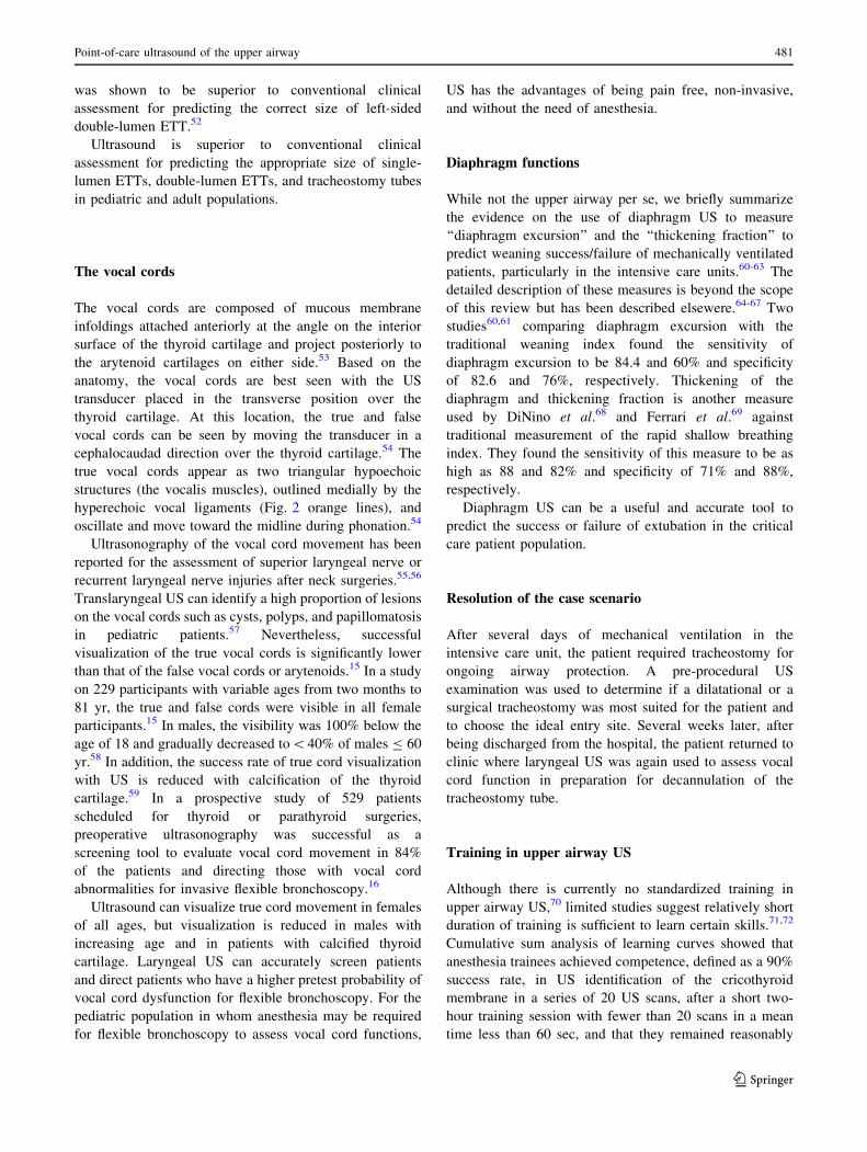
palpation in a cadaveric study that showed its ability to
heighten success and limit tube misplacement in
cricothyrotomy.8 Furthermore, this same technique can
be used to identify the optimal interspace between
tracheal rings for placement of a tracheostomy tube. We
recommend the longitudinal technique as the first to learn
and as the initial technique, such that every anesthesia
department dealing with difficult airways on a regular
basis should have the expertise to apply it. For patients
with a very short neck, or flexion-deformity of the neck
that leaves no space to place the US transducer in the
longitudinal position, we recommend the transverse
TACA technique to identify the cricothyroid membrane,
as in these subsets of patients, this may be the only
successful technique.27 Achieving a hundred percent
success rate of identifying the cricothyroid membrane is
possible when the longitudinal SOP technique is applied
in tandem with the transverse TACA technique.27
Performing the longitudinal ‘‘SOP’’ technique (String of
Pearls):27
1) The sternal bone is identified and the transducer is
placed transversely on the patient’s neck just cephalad to
the suprasternal notch to visualize the trachea (horseshoe-
shaped dark structure with a posterior white line) (Fig. 5,
first row).
2) The transducer is slid towards the patient’s right side
(towards the operator) so that the right border of the
transducer is positioned midline of the trachea and the US
image of the tracheal ring is thus truncated into half on the
screen (Fig. 5, second row).
3) The right end of the transducer is maintained over the
midline of the trachea, while the left end is rotated 90� into
Fig. 4 Transverse scan just cranial to the suprasternal notch and to
the patient’s left side of the trachea. Anterior part of tracheal cartilage
(light blue) is visualized as C-shaped hypoechoic (cartilage) and
hyperechoic (the tissue-air border) structures with shadowing.
Esophagus (purple) is seen as a round structure to the left of the
trachea without the hypo- and hyperechoic structures as seen in the
trachea. Carotid artery (red) is a distinct round structure lateral to the
trachea that is black because of absorption of sonographic waves by
the blood. Reproduced with permission from: Kristensen MS.
Ultrasonography in the management of the airway. Acta
Anaesthesiol Scand 2011; 55: 1155-734
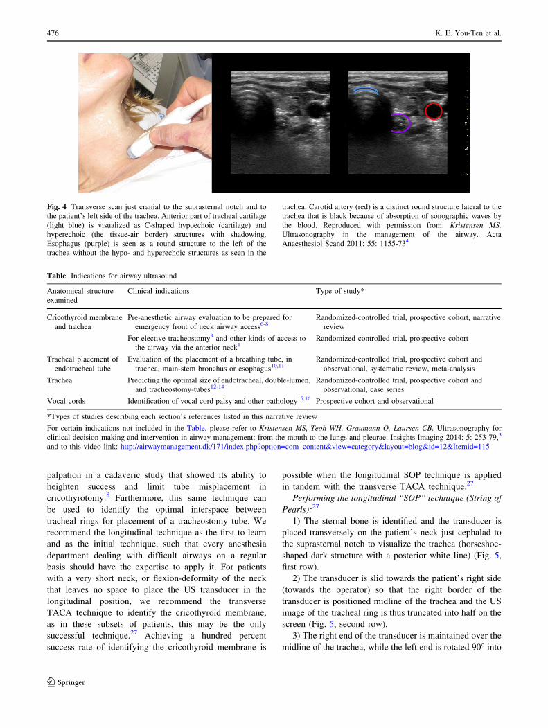
Table Indications for airway ultrasound
Anatomical structure
examined
Clinical indications Type of study*
Cricothyroid membrane
and trachea
Pre-anesthetic airway evaluation to be prepared for
emergency front of neck airway access6-8Randomized-controlled trial, prospective cohort, narrative
review
For elective tracheostomy9 and other kinds of access to
the airway via the anterior neck1Randomized-controlled trial, prospective cohort
Tracheal placement of
endotracheal tube
Evaluation of the placement of a breathing tube, in
trachea, main-stem bronchus or esophagus10,11Randomized-controlled trial, prospective cohort and
observational, systematic review, meta-analysis
Trachea Predicting the optimal size of endotracheal, double-lumen,
and tracheostomy-tubes12-14Randomized-controlled trial, prospective cohort and
observational, case series
Vocal cords Identification of vocal cord palsy and other pathology15,16 Prospective cohort and observational
*Types of studies describing each section’s references listed in this narrative review
For certain indications not included in the Table, please refer to Kristensen MS, Teoh WH, Graumann O, Laursen CB. Ultrasonography for
clinical decision-making and intervention in airway management: from the mouth to the lungs and pleurae. Insights Imaging 2014; 5: 253-79,5
and to this video link: http://airwaymanagement.dk/171/index.php?option=com_content&view=category&layout=blog&id=12&Itemid=115
476 K. E. You-Ten et al.
123

the sagittal plane resulting in a longitudinal scan of the
midline of the trachea. A number of dark (hypoechoic)
rings will be seen anterior to the white hyperechoic line
(air-tissue border), akin to a ‘‘string of pearls’’. The dark
hypoechoic ‘‘pearls’’ are the anterior part of the tracheal
rings (Fig. 5, third row).
4) The transducer is kept longitudinally in the midline
and slid cephalad until the cricoid cartilage comes into
view (seen as a larger, more elongated and anteriorly
placed dark ‘‘pearl’’ compared with the other tracheal
rings). Further cephalad, the distal part of the thyroid
cartilage can be seen as well (Fig. 5, fourth row). The
longitudinal course of the midline of the airway can be
marked with a pen.
5) While still holding the transducer, the other hand is
used to slide a needle (as a marker, for its ability to cast a
shadow in the US image) between the transducer and the
patient’s skin until the needle’s shadow is seen midway
between the caudal border of the thyroid cartilage and the
cephalad border of the cricoid cartilage (Fig. 5, fourth
row).
6) Now the transducer is removed and the needle marks
the centre of the cricothyroid membrane in the transverse
plane. This can be marked on the skin with a pen.
The technique is shown in this video: http://
airwaymanagement.dk/pearls.28
Performing the Transverse ‘‘TACA’’ technique (Thyroid
cartilage, Airline, Cricoid cartilage, Airline):27
1) Estimate the thyroid cartilage’s level on the neck and
place the US transducer transversely over it, scanning to
identify the thyroid cartilage as a hyperechoic triangular
structure (Fig. 6, first row).
Fig. 5 The longitudinal ‘‘String of Pearls’’ (SOP) technique for
identifying the cricothyroid membrane and the interspaces between
tracheal rings. See the text for details. Orange-red = tracheal ring;
light blue = the tissue-air border; green = the cricoid cartilage; purple
= the distal end of the thyroid cartilage. Yellow = the shadow from the
needle slid in between the transducer and the skin. With permission
from The Scandinavian Airway Management course ‘‘www.
airwaymanagement.dk’’
Point-of-care ultrasound of the upper airway 477
123

2) Move the transducer caudally until the cricothyroid
membrane is identified: this is recognizable as a
hyperechoic white line resulting from the echo of the
air-tissue border of the mucosal lining on the inside
of the cricothyroid membrane, often with parallel
white lines (reverberation artefacts) below (Fig. 6,
second row).
3) Move the transducer further caudally until the cricoid
cartilage is identified (a black ‘‘lying C’’ with a white
lining) (Fig. 6, third row).
4) Finally, move the transducer slightly back cephalad
until the centre of the cricothyroid membrane is identified
(Fig. 6, fourth row).
5) The centre can be marked both transversely and
sagittally on the skin with a pen. By identifying the highly
characteristic shapes of both the thyroid and cricoid
cartilages, both the cephalad and caudal borders of the
cricothyroid membrane can be identified.
The technique is shown in this video: http://
airwaymanagement.dk/taca.29
In conclusion, US-guided localization of the
cricothyroid membrane fills the void of very poor results
of accurate localization by visualization or palpation. It is
easily learned and should be considered, if not routinely,
then at least before embarking on management of
anticipated difficult airway situations.
Confirmation of tracheal intubation
The use of US for confirmation of endotracheal tube
placement has gained increasing popularity as an adjunct to
Fig. 6 The transverse ‘‘thyroid-airline-cricoid-airline’’ (TACA)
technique for identifying the cricothyroid membrane. See the text
for details. Blue triangle = thyroid cartilage; blue horizontal line = the
‘‘airline’’ = the cricothyroid membrane; blue ‘‘lying C’’ = the anterior
part of the cricoid cartilage. With permission from The Scandinavian
Airway Management course ‘‘ www.airwaymanagement.dk’’
478 K. E. You-Ten et al.
123

standards of practice, including end-tidal CO2,
visualization of the tube between cords, and endoscopic
visualization of tracheal rings through the endotracheal
tube (ETT). Currently, several prospective studies and
systematic reviews show the accuracy of US in confirming
the correct placement of the ETT by excluding esophageal
intubation.10,11,30 In a systematic review and meta-analysis,
Chou et al. studied the diagnostic accuracy of using
tracheal US to examine ETT placement during emergency
intubations by indirectly excluding esophageal
intubation.10 A total of 12 eligible studies involving adult
patients and cadaveric models were identified. For
detection of esophageal intubation, the pooled sensitivity
was 0.93 (95% confidence interval [CI], 0.86 to 0.96) and
the specificity was 0.97 (95% CI, 0.95 to 0.98).
Several prospective studies on human and cadaveric
models evaluated the sensitivity and specificity of US
confirmation of tube placement in the trachea.30-38 In a
meta-analysis of 969 intubations performed in emergency
and elective situations, Das et al. showed that transtracheal
ultrasonography’s pooled sensitivity and specificity were
0.98 (95% CI, 0.97 to 0.99) and 0.98 (95% CI, 0.95 to
0.99), respectively.11 In emergency scenarios, transtracheal
US showed an aggregate sensitivity and specificity of 0.98
(95% CI, 0.97 to 0.99) and 0.94 (95% CI, 0.86 to 0.98),
respectively.
Various US techniques have been used to visualize
diaphragmatic movements, lung sliding, methods, and
transtracheal identification of correct tube placement in
the trachea and its distinction from endobronchial
intubation.31-34 At present, there is a lack of data
comparing the accuracy of different US techniques.
Regardless of the technique chosen, US can be used to
evaluate proper ETT placement in the trachea by indirectly
ruling out an esophageal intubation.
Technique for confirmation of tracheal intubation with
ultrasonography
In the section below we explain the most consistently
described US technique for excluding esophageal
placement of the endotracheal tube. A curved US probe
may be used; however, tracheal rings are superficial and we
recommend using a high-frequency linear probe. If time
permits, one should visualize the normal airway anatomy
before placing the ETT (Fig. 1).
The US probe should be placed above the suprasternal
notch in a transverse direction. At this point, caution should
be exercised not to apply too much pressure as this may
distort the airway anatomy. As described earlier, on
ultrasonography a tracheal ring is visualized as C-shaped
hypoechoic (cartilage) and hyperechoic structures (the
tissue-air border) with shadowing (Fig. 4). The esophagus
is usually seen on one side of the trachea as an oval structure
with a hyperechoic wall and hypoechoic centre (Fig. 4).
Tracheal US can be performed in real time as the ETT is
passed. An esophageal intubation will reveal an adjacent
hyperechoic structure with shadowing posterolateral to the
trachea, consistent with the ETT location within the
esophagus. This has been referred to as the ‘‘double tract
sign’’39 (Fig. 7). It should be noted that if the esophagus is
located directly posterior to the trachea, an esophageal
intubation may be missed by US as this second hyperechoic
structure will be obscured by the shadowing from the trachea.
Current research suggests the use of focused US is
feasible to rapidly confirm tracheal or esophageal
placement of ETTs. Large prospective studies are needed
to investigate the learning curve of this technique by novice
operators before recommending its routine use in
anesthesia practice.
Tracheostomy
Ultrasonography is a useful adjunct for both surgical and
dilatational tracheostomy as it may increase the success
rate by helping to identify the trachea and the optimal
Tracheal lumen with �ssue-air hyperechoic lines.
Endotracheal tube in the esophagus with �ssue-air hyperechoic lines.
Fig. 7 Esophageal intubation. Transverse scan just cranial to the
suprasternal notch and to the left side of the patient’s trachea. An
esophageal intubation is shown as an adjacent hyperechoic structure
with shadowing posterolateral to the trachea, consistent with the
endotracheal tube within the esophagus. The tissue-air hyperechoic
lines are visualized in the trachea and esophagus (because of
esophageal intubation); this has been referred to as the ‘‘double
tract sign’’39
Point-of-care ultrasound of the upper airway 479
123

interspace between tracheal rings, determine the depth to
the tracheal lumen, and identify overlaying blood vessels or
other pathology prior to the procedure. Knowledge of these
factors may inform the decision whether to offer a surgical
or a dilatational tracheostomy. Ultrasonography can be
applied 1) before the procedure itself either to guide the
decision as to whether the best approach is a surgical one in
case of overlying large blood vessels, inability to identify
an appropriate interspace between the tracheal rings,
typically in very short necks, and subcutaneous
emphysema or a dilatational technique or to prepare for
the dilatational technique,9,40 2) before and during the
procedure instead of guidance by a flexible bronchoscope,
or 3) in combination with a flexible bronchoscope.
Identifying the trachea can be challenging in obese
patients; those with a short thick neck, neck mass, previous
surgery, or radiotherapy to the neck; thoracic pathology; or
other conditions resulting in tracheal deviation.41 Even the
addition of chest radiography and techniques of needle
aspiration to locate the trachea may be futile.42 Under such
circumstances, preoperative US for localization of the
trachea and the tracheal structures is extremely useful.42
Pre-procedural US often leads to a change in technique
from dilatational to surgical or the inverse9 and to a change
in the selection of the best interspace between the tracheal
rings.43
Following pre-procedural US scanning and marking of
appropriate interspace between tracheal rings, the
tracheostomy itself can be performed solely with clinical
guidance,9 with US guidance, or with flexible
bronchoscopy. Of these, only the flexible bronchoscope
allows intraluminal observation of the needle and
guidewire in the trachea; whereas only US allows real-
time measurement of the skin depth to the tracheal wall,
both measures prevent penetration of the posterior tracheal
wall. Occasionally, the obstruction of the lumen of the
airway caused by the flexible scope may interfere with
adequate ventilation and oxygenation of the patient; this
can be avoided by using a US-only technique.43,44
When comparing a landmark technique with the real-
time out-of-plane US-guided transverse technique, the
latter revealed a higher first-pass success (87% vs 58 %)
and less deviation from the midline with the US-guided
technique.45 The study included bronchoscopy during the
dilatational part of the procedure. When comparing US
with bronchoscopy guidance, Gobatto et al. found a 5%
rate of puncture of the orotracheal tube in the US group vs
1.7 % in the bronchoscopy group (P = 0.619) but
concluded that overall US guidance is an
acceptable alternative to bronchoscopy guidance.46
Real-time US guidance with visualization of the needle
path by means of a linear high-frequency transducer placed
transversely over the trachea was successful in a feasibility
study of 13 patients.47 Nevertheless, the real-time US
guidance of the procedure has a fairly long learning curve.
In a study of 85 consecutive patients, it was found that 20
procedures had to be performed before obtaining a low
frequency of major complications; a proficiency of 50
procedures is required before the combination of procedure
time and overall complication rate is fully satisfactory.48
We recommend a routine pre-emptive US examination
to determine if a dilatational or surgical tracheostomy is
most suited for the patient and to choose the ideal entry
site. This technique is easily learned. The information
obtained regarding the position of the ideal tracheal
interspace, the distance from the skin to luminal air, and
overlying blood vessels to avoid is then used to mark the
possible entry sites and guide the subsequent tracheostomy.
If the US examiner is experienced, US can be continued in
real time during needle insertion. Ideally, both US and
procedural flexible bronchoscopy are used in
combination—as this will add the possibility to observe
the needle entry into the tracheal lumen to avoid
penetration of the posterior wall, as a supplement to the
advantages obtained with US.
Tracheal diameter and appropriate sizing of
endotracheal and tracheostomy tubes
Ultrasound is a reliable tool for assessment of the
subglottic upper airway diameter and is validated against
magnetic resonance imaging49 and computed tomography
scans.14 In the pediatric population the subglottic
transverse diameter measured by US correlates well with
the endotracheal tube outer diameter and is superior to age-
and height-based formulas in estimating endotracheal tube
size.12,50,51 Moreover, US can predict the proper
tracheostomy tube size for exchange in children by
assessing the internal and external transverse tracheal
diameter and the depth of the trachea from the skin
surface.13 In this small series of four children, US
confirmed that a new larger fenestrated tracheostomy
tube could be replaced in one child but not in the other
three children because of lack of space to allow for a larger
tracheostomy tube.13
The diameter of the left mainstem bronchus, and thus
the proper size of a left-sided double-lumen ETT, can be
estimated by measuring the outer tracheal width with the
US probe placed in the transverse position just above the
sternoclavicular joint.14,52 In a series of 45 patients
undergoing intubation with a left-sided double-lumen
ETT, Sustic et al. showed a strong correlation between
tracheal width as measured by US and tracheal width (r =
0.882) and left main bronchus width (r = 0.832) as
measured by computed tomography.14 Furthermore, US
480 K. E. You-Ten et al.
123
was shown to be superior to conventional clinical
assessment for predicting the correct size of left-sided
double-lumen ETT.52
Ultrasound is superior to conventional clinical
assessment for predicting the appropriate size of single-
lumen ETTs, double-lumen ETTs, and tracheostomy tubes
in pediatric and adult populations.
The vocal cords
The vocal cords are composed of mucous membrane
infoldings attached anteriorly at the angle on the interior
surface of the thyroid cartilage and project posteriorly to
the arytenoid cartilages on either side.53 Based on the
anatomy, the vocal cords are best seen with the US
transducer placed in the transverse position over the
thyroid cartilage. At this location, the true and false
vocal cords can be seen by moving the transducer in a
cephalocaudad direction over the thyroid cartilage.54 The
true vocal cords appear as two triangular hypoechoic
structures (the vocalis muscles), outlined medially by the
hyperechoic vocal ligaments (Fig. 2 orange lines), and
oscillate and move toward the midline during phonation.54
Ultrasonography of the vocal cord movement has been
reported for the assessment of superior laryngeal nerve or
recurrent laryngeal nerve injuries after neck surgeries.55,56
Translaryngeal US can identify a high proportion of lesions
on the vocal cords such as cysts, polyps, and papillomatosis
in pediatric patients.57 Nevertheless, successful
visualization of the true vocal cords is significantly lower
than that of the false vocal cords or arytenoids.15 In a study
on 229 participants with variable ages from two months to
81 yr, the true and false cords were visible in all female
participants.15 In males, the visibility was 100% below the
age of 18 and gradually decreased to\40% of males B 60
yr.58 In addition, the success rate of true cord visualization
with US is reduced with calcification of the thyroid
cartilage.59 In a prospective study of 529 patients
scheduled for thyroid or parathyroid surgeries,
preoperative ultrasonography was successful as a
screening tool to evaluate vocal cord movement in 84%
of the patients and directing those with vocal cord
abnormalities for invasive flexible bronchoscopy.16
Ultrasound can visualize true cord movement in females
of all ages, but visualization is reduced in males with
increasing age and in patients with calcified thyroid
cartilage. Laryngeal US can accurately screen patients
and direct patients who have a higher pretest probability of
vocal cord dysfunction for flexible bronchoscopy. For the
pediatric population in whom anesthesia may be required
for flexible bronchoscopy to assess vocal cord functions,
US has the advantages of being pain free, non-invasive,
and without the need of anesthesia.
Diaphragm functions
While not the upper airway per se, we briefly summarize
the evidence on the use of diaphragm US to measure
‘‘diaphragm excursion’’ and the ‘‘thickening fraction’’ to
predict weaning success/failure of mechanically ventilated
patients, particularly in the intensive care units.60-63 The
detailed description of these measures is beyond the scope
of this review but has been described elsewere.64-67 Two
studies60,61 comparing diaphragm excursion with the
traditional weaning index found the sensitivity of
diaphragm excursion to be 84.4 and 60% and specificity
of 82.6 and 76%, respectively. Thickening of the
diaphragm and thickening fraction is another measure
used by DiNino et al.68 and Ferrari et al.69 against
traditional measurement of the rapid shallow breathing
index. They found the sensitivity of this measure to be as
high as 88 and 82% and specificity of 71% and 88%,
respectively.
Diaphragm US can be a useful and accurate tool to
predict the success or failure of extubation in the critical
care patient population.
Resolution of the case scenario
After several days of mechanical ventilation in the
intensive care unit, the patient required tracheostomy for
ongoing airway protection. A pre-procedural US
examination was used to determine if a dilatational or a
surgical tracheostomy was most suited for the patient and
to choose the ideal entry site. Several weeks later, after
being discharged from the hospital, the patient returned to
clinic where laryngeal US was again used to assess vocal
cord function in preparation for decannulation of the
tracheostomy tube.
Training in upper airway US
Although there is currently no standardized training in
upper airway US,70 limited studies suggest relatively short
duration of training is sufficient to learn certain skills.71,72
Cumulative sum analysis of learning curves showed that
anesthesia trainees achieved competence, defined as a 90%
success rate, in US identification of the cricothyroid
membrane in a series of 20 US scans, after a short two-
hour training session with fewer than 20 scans in a mean
time less than 60 sec, and that they remained reasonably
Point-of-care ultrasound of the upper airway 481
123
competent three months later.71 Following a brief 20-min
didactic teaching and a 30-min practice session on upper
airway US, emergency medicine fellows lacking formal
airway bedside US training were able to identify the
location and depth of a saline-filled endotracheal tube
above or at the suprasternal notch in an adult cadaver
model with a sensitivity of 96 % (23 of 24).72 These studies
suggest the training required for US identification of the
cricothyroid membrane and endotracheal tube seems to be
short even without prior airway US experience. The
number of supervised US examinations required to
maintain competence is debatable. Using the Objective
Structured Assessment of Ultrasound Skills is a valid and
reliable method68,69 that might also be valuable to assess
basic ultrasonography of the upper airway and may help
determine when trainees are qualified for independent
practice. Our recommendation is a minimum of 25
supervised US scans for each procedure or outcome. It is
particularly important to train in POCUS for cricothyroid
membrane identification, where existing modalities are
unreliable for the purpose, in contrast to other applications,
where POCUS is an additional option alongside existing
fairly reliable modalities. Further research regarding
training education in upper airway US is warranted.
Future directions
Upper airway US is useful in airway management because
of its portability, minimal invasiveness, cost effectiveness,
low radiation exposure, and accessibility. Modern
advancements in ultrasonography technologies such as
three-dimensional US may be useful in the complex
evaluation of upper and lower airway anatomy with
accurate prediction of difficult airways, diagnosis of
obstructive sleep apnea, and guidance of airway nerve
blocks.
Conclusions
Ultrasound has many advantages for imaging the upper
airway; it is safe, quick, portable, and accessible and
provides static and real-time dynamic images relevant for
various clinical indications of management of the airway.
Upper airway POCUS can be used dynamically for optimal
benefit in perioperative airway management, immediately
before, during, and after airway interventions. Acute
airway procedures under real-time US guidance may
become standard procedures in anesthesia, emergency,
and intensive care settings. With a growing body of
evidence in many clinical applications, there is a need to
incorporate upper airway US education and training of
personnel responsible for perioperative airway
management. POCUS of the upper airway has the
potential to become a first-line non-invasive airway
assessment tool.
Conflict of interest None declared.
Editorial responsibility This submission was handled by Dr.
Gregory L. Bryson, Deputy Editor-in-Chief, Canadian Journal of
Anesthesia.
Funding None.
References
1. Woodall NM, Cook TM. National census of airway management
techniques used for anaesthesia in the UK: first phase of the
Fourth National Audit Project at the Royal College of
Anaesthetists. Br J Anaesth 2011; 106: 266-71.
2. Cook TM, Woodall N, Harper J, Benger J, Fourth National Audit
Project. Major complications of airway management in the UK:
results of the Fourth National Audit Project of the Royal College
of Anaesthetists and the Difficult Airway Society. Part 2:
intensive care and emergency departments. Br J Anaesth 2011;
106: 632-42.
3. Moore CL, Copel JA. Point-of-care ultrasonography. N Engl J
Med 2011; 364: 749-57.
4. Kristensen MS. Ultrasonography in the management of the
airway. Acta Anaesthesiol Scand 2011; 55: 1155-73.
5. Kristensen MS, Teoh WH, Graumann O, Laursen CB.
Ultrasonography for clinical decision-making and intervention
in airway management: from the mouth to the lungs and pleurae.
Insights Imaging 2014; 5: 253-79.
6. Teoh WH, Kristensen MS. Prediction in airway management:
what is worthwhile, what is a waste of time and what about the
future? Br J Anaesth 2016; 117: 1-3.
7. Kristensen MS, Teoh WH, Rudolph SS. Ultrasonographic
identification of the cricothyroid membrane: best evidence,
techniques, and clinical impact. Br J Anaesth 2016; 117(Suppl
1): i39-48.
8. Siddiqui N, Arzola C, Friedman Z, Guerina L, You-Ten KE.
Ultrasound improves cricothyrotomy success in cadavers with
poorly defined neck anatomy: a randomized control trial.
Anesthesiology 2015; 123: 1033-41.
9. Even-Tov E, Koifman I, Rozentsvaig V, Livshits L, Gilbey P. Pre-
procedural ultrasonography for tracheostomy in critically ill
patients: a prospective study. Isr Med Assoc J 2017; 19: 337-40.
10. Chou EH, Dickman E, Tsou PY, et al. Ultrasonography for
confirmation of endotracheal tube placement: a systematic review
and meta-analysis. Resuscitation 2015; 90: 97-103.
11. Das SK, Choupoo NS, Haldar R, Lahkar A. Transtracheal
ultrasound for verification of endotracheal tube placement: a
systematic review and meta-analysis. Can J Anesth 2015; 62:
413-23.
12. Shibasaki M, Nakajima Y, Ishii S, Shimizu F, Shime N, Sessler DI.
Prediction of pediatric endotracheal tube size by ultrasonography.
Anesthesiology 2010; 113: 819-24.
13. Hardee PS, Ng SY, Cashman M. Ultrasound imaging in the
preoperative estimation of the size of tracheostomy tube required
in specialised operations in children. Br J Oral Maxillofac Surg
2003; 41: 312-6.
482 K. E. You-Ten et al.
123

14. Sustic A, Miletic D, Protic A, Ivancic A, Cicvaric T. Can
ultrasound be useful for predicting the size of a left double-lumen
bronchia ltube? Tracheal width as measured by ultrasonography
versus computed tomography. J Clin Anesth 2008; 20: 247-52.
15. Wang CP, Chen TC, Yang TL, et al. Transcutaneous ultrasound
for evaluation of vocal fold movement in patients with thyroid
disease. Eur J Radiol 2012; 81: e288-91.
16. Cheng SP, Lee JJ, Liu TP, Lee KS, Liu CL. Preoperative
ultrasonography assessment of vocal cord movement during
thyroid and parathyroid surgery. World J Surg 2012; 36: 2509-15.
17. Cook TM, Woodall N, Frerk C, Fourth National Audit Project.
Major complications of airway management in the UK: results of
the Fourth National Audit Project of the Royal College of
Anaesthetists and the Difficult Airway Society. Part 1:
anaesthesia. Br J Anaesth 2011; 106: 617-31.
18. Apfelbaum JL, Hagberg CA, Caplan RA, et al. Practice guidelines
for management of the difficult airway: an updated report by the
American Society of Anesthesiologists Task Force on
Management of the Difficult Airway. Anesthesiology 2013;
118: 251-70.
19. Law JA, Broemling N, Cooper RM, et al. The difficult airway
with recommendations for management–part 1–difficult tracheal
intubation encountered in an unconscious/induced patient. Can J
Anesth 2013; 60: 1089-118.
20. Hiller KN, Karni RJ, Cai C, Holcomb JB, Hagberg CA.
Comparing success rates of anesthesia providers versus trauma
surgeons in their use of palpation to identify the cricothyroid
membrane in female subjects: a prospective observational study.
Can J Anesth 2016; 63: 807-17.
21. Law JA. Deficiencies in locating the cricothyroid membrane by
palpation: we can’t and the surgeons can’t, so what now for the
emergency surgical airway? Can J Anesth 2016; 63: 791-6.
22. Lamb A, Zhang J, Hung O, et al. Accuracy of identifying the
cricothyroid membrane by anesthesia trainees and staff in a
Canadian institution. Can J Anesth 2015; 62: 495-503.
23. You-Ten KE, Desai D, Postonogova T, Siddiqui N. Accuracy of
conventional digital palpation and ultrasound of the cricothyroid
membrane in obese women in labour. Anaesthesia 2015; 70:
1230-4.
24. Erlandson MJ, Clinton JE, Ruiz E, Cohen J. Cricothyrotomy in
the emergency department revisited. J Emerg Med 1989; 7: 115-
8.
25. Kristensen MS, Teoh WH, Baker PA. Percutaneous emergency
airway access; prevention, preparation, technique and training. Br
J Anaesth 2015; 114: 357-61.
26. Kristensen MS, Teoh WH, Rudolph SS, et al. Structured approach
to ultrasound-guided identification of the cricothyroid membrane:
a randomized comparison with the palpation method in the
morbidly obese. Br J Anaesth 2015; 114: 1003-4.
27. Kristensen MS, Teoh WH, Rudolph SS, Hesselfeldt R, Borglum J,
Tvede MF. A randomised cross-over comparison of the transverse
and longitudinal techniques for ultrasound-guided identification
of the cricothyroid membrane in morbidly obese subjects.
Anaesthesia 2016; 71: 675-83.
28. Airway Management for Anaesthesiologists. Pearls. 2016.
Available from URL: http://www.airwaymanagement.dk/ http://
airwaymanagement.dk/pearls (accessed December 2017).
29. Airway Management for Anaesthesiologists. TACA. 2016.
Available from URL: http://www.airwaymanagement.dk/ http://
airwaymanagement.dk/taca (accessed December 2017).
30. Ramsingh D, Frank E, Haughton R, et al. Auscultation versus
Point-of-care Ultrasound to Determine Endotracheal versus
Bronchial Intubation: A Diagnostic Accuracy Study.
Anesthesiology 2016; 124: 1012-20.
31. Gottlieb M, Bailitz JM, Christian E, et al. Accuracy of a novel
ultrasound technique for confirmation of endotracheal intubation
by expert and novice emergency physicians. West J Emerg Med
2014; 15: 834-9.
32. Chou HC, Chong KM, Sim SS, et al. Real-time tracheal
ultrasonography for confirmation of endotracheal tube
placement during cardiopulmonary resuscitation. Resuscitation
2013; 84: 1708-12.
33. Chou HC, Tseng WP, Wang CH, et al. Tracheal rapid ultrasound
exam (T.R.U.E.) for confirming endotracheal tube placement
during emergency intubation. Resuscitation 2011; 82: 1279-84.
34. Hosseini JS, Talebian MT, Ghafari MH, Eslami V. Secondary
confirmation of endotracheal tube position by diaphragm motion
in right subcostal ultrasound view. Int J Crit Illn Inj Sci 2013; 3:
113-7.
35. Abbasi S, Farsi D, Zare MA, Hajimohammadi M, Rezai M,
Hafezimoghadam P. Direct ultrasound methods: a confirmatory
technique for proper endotracheal intubation in the emergency
department. Eur J Emerg Med 2015; 22: 10-6.
36. Muslu B, Sert H, Kaya A, et al. Use of sonography for rapid
identification of esophageal and tracheal intubations in adult
patients. J Ultrasound Med 2011; 30: 671-6.
37. Werner SL, Smith CE, Goldstein JR, Jones RA, Cydulka RK. Pilot
study to evaluate the accuracy of ultrasonography in confirming
endotracheal tube placement. Ann Emerg Med 2007; 49: 75-80.
38. Ma G, Davis DP, Schmitt J, Vilke GM, Chan TC, Hayden SR. The
sensitivity and specificity of transcricothyroid ultrasonography to
confirm endotracheal tube placement in a cadaver model. J Emerg
Med 2007; 32: 405-7.
39. Kristensen MS, Teoh WH. Ultrasound in confirming endotracheal
intubation. In: Rosenblatt WH, Popescu WM (Eds). Master
Techniques in Upper and Lower Airway Management. Wolters
Kluwer Health; 2015: 28-9.
40. Yavuz A, Yilmaz M, Goya C, Alimoglu E, Kabaalioglu A.
Advantages of US in percutaneous dilatational tracheostomy:
randomized controlled trial and review of the literature.
Radiology 2014; 273: 927-36.
41. Soni KD, Jindal M, Aggarwal R, Deganwa M, Prasad J.
Ultrasound and fibreoptic-guided percutaneous tracheostomy in
patient with deviated trachea. Anaesthesiol Intensive Ther 2016;
48: 148-9.
42. Munir N, Hughes D, Sadera G, Sherman IW. Ultrasound-guided
localisation of trachea for surgical tracheostomy. Eur Arch
Otorhinolaryngol 2010; 267: 477-9.
43. Sustic A, Kovac D, Zgaljardic Z, Zupan Z, Krstulovic B.
Ultrasound-guided percutaneous dilatational tracheostomy: a
safe method to avoid cranial misplacement of the tracheostomy
tube. Intensive Care Med 2000; 26: 1379-81.
44. Reilly PM, Sing RF, Giberson FA, et al. Hypercarbia during
tracheostomy: a comparison of percutaneous endoscopic,
percutaneous Doppler, and standard surgical tracheostomy.
Intensive Care Med 1997; 23: 859-64.
45. Rudas M, Seppelt I, Herkes R, Hislop R, Rajbhandari D,
Weisbrodt L. Traditional landmark versus ultrasound guided
tracheal puncture during percutaneous dilatational tracheostomy
in adult intensive care patients: a randomised controlled trial. Crit
Care 2014; 18: 514.
46. Gobatto AL, Besen BA, Tierno PF, et al. Ultrasound-guided
percutaneous dilational tracheostomy versus bronchoscopy-
guided percutaneous dilational tracheostomy in critically ill
patients (TRACHUS): a randomized noninferiority controlled
trial. Intensive Care Med 2016; 42: 342-51.
47. Rajajee V, Fletcher JJ, Rochlen LR, Jacobs TL. Real-time
ultrasound-guided percutaneous dilatational tracheostomy: a
feasibility study. Crit Care 2011; 15: R67.
48. Petiot S, Guinot PG, Diouf M, Zogheib E, Dupont H. Learning
curve for real-time ultrasound-guided percutaneous
tracheostomy. Anaesth Crit Care Pain Med 2017; 36: 279-83.
Point-of-care ultrasound of the upper airway 483
123

49. Lakhal K, Delplace X, Cottier JP, et al. The feasibility of
ultrasound to assess subglottic diameter. Anesth Analg 2007; 104:
611-4.
50. Bae JY, Byon HJ, Han SS, Kim HS, Kim JT. Usefulness of
ultrasound for selecting a correctly sized uncuffed tracheal tube
for paediatric patients. Anaesthesia 2011; 66: 994-8.
51. Kim EJ, Kim SY, Kim WO, Kim H, Kil HK. Ultrasound
measurement of subglottic diameter and an empirical formula
for proper endotracheal tube fitting in children. Acta Anaesthesiol
Scand 2013; 57: 1124-30.
52. Sustic A, Protic A, Cicvaric T, Zupan Z. The addition of a brief
ultrasound examination to clinical assessment increases the
ability to confirm placement of double-lumen endotracheal
tubes. J Clin Anesth 2010; 22: 246-9.
53. Standring S. Gray’s Anatomy: The Anatomical Basis of Clinical
Practice. 40th ed. Philadelphia: Churchill Livingstone; 2008 .
54. Singh M, Chin KJ, Chan VW, Wong DT, Prasad GA, Yu E. Use of
sonography for airway assessment: an observational study. J
Ultrasound Med 2010; 29: 79-85.
55. Miles KA. Ultrasound demonstration of vocal cord movements.
Br J Rodiol 1989; 62: 871-2.
56. Kundra P, Kumar K, Allampalli V, Anathkrishnan R,
Gopalakrishnan S, Elangovan S. Use of ultrasound to assess
superior and recurrent laryngeal nerve function immediately after
thyroid surgery. Anaesthesia 2012; 67: 301-2.
57. Bisetti MS, Segala F, Zappia F, Albera R, Ottaviani F, Schindler
A. Non-invasive assessment of benign vocal folds lesions in
children by means of ultrasonography. Int J Pediatr
Otorhinolaryngol 2009; 73: 1160-2.
58. Hu Q, Zhu SY, Luo F, Gao Y, Yang XY. High-frequency
sonographic measurements of true and false vocal cords. J
Ultrasound Med 2010; 29: 1023-30.
59. Carneiro-Pla D, Miller BS, Wilhelm SM, et al. Feasibility of
surgeon-performed transcutaneous vocal cord ultrasonography in
identifying vocal cord mobility: a multi-institutional experience.
Surgery 2014; 156: 1597-602.
60. Kim WY, Suh HJ, Hong SB, Koh Y, Lim CM. Diaphragm
dysfunction assessed by ultrasonography: influence on weaning
from mechanical ventilation. Crit Care Med 2011; 39: 2627-30.
61. Oliveira KF, Arzola C, Ye XY, Clivatti J, Siddiqui N, You-Ten KE.
Determining the amount of training needed for competency of
anesthesia trainees in ultrasonographic identification of the
cricothyroid membrane. BMC Anesthesiol 2017; 17: 74.
62. Uya A, Spear D, Patel K, Okada P, Sheeran P, McCreight A. Can
novice sonographers accurately locate an endotracheal tube with
a saline-filled cuff in a cadaver model? A pilot study. Acad
Emerg Med 2012; 19: 361-4.
63. Tolsgaard MG, Ringsted C, Dreisler E, et al. Reliable and
valid assessment of ultrasound operator competence in obstetrics
and gynecology. Ultrasound Obstet Gynecol 2014; 43: 437-
43.
64. El-Boghdadly K, Goffi A, Chan V. In reply: proper use and
interpretation of diaphragmatic ultrasonography. Can J Anesth
2017; 64: 550-1.
65. Sondekoppam RV, Naik L, Tsui J, Tsui BC. Proper use and
interpretation of diaphragmatic ultrasonography. Can J Anesth
2017; 64: 548-9.
66. El-Boghdadly K, Goffi A, Chan V. Point of care diaphragmatic
ultrasonography made easy. Can J Anesth 2017; 64: 327-8.
67. Naik LY, Sondekoppam RV, Tsui JJ, Tsui BC. An ultrasound-
guided ABCDE approach with a sniff test to evaluate
diaphragmatic function without acoustic windows. Can J
Anesth 2017; 63: 1199-200.
68. DiNino E, Gartman EJ, Sethi JM, McCool FD. Diaphragm
ultrasound as a predictor of successful extubation from
mechanical ventilation. Thorax 2014; 69: 423-7.
69. Ferrari G, De Filippi G, Elia F, Panero F, Volpicelli G, Apra F.
Diaphragm ultrasound as a new index of discontinuation from
mechanical ventilation. Crit Ultrasound J 2014; 6: 8.
70. Mok D, Schwarz SK, Rondi K. Point-of-care ultrasonography in
Canadian anesthesiology residency programs: a national survey
of program directors. Can J Anesth 2017; 64: 1023-36.
71. Todsen T, Tolsgaard MG, Olsen BH, et al. Reliable and valid
assessment of point-of-care ultrasonography. Ann Surg 2015;
261: 309-15.
72. Jiang JR, Tsai TH, Jerng JS, Yu CJ, Wu HD, Yang PC.
Ultrasonographic evaluation of liver/spleen movements and
extubation outcome. Chest 2004; 126: 179-85.
484 K. E. You-Ten et al.
123