Podoplanin promotes cell migration via the EGF-Src...
10
241 Abstract: Human podoplanin is a type-1 trans- membrane sialomucin-like glycoprotein that is involved in cell migration, tumor cell invasion and metastasis. Our recent study of oral squamous cell carcinoma (OSCC) demonstrated that the degree of immunohistochemical expression of podoplanin was correlated with the severity of epithelial dysplasia and significantly associated with a poor pathologic grade of differentiation. Furthermore, it has been reported that Src directly associates with the epidermal growth factor receptor (EGFR) in OSCC cells upon stimula- tion with EGF and phosphorylates Crk-associated substrate (Cas), podoplanin acting downstream of Src and Cas to promote cell migration. However, the molecular function of podoplanin remains unclear. In this study we performed real-time RT-PCR, Western blotting and scratch assay using OSCC cell lines in order to clarify the molecular biological function of podoplanin expression associated with various growth factors including EGF and with the Src-Cas signaling pathway. Podoplanin was found to have a marked influence on cancer cell migration and the expression of matrix metalloprotease-9 (MMP-9) in the oral cavity upon stimulation with EGF. Podo- planin promotes oral cancer cell migration, and the EGF-Src-Cas pathway is one of the possible mecha- nisms responsible for progression of cancer in the oral cavity. (J Oral Sci 54, 241-250, 2012) Keywords: oral squamous cell carcinoma; podoplanin; EGF; MMPs; EGF-Src-Cas pathway. Introduction Human podoplanin is a type I transmembrane sialo- mucin-like glycoprotein consisting of 162 amino acids (1). The protein was originally detected as a 38-kDa mucoprotein on the surface of podocytes in rats with puromycin-induced nephrosis, and was linked to the flat- tening of foot processes (2). Podoplanin may play a role in cellular contractility and cytoskeletal reorganization. Several studies have suggested that overexpression of podoplanin is associated with a poor clinical outcome in oral squamous cell carcinoma (OSCC) (3-5). Our previous study of OSCC also showed that the intensity Correspondence to Dr. Harumi Inoue, Division of Pathology, Department of Diagnostic & Therapeutic Sciences, Meikai University School of Dentistry, 1-1 Keyakidai, Sakado, Saitama 350-0283, Japan Tel: +81-49-279-2773 Fax: +81-49-286-6101 E-mail: [email protected] Journal of Oral Science, Vol. 54, No. 3, 241-250, 2012 Original Podoplanin promotes cell migration via the EGF-Src-Cas pathway in oral squamous cell carcinoma cell lines Harumi Inoue 1) , Yuji Miyazaki 1) , Kentaro Kikuchi 1) , Noriaki Yoshida 1) , Fumio Ide 1) , Yoshihiro Ohmori 2) , Akihito Tomomura 3) , Hideaki Sakashita 4) and Kaoru Kusama 1) 1) Division of Pathology, Department of Diagnostic & Therapeutic Sciences, Meikai University School of Dentistry, Sakado, Japan 2) Division of Microbiology, Department of Oral Biology & Tissue Engineering, Meikai University School of Dentistry, Sakado, Japan 3) Division of Biochemistry, Department of Oral Biology & Tissue Engineering, Meikai University School of Dentistry, Sakado, Japan 4) Division of Oral and Maxillofacial Surgery, Department of Diagnostic & Therapeutic Sciences, Meikai University School of Dentistry, Sakado, Japan (Received 8 June and accepted 10 July 2012)
Transcript of Podoplanin promotes cell migration via the EGF-Src...

241
Abstract: Human podoplanin is a type-1 trans-membrane sialomucin-like glycoprotein that is involved in cell migration, tumor cell invasion and metastasis. Our recent study of oral squamous cell carcinoma (OSCC) demonstrated that the degree of immunohistochemical expression of podoplanin was correlated with the severity of epithelial dysplasia and significantly associated with a poor pathologic grade of differentiation. Furthermore, it has been reported that Src directly associates with the epidermal growth factor receptor (EGFR) in OSCC cells upon stimula-tion with EGF and phosphorylates Crk-associated substrate (Cas), podoplanin acting downstream of Src and Cas to promote cell migration. However, the molecular function of podoplanin remains unclear. In this study we performed real-time RT-PCR, Western blotting and scratch assay using OSCC cell lines in order to clarify the molecular biological function of podoplanin expression associated with various
growth factors including EGF and with the Src-Cas signaling pathway. Podoplanin was found to have a marked influence on cancer cell migration and the expression of matrix metalloprotease-9 (MMP-9) in the oral cavity upon stimulation with EGF. Podo-planin promotes oral cancer cell migration, and the EGF-Src-Cas pathway is one of the possible mecha-nisms responsible for progression of cancer in the oral cavity. (J Oral Sci 54, 241-250, 2012)
Keywords: oral squamous cell carcinoma; podoplanin; EGF; MMPs; EGF-Src-Cas pathway.
IntroductionHuman podoplanin is a type I transmembrane sialo-
mucin-like glycoprotein consisting of 162 amino acids (1). The protein was originally detected as a 38-kDa mucoprotein on the surface of podocytes in rats with puromycin-induced nephrosis, and was linked to the flat-tening of foot processes (2). Podoplanin may play a role in cellular contractility and cytoskeletal reorganization.
Several studies have suggested that overexpression of podoplanin is associated with a poor clinical outcome in oral squamous cell carcinoma (OSCC) (3-5). Our previous study of OSCC also showed that the intensity
Correspondence to Dr. Harumi Inoue, Division of Pathology, Department of Diagnostic & Therapeutic Sciences, Meikai University School of Dentistry, 1-1 Keyakidai, Sakado, Saitama 350-0283, JapanTel: +81-49-279-2773Fax: +81-49-286-6101E-mail: [email protected]
Journal of Oral Science, Vol. 54, No. 3, 241-250, 2012
Original
Podoplanin promotes cell migration via the EGF-Src-Cas pathway in oral squamous cell carcinoma cell linesHarumi Inoue1), Yuji Miyazaki1), Kentaro Kikuchi1), Noriaki Yoshida1),
Fumio Ide1), Yoshihiro Ohmori2), Akihito Tomomura3), Hideaki Sakashita4)
and Kaoru Kusama1)
1)Division of Pathology, Department of Diagnostic & Therapeutic Sciences,Meikai University School of Dentistry, Sakado, Japan
2)Division of Microbiology, Department of Oral Biology & Tissue Engineering,Meikai University School of Dentistry, Sakado, Japan
3)Division of Biochemistry, Department of Oral Biology & Tissue Engineering,Meikai University School of Dentistry, Sakado, Japan
4)Division of Oral and Maxillofacial Surgery, Department of Diagnostic & Therapeutic Sciences,Meikai University School of Dentistry, Sakado, Japan
(Received 8 June and accepted 10 July 2012)

242
of immunohistochemical expression of podoplanin was correlated with the severity of epithelial dysplasia (P = 0.016) and significantly associated with a poor patho-logic grade of differentiation (P = 0.020). These findings suggested that podoplanin is associated with tumor development via the oral dysplasia-carcinoma sequence (6).
A developmental regulatory program, referred to as the “epithelial-mesenchymal transition” (EMT), has become prominently implicated in the ability of transformed epithelial cells to disseminate and acquire invasiveness and resistance to apoptosis (7-9). Two previous studies of the association of EMT with podoplanin expression have produced contrasting findings. One suggested that podoplanin plays a role in tumor invasion by binding to proteins such as ezrin, radixin and moesin to activate RhoA, thereby remodeling the actin cytoskeleton of tumor cells and contributing to their increased motility (10). The other study suggested that podoplanin shifts the pattern of invasion from that of single cells involving EMT to that involving large cell sheets in the absence of EMT (11). In our previous study we found that 62% of podoplanin-positive primary OSCCs with patho-logical node metastasis (pN+) showed EMT, whereas the remainder lacked EMT. These findings suggest that podoplanin expression in tumor cells does not interfere with the occurrence of EMT in OSCCs (6).
Src belongs to a family of 11 non-receptor tyrosine kinases known as the Src family kinases (SFKs) (12). SFKs play critical roles in a variety of cellular signal transduction pathways, regulating diverse processes including cell division, motility, adhesion, angiogenesis, and survival (13). SFKs are capable of inducing malig-nant transformation in a variety of cell types, and are frequently overexpressed in various kinds of epithelial and non-epithelial malignant neoplasms. The extent of increased Src family activity often correlates with malignant potential. Src acts as a signal transducer from cell surface receptors through sequential phosphoryla-tion of tyrosine residues on substrates (14). Binding to activator molecules such as receptor kinase adaptors including EGF, platelet-derived growth factor (PDGF), fibrous growth factor-basic (bFGF), and hepatocyte growth factor (HGF), or the activated receptor itself, or dephosphorylation of the C-terminal tyrosine by certain tyrosine phosphatases, converts SFKs to functionally active forms, resulting in phosphorylation of downstream effectors and leading to cellular responses (15).
p130Cas is a 130-kDa scaffolding protein that is recruited to focal adhesions upon ligation to integrin (16). The protein was so named because of its association
with, and phosphorylation by, Crk-associated substrate (17). p130Cas contains an SH3 domain, which regulates localization to the focal contact through interaction with focal adhesion kinase (FAK). The poly-proline-rich regions of p130Cas, located between the substrate domain and the C-terminal, mediate interaction with SFKs (18). In addition, in CHO cells, p130Cas acts downstream of FAK to promote cell migration (19), and in human pancreatic carcinoma cells interaction between p130Cas and Crk activates Rac, which is necessary for cell migra-tion. Therefore, the FAK/p130Cas interaction has been considered a “molecular switch” for cell migration (20). With regard to the association between the Src-Cas signaling pathway and podoplanin expression, Yongquan et al. suggested that podoplanin acts downstream of Src and Cas, and that Src utilizes Cas to induce podoplanin expression, thus promoting tumor cell migration and leading to cancer invasion and metastasis (21).
The aim of the present study was to clarify the molecular biological role of podoplanin in tumor cell migration and invasion upon stimulation with cancer microenvironmental growth factors such as bFGF, transforming growth factor-β1 (TGF-β1), HGF, PDGF, and EGF in OSCC cell lines, and to examine subsequent Src-Cas signaling activity.
Materials and MethodsCell lines and cell culture
The human oral squamous cell carcinoma-derived cell lines Ca9-22, HSC-2, -3, and -4 were provided by RIKEN BRC through the National Bio-Resource Project of MEXT, Japan. Each cell line was routinely grown in RPMI 1640 medium (Sigma-Aldrich, St. Louis, MO, USA) supplemented with 100 U/mL penicillin-strepto-mycin (GIBCO Invitrogen, Carlsbad, CA, USA), 10 U/mL fungizone (GIBCO Invitrogen), and 10% fetal bovine serum (GIBCO Invitrogen) in a humidified atmosphere of 5% CO2 at 37°C.
Real-time quantitative RT-PCR Total RNAs were extracted using a Protein and RNA
Extraction Kit for mammalian cells (PAREx; Takara, Tokyo, Japan) in accordance with the manufacturer’s instructions after each cell line had been treated with 100 ng/mL bFGF, TGF-b1, HGF, PDGF, or EGF for 24 h or 48 h. Real-time RT-PCR was performed using a Thermal Cycler Dice Real Time System (Takara) in accordance with the manufacturer’s instructions. A one-step SYBR primeScript RT-PCR Kit II (Takara) was used for the reaction. The primers, based on sequences for podoplanin, MMP-2, -7, -9 and glyceraldehyde 3

243
dehydrogenase (GAPDH), as an internal normalization control, are shown in Table 1. Each PCR mixture (final reaction volume, 20 µL) contained 10.0 µL of One Step SYBR RT-PCR Buffer 4, 0.8 µL of PrimeScript 1step Enzyme Mix 2, 0.8 µL of forward primer (100 pmol/µL), 0.8 µL of reverse primer (100 pmol/µL), and 7.6 µL of the RNA and sterile water. The PCR conditions were: 42°C / 5 min; 95°C / 10 s; 45 cycles of 95°C / 5 s, 60°C / 30 s. Dissociation was performed according to a melting program. Briefly, the RQ value for each sample was based on duplicate determination and compared to with the average value for GAPDH.
siRNA transfectionsiRNAs for podoplanin (Santa Cruz Biotechnology,
CA, USA), c-Src siRNA (Santa Cruz Biotechnology) and p130Cas (Santa Cruz Biotechnology) were diluted 1:1 with FuGENE transfection reagent (Roche Diagnos-tics, Mannheim, Germany), and mixed with RPMI1640 medium.
PP2, an inhibitor of Src PP2 was purchased from Enzo Life Science Ltd.
(Exeter, UK). A stock solution of this compound was prepared in DMSO (25 mg/mL, Sigma-Aldrich) and stored at -80°C.
Western blottingCellular proteins were extracted using PAREx (Takara)
after treatment with 100 ng/mL bFGF, TGF-b1, HGF, PDGF and EGF for 24 h and 48 h. For inhibition assay, the mixture of podoplanin siRNA (10 nM, for 24 h), c-src siRNA (50 nM and 100 nM, for 24 h) and p130Cas siRNA (50 nM and 100 nM, for 24 h) with PP2-DMSO solution (20 μM and 40 μM, for 15 min) was added, followed by treatment with 100 ng/mL EGF for 24 h and 48 h. Cell lysates were centrifuged at 12,000 rpm for 5 min at 4°C, and the supernatants were recovered for determination of protein content. Aliquots of proteins (30 μg/well) were subjected to 10% sodium dodecyl sulfate polyacrylamide gel electrophoresis (SDS-PAGE) and then transferred to polyvinylidene difluoride membranes (Bio-Rad, Hercules, CA), which were blocked in 1% (w/v) bovine serum albumin (BSA). Specific proteins on the membranes were detected by incubation with a specific primary antibody overnight at 4ºC, followed by a species-specific secondary antibody such as anti-mouse or anti-rabbit conjugated with peroxidase and 3,3’-diami-nobenzidine tetrahydrochloride (DAB)/H2O2 solution for 5-10 min. Primary antibodies used in this study are shown in Table 2.
Cell migration assay (scratch assay)Cell lines were seeded in 12-well tissue culture slides
Table 1 Base sequences of the primersProduct name Sequences (5’→3’)
PodoplaninForward TGACTCCAGGAACCAGCGAAGReverse GCGAATGCCTGTTACACTGTTGA
MMP-2Forward TACTGGATCTACTCAGCCAGCAReverse CTTCAGGTAATAGGCACCCTTG
MMP-7Forward GAGTGCCAGATGTTGCAGAAReverse AAATGCAGGGGGATCTCTTT
MMP-9Forward CCGAGCTGACTCGACGGTGATGGReverse GAGGTGCCGGATGCCATTCACGTC
GAPDHForward GCACCGTCAAGGCTGAGAACReverse TGGTGAAGACGCCAGTGGA
Table 2 Primary antibodies used in the studyAntibodies Dilution Sourceβ-actin 1:200 Santa Cruz Biotechnology, CA, USAGAPDH 1:500 Santa Cruz Biotechnology, CA, USApodoplanin 1:50 Santa Cruz Biotechnology, CA, USAp130Cas 1:100 Santa Cruz Biotechnology, CA, USASrc Rabbit mAb 1:1000 Cell Signaling Technology, Denvers, USANon-phospho-Src (Ty527) Antibody 1:1000 Cell Signaling Technology, Denvers, USAPhospho-Src (Ty527) Antibody 1:1000 Cell Signaling Technology, Denvers, USA

244
at 5 × 10⁵ cells/well and incubated at 37°C for 24 h. One microliter of mixture (final concentration of siRNA for podoplanin, 10 nM) was applied to each well of a 12-well tissue culture slide and incubated for 24 h. A scratch through the central axis of the plate was gently made using a pipette tip, and the cells were then incubated with EGF (100 ng/mL). Migration of the cells into the scratch was observed, and the migration distance was measured
at 0 h and 27 h.The study protocol was reviewed and approved by the
Research Ethics Committee of Meikai University School of Dentistry (A0832).
ResultsEffects of bFGF, TGF-β1, HGF, PDGF and EGF on podoplanin expression
Fig. 1 Effects of bFGF, TGF-β1, HGF, PDGF, and EGF on expression of podoplanin in four OSCC cell lines. (A) The expression of podoplanin mRNA was detected by real-time RT-PCR. Expression levels were normalized to that of control RNA. Total RNAs were prepared from Ca9-22, HSC-2, -3, and -4 cells treated with 100 ng/mL bFGF, TGF-β1, HGF, PDGF, and EGF for 24 h. Each bar represents the mean ± SD of three separate experiments with duplicate samples. (B) The expression of podoplanin and GAPDH was examined by Western blot analysis after treatment with 100 ng/mL bFGF, TGF-β1, HGF, PDGF, and EGF for 24 h.
Fig. 2 Effects of EGF on expression of podoplanin in four OSCC cell lines. (A) The expression of podoplanin mRNA was detected by real-time RT-PCR. Expression levels were normalized to that of control RNA. Total RNAs were prepared from Ca9-22, HSC-2, -3, and -4 cells treated with 100 ng/mL EGF for 24 h or 48 h. Each bar represents the mean ± SD of three separate experiments with duplicate samples. (B) The expression of podoplanin and GAPDH was examined by Western blot analysis after treatment with 100 ng/mL EGF for 24 h or 48 h.
245
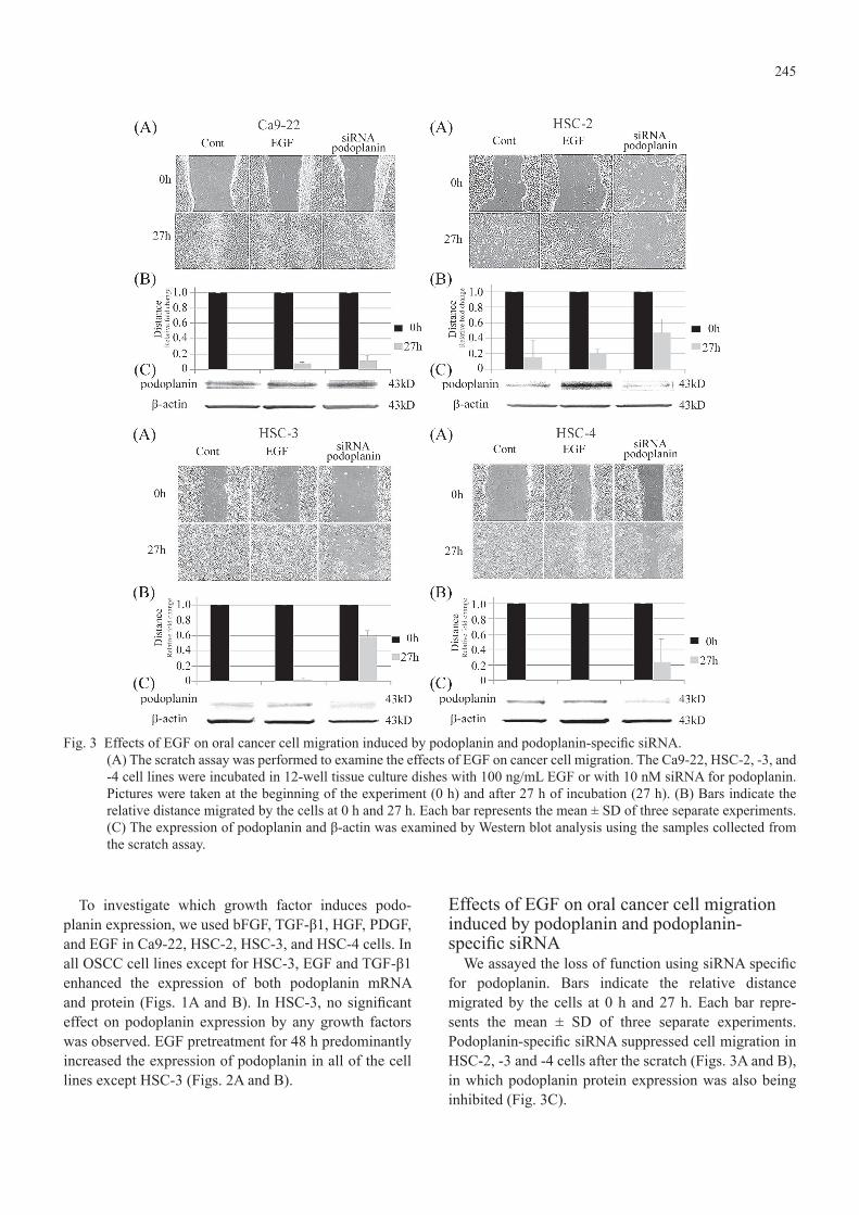
To investigate which growth factor induces podo-planin expression, we used bFGF, TGF-β1, HGF, PDGF, and EGF in Ca9-22, HSC-2, HSC-3, and HSC-4 cells. In all OSCC cell lines except for HSC-3, EGF and TGF-β1 enhanced the expression of both podoplanin mRNA and protein (Figs. 1A and B). In HSC-3, no significant effect on podoplanin expression by any growth factors was observed. EGF pretreatment for 48 h predominantly increased the expression of podoplanin in all of the cell lines except HSC-3 (Figs. 2A and B).
Effects of EGF on oral cancer cell migration induced by podoplanin and podoplanin-specific siRNA
We assayed the loss of function using siRNA specific for podoplanin. Bars indicate the relative distance migrated by the cells at 0 h and 27 h. Each bar repre-sents the mean ± SD of three separate experiments. Podoplanin-specific siRNA suppressed cell migration in HSC-2, -3 and -4 cells after the scratch (Figs. 3A and B), in which podoplanin protein expression was also being inhibited (Fig. 3C).
Fig. 3 Effects of EGF on oral cancer cell migration induced by podoplanin and podoplanin-specific siRNA.(A) The scratch assay was performed to examine the effects of EGF on cancer cell migration. The Ca9-22, HSC-2, -3, and -4 cell lines were incubated in 12-well tissue culture dishes with 100 ng/mL EGF or with 10 nM siRNA for podoplanin. Pictures were taken at the beginning of the experiment (0 h) and after 27 h of incubation (27 h). (B) Bars indicate the relative distance migrated by the cells at 0 h and 27 h. Each bar represents the mean ± SD of three separate experiments. (C) The expression of podoplanin and β-actin was examined by Western blot analysis using the samples collected from the scratch assay.
246
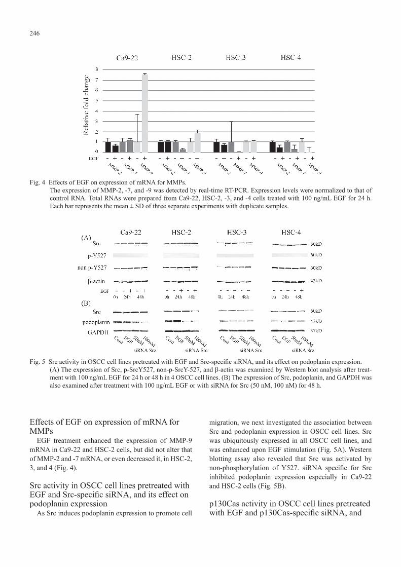
Effects of EGF on expression of mRNA for MMPs
EGF treatment enhanced the expression of MMP-9 mRNA in Ca9-22 and HSC-2 cells, but did not alter that of MMP-2 and -7 mRNA, or even decreased it, in HSC-2, 3, and 4 (Fig. 4).
Src activity in OSCC cell lines pretreated with EGF and Src-specific siRNA, and its effect on podoplanin expression
As Src induces podoplanin expression to promote cell
migration, we next investigated the association between Src and podoplanin expression in OSCC cell lines. Src was ubiquitously expressed in all OSCC cell lines, and was enhanced upon EGF stimulation (Fig. 5A). Western blotting assay also revealed that Src was activated by non-phosphorylation of Y527. siRNA specific for Src inhibited podoplanin expression especially in Ca9-22 and HSC-2 cells (Fig. 5B).
p130Cas activity in OSCC cell lines pretreated with EGF and p130Cas-specific siRNA, and
Fig. 4 Effects of EGF on expression of mRNA for MMPs.The expression of MMP-2, -7, and -9 was detected by real-time RT-PCR. Expression levels were normalized to that of control RNA. Total RNAs were prepared from Ca9-22, HSC-2, -3, and -4 cells treated with 100 ng/mL EGF for 24 h. Each bar represents the mean ± SD of three separate experiments with duplicate samples.
Fig. 5 Src activity in OSCC cell lines pretreated with EGF and Src-specific siRNA, and its effect on podoplanin expression.(A) The expression of Src, p-SrcY527, non-p-SrcY-527, and β-actin was examined by Western blot analysis after treat-ment with 100 ng/mL EGF for 24 h or 48 h in 4 OSCC cell lines. (B) The expression of Src, podoplanin, and GAPDH was also examined after treatment with 100 ng/mL EGF or with siRNA for Src (50 nM, 100 nM) for 48 h.

247
its effect on podoplanin expressionp130Cas downstream of Src and podoplanin was
investigated in OSCC. p130Cas was ubiquitously expressed in all OSCC cell lines, and enhanced upon EGF stimulation (Fig. 6A). siRNA specific for p130Cas inhibited the expression of both podoplanin mRNA and protein, especially in HSC-2 cells (Figs. 6B and C).
Effects of PP2 on podoplanin expressionPP2, known to be a Src inhibitor, did not affect the
expression of Src itself. Podoplanin expression was
inhibited in a dose-dependent manner in all of the OSCC cell lines (Fig. 7).
DiscussionAmong the growth factors related to receptor tyrosine
kinases, few studies have examined the association between HGF or PDGF and OSCC, in comparison with that of EGF. HGF acts mainly as a paracrine factor in head and neck SCC (HNSCC) cells, the HGF/c-Met pathway being frequently up-regulated and functional in HNSCC (22). c-Src and c-Met interactions are distinct
Fig. 6 p130Cas activity in OSCC cell lines pretreated with EGF and p130Cas-specific siRNA, and its effect on podoplanin expression.(A) The expression of p130Cas and β-actin was examined by Western blot analysis after treatment with 100 ng/mL EGF for 24 h or 48 h in 4 OSCC cell lines. (B) The expression of podoplanin mRNA was detected by real-time RT-PCR. Expression levels were normalized to that of control RNA. Total RNAs were prepared from Ca9-22, HSC-2, -3, and -4 cells treated with 100 ng/mL EGF or with siRNA for p130Cas (50 nM and 100 nM) for 48 h. Each bar represents the mean ± SD of three separate experiments with duplicate samples. (C) The expression of p130Cas, podoplanin, and GAPDH was examined by Western blot analysis after treatment with 100 ng/mL EGF and siRNA for p130Cas (50 nM and 100 nM) for 48 h.
Fig. 7 Effects of PP2 on podoplanin expression.The expression of Src, podoplanin, and GAPDH was examined by Western blot analysis after treatment with PP2, a known inhibitor of Src (0 μM, 20 μM, and 40 μM) for 48 h in 4 OSCC cell lines.

248
in HNSCC (23). HGF is mitogenic and motogenic for human HNSCC cell lines (24). Incubation of HNSCC cells in the presence of HGF leads to a rapid increase in phosphorylation of both FAK and the growth-promoting kinase Erk. ERK MAPKs, one of the major three subfam-ilies of mitogen-activated protein kinases (MAPKs), are located downstream from the growth factors EGF, NGF and PDGF (25). Nevertheless, our present study demon-strated that among the growth factors related to SFK receptors, only EGF markedly enhanced the expression of podoplanin, especially at 48 h after the start of treat-ment. In addition, it is noteworthy that TGF-β1, which is not one of the SFKs and is one of the most extensively investigated cytokines known to induce EMT (26), also showed an increase in its expression. This finding indi-cates that there may be a podoplanin-dependent signaling pathway via TGF-β1 in OSCC. A recent study has indicated that TGF-β1 and STAT-3 induce podoplanin expression in keratinocytes, which might be significantly involved in the process of wound healing (27).
The scratch assay and examination of MMP mRNA expression demonstrated obvious podoplanin-dependent cell migration in OSCC. MMPs, which are secreted from both tumor and/or stroma cells, play a role in tumor
invasion and metastasis by degrading the extracellular matrix (ECM) surrounding the tumor, especially the basement membranes (28). Emerging evidence indicates that MMPs can stimulate processes associated with EMT to enhance tumor cell invasion and metastasis potential (29). Most reports have suggested that predominant expression of MMP-2, -3 and -9 proteins is correlated with a poorer prognosis (30). MMP-9, known as type IV collagenase, is particularly abundant in basement membranes and the metastatic potential of carcinoma might correlate with the degree of enzymatic degradation of basement membrane type IV collagen (31,32). In the present study, the expression of MMP-9 was enhanced by EGF treatment in the Ca9-22 and HSC-2 cell lines, which was consistent with a previous study of HNSCC indi-cating that activation of EGFR promotes cell migration and invasion through production of MMP-9, followed by degradation of E-cadherin (33). Although emerging evidence suggests that SFKs may mediate MMP activity (34), further studies to clarify the correlation between Src and MMP activities in OSCC cell lines are required.
Podoplanin might facilitate tumor invasion through its ability to promote actin cytoskeleton remodeling of tumor cells, contributing to their increased motility
Fig. 8 The EGF-Src-Cas pathway in OSCC cell lines.The EGF-Src-Cas pathway in OSCC cell lines is related to cytoskeletal reorganization, resulting in enhancement of cell migration. EGF: epidermal growth factors, EGFR: epidermal growth factor receptor, Y-527: the 527th tyrosine, SH2: Src homology 2, SH3: Src homology 3, ERM: ERM proteins such as ezrin, radixin and moesin.

249
(35). The association between podoplanin and the actin cytoskeleton seems to be mediated by ezrin, which is markedly phosphorylated in the presence of podoplanin overexpression (11,36,37). Our series of studies on the EGF-Src-Cas-podoplanin signaling pathway have revealed that oral cancer cells express Src and p130Cas ubiquitously. Activation of both Src and p130Cas is important for inducing podoplanin expression. There-fore, podoplanin utilizes Src and p130Cas to reorganize the cancer cell cytoskeleton and may be associated with tumor cell invasion in the oral cavity.
The biological function of podoplanin may vary among different types of cancer. It has been reported that podoplanin can induce Rho A activation and EMT in MDCK cells (37), whereas it attenuates Rho A activity and does not induce EMT in a breast carcinoma cell line (11). These contradictory findings suggest that the func-tions of podoplanin are cell type-specific (38). The results of our experimental studies suggest that the expression of podoplanin promotes cell migration, and that the EGF-Src-Cas pathway (Fig. 8) is one of the mechanisms involved in cancer progression in the oral cavity.
References 1. Schacht V, Ramirez MI, Hong YK, Hirakawa S,
Feng D, Harvey N, Williams M, Dvorak AM, Dvorak HF, Oliver G, Detmar M (2003) T1α/podoplanin deficiency disrupts normal lymphatic vasculature formation and causes lymphedema. EMBO J 22, 3546-3556.
2. Breiteneder-Geleff S, Matsui K, Soleiman A, Meraner P, Poczewski H, Kalt R, Schaffner G, Kerjaschki D (1997) Podoplanin, novel 43-kd membrane protein of glomerular epithelial cells, is down-regulated in puromycin nephrosis. Am J Pathol 151, 1141-1152.
3. Erovic BM, Neuchrist C, Kandutsch S, Woegerbauer M, Pammer J (2003) CD9 expression on lymphatic vessels in head and neck mucosa. Mod Pathol 16, 1028-1034.
4. Schacht V, Dadras SS, Johnson LA, Jackson DG, Hong YK, Detmar M (2005) Up-regulation of the lymphatic marker podoplanin, a mucin-type transmembrane glycoprotein, in human squamous cell carcinomas and germ cell tumors. Am J Pathol 166, 913-921.
5. Yuan P, Temam S, El-Naggar A, Zhou X, Liu DD, Lee JJ, Mao L (2006) Overexpression of podo-planin in oral cancer and its association with poor clinical outcome. Cancer 107, 563-569.
6. Inoue H, Miyazaki Y, Kikuchi K, Yoshida N, Ide
F, Ohmori Y, Tomomura A, Sakashita H, Kusama K (2012) Podoplanin expression during dysplasia-carcinoma sequence in the oral cavity. Tumor Biol 33, 183-194.
7. Klymkowsky MW, Savagner P (2009) Epithelial-mesenchymal transition: a cancer researcher’s conceptual friend and foe. Am J Pathol 174, 1588-1593.
8. Polyak K, Haviv I, Campbell IG (2009) Co-evolu-tion of tumor cells and their microenvironment. Trends Genet 25, 30-38.
9. Thiery JP, Acloque H, Huang RY, Nieto MA (2009) Epithelial-mesenchymal transitions in develop-ment and disease. Cell 139, 871-890.
10. Martín-Villar E, Scholl FG, Gamallo C, Yurrita MM, Muñoz-Guerra M, Cruces J, Quintanilla M (2005) Characterization of human PA2.26 antigen (T1α-2, podoplanin), a small membrane mucin induced in oral squamous cell carcinomas. Int J Cancer 113, 899-910.
11. Wicki A, Lehembre F, Wick N, Hantusch B, Kerjaschki D, Christofori G (2006) Tumor inva-sion in the absence of epithelial-mesenchymal transition: podoplanin-mediated remodeling of the actin cytoskeleton. Cancer Cell 9, 261-272.
12. Sen B, Johnson FM (2011) Regulation of Src family kinases in human cancers. J Signal Trans-duct 2011, 865819.
13. Summy JM, Gallick GE (2003) Src family kinases in tumor progression and metastasis. Cancer Metastasis Rev 22, 337-358.
14. Abram CL, Courtneidge SA (2000) Src family tyrosine kinases and growth factor signaling. Exp Cell Res 254, 1-13.
15. Kasai A, Shima T, Okada M (2005) Role of Src family tyrosine kinases in the down-regulation of epidermal growth factor signaling in PC12 cells. Genes Cells 10, 1175-1187.
16. O’Neill GM, Fashena SJ, Golemis EA (2000) Inte-grin signalling: a new Cas(t) of characters enters the stage. Trends Cell Biol 10, 111-119.
17. Sakai R, Iwamatsu A, Hirano N, Ogawa S, Tanaka T, Mano H, Yazaki Y, Hirai H (1994) A novel signaling molecule, p130, forms stable complexes in vivo with v-Crk and v-Src in a tyrosine phosphorylation-dependent manner. EMBO J 13, 3748-3756.
18. Sakai R, Nakamoto T, Ozawa K, Aizawa S, Hirai H (1997) Characterization of the kinase activity essential for tyrosine phosphorylation of p130Cas in fibroblasts. Oncogene 14, 1419-1426.
250
19. Cary LA, Han DC, Polte TR, Hanks SK, Guan JL (1998) Identification of p130Cas as a mediator of focal adhesion kinase-promoted cell migration. J Cell Biol 140, 211-221.
20. Klemke RL, Leng J, Molander R, Brooks PC, Vuori K, Cheresh DA (1998) CAS/Crk coupling serves as a “molecular switch” for induction of cell migration. J Cell Biol 140, 961-972.
21. Shen Y, Chen CS, Ichikawa H, Goldberg GS (2010) SRC induces podoplanin expression to promote cell migration. J Biol Chem 285, 9649-9656.
22. Knowles LM, Stabile LP, Egloff AM, Rothstein ME, Thomas SM, Gubish CT, Lerner EC, Seethala RR, Suzuki S, Quesnelle KM, Morgan S, Ferris RL, Grandis JR, Siegfried JM (2009) HGF and c-Met participate in paracrine tumorigenic path-ways in head and neck squamous cell cancer. Clin Cancer Res 15, 3740-3750.
23. Sen B, Peng S, Saigal B, Williams MD, Johnson FM (2011) Distinct interactions between c-Src and c-Met in mediating resistance to c-Src inhibition in head and neck cancer. Clin Cancer Res 17, 514-524.
24. Fleigel J, Sedwick J, Kornberg LJ (2002) Hepa-tocyte growth factor/scatter factor stimulates mitogenesis and migration of a head and neck squamous cell carcinoma cell line. Otolaryngol Head Neck Surg 127, 271-278.
25. Mishima K, Inoue K, Hayashi Y (2002) Overex-pression of extracellular-signal regulated kinases on oral squamous cell carcinoma. Oral Oncol 38, 468-474.
26. Richter P, Umbreit C, Franz M, Berndt A, Grimm S, Uecker A, Böhmer FD, Kosmehl H, Berndt A (2011) EGF/TGFβ1 co-stimulation of oral squamous cell carcinoma cells causes an epithelial-mesenchymal transition cell phenotype expressing laminin 332. J Oral Pathol Med 40, 46-54.
27. Honma M, Minami-Hori M, Takahashi H, Iizuka H (2012) Podoplanin expression in wound and hyperproliferative psoriatic epidermis: regulation by TGF-β and STAT-3 activating cytokines, IFN-γ, IL-6, and IL-22. J Dermatol Sci 65, 134-140.
28. Tokuraku M, Sato H, Murakami S, Okada Y, Watanabe Y, Seiki M (1995) Activation of the precursor of gelatinase A/72 kDa type IV collage-nase/MMP-2 in lung carcinomas correlates with the expression of membrane-type matrix metal-loproteinase (MT-MMP) and with lymph node metastasis. Int J Cancer 64, 355-359.
29. Orlichenko LS, Radisky DC (2008) Matrix metal-loproteinases stimulate epithelial-mesenchymal transition during tumor development. Clin Exp Metastasis 25, 593-600.
30. Lin CY, Tsai PH, Kandaswami CC, Lee PP, Huang CJ, Hwang JJ, Lee MT (2011) Matrix metallo-proteinase-9 cooperates with transcription factor Snail to induce epithelial-mesenchymal transition. Cancer Sci 102, 815-827.
31. Thomas GT, Lewis MP, Speight PM (1999) Matrix metalloproteinases and oral cancer. Oral Oncol 35, 227-233.
32. Kurahara S, Shinohara M, Ikebe T, Nakamura S, Beppu M, Hiraki A, Takeuchi H, Shirasuna K (1999) Expression of MMPs, MT-MMP, and TIMPs in squamous cell carcinoma of the oral cavity: correlations with tumor invasion and metastasis. Head Neck 21, 627-638.
33. Zuo JH, Zhu W, Li MY, Li XH, Yi H, Zeng GQ, Wan XX, He QY, Li JH, Qu JQ, Chen Y, Xiao ZQ (2011) Activation of EGFR promotes squamous carcinoma SCC10A cell migration and invasion via inducing EMT-like phenotype change and MMP-9-mediated degradation of E-cadherin. J Cell Biochem 112, 2508-2517.
34. Razandi M, Pedram A, Park ST, Levin ER (2003) Proximal events in signaling by plasma membrane estrogen receptors. J Biol Chem 278, 2701-2712.
35. Moustakas A, Heldin CH (2007) Signaling networks guiding epithelial-mesenchymal transitions during embryogenesis and cancer progression. Cancer Sci 98, 1512-1520.
36. Scholl FG, Gamallo C, Vilaró S, Quintanilla M (1999) Identification of PA2.26 antigen as a novel cell-surface mucin-type glycoprotein that induces plasma membrane extensions and increased motility in keratinocytes. J Cell Sci 112, 4601-4613.
37. Martín-Villar E, Megías D, Castel S, Yurrita MM, Vilaró S, Quintanilla M (2006) Podoplanin binds ERM proteins to activate RhoA and promote epithelial-mesenchymal transition. J Cell Sci 119, 4541-4553.
38. Suzuki H, Onimaru M, Yonemitsu Y, Maehara Y, Nakamura S, Sueishi K (2010) Podoplanin in cancer cells is experimentally able to attenuate prolymphangiogenic and lymphogenous meta-static potentials of lung squamoid cancer cells. Mol Cancer 9, 287.