PNS in the Nervous System - Sinoe Medical Association...
27
1 The Peripheral Nervous System (PNS) PNS in the Nervous System Peripheral Nervous System (PNS) § PNS – all neural structures outside the brain and spinal cord § Includes sensory receptors, peripheral nerves, associated ganglia, and motor endings § Provides links to and from the external environment PNS in the Nervous System Sensory Receptors § Structures specialized to respond to stimuli § Activation of sensory receptors results in depolarizations that trigger impulses to the CNS § The realization of these stimuli, sensation and perception, occur in the brain Receptor Classification by Stimulus Type § Mechanoreceptors – respond to touch, pressure, vibration, stretch, and itch § Thermoreceptors – sensitive to changes in temperature § Photoreceptors – respond to light energy (e.g., retina) § Chemoreceptors – respond to chemicals (e.g., smell, taste, changes in blood chemistry) § Nociceptors – sensitive to paincausing stimuli Receptor Class by Location: Exteroceptors § Respond to stimuli arising outside the body § Found near the body surface § Sensitive to touch, pressure, pain, and temperature § Include the special sense organs Receptor Class by Location: Interoceptors § Respond to stimuli arising within the body § Found in internal viscera and blood vessels § Sensitive to chemical changes, stretch, and temperature changes
Transcript of PNS in the Nervous System - Sinoe Medical Association...
1
The Peripheral Nervous System (PNS)
PNS in the Nervous System
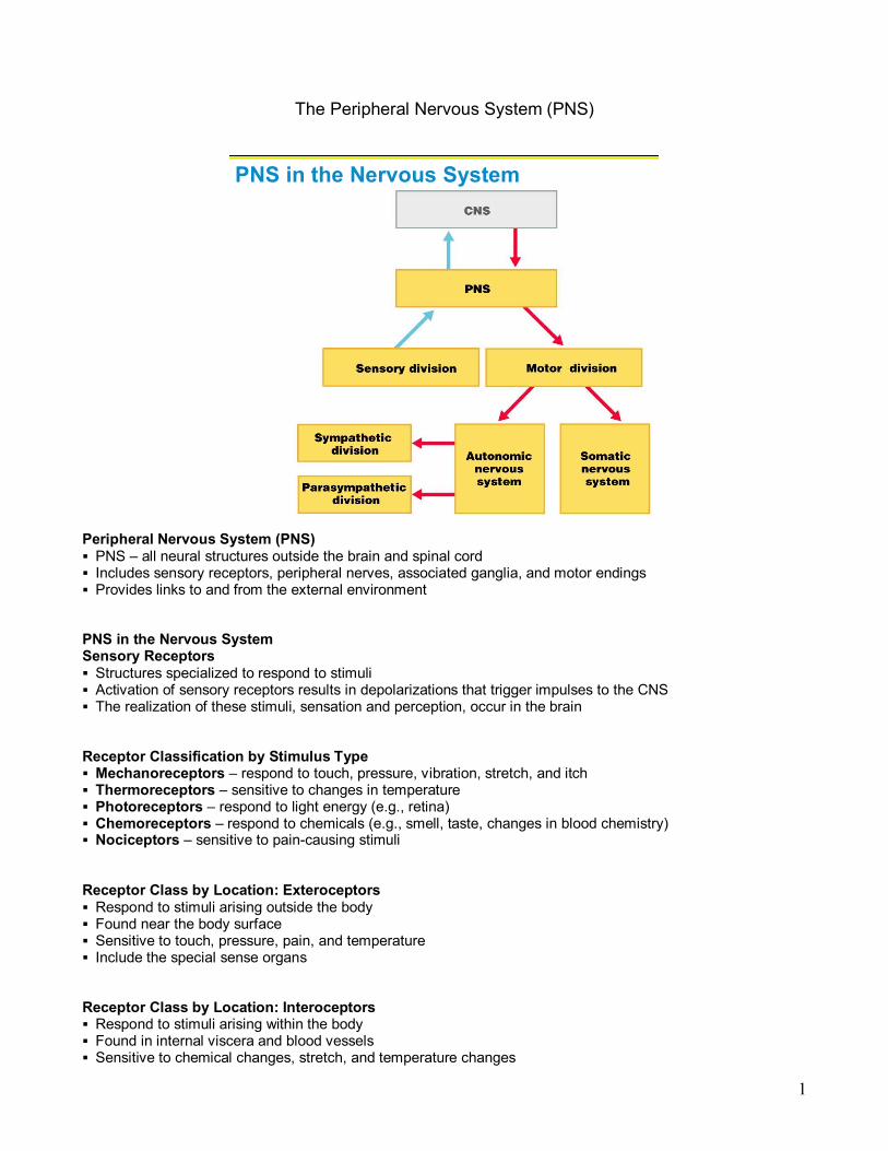
Peripheral Nervous System (PNS) § PNS – all neural structures outside the brain and spinal cord § Includes sensory receptors, peripheral nerves, associated ganglia, and motor endings § Provides links to and from the external environment
PNS in the Nervous System Sensory Receptors § Structures specialized to respond to stimuli § Activation of sensory receptors results in depolarizations that trigger impulses to the CNS § The realization of these stimuli, sensation and perception, occur in the brain
Receptor Classification by Stimulus Type § Mechanoreceptors – respond to touch, pressure, vibration, stretch, and itch § Thermoreceptors – sensitive to changes in temperature § Photoreceptors – respond to light energy (e.g., retina) § Chemoreceptors – respond to chemicals (e.g., smell, taste, changes in blood chemistry) § Nociceptors – sensitive to paincausing stimuli
Receptor Class by Location: Exteroceptors § Respond to stimuli arising outside the body § Found near the body surface § Sensitive to touch, pressure, pain, and temperature § Include the special sense organs
Receptor Class by Location: Interoceptors § Respond to stimuli arising within the body § Found in internal viscera and blood vessels § Sensitive to chemical changes, stretch, and temperature changes

2
Receptor Class by Location: Proprioceptors § Respond to degree of stretch of the organs they occupy § Found in skeletal muscles, tendons, joints, ligaments, and connective tissue coverings of bones and muscles § Constantly “advise” the brain of one’s movements
Receptor Classification by Structural Complexity § Receptors are structurally classified as either simple or complex § Most receptors are simple and include encapsulated and unencapsulated varieties § Complex receptors are special sense organs
Simple Receptors: Encapsulated
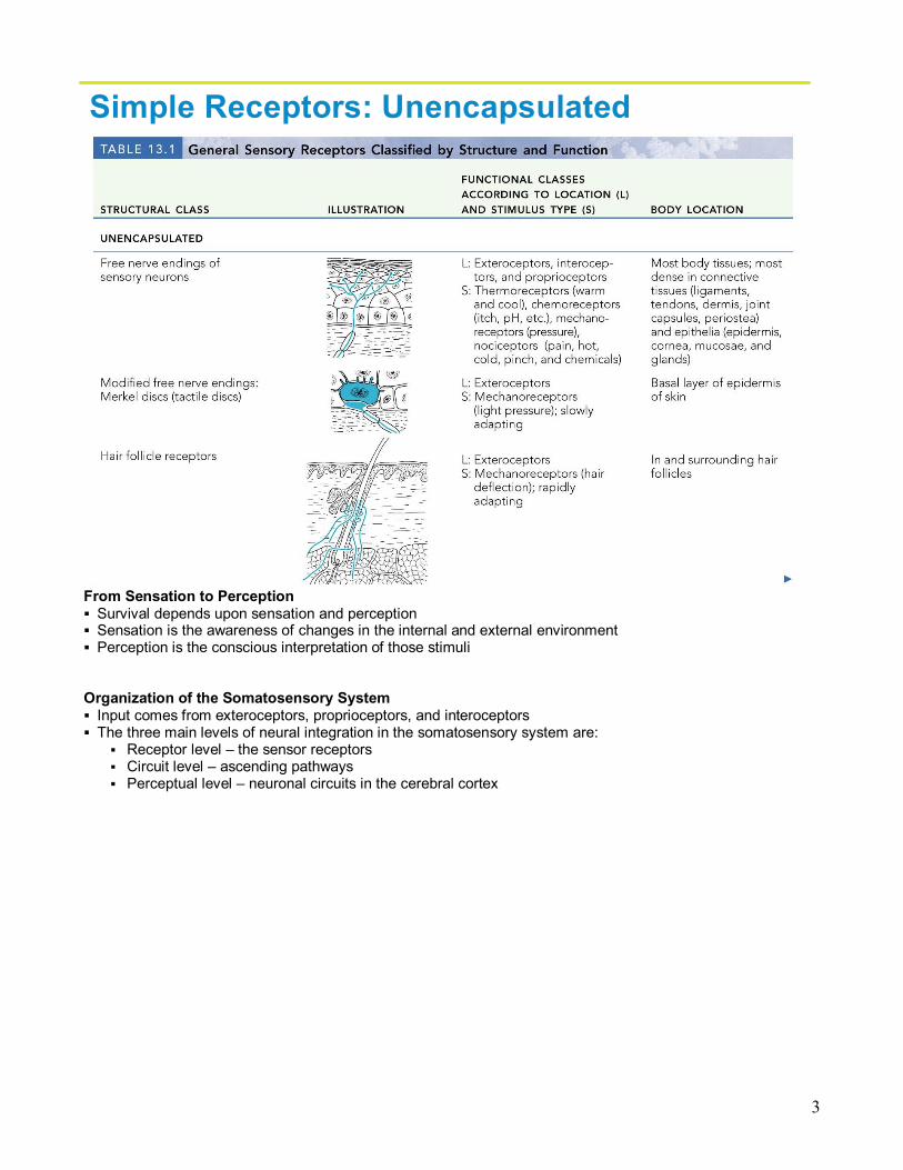
Table 13.1.2 Simple Receptors: Unencapsulated § Free dendritic nerve endings
§ Respond chiefly to temperature and pain § Merkel (tactile) discs § Hair follicle receptors § Meissner’s corpuscles (tactile corpuscles) § Pacinian corpuscles (lamellated corpuscles) § Muscle spindles, Golgi tendon organs, and Ruffini’s corpuscles § Joint kinesthetic receptors
3
Simple Receptors: Unencapsulated
From Sensation to Perception § Survival depends upon sensation and perception § Sensation is the awareness of changes in the internal and external environment § Perception is the conscious interpretation of those stimuli
Organization of the Somatosensory System § Input comes from exteroceptors, proprioceptors, and interoceptors § The three main levels of neural integration in the somatosensory system are:
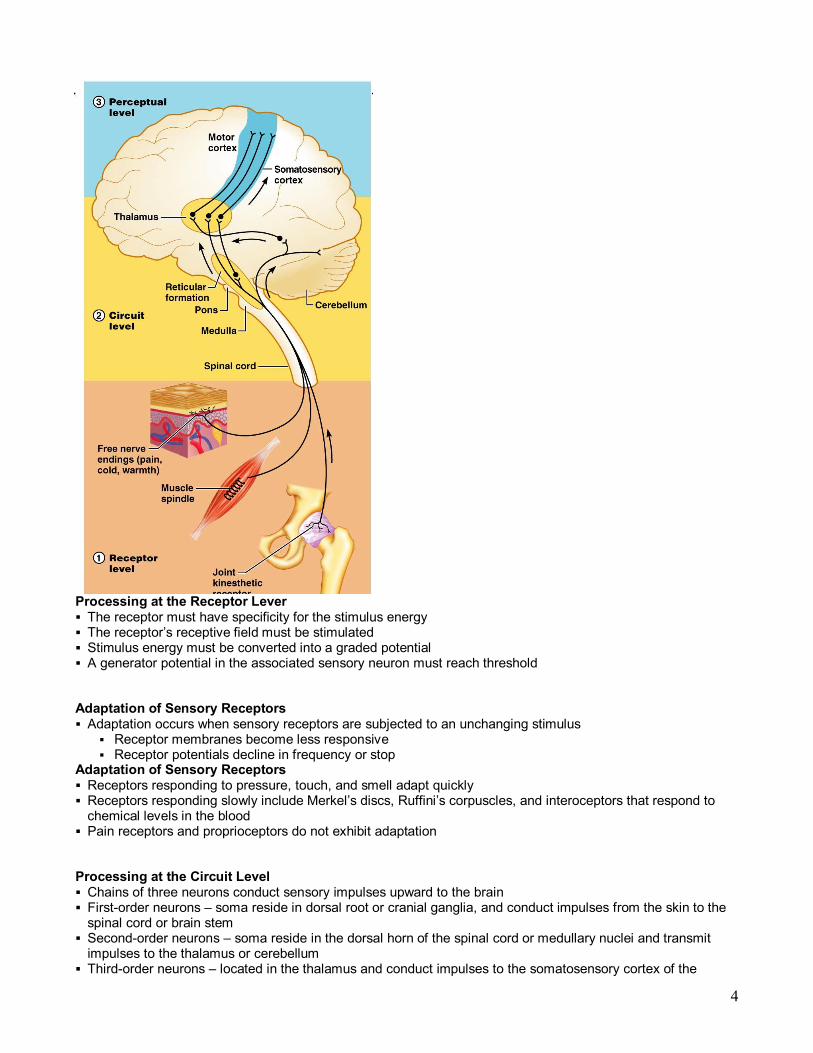
§ Receptor level – the sensor receptors § Circuit level – ascending pathways § Perceptual level – neuronal circuits in the cerebral cortex
4
Processing at the Receptor Lever § The receptor must have specificity for the stimulus energy § The receptor’s receptive field must be stimulated § Stimulus energy must be converted into a graded potential § A generator potential in the associated sensory neuron must reach threshold
Adaptation of Sensory Receptors § Adaptation occurs when sensory receptors are subjected to an unchanging stimulus
§ Receptor membranes become less responsive § Receptor potentials decline in frequency or stop
Adaptation of Sensory Receptors § Receptors responding to pressure, touch, and smell adapt quickly § Receptors responding slowly include Merkel’s discs, Ruffini’s corpuscles, and interoceptors that respond to chemical levels in the blood
§ Pain receptors and proprioceptors do not exhibit adaptation
Processing at the Circuit Level § Chains of three neurons conduct sensory impulses upward to the brain § Firstorder neurons – soma reside in dorsal root or cranial ganglia, and conduct impulses from the skin to the spinal cord or brain stem
§ Secondorder neurons – soma reside in the dorsal horn of the spinal cord or medullary nuclei and transmit impulses to the thalamus or cerebellum
§ Thirdorder neurons – located in the thalamus and conduct impulses to the somatosensory cortex of the

5
cerebrum § The thalamus projects fibers to:
§ The somatosensory cortex § Sensory association areas
§ First one modality is sent, then those considering more than one § The result is an internal, conscious image of the stimulus
Main Aspects of Sensory Perception § Perceptual detection – detecting that a stimulus has occurred and requires summation § Magnitude estimation – how much of a stimulus is acting § Spatial discrimination – identifying the site or pattern of the stimulus § Feature abstraction – used to identify a substance that has specific texture or shape § Quality discrimination – the ability to identify submodalities of a sensation (e.g., sweet or sour tastes) § Pattern recognition – ability to recognize patterns in stimuli (e.g., melody, familiar face)
Structure of a Nerve § Nerve – cordlike organ of the PNS consisting of peripheral axons enclosed by connective tissue § Connective tissue coverings include:
§ Endoneurium – loose connective tissue that surrounds axons § Perineurium – coarse connective tissue that bundles fibers into fascicles § Epineurium – tough fibrous sheath around a nerve
Classification of Nerves § Sensory and motor divisions § Sensory (afferent) – carry impulse to the CNS § Motor (efferent) – carry impulses from CNS § Mixed – sensory and motor fibers carry impulses to and from CNS; most common type of nerve § Mixed nerves – carry somatic and autonomic (visceral) impulses § The four types of mixed nerves are:
§ Somatic afferent and somatic efferent § Visceral afferent and visceral efferent
§ Peripheral nerves originate from the brain or spinal column
Regeneration of Nerve Fibers § Damage to nerve tissue is serious because mature neurons are amitotic § If the soma of a damaged nerve remains intact, damage can be repaired § Regeneration involves coordinated activity among:
§ Macrophages – remove debris § Schwann cells – form regeneration tube and secrete growth factors § Axons – regenerate damaged part
Cranial Nerves
6
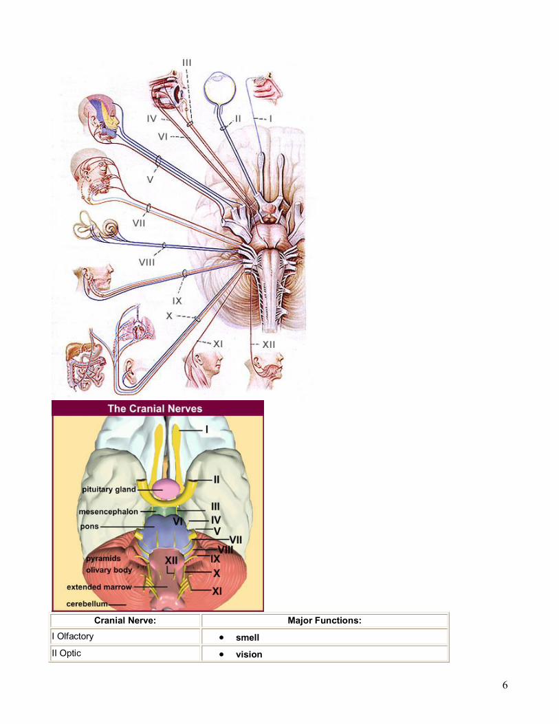
Cranial Nerve: Major Functions: I Olfactory • smell II Optic • vision

7
III Oculomotor • eyelid and eyeball movement
IV Trochlear • innervates superior oblique
• turns eye downward and laterally
V Trigeminal • chewing face & mouth touch & pain
VI Abducens • turns eye laterally
VII Facial
• controls most facial expressions
• secretion of tears & saliva
• taste
VIII Vestibulocochlear (auditory)
• hearing
• equillibrium sensation
IX Glossopharyngeal • taste
• senses carotid blood pressure
X Vagus
• senses aortic blood pressure
• slows heart rate
• stimulates digestive organs
• taste
XI Spinal Accessory • controls trapezius & sternocleidomastoid
• controls swallowing movements XII Hypoglossal • controls tongue movements
Nerves in Order Modality Function Olfactory Special Sensory Smell
Optic Special Sensory Vision
Oculomotor Somatic Motor
Visceral Motor
Levator palpebrae, superioris, superior, medial & inferior recti muscles
Parasympathetic to ciliary & pupillary constrictor muscles
Trochlear Somatic Motor Superior oblique muscle
Trigeminal Branchial Motor Muscles of mastication

8
General Sensory Sensory for head/neck, sinuses, meninges, & external surface of tympanic membrane
Abducens Somatic Motor Lateral rectus muscle Facial Branchial Motor
Visceral Motor
General Sensory
Special Sensory
Muscles of facial expression
Parasympathetic to all glands of head except the parotid
Sensory for ear and tympanic membrane
Taste anterior twothirds of tongue Vestibulocochlear Special Sensory Hearing and Balance
Glossopharyngeal
Branchial Motor
Visceral Motor
Visceral Sensory
General Sensory
Special Sensory
Stylopharyngeus muscle
Parotid Gland
Carotid Body
Sensation posterior onethird tongue & internal surface of tympanic membrane.
Taste posterior onethird tongue
Vagus
Branchial Motor
Visceral Motor
Visceral Sensory
Special Sensory
Muscles pharynx & larynx
Parasympathetic to neck, thorax, & abdomen
Sensory from pharynx, larynx & viscera
Sensory from external ear Spinal Accessory Branchial Motor Trapezius & sternocleidomastoid muscles Hypoglossal Somatic Motor Tongue muscles except palatoglossal
Cranial Nerves § Twelve pairs of cranial nerves arise from the brain § They have sensory, motor, or both sensory and motor functions § Each nerve is identified by a number (I through XII) and a name § Four cranial nerves carry parasympathetic fibers that serve muscles and glands
"On Old Olympic Towering Tops A Finn And German Viewed Some Hops"

9
Cranial Nerves
Summary of Function of Cranial Nerves
Summary of Function of Cranial Nerves
10
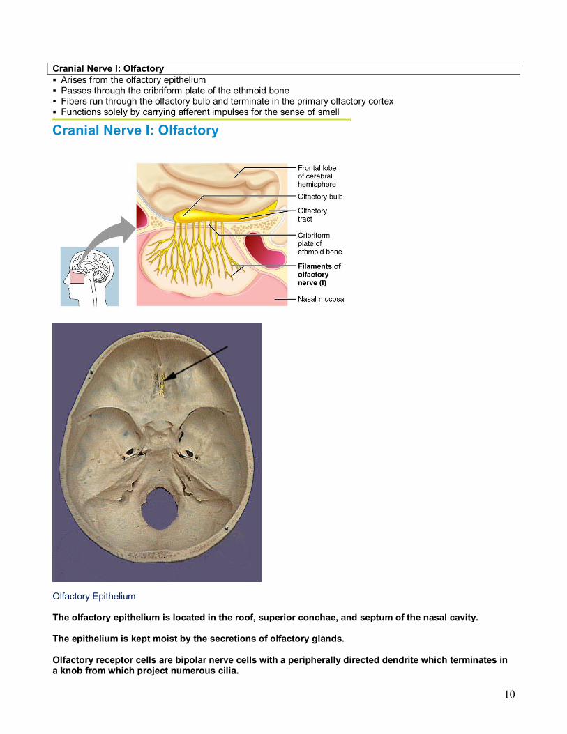
Cranial Nerve I: Olfactory § Arises from the olfactory epithelium § Passes through the cribriform plate of the ethmoid bone § Fibers run through the olfactory bulb and terminate in the primary olfactory cortex § Functions solely by carrying afferent impulses for the sense of smell
Cranial Nerve I: Olfactory
Olfactory Epithelium
The olfactory epithelium is located in the roof, superior conchae, and septum of the nasal cavity.
The epithelium is kept moist by the secretions of olfactory glands.
Olfactory receptor cells are bipolar nerve cells with a peripherally directed dendrite which terminates in a knob from which project numerous cilia.

11
The olfactory chemoreceptors are located on these cilia.
Sensory Transduction and Peripheral Course of the Olfactory Nerve
Inhaled aromatic molecules dissolve in the moisture lining the olfactory epithelium and stimulate its chemoreceptors.
Olfactory receptor cells initiate action potentials in response to these chemical stimuli. Intracellular studies show the presence of a slow rising receptor (generator) potential followed by a spike discharge from the receptor cell.
The peripheral processes of the receptor cells assemble into small bundles and pass through the cribiform plate of the ethmoid bone to synapse on secondary sensory neurons in the olfactory bulb.

12
Copyright © 2006 Pearson Education, Inc., publishing as Benjamin Cummings
Olfactory Receptors
Figure 15.21
Cranial Nerve II: Optic § Arises from the retina of the eye § Optic nerves pass through the optic canals and converge at the optic chiasm § They continue to the thalamus where they synapse § From there, the optic radiation fibers run to the visual cortex § Functions solely by carrying afferent impulses for vision
Cranial Nerve II: Optic
Sensory Transduction
Light passing through the cornea and aqueous humor and entering the pupil travels through the lens and

13
vitreous body to reach the retina at the back of the eye.
The process of converting photons of light into electrical signals occurs in a deep layer of the retina which contains the photoreceptor cells the rods and cones.
Rods and cones are specialized cells which have stacks of plasma membrane associated with visual pigments making them sensitive to light.
The differences between the rod system and the cone system are described in the table below:
Rod system Cone system
High sensitivity, specialized for night vision
Lower sensitivity, specialized for day vision
Saturate in daylight Saturate only in intense light
Achromatic Chromatic, mediate color vision
Low acuity High acuity
Not present in central fovea
Concentrated in central fovea
Present in larger number than cones
Present in smaller number than rods
Sensory Transduction
Light incident on the photoreceptor cells triggers a series of chemical reactions which alter plasma membrane permeability resulting in a hyperpolarization of the rod or cone.
This hyperpolarization of the photoreceptor cell can produce either an excitatory (depolarization) or inhibitory (hyperpolarization) response by the bipolar cell dependent on the nature of the synapse between the two cells.
The bipolar cells are the primary sensory neurons of the visual pathway. They synapse with and either excite or inhibit the action potential firing rate of the secondary sensory neurons the ganglion cells.
The axons of the ganglion cells converge at the optic disc near the center of the retina to exit the eye as the optic nerve.
At the optic chiasm approximately 1/2 of the fibers from each optic nerve cross the midline and exit the chiasm in the opposite optic tract.
The fibers of the optic tracts continue posteriorly around the cerebral peduncles of the midbrain with most synapsing in the lateral geniculate nucleus of their respective thalamus. A small portion of the fibers enter the pretectal region of the midbrain and participate in the pupillary light reflex.
Cells of the lateral geniculate nuclei are tertiary sensory neurons which project to the primary visual cortex in the occipital lobe via the optic radiation (geniculocalcarine tract). Note that the axons of the

14
optic radiation fan out to pass above and lateral to the inferior horn of the lateral ventricles enroute to the visual cortex. The fibers that course anteriorly toward the pole of the temporal lobe before turning posteriorly are referred to as Meyer's loop.
Central course
The entire area seen by an eye when it is focused on a central point is called the visual field of that eye.
Because rays of light reach the retina by converging and passing through the small opening of the pupil, the image of the entire visual field is projected onto the retina upsidedown and reversed.
Therefore:
• The right half of the retina receives stimuli from the left visual field.
• The left half of the retina receives stimuli from the right half of the visual field.
• The upper half of the retina receives stimuli from the lower half of the visual field.
• The lower half of the retina receives stimuli from the upper half of the visual field. Retinal Projections to the Primary Visual Cortex
Because fibers from different quadrants of the retina project to the primary visual cortex via predictable portions of the optic nerves, chiasm, and tracts it is convenient and clinically useful to divide the retina (and therefore the visual field) of each eye into nasal and temporal halves as well as superior and inferior halves yielding four quadrants.
The regions of the retina are referenced to the midline. The nasal hemiretina lies medial to the fovea, while the temporal hemiretina lies lateral to the fovea. The superior and inferior halves of the retina are also referenced to the fovea.
Axons of ganglion cells from the nasal hemiretina (lateral visual field) decussate at the optic chiasm and project to the contralateral lateral geniculate nucleus and midbrain.
Axons from the temporal hemiretina (medial visual field) remain ipsilateral throughout their course.
Axons from the inferior half of the retina (upper visual field) project via the Meyer's loop/temporal lobe portion of the optic radiation to the primary visual cortex below the calcarine fissure.
Axons from the superior half of the retina (lower visual field) project via the parietal lobe portion of the optic radiation to the primary visual cortex above the calcarine fissure.
Ganglion cells from the center of the retina (fovea) project to the tip of the occipital pole.

15
Clinical Correlation Visual Deficits & Damage to the Retina
Visual Deficits
Armed with knowledge of the anatomy of the visual system, one can predict the deficits associated with a lesion at a particular point in the central visual pathway.
Damage to the retina
Results in a loss of input from the affected portion of the retina leading to a monocular field deficit.
Since axons of the ganglion cells converge toward the optic disc, damage to a portion of the retina closer to the optic disc will affect a greater number of neurons than would the same amount of damage in the peripheral retina leading to a larger visual field defect in that eye.
Since the cones are concentrated in the fovea, damage to the fovea results in a greater visual handicap than

16
damage to peripheral regions of the retina.
Damage to the optic nerve will also result in a monocular visual defect due to loss of input from the ipsilateral eye. The patient will complain of blindness in that eye.

17
Visual Pathways
damage to the optic nerve
Damage to the optic nerve will also result in a monocular visual defect due to loss of input from the ipsilateral eye. The patient will complain of blindness in that eye.
Damage to the optic nerve ipsilateral blindness.
Cranial Nerve III: Oculomotor § Fibers extend from the ventral midbrain, pass through the superior orbital fissure, and go to the extrinsic eye muscles
§ Functions in raising the eyelid, directing the eyeball, constricting the iris, and controlling lens shape § Parasympathetic cell bodies are in the ciliary ganglia

18
Cranial Nerve III: Oculomotor
Cranial Nerve IV: Trochlear § Fibers emerge from the dorsal midbrain and enter the orbits via the superior orbital fissures; innervate the superior oblique muscle
§ Primarily a motor nerve that directs the eyeball
Cranial Nerve IV: Trochlear
Cranial Nerve V: Trigeminal § Three divisions: ophthalmic (V1), maxillary (V2), and mandibular (V3) § Fibers run from the face to the pons via the superior orbital fissure (V1), the foramen rotundum (V2), and the foramen ovale (V3)
§ Conveys sensory impulses from various areas of the face (V1) and (V2), and supplies motor fibers (V3) for mastication

19
Cranial Nerve V: Trigeminal

20
• The trigeminal nerve as the name indicates is composed of three large branches.
• They are the ophthalmic (V1, sensory), maxillary (V2, sensory) and mandibular (V3, motor and sensory) branches.
• The large sensory root and smaller motor root leave the brainstem at the midlateral surface of pons.
• The sensory root terminates in the largest of the cranial nerve nuclei which extends from the pons all the way down into the second cervical level of the spinal cord. The sensory root joins the trigeminal or semilunar ganglion between the layers of the dura mater in a depression on the floor of the middle crania fossa.
• This depression is the location of the so called Meckle's cave.
• The motor root originates from cells located in the masticator motor nucleus of trigeminal nerve located in the midpons of the brainstem.
• The motor root passes through the trigeminal ganglion and combines with the corresponding sensory root to become the mandibular nerve. It is distributed to the muscles of mastication, the mylohyoid muscle and the anterior belly of the digastric.
• The mandibular nerve also innervates the tensor veli palatini and tensor tympani muscles.
• The three sensory branches of the trigeminal nerve emanate from the ganglia to form the three branches of the trigeminal nerve.
• The ophthalmic and maxillary branches travel in the wall of the cavernous sinus just prior to leaving the cranium.
• The ophthalmic branch travels through the superior orbital fissure and passes through the orbit to reach the skin of the forehead and top of the head.
• The maxillary nerve enters the cranium through the foramen rotundum via the pterygopalatine fossa. Its sensory branches reach the pterygopalatine fossa via the inferior orbital fissure (face, cheek and upper teeth) and pterygopalatine canal (soft and hard palate, nasal cavity and pharynx).
• There are also meningeal sensory branches that enter the trigeminal ganglion within the cranium.
• The sensory part of the mandibular nerve is composed of branches that carry general sensory information from the mucous membranes of the mouth and cheek, anterior twothirds of the tongue, lower teeth, skin of the lower jaw, side of the head and scalp and meninges of the anterior and middle cranial fossae.
OPHTALMIC BRANCH A. Infratrochlear B. Anterior Ethmoid C. Posterior Ethmoid D. Lacrimal E. Supraorbital F. Supratrochlear

21
G. Nasociliary
Maxillary Nerve Branches
A. Zygoticaticotemporal B. Zygomaticofacial C. Post. Sup. Alveolar Brs D. Nasopalatine E. Greater Palatine F. Lesser Palatine G. Mid. & Ant. Alveolar Brs H. Infraorbital
Mandibular Nerve Branches

22
A. Auriculotemporal B. Lingual C. Inferior Alveolar D. N. to the Mylohyoid E. Mental F. Buccal
Cranial Nerve VI: Abdcuens § Fibers leave the inferior pons and enter the orbit via the superior orbital fissure § Primarily a motor nerve innervating the lateral rectus muscle
Cranial Nerve VI: Abdcuens
§ Fibers leave the inferior pons and enter the orbit via the superior orbital fissure
§ Primarily a motor nerve innervating the lateral rectus muscle
Cranial Nerve VII: Facial § Fibers leave the pons, travel through the internal acoustic meatus, and emerge through the stylomastoid foramen to the lateral aspect of the face
§ Mixed nerve with five major branches § Motor functions include facial expression, and the transmittal of autonomic impulses to lacrimal and salivary glands
§ Sensory function is taste from the anterior twothirds of the tongue

23
Cranial Nerve VIII: Vestibulocochlear § Fibers arise from the hearing and equilibrium apparatus of the inner ear, pass through the internal acoustic meatus, and enter the brainstem at the ponsmedulla border
§ Two divisions – cochlear (hearing) and vestibular (balance) § Functions are solely sensory – equilibrium and hearing
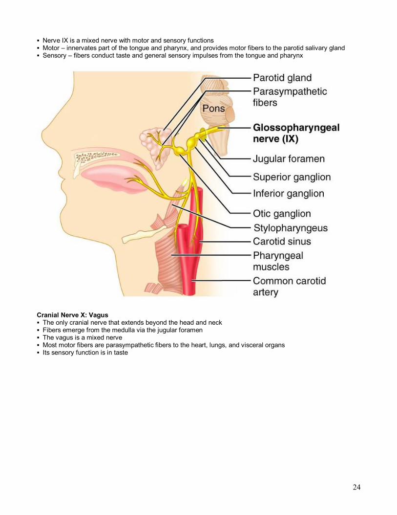
Cranial Nerve IX: Glossopharyngeal § Fibers emerge from the medulla, leave the skull via the jugular foramen, and run to the throat
24
§ Nerve IX is a mixed nerve with motor and sensory functions § Motor – innervates part of the tongue and pharynx, and provides motor fibers to the parotid salivary gland § Sensory – fibers conduct taste and general sensory impulses from the tongue and pharynx
Cranial Nerve X: Vagus § The only cranial nerve that extends beyond the head and neck § Fibers emerge from the medulla via the jugular foramen § The vagus is a mixed nerve § Most motor fibers are parasympathetic fibers to the heart, lungs, and visceral organs § Its sensory function is in taste

25
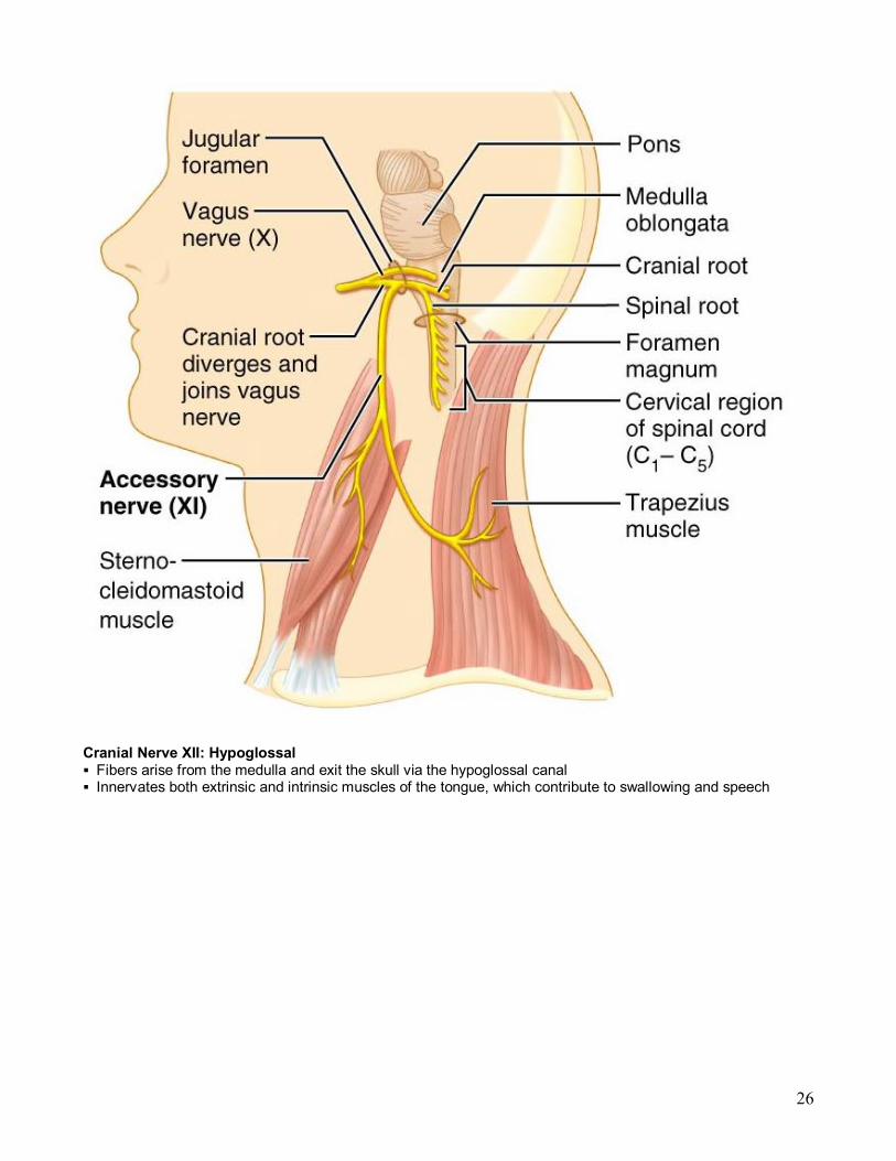
Cranial Nerve XI: Accessory § Formed from a cranial root emerging from the medulla and a spinal root arising from the superior region of the spinal cord
§ The spinal root passes upward into the cranium via the foramen magnum § The accessory nerve leaves the cranium via the jugular foramen § Primarily a motor nerve
§ Supplies fibers to the larynx, pharynx, and soft palate § Innervates the trapezius and sternocleidomastoid, which move the head and neck
26
Cranial Nerve XII: Hypoglossal § Fibers arise from the medulla and exit the skull via the hypoglossal canal § Innervates both extrinsic and intrinsic muscles of the tongue, which contribute to swallowing and speech

27