PMH CLINICAL PRACTICE GUIDELINES · PDF fileDIAGNOSIS 3 5. PATHOLOGY 5 6. ... alveolar soft...
12
PRINCESS MARGARET CANCER CENTRE CLINICAL PRACTICE GUIDELINES SARCOMA SOFT TISSUE SARCOMA
Transcript of PMH CLINICAL PRACTICE GUIDELINES · PDF fileDIAGNOSIS 3 5. PATHOLOGY 5 6. ... alveolar soft...

PRINCESS MARGARET CANCER CENTRE
CLINICAL PRACTICE GUIDELINES
SARCOMA
SOFT TISSUE SARCOMA

2
Last Revision Date – October 2015
Site Group: Sarcoma – Soft Tissue Sarcoma
Author: Dr. Peter Ferguson
1. INTRODUCTION 3
2. PREVENTION 3
3. SCREENING AND EARLY DETECTION 3
4. DIAGNOSIS 3
5. PATHOLOGY 5
6. MANAGEMENT 6
6.1 MANAGEMENT ALGORITHMS 6 6.2 SURGERY 9 6.3 CHEMOTHERAPY 9 6.4 RADIATION THERAPY 10 6.5 OTHER THERAPY 10 6.6 ONCOLOGY NURSING PRACTICE 10
7. SUPPORTIVE CARE 11
7.1 PATIENT EDUCATION 11 7.2 PSYCHOSOCIAL CARE 11 7.3 SYMPTOM MANAGEMENT 11 7.4 CLINICAL NUTRITION 11 7.5 PALLIATIVE CARE 11 7.6 OTHER 11
8. FOLLOW-UP CARE 12

3
Last Revision Date – October 2015
1. Introduction
Soft tissue sarcomas are rare tumours arising from mesenchymal tissues, representing
approximately 1% of all adult malignancies. They are an extremely heterogenous group
of tumours, composed of over 50 subtypes with distinct pathologic features and clinical
behaviours. The commonest subtypes are liposarcoma, leiomyosarcoma, synovial
sarcoma, malignant peripheral nerve sheath tumour and undifferentiated pleomorphic
sarcomas. The majority of soft tissue sarcomas arise in the extremities and trunk (80%),
with 15% arising in the retroperitoneum. Soft tissue sarcomas preferentially metastasize
to the lungs (90%), and regional lymph nodes (5%).
2. Prevention
Not applicable to soft tissue sarcoma.
3. Screening and Early Detection
Not applicable to soft tissue sarcoma.
4. Diagnosis
All patients with soft tissue sarcoma should be assessed in a multidisciplinary clinic with
expertise in sarcoma. Local imaging of the primary tumour must include MRI for extremity and
trunk locations and MRI with CT for retroperitoneal tumours. Chest CT must be done on all
patients. Other staging studies are indicated for specific subtypes – Abdominal/pelvic CT for
myxoid/round cell liposarcoma; CT imaging of regional lymph nodes for epithelioid sarcoma,
clear cell sarcoma, angiosarcoma and rhabdomyosarcoma and synovial sarcoma.
Important staging information to be gained from imaging of the primary site includes the
maximal tumour diameter (< or > 5 cm) and the relationship to the superficial fascia (deep or
superficial). Other information pertinent to local treatment planning includes involvement of
major vascular structures, major motor nerves, bone and pelvic/abdominal organs.
Tissue diagnosis must be obtained by core needle or open incisional biopsy. If open biopsy is
undertaken, the incision must be longitudinal in the extremities and in line with the eventual
definitive resection incision, with minimal contamination of the surrounding tissues. The crucial
information from biopsy includes tumour grade (low, medium and high grade) and sarcoma
subtype.
Staging is carried out according to the AJCC staging system, which considers the factors of
tumour size and location, grade, lymph node involvement and distant metastases:

4
Last Revision Date – October 2015

5
Last Revision Date – October 2015
5. Pathology
All sarcoma biopsies and resection specimens should undergo pathologic assessment by an
experienced sarcoma pathologist. Biopsies should establish a malignant diagnosis, sarcoma
subtype and grade. Ancillary tests such as immunohistochemistry, EM, cytogenetics and
molecular genetic testing may be used in addition to morphologic assessment to establish a
diagnosis, so assessment should be carried out in a site with access to the ancillary techniques.
The pathology synoptic report should contain the following information: tumour location,
primary diagnosis (WHO classification), depth (superficial or deep), size, histologic grade (using
FNCLCC or NCIC classifications), necrosis, status of margins, status of lymph nodes, results of
ancillary tests, additional features if present (mitoses, vascular invasion, character of tumour
margin) and TNM stage.
Although morphologic examination is the gold standard for most sarcoma diagnoses, molecular
genetic testing has emerged as an important ancillary technique as many sarcomas feature typical
and characteristic genetic abnormalities. Sarcomas whose diagnosis are confirmed using genetic
testing using FISH or PCR include: Ewing’s sarcoma, desmoplastic small round cell tumour,
alveolar rhabdomyosarcoma, myxoid/round cell liposarcoma, atypical lipomatous tumour,
dedifferentiated liposarcoma, alveolar soft part sarcoma, clear cell sarcoma, dermatofibrosarcoma
protuberans, extraskeletal myxoid chondrosarcoma, fibromyxoid sarcoma and synovial sarcoma.

6
Last Revision Date – October 2015
6. Management
6.1 Management Algorithms

7
Last Revision Date – October 2015
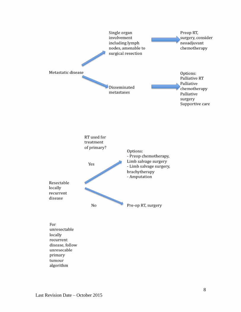
8
Last Revision Date – October 2015
9
Last Revision Date – October 2015
6.2 Surgery
Biopsy of extremity, truncal, head and neck and retroperitoneal sarcomas should be done in
consultation with an experienced sarcoma surgeon (for open incisional biopsy) or musculskeletal
radiologist (for core needle biopsy). Fine needle aspirate has little role in the diagnosis of
sarcoma. The incision or needle track should be in line with the eventual definitive surgical
incision and should be excised at the time of tumour resection.
The goal of sarcoma surgery should be resection with negative margins. If, on preoperative
imaging, margins of 1-2 cm or intact fascia can be obtained without sacrificing critical structures
such as bone, major motor nerves or major blood vessels, or vital organs, then surgery alone may
be all that is necessary. This is usually only possible in T1a tumours.
For tumours that are close to the above structures, surgery is used with adjuvant radiotherapy and
occasionally chemotherapy. When these adjuvants are used, very close negative margins are
acceptable. These may be blood vessel adventitia, nerve epineurium and bone periosteum. Entire
muscle compartments need not be resected unless the sarcoma is extensive.
If surgical margins are positive against critical structures such as nerves, blood vessels or bone,
re-resection generally is not necessary provided adjuvant radiotherapy is used. The local
recurrence rate in this situation is low. If surgical margins are grossly positive or positive
unexpectedly in other anatomical areas, re-excision should be considered to minimize the risk of
local recurrence.
Limb preservation should be the goal of surgery for extremity sarcomas. In cases where multiple
motor nerves, blood vessels or bone require resection for negative margins, or if anticipated
functional outcome with limb salvage surgery is expected to be very poor, amputation should be
considered.
6.3 Chemotherapy
Chemotherapy is absolutely indicated for certain subtypes of soft tissue sarcoma, most notably
soft tissue Ewing’s sarcoma and embryonal rhabdomyosarcoma. Other subtypes that are often
responsive include synovial sarcoma, leiomyosarcoma and some subtypes of liposarcoma
(myxoid, pleomorphic, dedifferentiated). These should be given in a neoadjuvant fashion.
Agents with activity in Ewing’s sarcoma and rhabdomyosarcoma include vincristine,
doxorubicin, cyclophosphamide, ifosfamide and etoposide. In other sarcomas, the main active
agents are doxorubicin and ifosfamide.
The benefit of chemotherapy in sarcoma is unclear. Randomized trials of doxorubicin based
chemotherapy shows a very small overall survival advantage. Some trials have shows decreased
distant and local recurrence, albeit marginally. Chemotherapy may be utilized in some centers to
try to improve resectability of stage II and III tumours.
For unresectable tumours chemotherapy may be utilized as the initial treatment to try to
downstage the tumour. In some situations the tumour may become resectable and limb salvage
may be undertaken.
In patients with metastatic disease, single agent chemotherapy may be utilized as the initial
treatment and if there is appreciable response, resection of the primary tumour and metastases
10
Last Revision Date – October 2015
may be undertaken providing serious functional consequences will not ensue. In these situations
single agents such as doxorubicin or ifosfamide may be used.
6.4 Radiation Therapy
Radiation therapy is used in addition to surgery is the vast majority of patients with soft tissue
sarcoma. The addition of radiation to surgery has allowed for limb salvage surgery in most
patients, due to a much narrower surgical margin without concomitant increase in local
recurrence.
Radiotherapy can be administered preoperatively or postoperatively. There is no difference in
local control between the two. The main advantage to preoperative radiation is that a smaller dose
(50 Gy vs. 66 Gy) and volume are utilized, both of which lead to lower rates of long term
extremity fibrosis and edema, and better functional outcome scores. The main disadvantage to
preoperative radiation is a near doubling of the wound complication rate (17% in postop vs. 35%
in preop). Wound complications however are also modifiable. Other problems with postoperative
radiation include a higher risk of radiation associated fractures and secondary sarcomas. For these
reasons, many centers now utilize preoperative radiation in soft tissue sarcomas.
In T1a and T2a patients (superficial tumours), radiation many not be necessary if the expected
surgical margins will be greater than 1-2 cm or intact fascia. If the margins are closer than
expected on final pathologic review then postoperative radiation may be undertaken. Otherwise,
if the expected surgical margins are to be less than 1 cm, in order to save critical functional
structures, radiation should be considered even in low-grade tumours.
In unresectable tumours, radiation may be utilized to attempt to downstage the tumour and limb
salvage surgery may then be undertaken if the tumour becomes resectable.
In patients with metastatic disease, radiation may be utilized as a palliative means to control pain
and other symptoms.
In patients with locally recurrent sarcoma, the decision to utilize radiation is dependent on
whether it was administered to treat the primary tumour. If not, then radiation should be given. If
radiation was previously utilized, the previous radiation plan should be consulted and if further
treatment often in the form of intensity modulated radiotherapy (IMRT) can be administered
without exceeding the critical dose to major anatomic structures, then it may be utilized.
Otherwise, chemotherapy may be utilized as an adjuvant, or surgery alone, either limb salvage or
amputation, may be undertaken.
6.5 Other Therapy
This section not applicable to soft tissue sarcoma.
6.6 Oncology Nursing
Specialized nursing care is essential in the management of patients with soft tissue sarcoma.
Because of the rarity of the condition, nurses knowledgeable about and experienced in caring for
patients with soft tissue sarcoma contribute significantly to their outcome. Specialized surgical
nursing care is important in dealing with postoperative complications and rehabilitation issues.
11
Last Revision Date – October 2015
Similarly, chemotherapy and radiotherapy nursing care by individuals knowledgeable about
sarcoma are important in managing toxicity issues and some symptom control.
7. Supportive Care
7.1 Patient Education
Because of the rarity of soft tissue sarcoma, patients often benefit from a significant educational
component to their treatment. Patient based support groups are often utilized for new patients to
gain insight into their disease. Educational materials prior to chemotherapy, radiation and surgery
are ideal to help patients prepare for complications and toxicity.
7.2 Psychosocial Care
Social work and psychiatry and invaluable in providing support for psychosocial issues. Because
many patients often travel great distances for sarcoma treatment when it is provided in quaternary
care centres, social work is often important in managing social and family issues. Psychiatry may
be beneficial in helping often young patients and families with coping strategies.
7.3 Symptom Management
Although most patients with soft tissue sarcoma present with painless masses, some patients may
have significant symptoms if major nerves or bone are involved or if tumours are extremely large.
In these situations, involvement of pain management specialized may be beneficial. The often
large surgical resections for sarcoma may be associated with significant postoperative pain and
the same individuals may be involved in this situation.
7.4 Clinical Nutrition
This section not applicable to soft tissue sarcoma.
7.5 Palliative Care
Patients with advanced soft tissue sarcoma often remain ambulatory and mobile. The usual course
of events with patients with metastatic sarcoma is respiratory failure from multiple pulmonary
metastases. Early involvement of palliative care physicians with patients with metastatic sarcoma
often provides a high quality end of life experience.
7.6 Other
Patients who have undergone limb salvage surgery for sarcoma should be referred for
physiotherapy/ rehabilitation. This should continue until the patient reaches their maximal
functional recovery, which may take several months.
12
Last Revision Date – October 2015
8. Follow-up Care
For the first 2 years after initial treatment, patients undergo physical examination of the primary
disease site and chest x-ray. Follow-up appointments then take place every 6 months until 5 years
after initial treatment and then annually until 10 years after initial treatment.
For patients who develop locally recurrent or metastatic disease, follow-up takes place at the
same intervals as presentation with primary disease.
For patients who have developed metastatic disease, consideration should be given to performing
intermittent chest CT scans in follow-up.
For patients who have had primary tumours in anatomic areas that are difficult to examine
(retroperitoneum, pelvis) consideration should be given to performing intermittent MRI of the
area.