Pleomorphic adenoma of major and minor salivary glands ...
4
948 Pleomorphic adenoma of major and minor salivary glands: Report of 5 cases Sujatha D 1 , Anuradha Pai 2 , Nidhin J Valappila 3 ABSTRACT: Salivary gland tumors constitute approximately 3% of all head and neck tumors. Pleomorphic adenoma is the most common tumor of the salivary glands. Pleomorphic adenomas constitute 60% of all salivary gland tumors. It's more commonly seen in parotid, with less than 10% in submandibular, sublingual and minor salivary glands. Here, we report a series of 5 cases of pleomorphic adenoma. Key words: Pleomorphic adenoma, salivary glands, diagnosis, tumour CASE REPORT doi: 10.5866/4.3.948 1&2 Professor 3 Post graduate student Department of Oral Medicine & Radiology The Oxford dental college, hospital & research centre, Bommanahalli, Hosur road, Bangalore 560068, Karnataka, India Article Info: Received: July 11, 2012; Review Completed: August, 10, 2012; Accepted: September 10, 2012 Published Online: October, 2012 (www. nacd. in) © NAD, 2012 - All rights reserved Email for correspondence: [email protected] Quick Response Code Introduction Pleomorphic adenoma is a benign tumour arising from cells of salivary gland tissue. It’s also known as mixed tumour because of presence of epithelial and mesenchymal elements. 63% of pleomorphic adenoma occurs in parotid gland, 1, 2, 5,6,10 followed by 38% in minor salivary glands and 10% in submandibular glands. 10 Since asymptomatic patients visit dentist in later stage, diagnosis will be an incidental finding. The parotid gland and the palate are the most commonly affected sites. 2, 3, 8 Pleomorphic adenomas are generally slow growing and painless tumour. 4 Cases of rapid growth after a long period of quiescence have been reported and malignant transformation occasionally occurs. 4, 8 Pleomorphic adenoma commonly presents in middle age women. 2, 3,5,7,9 Although it occurs most commonly in the major salivary glands, it may also occur in the minor salivary glands and extra-salivary tissue. It is usually encapsulated when it arises in the major salivary glands but not in the minor salivary glands. INDIAN JOURNAL OF DENTAL ADVANCEMENTS Journal homepage: www. nacd. in Indian J Dent Adv 2012; 4(3): 948-951
Transcript of Pleomorphic adenoma of major and minor salivary glands ...

948
Pleomorphic adenoma of majorand minor salivary glands:
Report of 5 casesSujatha D1, Anuradha Pai2, Nidhin J Valappila3
ABSTRACT:
Salivary gland tumors constitute approximately 3% of all head
and neck tumors. Pleomorphic adenoma is the most common
tumor of the salivary glands. Pleomorphic adenomas constitute
60% of all salivary gland tumors. It's more commonly seen in
parotid, with less than 10% in submandibular, sublingual and
minor salivary glands. Here, we report a series of 5 cases of
pleomorphic adenoma.
Key words: Pleomorphic adenoma, salivary glands, diagnosis,
tumour
C A S E R E P O R T
doi: 10.5866/4.3.948
1&2Professor3Post graduate studentDepartment of Oral Medicine & RadiologyThe Oxford dental college, hospital & research centre,Bommanahalli, Hosur road, Bangalore 560068,Karnataka, India
Article Info:
Received: July 11, 2012;Review Completed: August, 10, 2012;Accepted: September 10, 2012Published Online: October, 2012 (www. nacd. in)© NAD, 2012 - All rights reserved
Email for correspondence:[email protected]
Quick Response Code
Introduction
Pleomorphic adenoma is a benign tumour arising from cells of salivary gland tissue. It’s also known asmixed tumour because of presence of epithelial and mesenchymal elements. 63% of pleomorphic adenomaoccurs in parotid gland,1, 2, 5,6,10 followed by 38% in minor salivary glands and 10% in submandibular glands.10
Since asymptomatic patients visit dentist in later stage, diagnosis will be an incidental finding. The parotidgland and the palate are the most commonly affected sites.2, 3, 8 Pleomorphic adenomas are generally slowgrowing and painless tumour.4 Cases of rapid growth after a long period of quiescence have been reportedand malignant transformation occasionally occurs.4, 8
Pleomorphic adenoma commonly presents in middle age women.2, 3,5,7,9 Although it occurs most commonlyin the major salivary glands, it may also occur in the minor salivary glands and extra-salivary tissue. It isusually encapsulated when it arises in the major salivary glands but not in the minor salivary glands.
INDIAN JOURNAL OF DENTAL ADVANCEMENTS
Jour nal homepage: www. nacd. in
Indian J Dent Adv 2012; 4(3): 948-951

949
Epithelial cells give rise to ductal structures andmesenchymal elements give rise to myxoid, hyaline,cartilaginous and osseous changes .Based on thepresence of amount of different epithelial andmesenchymal elements and anatomical site ofoccurrence the tumour will show diversity in clinical,radiological and histopathological presentation.10
Here we are describing 5 cases of pleomorphicadenoma of major and minor salivary gland reportedin our institution over a period of 2 years.
Case Series
Case 1
A 42 years, old male patient reported tooutpatient department with a chief complaint of slowgrowing asymptomatic swelling (Fig 1a, 1b) in theright side of face with 3 years evolution.Examination revealed a well defined solitaryswelling in the region of angle of mouth andelevating the ear lobe measuring about 5 x 4 cmcovered with the normal skin and was firm inconsistency. Fine needle aspiration cytology wasperformed which was suggestive of pleomorphicadenoma of right parotid gland. The patient wasreferred to oral surgery for the surgical removal oftumour. Diagnosis was confirmed after thehistopathological study of excised tissue.
Case 2
A male patient aged 60 years came to ourhospital with a chief complaint of swelling on theright side of neck of duration 12 years. It wasasymptomatic, slow growing and reached thepresent size (Fig 2). Extra oral examination revealeda well defined swelling of size approximately 4 x 6cm in the right submandibular region extending intoadjacent areas. Swelling was firm in consistency andwas not fixed to underlying structures.Ultrasonography and FNAC were done which wassuggestive of pleomorphic adenoma of rightsubmandibular gland. Tumour was excised undergeneral anesthesia in the dept of oral surgery andspecimen was sent for histopathological examinationwhich confirmed our diagnosis.
Case 3
Another wise healthy 55 years old male wasreferred to our outpatient clinic with a compliant oflarge mass in the palate (Fig 3) since 3 years whichwas affecting his speech. Swelling was slow growingand attained present size. On intra oral examination
a well defined swelling was noticed in the midpalatal region extending from rugae region to softpalate measuring approximately 7x5 cm. Swellingwas with normal overlying mucosa and was firm inconsistency. FNAC was done which was suggestiveof pleomorphic adenoma of minor salivary gland.Tumour excision and biopsy of specimen confirmedthe diagnosis.
Case 4
A female patient of 66 years came to ourinstitution with a complaint of swelling in upperright back teeth region of 1 month duration. Swellingwas small, gradually growing and reached sizeapproximately 3x3 cm (Fig 4). Surface of swellingwas normal without any surface changes. Swellingwas firm in consistency and nontender on palpation.FNAC was done which was suggestive ofpleomorphic adenoma of minor salivary gland.Surgical excision and biopsy confirmed our diagnosis
Case 5
A male patient of 32 years reported to ourcollege with a chief complaint of swelling in upperright back teeth region of 1 month duration. Swellingwas small and gradually growing and reached sizeof about 3x3 cm (Fig 5, 6). Surface of swelling wasnormal without any surface changes. Swelling wasfirm in consistency and non tender on palpation.FNAC was suggestive pleomorphic adenoma ofminor salivary gland. Surgical excision and biopsyconfirmed the diagnosis.
Discussion
Pleomorphic adenoma is an insidious tumor thatcan reach into great proportion if not treated4. Fourof five salivary gland tumors of the parotid glandand more than half the tumors of submandibulargland and the palate are pleomorphic adenomas.8
Pleomorphic adenoma occurs less commonly outsidesalivary gland tissue and may arise from anyglandular tissue with myoepithelial cells.10 It canoccur in the lacrimal glands, external auditory canal,skin, breast tissue, and vulva, and accounts for halfof lacrimal gland tumours; the other half aremalignant.10 Late presentation is common; themajority of patients reported here had tumours inexcess of 3 cm in diameter at the time of ourdiagnosis. This could be attributed to asymptomaticnature of tumour.
Pleomorphic adenoma of major and minor Sujatha, et, al.
Indian J Dent Adv 2012; 4(3): 948-951

950
Diagnosis can be made using various techniquesavailable. A radiograph doesn’t show any bonychanges hence it can be used for ruling out othertype of lesions from pleomorphic adenoma. Fineneedle aspiration cytology, with or withoutultrasound guidance, is often used in investigationsof superficial salivary gland masses, and provideshigh diagnostic accuracy.6, 12 FNAC is economic andeasy to perform and required minimum instruments.Differentiation from adenoid cystic carcinoma andlow grade adenocarcinoma may be difficult with fineneedle aspiration alone.13
Radiologically it is difficult to distinguishpleomorphic adenoma from its variants,myoepithelioma and basal cell adenoma.14 Imagingtechniques ultrasound, magnetic resonance (MRI),or computed tomography for diagnosis of salivarygland tumour depends on the site & size of thetumor.6,15 Superficially placed tumours can bediagnosed easily using ultrasonography where astumours placed deep inside structures can bediagnosed by MRI. Because pleomorphic adenomais commonly located in the superficial lobe of parotidgland; high resolution ultrasound is suited fordifferentiating it from commonly found smallreactive nodes within the parotid gland. Onultrasound it appears as a hypoechoic, homogenouswell circumscribed mass with posterior acousticenhancement.10 It also helps to guide fine-needleaspiration (FNA) for cytological diagnosis. Adenomaarising from accessory parotid tissue lying anteriorto main body of the gland or along the main ductclinically presents as cheek lump or buccomassetricmass that can be elevated with ultrasound. MRI isthe method of choice for deep lobe parotid tumourwhich clinically presents as a preauricular ororopharyngeal mass.15 It must be differentiatedradiologically from adjacent deep neck spaces(Parapharyngeal and masticator space). Table 1.
Surgical excision is the treatment of choice forpleomorphic adenoma. Malignant transformationmay take place in long standing untreated tumorsand in recurrent tumors.2, 4, 7 Recurrence rate is 1 to5 %.2,16 Histologically, tumours that have anirregular border with “tongues” of tumour growinginto surrounding tissue are associated with a highrisk of local recurrence after excision.18 Even thoughit is histologically benign tumour, in some cases itmay invade local blood vessels in the absence of anyother features associated with malignancy.19
Metastasis from cases of “benign” pleomorphicadenoma to lymph nodes, bone, skin, liver and lunghave been reported, but the metastatic deposits donot show any malignant histological features.16
There is no recognized feature for the primarytumour to predict its metastatic tendency.Carcinoma ex-pleomorphic adenoma is a malignantneoplasm arising from a coexisting or previouslyexcised pleomorphic adenoma.2,3,5,10 Thecarcinomatous element arises from the epithelialcomponent of the benign tumour. On imaging, it maylook similar to a pleomorphic adenoma, or may showinfiltrative margins, necrotic areas, and regionallymph node metastases. For diagnosis histologicalevidence is needed that shows carcinoma ex-pleomorphic adenoma is arising from a pre-existingbenign pleomorphic adenoma.
Conclusion
Pleomorphic adenomas may present in a varietyof ways. As Oral physician we need to be aware ofits diverse presentation as sound knowledge of thistumour will help in its early diagnosis and treatmentthus prevent or reduce the mortality associated withthe tumour.
REFERENCES
1) Ellis GL, Auclair PL. Tumors of the salivary glands, atlasof tumor pathology. 3rd series, Facsicle 17. Washington, DC:Armed Forces Institute of Pathology; 1996.
2) R.Rajendran, editor. Shafer’s Textbook of Oral Pathology.5th
ed. Elsevier; 2006.
3) Neville. Oral and Maxillofacial Pathology.2nd ed. Elsevier;2004.
4) Cawson RA. Essentials of Oral Pathology and OralMedicine.7th ed. Churchill Livingstone; 2006.
5) Regezi. Oral Pathology Clinical Pathologic Correlations.5th
ed. Saunders; 2008.
6) Greenberg. Burket’s Oral Medicine.11th ed.Bc Decker; 2008.
7) Ravikiran. Textbook of Oral Medicine, Oral Diagnosis andOral Radiology. Elsevier; 2010.
8) Chidzonga MM, Perez VM. Pleomorphic adenoma of thesalivary glands, Clinicopathologic study of 206 cases inZimbabwe. Oral Surg Oral Med Oral Pathol Oral RadiolEndod 1995;79:747-749.
9) Ojha J, Bhattacharyya I. Intraosseous pleomorphicadenoma of the mandible: report of a case and review of theliterature. Oral Surg Oral Med Oral Pathol Oral RadiolEndod 2007; 104: 21-26.
10) Lingam RK, Daghir AA. Pleomorphic adenoma (benignmixed tumour) of the salivary glands: its diverse clinical,radiological, and histopathological presentation. BritishJournal of Oral and Maxillofacial Surgery 2011; 49:14-20.
Pleomorphic adenoma of major and minor Sujatha, et, al.
Indian J Dent Adv 2012; 4(3): 948-951
951
11) Coelho C, Cabrita C. Pleomorphic adenoma of minor salivaryglands: Report of two cases. Journal of Cranio-MaxillofacialSurgery 2006; 34: Suppl. S1: P 240.
12) BajajY, Singh S, Cozens N, Sharp J. Critical clinicalappraisal of the role of ultrasound guided fine needleaspiration cytology in the management of parotid tumours.J Laryngol Otol 2005; 119:289-292.
13) Das DK, Anim JT. Pleomorphic adenoma of salivary gland:to what extent does fine needle aspiration cytology reflecthistopathological features? Cytopathology 2005; 16:65-70.
14) Takeshita T, Tanaka H, Harasawa A, Kaminaga T,Imamura T, Furui S.CT and MRI findings of basal celladenoma of the parotid gland. Radiat Med 2004;22:260-264.
15) Motoori K, Yamamoto S,et al. Inter- and intratumoralvariability in magnetic resonance imaging of pleomorphic
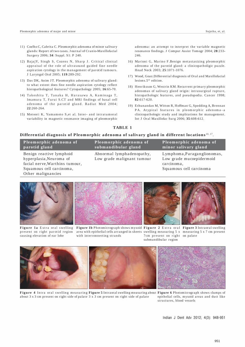
TABLE 1
Differential diagnosis of Pleomorphic adenoma of salivary gland in different locations10, 17.
Pleomorphic adenoma of Pleomorphic adenoma of Pleomorphic adenoma ofparotid gland submandibular gland minor salivary gland
Benign reactive lymphoid Abnormal lymphadenopathy, Lymphoma,Paraganglionomas,hyperplasia,Neuroma of Low grade malignant tumour Low grade mucoepidermoidfacial nerve,Warthins tumour, carcinoma,Squamous cell carcinoma, Squamous cell carcinomaOther malignancies
adenoma: an attempt to interpret the variable magneticresonance findings. J Comput Assist Tomogr 2004; 28:233-246.
16) Marioni G, Marino F.Benign metastasizing pleomorphicadenoma of the parotid gland: a clinicopatholigic puzzle.Head Neck 2003; 25:1071-1076.
17) Wood, Goaz.Differential diagnosis of Oral and Maxillofaciallesions.5th edition.
18) Henriksson G, Westrin KM. Recurrent primary pleomorphicadenomas of salivary gland origin: intrasurgical rupture,histopathologic features, and pseudopodia. Cancer 1998;82:617-620.
19) Ethunandan M, Witton R, Hoffman G, Spedding A, BrennanPA. Atypical features in pleomorphic adenoma–aclinicopathologic study and implications for management.Int J Oral Maxillofac Surg 2006; 35:608-612.
Figure 1a Extra oral swellingpresent on right parotid regioncausing elevation of ear lobe
Figure 1b Photomicrograph shows myxoidarea with epithelial cells arranged in sheetswith interconnecting strands
Figure 2 Extra oralswelling measuring 5 x7cm present on rightsubmandibular region
Figure 4 Intra oral swelling measuringabout 3 x 3 cm present on right side of palate
Figure 5 Intraoral swelling measuring about3 x 3 cm present on right side of palate
Figure 6 Photomicrograph shows clumps ofepithelial cells, myxoid areas and duct likestructures, blood vessels
Figure 3 Intraoral swellingmeasuring 5 x 7 cm presenton palate
Pleomorphic adenoma of major and minor Sujatha, et, al.
Indian J Dent Adv 2012; 4(3): 948-951


![[PAPER] Pleomorphic Adenoma Print.docx](https://static.fdocuments.net/doc/165x107/56d6bd9b1a28ab30168ea546/paper-pleomorphic-adenoma-printdocx.jpg)