Platelet ITAM signaling is critical for vascular · tive feedback pathway for sustained platelet...
10
Platelet ITAM signaling is critical for vascular integrity in inflammation Yacine Boulaftali, … , Mark L. Kahn, Wolfgang Bergmeier J Clin Invest. 2013; 123(2):908-916. https://doi.org/10.1172/JCI65154. Platelets play a critical role in maintaining vascular integrity during inflammation, but little is known about the underlying molecular mechanisms. Here we report that platelet immunoreceptor tyrosine activation motif (ITAM) signaling, but not GPCR signaling, is critical for the prevention of inflammation-induced hemorrhage. To generate mice with partial or complete defects in these signaling pathways, we developed a protocol for adoptive transfer of genetically and/or chemically inhibited platelets into thrombocytopenic (TP) mice. Unexpectedly, platelets with impaired GPCR signaling, a crucial component of platelet plug formation and hemostasis, were indistinguishable from WT platelets in their ability to prevent hemorrhage at sites of inflammation. In contrast, inhibition of GPVI or genetic deletion of Clec2, the only ITAM receptors expressed on mouse platelets, significantly reduced the ability of platelets to prevent inflammation-induced hemorrhage. Moreover, transfusion of platelets without ITAM receptor function or platelets lacking the adapter protein SLP-76 into TP mice had no significant effect on vascular integrity during inflammation. These results indicate that the control of vascular integrity is a major function of immune-type receptors in platelets, highlighting a potential clinical complication of novel antithrombotic agents directed toward the ITAM signaling pathway. Research Article Hematology Find the latest version: http://jci.me/65154-pdf
Transcript of Platelet ITAM signaling is critical for vascular · tive feedback pathway for sustained platelet...

Platelet ITAM signaling is critical for vascularintegrity in inflammation
Yacine Boulaftali, … , Mark L. Kahn, Wolfgang Bergmeier
J Clin Invest. 2013;123(2):908-916. https://doi.org/10.1172/JCI65154.
Platelets play a critical role in maintaining vascular integrity during inflammation, but little isknown about the underlying molecular mechanisms. Here we report that plateletimmunoreceptor tyrosine activation motif (ITAM) signaling, but not GPCR signaling, iscritical for the prevention of inflammation-induced hemorrhage. To generate mice withpartial or complete defects in these signaling pathways, we developed a protocol foradoptive transfer of genetically and/or chemically inhibited platelets into thrombocytopenic(TP) mice. Unexpectedly, platelets with impaired GPCR signaling, a crucial component ofplatelet plug formation and hemostasis, were indistinguishable from WT platelets in theirability to prevent hemorrhage at sites of inflammation. In contrast, inhibition of GPVI orgenetic deletion of Clec2, the only ITAM receptors expressed on mouse platelets,significantly reduced the ability of platelets to prevent inflammation-induced hemorrhage.Moreover, transfusion of platelets without ITAM receptor function or platelets lacking theadapter protein SLP-76 into TP mice had no significant effect on vascular integrity duringinflammation. These results indicate that the control of vascular integrity is a major functionof immune-type receptors in platelets, highlighting a potential clinical complication of novelantithrombotic agents directed toward the ITAM signaling pathway.
Research Article Hematology
Find the latest version:
http://jci.me/65154-pdf
Research article
908 The Journal of Clinical Investigation http://www.jci.org Volume 123 Number 2 February 2013
Platelet ITAM signaling is critical for vascular integrity in inflammation
Yacine Boulaftali,1 Paul R. Hess,2 Todd M. Getz,1 Agnieszka Cholka,1 Moritz Stolla,3 Nigel Mackman,1 A. Phillip Owens III,1 Jerry Ware,4 Mark L. Kahn,2 and Wolfgang Bergmeier1,5
1McAllister Heart Institute, University of North Carolina at Chapel Hill, Chapel Hill, North Carolina, USA. 2Department of Medicine, University of Pennsylvania, Philadelphia, Pennsylvania, USA. 3Cardeza Foundation for Hematologic Research and Department of Medicine, Thomas Jefferson University, Philadelphia,
Pennsylvania, USA. 4Department of Physiology and Biophysics, University of Arkansas for Medical Sciences, Little Rock, Arkansas, USA. 5Department of Biochemistry and Biophysics, University of North Carolina at Chapel Hill, Chapel Hill, North Carolina, USA.
Platelets play a critical role in maintaining vascular integrity during inflammation, but little is known about the underlying molecular mechanisms. Here we report that platelet immunoreceptor tyrosine activation motif (ITAM) signaling, but not GPCR signaling, is critical for the prevention of inflammation-induced hemor-rhage. To generate mice with partial or complete defects in these signaling pathways, we developed a protocol for adoptive transfer of genetically and/or chemically inhibited platelets into thrombocytopenic (TP) mice. Unexpectedly, platelets with impaired GPCR signaling, a crucial component of platelet plug formation and hemostasis, were indistinguishable from WT platelets in their ability to prevent hemorrhage at sites of inflam-mation. In contrast, inhibition of GPVI or genetic deletion of Clec2, the only ITAM receptors expressed on mouse platelets, significantly reduced the ability of platelets to prevent inflammation-induced hemorrhage. Moreover, transfusion of platelets without ITAM receptor function or platelets lacking the adapter protein SLP-76 into TP mice had no significant effect on vascular integrity during inflammation. These results indicate that the control of vascular integrity is a major function of immune-type receptors in platelets, highlighting a potential clinical complication of novel antithrombotic agents directed toward the ITAM signaling pathway.
IntroductionPlatelets play an essential role in the prevention of excessive blood loss after injury (hemostasis), but can also form occlusive thrombi at sites of atherosclerotic plaque rupture (thrombosis) (1). Platelet plug formation strongly depends on the soluble agonists throm-bin, ADP, and thromboxane A2 (TxA2), which induce cellular acti-vation via GPCRs (1). Thrombin, generated in response to the exposure of blood to tissue factor in the damaged vascular wall, is crucial in the initiation of thrombus formation. ADP and TxA2 are released from activated platelets and provide an important posi-tive feedback pathway for sustained platelet activation and platelet recruitment to the growing thrombus. The importance for throm-bosis and hemostasis of the various platelet GPCR receptors and their agonists is documented by the protection from thrombotic complications and the increased bleeding tendency observed in patients treated with heparin (which inhibits thrombin), aspi-rin (which blocks TxA2 generation), and/or clopidogrel bisulfate (which inhibits one of the ADP receptors, P2Y12) (2). Consistent-ly, mice lacking the major platelet receptors for thrombin and/or ADP exhibit strong protection from experimental thrombosis and markedly prolonged tail bleeding times (3).
The platelet receptor for collagen and laminin, glycoprotein VI (GPVI), is also critical for the initiation of thrombus formation at sites of vascular injury. GPVI belongs to the Ig superfamily of surface receptors and depends on its association with the immuno receptor
tyrosine activation motif–containing (ITAM-containing) FcR γ chain for signaling (4). Mice deficient in GPVI exhibit a mild defect in hemostasis and moderate to strong protection from thrombosis (5). Patients with impaired GPVI expression/function also show a mild bleeding diathesis (6, 7). Inhibitors of GPVI are currently being evaluated as novel antiplatelet drugs (2). Platelets also express the ITAM receptor C-type lectin–2 (CLEC2), but its role in hemostasis and thrombosis is not well understood. Whereas some investigators demonstrated impaired thrombus formation in the absence of func-tional CLEC2 (8, 9), others could not reproduce these findings (10). Platelet CLEC2, however, plays a critical role during development. Mice lacking CLEC2 (11, 12) or its ligand, podoplanin (13, 14), have defective separation of their lymphatic and vascular endothelial cells that causes petechia in utero. Similar defects were observed in mice deficient in Syk, Src-homology leucocyte protein 76 (SLP-76), and PLC-γ2, which are components of the CLEC2 signaling pathway (4).
More recent studies suggested that platelets are also critical for other physiological and pathological processes, such as angio-genesis (15), development (16, 17), immunity (18), viral hepatitis (19), arthritis (20), atherosclerosis (21), and sepsis (22). At sites of inflammation, platelets support leukocyte extravasation (23), but, importantly, they also prevent local hemorrhage. Consequently, thrombocytopenia (TP) is associated with inflammation-induced hemorrhage (24–26). Interestingly, the ability of platelets to safe-guard vascular integrity at sites of inflammation is independent of their ability to form a platelet plug, as inflammation-induced hemorrhage was not observed in mice deficient in the major sur-face adhesion receptors GPIb-V-IX, integrin αIIbβ3, or P-selectin (26, 27). Additional mechanistic studies suggest (a) that the plate-let support of vascular integrity in inflammation depends on intracellular storage granules and release of the vasoactive com-pounds stored therein (28), and (b) that platelets protect vascular
Conflict of interest: The authors have declared that no conflict of interest exists.
Note regarding evaluation of this manuscript: Manuscripts authored by scientists associated with Duke University, The University of North Carolina at Chapel Hill, Duke-NUS, and the Sanford-Burnham Medical Research Institute are handled not by members of the editorial board but rather by the science editors, who consult with selected external editors and reviewers.
Citation for this article: J Clin Invest. 2013;123(2):908–916. doi:10.1172/JCI65154.

research article
The Journal of Clinical Investigation http://www.jci.org Volume 123 Number 2 February 2013 909
integrity by dampening the inflammatory response (29, 30). How-ever, no information is available on the agonist receptors or the intracellular signaling pathways required for the vessel-stabilizing activity of platelets at sites of inflammation.
TP mice have been a very powerful tool for the identification of novel platelet functions in health and disease (see above). The method most commonly used to render mice TP is based on our previous studies on the cytotoxic effects of antibodies directed toward major platelet surface receptors (31), which demonstrated that antibodies against the glycoprotein Ibα (GPIbα) subunit of the GPIb-V-IX receptor complex and the major integrin recep-tor on platelets, αIIbβ3, cause virtually complete TP in mice (31). However, while mice receiving anti-GPIbα antibodies do not show any signs of distress, mice receiving anti-αIIbβ3 antibodies develop an anaphylaxis-like reaction, with symptoms that include hypo-thermia and a decrease in hematocrit due to internal bleeding. Thus, antibodies against GPIbα have emerged as the most reliable method to safely render mice TP for studies on the role of platelets in disease. The disadvantage of this method, however, is that the antibody remains in circulation and also targets platelets that are transfused into the TP mice. Consequently, comprehensive stud-ies on platelet signaling molecules that are important for vascu-lar integrity in inflammation would require deletion of multiple genes specifically in the megakaryocyte/platelet lineage using the loxP/PF4-Cre system (32). The technical difficulties, the costs, and the time associated with generation of these mice call for an alternative, more efficient method to generate mice with defects in multiple platelet signaling molecules.
In this study, we used a novel method to generate mice with plate-let-specific signaling defects, based on adoptive transfer of platelets into TP mice, to study the platelet signaling responses required for the maintenance of vascular integrity in inflammation.
ResultsAdoptive transfer of WT platelets into TP mice. Studying the role of platelet GPCR and ITAM signaling for vascular integrity in inflam-mation requires a method that allows for the rapid and efficient generation of mice with platelet-specific defects in multiple sig-
naling molecules. Thus, we first established a protocol for the adoptive transfer of genetically modified and/or inhibitor-treated platelets into TP mice. We hypothesized that Tg mice expressing a chimeric human IL-4 receptor α/GPIbα (hIL-4Rα/GPIbα) protein instead of GPIbα on the platelet surface (33) could be rendered TP without side effects by infusion of antibodies against hIL-4Rα. Injection of anti–hIL-4Rα antibody led to virtually complete TP, but not neutropenia, in the hIL-4Rα/GPIbα–Tg mice (Figure 1A). As was the case using antibodies against GPIbα in WT mice (31), anti–IL-4R–induced TP in hIL-4Rα/GPIbα–Tg mice was not asso-ciated with hypothermia (Figure 1B). In contrast, infusion of an antibody against αIIbβ3 into hIL-4Rα/GPIbα–Tg mice induced a significant drop in platelet count and body temperature within 30 minutes (Figure 1B). We next tested whether WT platelets could be successfully transfused into TP hIL-4R/GPIb–Tg mice. Transfusion of 2 × 10⁸, 4 × 10⁸, and 8 × 10⁸ WT platelets into TP hIL-4Rα/GPIbα–Tg mice raised the peripheral platelet count to 18.00% ± 5.76%, 29.50% ± 7.32%, and 56.25% ± 5.81%, respectively, of the level in untreated control hIL-4Rα/GPIbα–Tg mice (Figure 1C). In contrast, WT platelets infused into WT mice that received an anti-GPIbα antibody to induce TP were immediately cleared from the circulation (Figure 1D). In summary, we demonstrated that an anti–hIL-4R antibody can be used to safely deplete circulat-ing platelets in hIL-4Rα/GPIbα–Tg mice, facilitating the adoptive transfer of WT platelets into TP mice.
To test the ability of transfused WT platelets to form thrombi at sites of vascular injury, we next studied platelet adhesion to endothelium damaged by laser injury (34). In this model, a throm-bus consisting of platelets and fibrin formed rapidly at the site of vascular injury in control WT mice (Figure 2A). We detected fibrin, but not platelets, in injured venules of TP hIL-4Rα/GPIbα–Tg mice (Figure 2A). WT platelets transfused into TP hIL-4Rα/GPIbα–Tg mice were indistinguishable from endogenous plate-lets in WT control mice in their ability to adhere to and aggregate at sites of vascular injury (Figure 2A). Importantly, transfused platelets also prevented a loss of vascular integrity in models of immune complex–mediated inflammation in the skin (reverse pas-sive Arthus [rpA] reaction) and LPS-induced inflammation in the
Figure 1Adoptive transfer of WT platelets into TP hIL-4Rα/GPIbα–Tg mice. (A) TP was induced in hIL-4Rα/GPIbα–Tg mice by injection of antibodies against hIL-4R. Peripheral plate-let and neutrophil counts were determined at the indicated time points after injection of anti–hIL-4R antibodies (n = 5). Results are expressed as percent of count before antibody injection. ***P < 0.001 versus 0 minutes. (B) Body tempera-ture was measured over time after injection of anti–hIL-4R or anti-αIIbβ3 monoclonal antibodies (MoAb) into hIL-4Rα/GPIbα–Tg mice or injection of anti-GPIbα monoclonal anti-bodies into WT controls (n = 5 per antibody). **P < 0.01, *P < 0.05 versus PBS. (C and D) Peripheral platelet count in TP hIL-4Rα/GPIbα–Tg (TP Tg; C) or TP WT (D) mice transfused with the indicated amounts of WT platelets (n = 4). Results are expressed as percent of count before antibody injection (“Tg” and “WT” groups).

research article
910 The Journal of Clinical Investigation http://www.jci.org Volume 123 Number 2 February 2013
lung (Figure 2, B and C). In the rpA reaction, mice were challenged by intravenous injection of BSA and intradermal application of anti-BSA antibodies, and bleeding at the site of inflammation was assessed by quantification of tissue hemoglobin (Hb). Compared with unchallenged (0.10 ± 0.008 mg/cm2; data not shown) or rpA-challenged (0.25 ± 0.05 mg/cm2) hIL-4Rα/GPIbα–Tg control mice, tissue Hb content was significantly increased in TP hIL-4Rα/GPIbα–Tg mice 4 hours after rPA challenge (1.28 ± 0.08 mg/cm2; Figure 2B). Transfusion of 2 × 10⁸, 4 × 10⁸, and 8 × 10⁸ WT plate-lets into TP hIL-4Rα/GPIbα–Tg mice dose-dependently decreased tissue Hb at sites of inflammation (0.76 ± 0.10, 0.47 ± 0.04, and 0.29 ± 0.03 mg/cm2, respectively). To induce inflammation in the lung, mice were challenged by intranasal administration of 7 μg LPS, and Hb levels were determined in the lavage fluid 6 hours later. Compared with control hIL-4Rα/GPIbα–Tg mice (0.12 ± 0.02 mg/ml), significantly increased Hb levels were observed in the lavage fluid of LPS-challenged TP hIL-4Rα/GPIbα–Tg mice (2.77 ± 0.30 mg/ml; Figure 2C). Consistent with our findings in the rpA model, transfusion of 8 × 10⁸ WT platelets into TP hIL-4Rα/GPIbα–Tg mice before LPS challenge completely prevented hemorrhage in lungs (0.14 ± 0.05 mg/ml; Figure 2C).
Platelet GPCR signaling and vascular integrity in inflammation. To better understand how platelets secure vascular integrity at sites of inflammation, we investigated the roles of platelet GPCRs and ITAM receptors in this process. We first studied platelet prepara-tions with defects in GPCR signaling: (a) platelets isolated from
mice deficient in the thrombin receptor Par4 (35), (b) platelets isolated from mice treated with clopidogrel to irreversibly inhibit P2Y12, the main receptor for ADP, (c) platelets treated with aspirin to irreversibly inhibit cyclooxygenase-dependent TxA2 formation, and (d) Par4-deficient platelets treated with both clopidogrel and aspirin (referred to herein as Par4–/–/clopidogrel/aspirin plate-lets). To confirm the lack of GPCR response in these platelets, we tested their ability for integrin inside-out activation and α granule release in vitro. As shown in Figure 3A, Par4–/–/clopidogrel/aspi-rin platelets were unresponsive to stimulation with PAR4 recep-tor–activating peptide (PAR4p) or the combination of the second-wave mediators ADP and TxA2 (U46619). Both integrin activation and α granule release in Par4–/–/clopidogrel/aspirin platelets were partially reduced in response to activation with the snake toxin convulxin and with podoplanin (Figure 3A), which confirmed that full platelet activation via the ITAM receptors GPVI and CLEC2, respectively, requires feedback activation by ADP and TxA2 (4). Consistent with these in vitro studies, platelets with impaired GPCR signaling were markedly impaired in their ability to form 3-dimensional thrombi at sites of vascular injury (Figure 3B).
Next, all platelet preparations with defects in GPCR signaling were tested for their ability to prevent hemorrhage at sites of rPA-induced inflammation (Figure 4, A and B). 8 × 10⁸ platelets iso-lated from WT mice, Par4–/– mice, clopidogrel-treated WT mice, or clopidogrel-treated Par4–/– mice were incubated in the presence or absence of aspirin, washed, and transfused into TP hIL-4Rα/
Figure 2In vivo function of WT platelets transfused into TP hIL-4Rα/GPIbα–Tg mice. (A) Representative images of cremasteric venules taken 50 seconds after laser injury in WT mice, TP hIL-4Rα/GPIbα–Tg mice, and TP hIL-4Rα/GPIbα–Tg mice transfused with 8 × 10⁸ WT platelets. Mice were inject-ed with Alexa Fluor 488–labeled antibodies against GPIX to label circulating platelets (green) and Alexa Fluor 647–labeled antibodies against fibrin (blue). Note the complete lack of platelet accumulation in TP hIL-4Rα/GPIbα–Tg mice. Fibrin generation in the same mice demonstrated successful vascular injury. Scale bars: 10 μm. (B) Hb levels in skin lesions 4 hours after rpA challenge in hIL-4Rα/GPIbα–Tg mice and in TP hIL-4Rα/GPIbα–Tg mice reconstituted or not with the indicated amounts of WT platelets (n = 12 spots per concentration). Representative lesion sites are also shown. (C) Hb levels in BAL 6 hours after LPS challenge in hIL-4Rα/GPIbα–Tg mice and in TP hIL-4Rα/GPIbα–Tg mice reconstituted or not with 8 × 10⁸ WT platelets (n = 6 per group). Representative images of BAL are also shown. *P < 0.05, **P < 0.01, ***P < 0.001 versus TP Tg with no platelet reconstitution, or as indicated by brackets.

research article
The Journal of Clinical Investigation http://www.jci.org Volume 123 Number 2 February 2013 911
GPIbα–Tg mice before rpA challenge. All platelet transfusions significantly increased the peripheral platelet counts in TP hIL-4Rα/GPIbα–Tg mice to approximately 50% of that in control mice (Supplemental Figure 1, A and C). Surprisingly, clopidogrel-inhib-ited WT platelets, aspirin-inhibited WT platelets, Par4–/– platelets, and Par4–/–/clopidogrel/aspirin platelets all reduced hemorrhage at sites of rpA-induced inflammation — as assessed by tissue Hb levels (0.26 ± 0.03, 0.20 ± 0.02, 0.27 ± 0.04, and 0.21 ± 0.03 mg/cm2, respectively) and macroscopic evaluation — to a level comparable to that observed in TP hIL-4Rα/GPIbα–Tg mice transfused with WT platelets (0.29 ± 0.02 mg/cm2; Figure 4, A and B). These results suggested that GPCR signaling in platelets is not critical for main-taining vascular integrity during inflammation. In support of this conclusion, Par4–/–/clopidogrel/aspirin platelets were also indis-tinguishable from WT platelets in their ability to prevent pul-monary hemorrhage in TP hIL-4Rα/GPIbα–Tg mice after intra-nasal administration of LPS (0.15 ± 0.07 and 0.14 ± 0.05 mg/ml Hb, respectively; Figure 4, C and D).
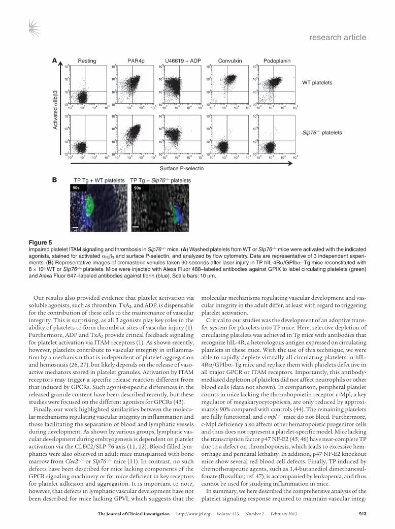
Platelet ITAM signaling and vascular integrity in inflammation. While the GPCR signaling pathway is important for platelet activation by soluble agonists, signaling via ITAM receptors is required for platelet activation by components of the ECM and extra-vascular cells. Mouse platelets express 2 ITAM receptors: GPVI, a receptor for collagen and laminin in the ECM, and CLEC2, a receptor for podoplanin expressed on extravascular cells (4). To study the role of platelet ITAM signaling in the maintenance of vascular integrity, we tested 4 different platelet preparations: (a) platelets treated in vitro with JAQ1, a blocking monoclonal anti-
body against GPVI (36), (b) platelets isolated from Clec2–/– mice, (c) JAQ1-treated Clec2–/– platelets, and (d) platelets isolated from mice lacking Slp76, known to be downstream of ITAM signaling receptors (37). In vitro, all platelet preparations with defects in ITAM signaling showed normal integrin activation and α granule release in response to the GPCR agonists PAR4p and ADP plus U46619 (Supplemental Figure 2 and Figure 5A). JAQ1-treated WT and Clec2–/– platelets were impaired in their responses to the GPVI agonist convulxin and the CLEC2 agonist podoplanin, respectively (Supplemental Figure 2). As expected, integrin activation and α granule release in response to convulxin or podoplanin were abol-ished in JAQ1-treated Clec2–/– and Slp76–/– platelets (Supplemental Figure 2 and Figure 5A). As we observed in platelets with impaired GPCR signaling (Figure 3B), Slp76–/– platelets were markedly impaired in their ability to form 3-dimensional thrombi in crem-asteric venules after laser injury (Figure 5B).
Upon transfusion into TP hIL-4Rα/GPIbα–Tg mice, JAQ1-treated WT platelets only partially restored vascular integrity compared with untreated WT platelets at sites of inflammation induced by rpA (0.60 ± 0.10 versus 0.29 ± 0.03 mg/cm2 Hb; Fig-ure 6, A and B) or by LPS (1.07 ± 0.10 versus 0.14 ± 0.05 mg/ml Hb; Figure 6, C and D). Platelets isolated from Clec2–/– mice also partially reduced hemorrhage in TP hIL-4Rα/GPIbα–Tg mice induced by rpA (0.63 ± 0.72 mg/cm2 Hb) and LPS (1.62 ± 0.20 mg/ml Hb). Transfusion of JAQ1-treated Clec2–/– platelets into TP hIL-4Rα/GPIbα–Tg mice had no significant effect on hemor-rhage induced by rpA (1.06 ± 0.13 mg/cm2 Hb) and LPS (2.42 ± 0.27 mg/ml Hb), and neither did transfusion of Slp76–/– platelets
Figure 3Characterization of platelets with impaired GPCR signaling. (A) WT or Par4–/–/clopidogrel/aspirin (P/C/A) platelets were activated with the indicat-ed agonists, stained for activated αIIbβ3 and surface P-selectin, and analyzed directly by flow cytometry. Data are representative of 3 independent experiments. (B) Representative images of cremasteric venules taken 90 seconds after laser injury in TP hIL-4Rα/GPIbα–Tg mice reconstituted with 8 × 10⁸ WT or Par4–/–/clopidogrel/aspirin platelets. Mice were injected with Alexa Fluor 488–labeled antibodies against GPIX to label circulat-ing platelets (green) and Alexa Fluor 647–labeled antibodies against fibrin (blue). Scale bars: 10 μm.

research article
912 The Journal of Clinical Investigation http://www.jci.org Volume 123 Number 2 February 2013
(Figure 6, A–D). Peripheral platelet counts were similar in mice transfused with the respective platelet preparations (Supplemen-tal Figure 1, B and D), excluding the possibility that hemorrhage at sites of inflammation was the result of impaired survival of platelets after adoptive transfer. These results demonstrated that the protective effect of platelets in the maintenance of vascular integrity during inflammation depends on the 2 ITAM receptors GPVI and CLEC2 and the adapter protein SLP-76.
DiscussionOur studies using a novel method to generate mice with multiple platelet-specific signaling defects identified platelet ITAM sig-naling as a critical event in the maintenance of vascular integrity at sites of inflammation. Platelets defective in ITAM signaling were unable to prevent hemorrhage at sites of immune complex–induced inflammation in the skin as well as LPS-induced inflam-mation in the lung. In contrast, platelets defective in GPCR signal-ing were indistinguishable from WT platelets in their ability to support vascular integrity in both models. Our findings provided evidence that the platelet signaling response required to prevent hemorrhage in inflammation can be generalized for different trig-gers and vascular beds, although this conclusion will need to be confirmed in other models of inflammation.
The central finding of our work was that the platelet ITAM sig-naling pathway is critical for the maintenance of vascular integ-rity in inflammation. Mouse platelets express 2 ITAM receptors, GPVI and CLEC2. Inhibition of GPVI or deficiency in CLEC2 par-tially reduced the ability of platelets to maintain vascular integrity in immune complex–induced inflammation in the skin or LPS-induced inflammation in the lung. A defect in both GPVI and
CLEC2 signaling, or genetic deletion of the downstream adapter protein SLP-76, completely abolished the positive effect of plate-lets on vascular integrity. GPVI signaling is likely activated at sites of inflammation by collagen and/or laminin, the 2 physiological ligands for GPVI found in the vessel wall (38). More difficult to explain, however, is how CLEC2 on platelets is activated in these situations. Biochemical and genetic studies have established the transmembrane protein podoplanin as the major CLEC2 ligand. Podoplanin expression was first documented in glomerular podo-cytes, lymphatic endothelium, and the brain (39). However, podo-planin was not found in blood endothelial cells or pericytes, and smooth muscle podoplanin was identified in advanced, but not early, atherosclerotic lesions (40). Thus, podoplanin is not consti-tutively expressed in the vessel wall. In LPS-induced inflammation in the lung, platelet CLEC2 may engage podoplanin expressed at high levels in type I epithelial cells in alveolae (41). However, a cellular source for podoplanin has not yet been identified in the skin. Alternatively, podoplanin could be “delivered” to sites of inflammation by infiltrating macrophages, a recently identified source of this molecule (42). Our observations that both GPVI and CLEC2 contributed to a similar extent to platelet-dependent vascular integrity in the lung and skin are consistent with a tis-sue-independent source of podoplanin as the trigger of platelet CLEC2 signaling. Finally, it is also possible that platelet CLEC2 receptors respond to an as yet unidentified ligand expressed in these tissues or in the vessel wall. Future studies in mice with con-ditional deletion of podoplanin in various tissues/cell types, such as alveolar cells, lymphatic endothelial cells, monocytes/macro-phages, or smooth muscle cells, will be required to distinguish among these possible mechanisms.
Figure 4Platelet GPCR signaling is not required for the maintenance of vascular integrity in inflammation. (A and B) rpA reaction in skin of TP hIL-4Rα/GPIbα–Tg mice reconstituted or not (–) with the indicated platelet preparations (8 × 10⁸ platelets/mouse; n = 15–20 spots per group). Clop, clopidogrel; ASA, aspirin. (A) Hb levels in skin lesions 4 hours after rpA challenge. Intradermal injection of PBS (without rpA) is also shown. (B) Representative images of rpA reaction sites (indicated by dashed outlines and arrows). (C and D) LPS-induced inflammation in lungs of TP hIL-4Rα/GPIbα–Tg mice reconstituted or not with the indicated platelet preparations (8 × 10⁸ platelets/mouse; n = 5 per group). (C) Hb levels in BAL 6 hours after LPS challenge. Intradermal injection of PBS (without LPS) is also shown. (D) Representative images of BAL. ***P < 0.001 versus no platelet reconstitution.
research article
The Journal of Clinical Investigation http://www.jci.org Volume 123 Number 2 February 2013 913
Our results also provided evidence that platelet activation via soluble agonists, such as thrombin, TxA2, and ADP, is dispensable for the contribution of these cells to the maintenance of vascular integrity. This is surprising, as all 3 agonists play key roles in the ability of platelets to form thrombi at sites of vascular injury (1). Furthermore, ADP and TxA2 provide critical feedback signaling for platelet activation via ITAM receptors (1). As shown recently, however, platelets contribute to vascular integrity in inflamma-tion by a mechanism that is independent of platelet aggregation and hemostasis (26, 27), but likely depends on the release of vaso-active mediators stored in platelet granules. Activation by ITAM receptors may trigger a specific release reaction different from that induced by GPCRs. Such agonist-specific differences in the released granule content have been described recently, but these studies were focused on the different agonists for GPCRs (43).
Finally, our work highlighted similarities between the molecu-lar mechanisms regulating vascular integrity in inflammation and those facilitating the separation of blood and lymphatic vessels during development. As shown by various groups, lymphatic vas-cular development during embryogenesis is dependent on platelet activation via the CLEC2/SLP-76 axis (11, 12). Blood-filled lym-phatics were also observed in adult mice transplanted with bone marrow from Clec2–/– or Slp76–/– mice (11). In contrast, no such defects have been described for mice lacking components of the GPCR signaling machinery or for mice deficient in key receptors for platelet adhesion and aggregation. It is important to note, however, that defects in lymphatic vascular development have not been described for mice lacking GPVI, which suggests that the
molecular mechanisms regulating vascular development and vas-cular integrity in the adult differ, at least with regard to triggering platelet activation.
Critical to our studies was the development of an adoptive trans-fer system for platelets into TP mice. Here, selective depletion of circulating platelets was achieved in Tg mice with antibodies that recognize hIL-4R, a heterologous antigen expressed on circulating platelets in these mice. With the use of this technique, we were able to rapidly deplete virtually all circulating platelets in hIL-4Rα/GPIbα–Tg mice and replace them with platelets defective in all major GPCR or ITAM receptors. Importantly, this antibody-mediated depletion of platelets did not affect neutrophils or other blood cells (data not shown). In comparison, peripheral platelet counts in mice lacking the thrombopoietin receptor c-Mpl, a key regulator of megakaryocytopoiesis, are only reduced by approxi-mately 90% compared with controls (44). The remaining platelets are fully functional, and c-mpl–/– mice do not bleed. Furthermore, c-Mpl deficiency also affects other hematopoietic progenitor cells and thus does not represent a platelet-specific model. Mice lacking the transcription factor p47 NF-E2 (45, 46) have near-complete TP due to a defect on thrombopoiesis, which leads to excessive hem-orrhage and perinatal lethality. In addition, p47 NF-E2 knockout mice show several red blood cell defects. Finally, TP induced by chemotherapeutic agents, such as 1,4-butanediol dimethanesul-fonate (Busulfan; ref. 47), is accompanied by leukopenia, and thus cannot be used for studying inflammation in mice.
In summary, we here described the comprehensive analysis of the platelet signaling response required to maintain vascular integ-
Figure 5Impaired platelet ITAM signaling and thrombosis in Slp76–/– mice. (A) Washed platelets from WT or Slp76–/– mice were activated with the indicated agonists, stained for activated αIIbβ3 and surface P-selectin, and analyzed by flow cytometry. Data are representative of 3 independent experi-ments. (B) Representative images of cremasteric venules taken 90 seconds after laser injury in TP hIL-4Rα/GPIbα–Tg mice reconstituted with 8 × 10⁸ WT or Slp76–/– platelets. Mice were injected with Alexa Fluor 488–labeled antibodies against GPIX to label circulating platelets (green) and Alexa Fluor 647–labeled antibodies against fibrin (blue). Scale bars: 10 μm.

research article
914 The Journal of Clinical Investigation http://www.jci.org Volume 123 Number 2 February 2013
rity in inflammation. By generating mice with multiple platelet-specific signaling defects, we demonstrated that vascular integrity in inflammation depended on both ITAM receptors, GPVI and CLEC2, as well as the adapter protein SLP-76. In contrast, signaling via the major GPCRs was dispensable for this response. Our studies highlight potential complications associated with novel antiplate-let drugs targeting the ITAM signaling pathway, which may lead to blood-lymph mixing or to bleeding at sites of inflammation.
MethodsAnimals. 6- to 8-week-old C57BL/6 WT mice were purchased from Jackson laboratory. Par4–/– mice (35) and hIL-4Rα/GPIbα–Tg mice (33) were bred and housed in our animal facility. Where indicated, WT and Par4–/– mice were treated with clopidogrel 24 and 3 hours before the experiment at a dosage of 75 mg/kg body weight (34). Clec2–/– and Slp76fl/fl;PF4-Cre chime-ric (Slp76–/–) mice (11, 37) were generated by transplant of the respective bone marrow into WT mice lethally irradiated with 2 doses of 6.5 Gy 4 hours apart (13 Gy total) using a Cs137 irradiator (J.L. Shepherd).
Reagents and antibodies. Low–molecular weight enoxaparin sodium (Lovenox) and clopidogrel (Plavix) were purchased from Sanofi-Aventis, and heparin-coated capillaries were purchased from VWR. Aspirin was purchased from Bayer. Hb, BSA, prostacyclin (PGI2), LPS, ADP, red blood cell lysis buffer, and formic acid were purchased from Sigma-Aldrich. The following antibodies were used: anti-GPIbα, GPIX, GPVI, and JON/A-PE (Emfret Analytics); recombinant mouse podoplanin/Fc chimera protein and anti–hIL-4R (clone 25463; R&D Systems); anti-αIIbβ3, Ly6G, and FITC-conjugated anti–P-selectin (BD Biosciences); anti-BSA (MP Biomedical). Alexa Fluor 647–labeled antibodies against fibrin were a gift from R. Camire (Children’s Hospital of Philadelphia, Philadelphia, Pennsylvania,
USA). Convulxin was provided by K.J. Clemetson (Theodor Kocher Insti-tute, Bern, Switzerland), and PAR4p was purchased from Advanced Chem-tech. U46619 was purchased from Cayman Chemical.
Platelet depletion. TP was induced in WT or hIL-4Rα/GPIbα–Tg mice by retro-orbital injection of antibodies against GPIbα and hIL-4R, respective-ly (2.5 μg/g body weight). Peripheral platelet and neutrophil counts were assessed by flow cytometry 4 hours after antibody injection (see below).
Platelet transfusion. Blood was drawn into heparinized tubes from the retro-orbital plexus of sedated mice (7.7 μl/g body weight). Platelet-rich plasma (PRP) was obtained by centrifugation at 100 g for 5 minutes. PRP was centrifuged at 700 g in the presence of PGI2 (2 μg/ml) for 5 minutes at room temperature. After 1 washing step, pelleted platelets were resus-pended in modified Tyrode’s buffer (137 mM NaCl, 0.3 mM Na2HPO4, 2 mM KCl, 12 mM NaHCO3, 5 mM N-2-hydroxyethylpiperazine-N′-2-ethanesulfonic acid, 5 mM glucose, pH 7.3). Platelets from several donor mice were pooled, and the platelet count was adjusted to 1 × 10⁹, 2 × 10⁹, or 4 × 10⁹ platelets/ml. BSA was added to purified platelets to yield a final BSA concentration of 150 μg/g body weight in mice. Recipient mice were injected with 200 μl washed platelets and BSA.
Flow cytometry. For determination of endogenous and transfused platelet counts, diluted whole blood was stained with antibodies against GPIbα (phy-coerythrin labeled; 2 μg/ml) and GPIX (Alexa Fluor 488 labeled; 2 μg/ml), and counts were assessed by flow cytometry (Accuri C6; BD Biosciences). To document the selective depletion of hIL-4Rα/GPIb–Tg platelets by anti–hIL-4R antibodies, we transfused WT platelets into hIL-4Rα/GPIbα–Tg mice, fol-lowed by injection of anti–hIL-4R antibodies (Supplemental Figure 3).
For determination of peripheral neutrophil counts, 50 μl heparinized whole blood was lysed in red cell lysis buffer for 10 minutes at room tem-perature. The lysed cells were centrifuged at 250 g, and the supernatant
Figure 6Platelet ITAM signaling is critical for the maintenance of vascular integrity during inflammation. (A and B) Hemorrhage in skin lesions of TP hIL-4Rα/GPIbα–Tg mice reconstituted or not with the indicated platelet preparations (8 × 10⁸ platelets/mouse), with or without treatment with JAQ1 antibody (n = 15–20 spots per group). (A) Hb levels 4 hours after rpA challenge. Intradermal injection of PBS (without rpA) is also shown. (B) Representative images of lesions (dashed outlines and arrowheads). (C and D) LPS-induced inflammation in lungs of TP hIL-4Rα/GPIbα–Tg mice reconstituted or not with the indicated platelet preparations (8 × 10⁸ platelets/mouse), with or without treatment with JAQ1 antibody (n = 3–5 per group). (C) Hb levels in BAL after LPS challenge. Intradermal injection of PBS (without LPS) is also shown. (D) Representative images of BAL. **P < 0.01, ***P < 0.001 versus WT platelets; #P < 0.001 versus no platelet reconstitution.
research article
The Journal of Clinical Investigation http://www.jci.org Volume 123 Number 2 February 2013 915
1. Broos K, Feys HB, De Meyer SF, Vanhoorelbeke K, Deckmyn H. Platelets at work in primary hemosta-sis. Blood Rev. 2011;25(4):155–167.
2. De Meyer SF, Vanhoorelbeke K, Broos K, Salles RI, Deckmyn H. Antiplatelet drugs. Br J Haematol. 2008;142(4):515–528.
3. Cornelissen I, et al. Roles and interactions among protease-activated receptors and P2ry12 in hemo-stasis and thrombosis. Proc Natl Acad Sci U S A. 2010;107(43):18605–18610.
4. Watson SP, Herbert JM, Pollitt AY. GPVI and CLEC-2 in hemostasis and vascular integrity. J Thromb Haemost. 2010;8(7):1456–1467.
5. Kato K, et al. The contribution of glycoprotein VI to stable platelet adhesion and thrombus forma-tion illustrated by targeted gene deletion. Blood. 2003;102(5):1701–1707.
6. Hermans C, Wittevrongel C, Thys C, Smethurst PA, Van Geet C, Freson K. A compound heterozygous mutation in glycoprotein VI in a patient with a bleeding disorder. J Thromb Haemost. 2009;7(8):1356–1363.
7. Dumont B, et al. Absence of collagen-induced plate-let activation caused by compound heterozygous GPVI mutations. Blood. 2009;114(9):1900–1903.
8. May F, et al. CLEC-2 is an essential platelet-activat-ing receptor in hemostasis and thrombosis. Blood. 2009;114(16):3464–3472.
9. Suzuki-Inoue K, et al. Essential in vivo roles of the C-type lectin receptor CLEC-2: embryonic/neona-
tal lethality of CLEC-2-deficient mice by blood/lymphatic misconnections and impaired throm-bus formation of CLEC-2-deficient platelets. J Biol Chem. 2010;285(32):24494–24507.
10. Hughes CE, Navarro-Nunez L, Finney BA, Mourao-Sa D, Pollitt AY, Watson SP. CLEC-2 is not required for platelet aggregation at arteriolar shear. J Thromb Haemost. 2010;8(10):2328–2332.
11. Bertozzi CC, et al. Platelets regulate lymphatic vas-cular development through CLEC-2-SLP-76 signal-ing. Blood. 2010;116(4):661–670.
12. Finney BA, et al. CLEC-2 and Syk in the mega-karyocytic/platelet lineage are essential for devel-opment. Blood. 2012;119(7):1747–1756.
13. Uhrin P, et al. Novel function for blood plate-lets and podoplanin in developmental separa-tion of blood and lymphatic circulation. Blood. 2010;115(19):3997–4005.
14. Fu J, et al. Endothelial cell O-glycan deficiency causes blood/lymphatic misconnections and con-sequent fatty liver disease in mice. J Clin Invest. 2008;118(11):3725–3737.
15. Kisucka J, et al. Platelets and platelet adhesion sup-port angiogenesis while preventing excessive hemor-rhage. Proc Natl Acad Sci U S A. 2006;103(4):855–860.
16. Echtler K, et al. Platelets contribute to postna-tal occlusion of the ductus arteriosus. Nat Med. 2010;16(1):75–82.
17. Carramolino L, Fuentes J, Garcia-Andres C, Azcoi-tia V, Riethmacher D, Torres M. Platelets play an
essential role in separating the blood and lymphat-ic vasculatures during embryonic angiogenesis. Circ Res. 2010;106(7):1197–1201.
18. Elzey BD, et al. Platelet-mediated modulation of adaptive immunity. A communication link between innate and adaptive immune compart-ments. Immunity. 2003;19(1):9–19.
19. Iannacone M, et al. Platelets mediate cytotoxic T lymphocyte-induced liver damage. Nat Med. 2005;11(11):1167–1169.
20. Boilard E, et al. Platelets amplify inflammation in arthritis via collagen-dependent microparticle pro-duction. Science. 2010;327(5965):580–583.
21. Massberg S, et al. A critical role of platelet adhesion in the initiation of atherosclerotic lesion forma-tion. J Exp Med. 2002;196(7):887–896.
22. Clark SR, et al. Platelet TLR4 activates neutrophil extracellular traps to ensnare bacteria in septic blood. Nat Med. 2007;13(4):463–469.
23. Zarbock A, Polanowska-Grabowska RK, Ley K. Platelet-neutrophil-interactions: linking hemosta-sis and inflammation. Blood Rev. 2007;21(2):99–111.
24. Kitchens CS, Weiss L. Ultrastructural changes of endothelium associated with thrombocytopenia. Blood. 1975;46(4):567–578.
25. Aursnes I, Pedersen OO. Petechial hemorrhage in the ciliary processes of thrombocytopenic rab-bits. An electron microscopic study. Microvasc Res. 1979;17(1):12–21.
26. Ho-Tin-Noe B, Demers M, Wagner DD. How plate-
Laser injury–induced thrombosis model. Laser-induced thrombosis in the cre-master muscle microcirculation was performed as described previously (34). Briefly, male mice (12–14 weeks of age) were anesthetized by intraperitoneal injection of Avertin (0.25 mg/g body weight). The cremaster muscle was iso-lated, and the microvessels were studied with a Zeiss Examiner Z1 micro-scope. Laser injuries were inflicted with an Ablate 3i system. 5 minutes prior to injury, mice were injected intravenously with Alexa Fluor 488–labeled antibodies against GPIX (0.3 μg/g body weight) and Alexa Fluor 647–labeled antibodies against fibrin (0.45 μg/g body weight). Images were collected for 5 minutes at a speed of 1 frame per second using Slidebook software.
Statistics. 2-group comparison of parametric data was compared using a 2-tailed Student’s t test. Statistical significance among multiple groups was assessed by 1-way ANOVA with Bonferroni post-hoc analysis. Statistical analyses were performed using Prism software (GraphPad). A P value of 0.05 or less was considered significant. All data are presented as mean ± SEM.
Study approval. All animal experiments and protocols were reviewed and approved by, and performed in accordance with, the Animal Care and Use Committee of the University of North Carolina at Chapel Hill.
AcknowledgmentsThis work was funded by a Fellowship from the French Founda-tion for the Medical Research; by the American Heart Association (12POST12040088, to Y. Boulaftali; and 10IRG4100001, to W. Bergmeier); by National Heart, Lung, and Blood Institute, NIH, grants R01 HL094594 and HL106009 (to W. Bergmeier), HL50545 (to J. Ware), HL072798 (to M.L. Kahn), HL006350 (to N. Mack-man), and F32 HL099175 (to A.P. Owens III).
Received for publication June 4, 2012, and accepted in revised form November 27, 2012.
Address correspondence to: Wolfgang Bergmeier, Department of Biochemistry and Biophysics, McAllister Heart Institute, 98 Man-ning Drive, 306 Mary Ellen Jones Bldg., University of North Caroli-na, Chapel Hill, North Carolina 27599, USA. Phone: 919.962.7331; Fax: 919.966.7639; E-mail: [email protected].
was carefully discarded. The pellet was resuspended in 20 μl PBS, and cells were stained with 2 μg/ml Alexa Fluor 488–labeled anti-Ly6G antibody (10 minutes at room temperature). Cells were analyzed immediately by flow cytometry. Results were expressed relative to neutrophil count before antibody injection.
Platelet activation. Platelets were washed and diluted with Tyrode’s buffer containing 1 mM CaCl2; stimulated with PAR4p (300 μM), ADP (10 μM) plus U46619 (5 μM), convulxin (300 ng/ml), or recombinant mouse podoplanin/Fc (1 μg/ml) plus anti-Fc IgG for 10 minutes; and stained for activated αIIbβ3 (JON/A-PE; 2 μg/ml) and surface expression of P-selectin (anti–P-selectin–FITC; 2 μg/ml). Where indicated, diluted washed platelets were incubated with JAQ1 (20 μg/ml) or aspirin (1 mg/ml) for 20 minutes prior to activation. The samples were directly analyzed on an Accuri C6 flow cytometer.
rpA reaction. The rpA reaction was triggered by intradermal injection of anti-BSA antibodies (60 μg diluted in PBS) followed by retro-orbital injection of Tyrode’s buffer or washed platelets resuspended in Tyrode’s buffer containing BSA, to reach a BSA concentration of 150 μg/g body weight. 4 hours later, mice were sacrificed, skin biopsies (8-mm punch) were homogenized in 500 μl PBS and spun at 15,000 g for 10 minutes, and the supernatant was analyzed. Formic acid was added, and the opti-cal density at 405 nm was measured. Diluted bovine Hb was used to set up a standard curve.
Lung inflammation model. Mice were sedated and inoculated intranasally with 7 μg Pseudomonas aeruginosa LPS or PBS vehicle, as described previ-ously (27). 10 minutes after intranasal application, washed platelets were injected retro-orbitally into mice. BAL was performed 6 hours later by can-nulating the trachea with an 18-gauge angiocath, and lungs were lavaged 3 times with 1 ml cold sterile PBS. Lavage fluids were pooled, and Hb con-centration was measured as described above.
Inhibitor studies. The collagen receptor GPVI was inhibited by incubation of platelets in vitro with the blocking antibody JAQ1 (100 μg/ml; 15 min-utes). Unbound JAQ1 antibody was washed away prior to platelet trans-fusion. To inhibit cyclooxygenase-mediated generation of TxA2, platelets were treated with aspirin (1 mg/ml) for 20 minutes at 37°C, washed, and then injected into recipient mice.

research article
916 The Journal of Clinical Investigation http://www.jci.org Volume 123 Number 2 February 2013
lets safeguard vascular integrity. J Thromb Haemost. 2011;9:56–65.
27. Goerge T, et al. Inflammation induces hemorrhage in thrombocytopenia. Blood. 2008;111(10):4958–4964.
28. Ho-Tin-Noe B, Goerge T, Cifuni SM, Duerschmied D, Wagner DD. Platelet granule secretion continu-ously prevents intratumor hemorrhage. Cancer Res. 2008;68(16):6851–6858.
29. Hirahashi J, et al. Mac-1 (CD11b/CD18) links inflammation and thrombosis after glomerular injury. Circulation. 2009;120(13):1255–1265.
30. Ho-Tin-Noe B, Carbo C, Demers M, Cifuni SM, Goerge T, Wagner DD. Innate immune cells induce hemorrhage in tumors during thrombocytopenia. Am J Pathol. 2009;175(4):1699–1708.
31. Nieswandt B, Bergmeier W, Rackebrandt K, Gess-ner JE, Zirngibl H. Identification of critical anti-gen-specific mechanisms in the development of immune thrombocytopenic purpura in mice. Blood. 2000;96(7):2520–2527.
32. Tiedt R, Schomber T, Hao-Shen H, Skoda RC. Pf4-Cre transgenic mice allow the generation of lineage-restricted gene knockouts for studying megakaryocyte and platelet function in vivo. Blood. 2007;109(4):1503–1506.
33. Kanaji T, Russell S, Ware J. Amelioration of
the macrothrombocytopenia associated with the murine Bernard-Soulier syndrome. Blood. 2002;100(6):2102–2107.
34. Stolla M, et al. The kinetics of alphaIIbbeta3 activa-tion determines the size and stability of thrombi in mice: implications for antiplatelet therapy. Blood. 2011;117(3):1005–1013.
35. Sambrano GR, Weiss EJ, Zheng YW, Huang W, Coughlin SR. Role of thrombin signalling in platelets in haemostasis and thrombosis. Nature. 2001;413(6851):74–78.
36. Nieswandt B, Bergmeier W, Schulte V, Rackebrandt K, Gessner JE, Zirngibl H. Expression and function of the mouse collagen receptor glycoprotein VI is strict-ly dependent on its association with the FcRgamma chain. J Biol Chem. 2000;275(31):23998–24002.
37. Abtahian F, et al. Regulation of blood and lym-phatic vascular separation by signaling proteins SLP-76 and Syk. Science. 2003;299(5604):247–251.
38. Ozaki Y, Suzuki-Inoue K, Inoue O. Novel interac-tions in platelet biology: CLEC-2/podoplanin and laminin/GPVI. J Thromb Haemost. 2009;7:191–194.
39. Astarita JL, Acton SE, Turley SJ. Podoplanin: emerging functions in development, the immune system, and cancer. Front Immunol. 2012;3:283.
40. Hatakeyama K, et al. Podoplanin expression in
advanced atherosclerotic lesions of human aortas. Thromb Res. 2012;129(4):e70–e76.
41. Vanderbilt JN, et al. Directed expression of trans-genes to alveolar type I cells in the mouse. Am J Respir Cell Mol Biol. 2008;39(3):253–262.
42. Kerrigan AM, et al. Podoplanin-expressing inflam-matory macrophages activate murine platelets via CLEC-2. J Thromb Haemost. 2012;10(3):484–486.
43. Italiano JE Jr, et al. Angiogenesis is regulated by a novel mechanism: pro- and antiangiogenic proteins are organized into separate platelet alpha granules and differentially released. Blood. 2008;111(3):1227–1233.
44. Alexander WS, Roberts AW, Nicola NA, Li R, Met-calf D. Deficiencies in progenitor cells of multiple hematopoietic lineages and defective megakaryocy-topoiesis in mice lacking the thrombopoietic recep-tor c-Mpl. Blood. 1996;87(6):2162–2170.
45. Shivdasani RA, et al. Transcription factor NF-E2 is required for platelet formation independent of the actions of thrombopoietin/MGDF in megakaryo-cyte development. Cell. 1995;81(5):695–704.
46. Levin J, et al. Pathophysiology of thrombocytope-nia and anemia in mice lacking transcription factor NF-E2. Blood. 1999;94(9):3037–3047.
47. Lesurtel M, et al. Platelet-derived serotonin mediates liver regeneration. Science. 2006;312(5770):104–107.