Nadezhda Mihaylova, Librarian at Hristo Botev 1907 Public Chitalishte in Roman

Central Archives of Stem Cell Research
Cite this article: Mihaylova Z , Stanimirov P , Mitev V , Ishkitiev N (2015) Platelet-Derived Products and Periodontal Ligament Stem Cells. Arch Stem Cell Res 2(1): 1006.
*Corresponding authorIshkitiev N, Department of Pediatric Dentistry, Faculty of Dental Medicine, Medical University – Sofia, 1431 G. Sofiyski Str, Sofia, Bulgaria, Tel: 359885108620; E-mail:
Submitted: 27 February 2015
Accepted: 10 April 2015
Published: 20 April 2015
Copyright© 2014 Ishkitiev et al.
OPEN ACCESS
Keywords• Stem cells• Periodontal ligament• Growth factors• Platelet-derived growth factor• Platelet-rich plasma• Platelet-rich fibrin
Review Article
Platelet-Derived Products and Periodontal Ligament Stem CellsMihaylova Z1, Stanimirov P1, Mitev V2 and Ishkitiev N3*1Department of Oral and Maxillofacial surgery, Medical University – Sofia, Bulgaria2Department of Medical Chemistry and Biochemistry, Medical University – Sofia, Bulgaria3Department of Pediatric Dentistry, Medical University – Sofia, Bulgaria
Abstract
Stem cells represent a remarkable cell type with the ability of self-renew and differentiation into a great number of cell lineages. They provide new therapeutic approaches for regeneration of damaged or missing tissues. Stem cells have been found in various tissues, including in the oral and maxillofacial region. Six different types of mesenchymal stem cells (MSC) are described in the oral cavity. Some of them, called periodontal ligament stem cells (PDLSC), are capable in appropriate conditions to maintain the regeneration of tooth-supporting structures. These cells are located in the periodontal ligament (PDL) surrounding the roots of the teeth. Main problem in the field of dentistry is the tooth – supporting structure loss. For that reason, many scientists have focused on examination of PDLSC application in tissue regeneration. However, the adequate restoration of tissues is provided by strictly regulated interactions between cells, growth factors and extracellular matrix. Growth factors provide the information transfer between cells and extracellular matrix and usually enhance the proliferative and differentiation potency of cells. Thus they support the healing process after injury. Growth factors are located in a highest amount in the alpha-granules of the platelets. Therefore, a different techniques for platelet concentrates delivery have been developed.
ABBREVIATIONSPDL: Periodontal Ligament; MSC: Mesenchymal Stem Cells;
BMSC: Bone Marrow Stem Cells; IPS: Induced Pluripotent Stem cells; PDLSC: Periodontal Ligament Stem Cells; DPSC: Dental Pulp Stem Cells; SHED: Stem cells from Human Exfoliated Deciduous teeth; DFSC: Dental Follicle Stem Cells; SCAP: Stem Cells from Apical Papilla; ISCT: International Society of Cellular Therapy; PDGF: Platelet-Derived Growth Factor; PRP: Platelet-Rich Plasma; PRF: Platelet-Rich Fibrin.
INTRODUCTIONThe increasing frequency of neoplasms and traumas and the
tendency for establishment of new more effective therapeutic approaches, contribute to the progressive development of tissue engineering and regenerative medicine. The progress in tissue engineering starts from the beginning of the 90s and in the past decade a marked step forward has been made in the field of regenerative medicine [1]. Tissue regeneration is a combination of processes, related to specifically regulated interactions between cells, system and local growth factors and extracellular matrix [2-4]. Cells, capable to differentiate into various cell
lineages, stimulating the extracellular matrix formation and the regeneration of damaged tissues are basic for the tissue regeneration [5-7]. Stem cells are very attractive area of research today and have been used in the field of preclinical and clinical regenerative medicine with the aim to treat many degenerative diseases, including in the oral cavity [8]. The advancement in stem cells research significantly increases the opportunities for tissue regeneration [9] and leads to development of new therapeutic approaches for tissue preservation.
Tissue injury and hemorrhage are followed by activation of coagulation cascade and formation of blood cloth. Platelets aggregate and release their specialized granules, which are reservoirs of various growth factors [10], at the site of the blood cloth. Growth factors and cytokines can promote and maintain mesenchymal stem cells self-renewal in vitro, as it is demonstrated by the expression of gene markers associated with stemness – oct-4, sox-2, rex-1 [11].
The reveal of the intimate mechanisms of interactions between stem cells, growth factors and extracellular matrix gives an impetus for regeneration development research.
Central
Ishkitiev et al. (2015)Email:
Arch Stem Cell Res 2(1): 1006 (2015) 2/7
Stem cells
Stem cells represent a special type of cells, characterized with properties like clonogenicity, self-renewal, multi-lineage differentiation [12]. They have the ability to differentiate into numerous cell lineages like osteoblasts, chondroblasts, adipocyte-like cells, pancreatic [13], hepatocyte-like cells [14], etc. Stem cells could be divided into three general types - embryonic stem cells, adult stem cells and induced pluripotent stem cells (IPS) [15]. Embryonic stem cells are pluripotent cells delivered from the blastocyst which can differentiate into the three germ layers – ecto, endo and mesoderm [16], but they cannot differentiate into extra-embryonal tissues [17].
The adult stem cells are dispersed in between other differentiated tissues and organs and their differentiation capacity is more limited compared to the embryonic stem cells; they support restoration of damaged tissues [15].
Induced pluripotent stem cells are delivered through gene transfection and cell reprogramming of somatic cells, using appropriate vectors to acquire similar features like embryonic stem cells [18].
Adult stem cells
The adult stem cells are also called somatic or postnatal stem cells. Investigating the regenerative mechanism of bone marrow after radiation exposure Till and McCulloch determined the presence of stem cells in adult organisms [19]. That kind of fibroblast-like cells have been called mesenchymal stem cells (MSC) [20]. Many investigators are researching different biological materials and tissues as a source of stem cells, like bone marrow [21], placenta [22], blood [23], adipose tissue [24], etc.
Adult stem cells can divide either symmetrically (allowing the increase of stem cell number) or asymmetrically. Asymmetric divisions keep the number of stem cells unaltered and are responsible for the generation of cells with different properties [25]. Most adult stem cells are multipotent, which means they can only differentiate into a limited number of cell types. By the proposal of International Society of Cellular Therapy (ISCT), the minimal criteria for MSC definition has been established. In accordance to that criteria, MSC must be adherent to tissue-culture-treated plastic in standard culture conditions [26], must express CD105, CD73 and CD90 and lack the expression of CD45, CD34, CD14 or CD11b, CD79a or CD19 and HLA-DR surface molecules and also to have the ability to differentiate to osteoblasts, chondroblasts, adipocytes in vitro [27]. Studies in field of dentistry, from the past few years have identified different adult stem cells sources in oral and maxillofacial region. Dental adult stem cells might be excellent candidates for cell-based therapy.
Dental stem cells
Six different types of mesenchymal stem cells have been isolated from the oral cavity – dental pulp stem cells (DPSC) [28], stem cells from human exfoliated deciduous teeth (SHED) [29], dental follicle stem cells (DFSC) [30], stem cells from apical papilla (SCAP) [31], periodontal ligament stem cells (PDLSC) and alveolar bone stem cells. Seo et al. in 2004 isolated for the first time PDLSC and showed that in appropriate conditions
these cells have the ability to express stem-cell-surface markers, to proliferate, to differentiate into various cell lineages and the potential to participate in the regenerative mechanism of the periodontal tissues [32].
Periodontal ligament and PDLSC
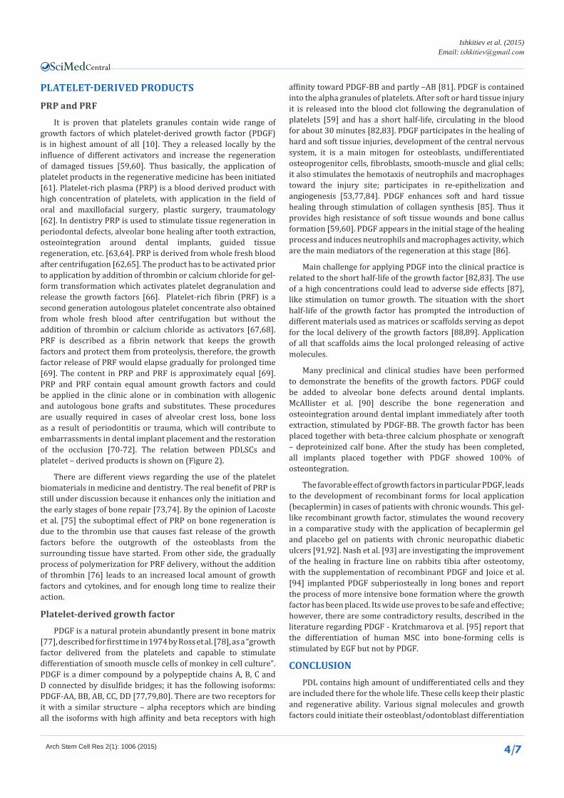
Periodontal ligament is a highly specialized connective tissue localized between the cementum covering the root surface of the tooth and the alveolar bone and comprises cells, collagen fibers and extracellular matrix. It’s main function is to provide the tooth stability in the alveolar bone and the resistance to mastication forces. Cells in the periodontium are heterogeneous population including osteoblasts and osteoclasts (localized in the periodontal ligament but functionally connected to the bone), fibroblasts, epithelial rests of Malassez, macrophages, undifferentiated mesenchymal stem cells and cementoblasts (functionally related to the cementum) [33]. On (Figure 1) are shown some of the tooth structures, including the periodontal ligament.
Serious problem in periodontology and dental implantology is the destruction of periodontal tissues – alveolar bone, cementum and periodontal ligament. Periodontitis is a very common inflammatory disease which leads to the lost of the tissues supporting teeth. Nearly 85-90% of the adults are suffering from periodontitis; however most of them are affected by mild or average and 15% are with severe form of the disease [34,35].
Many authors like Gay IC et al. [36] have been investigating PDLSC proliferative and differentiation capacity. Kim et al. determine and compare the process of osteogenesis around dental implant induced by PDLSC and bone marrow derived stem
Figure 1 Tooth and periodontal structures; periodontal ligament is the source of periodontal ligament stem cells.

Central
Ishkitiev et al. (2015)Email:
Arch Stem Cell Res 2(1): 1006 (2015) 3/7
cells (BMSC) [37]. Wada et al. are investigating PDLSC, BMSC and DPSC immunomodulatory and osteogenic differentiation properties [38].
Exactly like BMSC, PDLSC have self-renewal capacity and stem cell related surface markers expression – they express STRO – 1 (putative stem cell marker), CD146 (perivascular cell marker), CD13, CD29 (integrin-1), CD44, CD90, CD105 (endoglin), CD106 (vascular cell adhesion molecule-1), CD166 (activated leukocyte cell adhesion molecule) and embryonic markers like: (OCT)-4, (SSEA)-1, (SSEA)-4, etc. [39].
When stimulated in an appropriate conditions, PDLSC can attain the formation of new bone, cementum and periodontal ligament [40]. Investigating the potential of PDLSC to undergo cementoblast / osteoblast differentiation shows their ability to form mineralized nodules with high calcium content in the extracellular matrix and the expression of a number osteoblast/cementoblast markers – alkaline phosphatase, matrix extracellular phosphoglycoprotein, bone sialoprotein, osteocalcin, TGF-beta receptor type I [32].
Sonoyama et al. are implanting autologous mesenchymal stem cells from PDL with the aim to create tooth root-periodontal complex in swines. They show this newly-formed structure capable to support a ceramic crown stability and thus to achieve a normal function of the tooth [41]. PDLSC could also participate
in so called “hybrid” teeth formation together with some other type of stem and progenitor cells. Zhang et al. create such hybrid” teeth formation by implant autologous stem cells derived from teeth structures (including PDLS) together with BMSC (from iliac crest) in artificially created bone defect in the lower jaw of swines and prove the formation of tooth-like structures [42].
Several surgical techniques, like guided tissue regeneration, bone grafts and substitutes placement and use of enamel matrix derivate, are often performed for the clinical preservation of periodontal tissues vitality and function, but their success is not absolutely predictable. Guided tissue regeneration is based on the use of non-resorbable barrier membranes ensuring the blood clot integrity; thus the target area would be colonized by no other cells but PDL-cells [43]. The method of guided bone regeneration could applied, when the alveolar bone needs to be prepared before the dental implant placement [44]. Another therapeutic approach in oral, maxillofacial and periodontal surgery is the use of platelet concentrates – platelet rich plasma and platelet rich fibrin. In one study Zhao et al. [45] construct a cell transplant method consisting of cell sheet fragments of PDLSC and platelet rich fibrin - granules to enhance periodontal healing in avulsed tooth re-implantation.
Growth factors
The appropriate application of the triad – cells, scaffold and signal molecules or growth factors, aims the adequate restoration of tissues [46,47]. A special part of these biological events is taken by the growth factors. They provide the information transfer between cells and extracellular matrix, stimulate endogenous regenerative mechanisms and improve the damaged tissues functions [48,49]. It’s proven that growth factors are supporting the exact regulation and enhancement of tissue healing [50-52] by binding to a specific receptors on the target cell’s membrane [53] and initiating an intracellular cascade of changes leading to specific genes expression. A great number of in vitro and in vivo studies demonstrate the growth factors ability to stimulate tissue regeneration through the enhancement of hemotaxis, proliferation, differentiation of cells, extracellular matrix secretion [2,54-56]. For being therapeutically effective, growth factors need to get to their specific targets and remain there long enough, without being unfenced, to realize their mechanism of action [57]. The effects of growth factors on the cells could be generally described as hemotactic effect (onset of direct migration), mitogenic effect (stimulate cell-division), morphogenic effect (stimulate cell-differentiation), apoptosis (programmed cell death), metabolic effects, a combination on two or more of the previous effects. There are different ways and mechanisms for the growth factors to realize their effectiveness according to the time, concentration and the target cell phenotype [47]. As a part of very big group of polipeptide regulator molecules, growth factors are released from normal healthy cell lineages to guide cell maturation and regeneration after tissue injury [58]. Growth factors are working through autocirne, paracrine and telecrine secretory pathways [53]. They play main role in wound healing from the initiation of the injury to the complete recovery. Growth factors participate in the healing and regeneration of virtually all tissues [48,49,57].
Figure 2 Platelet-derived products are capable to influence Periodontal ligament stem cells (PDLSCs) proliferation, differentiation, collagen synthesis in cell culture. Stem cell delivery-based therapy with or without the use of scaffold biomaterials could enhance tissue regeneration in case of periodontal disease, osseus deffects around dental implants, avulced tooth replantation, etc.
Central
Ishkitiev et al. (2015)Email:
Arch Stem Cell Res 2(1): 1006 (2015) 4/7
PLATELET-DERIVED PRODUCTS
PRP and PRF
It is proven that platelets granules contain wide range of growth factors of which platelet-derived growth factor (PDGF) is in highest amount of all [10]. They a released locally by the influence of different activators and increase the regeneration of damaged tissues [59,60]. Thus basically, the application of platelet products in the regenerative medicine has been initiated [61]. Platelet-rich plasma (PRP) is a blood derived product with high concentration of platelets, with application in the field of oral and maxillofacial surgery, plastic surgery, traumatology [62]. In dentistry PRP is used to stimulate tissue regeneration in periodontal defects, alveolar bone healing after tooth extraction, osteointegration around dental implants, guided tissue regeneration, etc. [63,64]. PRP is derived from whole fresh blood after centrifugation [62,65]. The product has to be activated prior to application by addition of thrombin or calcium chloride for gel-form transformation which activates platelet degranulation and release the growth factors [66]. Platelet-rich fibrin (PRF) is a second generation autologous platelet concentrate also obtained from whole fresh blood after centrifugation but without the addition of thrombin or calcium chloride as activators [67,68]. PRF is described as a fibrin network that keeps the growth factors and protect them from proteolysis, therefore, the growth factor release of PRF would elapse gradually for prolonged time [69]. The content in PRP and PRF is approximately equal [69]. PRP and PRF contain equal amount growth factors and could be applied in the clinic alone or in combination with allogenic and autologous bone grafts and substitutes. These procedures are usually required in cases of alveolar crest loss, bone loss as a result of periodontitis or trauma, which will contribute to embarrassments in dental implant placement and the restoration of the occlusion [70-72]. The relation between PDLSCs and platelet – derived products is shown on (Figure 2).
There are different views regarding the use of the platelet biomaterials in medicine and dentistry. The real benefit of PRP is still under discussion because it enhances only the initiation and the early stages of bone repair [73,74]. By the opinion of Lacoste et al. [75] the suboptimal effect of PRP on bone regeneration is due to the thrombin use that causes fast release of the growth factors before the outgrowth of the osteoblasts from the surrounding tissue have started. From other side, the gradually process of polymerization for PRF delivery, without the addition of thrombin [76] leads to an increased local amount of growth factors and cytokines, and for enough long time to realize their action.
Platelet-derived growth factor
PDGF is a natural protein abundantly present in bone matrix [77], described for first time in 1974 by Ross et al. [78], as a “growth factor delivered from the platelets and capable to stimulate differentiation of smooth muscle cells of monkey in cell culture”. PDGF is a dimer compound by a polypeptide chains A, B, C and D connected by disulfide bridges; it has the following isoforms: PDGF-AA, BB, AB, CC, DD [77,79,80]. There are two receptors for it with a similar structure – alpha receptors which are binding all the isoforms with high affinity and beta receptors with high
affinity toward PDGF-BB and partly –AB [81]. PDGF is contained into the alpha granules of platelets. After soft or hard tissue injury it is released into the blood clot following the degranulation of platelets [59] and has a short half-life, circulating in the blood for about 30 minutes [82,83]. PDGF participates in the healing of hard and soft tissue injuries, development of the central nervous system, it is a main mitogen for osteoblasts, undifferentiated osteoprogenitor cells, fibroblasts, smooth-muscle and glial cells; it also stimulates the hemotaxis of neutrophils and macrophages toward the injury site; participates in re-epithelization and angiogenesis [53,77,84]. PDGF enhances soft and hard tissue healing through stimulation of collagen synthesis [85]. Thus it provides high resistance of soft tissue wounds and bone callus formation [59,60]. PDGF appears in the initial stage of the healing process and induces neutrophils and macrophages activity, which are the main mediators of the regeneration at this stage [86].
Main challenge for applying PDGF into the clinical practice is related to the short half-life of the growth factor [82,83]. The use of a high concentrations could lead to adverse side effects [87], like stimulation on tumor growth. The situation with the short half-life of the growth factor has prompted the introduction of different materials used as matrices or scaffolds serving as depot for the local delivery of the growth factors [88,89]. Application of all that scaffolds aims the local prolonged releasing of active molecules.
Many preclinical and clinical studies have been performed to demonstrate the benefits of the growth factors. PDGF could be added to alveolar bone defects around dental implants. McAllister et al. [90] describe the bone regeneration and osteointegration around dental implant immediately after tooth extraction, stimulated by PDGF-BB. The growth factor has been placed together with beta-three calcium phosphate or xenograft – deproteinized calf bone. After the study has been completed, all implants placed together with PDGF showed 100% of osteontegration.
The favorable effect of growth factors in particular PDGF, leads to the development of recombinant forms for local application (becaplermin) in cases of patients with chronic wounds. This gel-like recombinant growth factor, stimulates the wound recovery in a comparative study with the application of becaplermin gel and placebo gel on patients with chronic neuropathic diabetic ulcers [91,92]. Nash et al. [93] are investigating the improvement of the healing in fracture line on rabbits tibia after osteotomy, with the supplementation of recombinant PDGF and Joice et al. [94] implanted PDGF subperiosteally in long bones and report the process of more intensive bone formation where the growth factor has been placed. Its wide use proves to be safe and effective; however, there are some contradictory results, described in the literature regarding PDGF - Kratchmarova et al. [95] report that the differentiation of human MSC into bone-forming cells is stimulated by EGF but not by PDGF.
CONCLUSIONPDL contains high amount of undifferentiated cells and they
are included there for the whole life. These cells keep their plastic and regenerative ability. Various signal molecules and growth factors could initiate their osteoblast/odontoblast differentiation
Central
Ishkitiev et al. (2015)Email:
Arch Stem Cell Res 2(1): 1006 (2015) 5/7
and collagen synthesis. PDGF is a main mitogen for the cell processes. It is capable to stimulate angiogenesis, tissue re-epithelisation, participates in wound healing, but also could have adverse side effects. The reveal of intimate mechanisms of interaction between PDGF and PDLSC will contribute for the overall understanding of molecule mechanisms of tissue regeneration, the advantages of using the growth factor in the clinic.
ACKNOWLEDGEMENTSFinancial support for this study was provided by Bulgarian
Ministry of Education Grant № DFNI B02/15, 14.12.2014.
Conflict of Interest
The authors report no financial affiliation (e.g., employment, direct payment, stock holdings, retainers, consultantships, patent licensing arrangements or honoraria), or involvement with any commercial organization with direct financial interest in the subject or materials discussed in this manuscript, nor have any such arrangements existed in the past three years. Any other potential conflict of interest is disclosed.
REFERENCES1. Langer R, Vacanti JP. Tissue engineering. Science. 1993; 260: 920-926.
2. Lee SJ, Park YJ, Park SN, Lee YM, Seol YJ, Ku Y. Molded porous poly (L-lactide) membranes for guided bone regeneration with enhanced effects by controlled growth factor release. J Biomed Mater Res. 2001; 55: 295-303.
3. Shimabukuro Y, Terashima H, Takedachi M, Maeda K, Nakamura T, Sawada K. Fibroblast growth factor-2 stimulates directed migration of periodontal ligament cells via PI3K/AKT signaling and CD44/hyaluronan interaction. J Cell Physiol. 2011; 226: 809-821.
4. Powell CA, Bannister SR, Mackey SA, Maller SC, McDonnell HT, Deas DE. Periodontal wound healing with and without platelet-rich plasma: histologic observations and assessment of flap tensile strength. J Periodontol. 2009; 80: 985-992.
5. McCulloch CAG. Basic considerations in periodontal wound healing to achieve regeneration. Periodontol 2000. 1993; 1: 16-25.
6. McCulloch CA. Origins and functions of cells essential for periodontal repair: the role of fibroblasts in tissue homeostasis. Oral Dis. 1995; 1: 271-278.
7. Beertsen W, McCulloch CA, Sodek J. The periodontal ligament: a unique, multifunctional connective tissue. Periodontol 2000. 1997; 13: 20-40.
8. Ding G, Liu Y, Wang W, Wei F, Liu D, Fan Z. Allogeneic periodontal ligament stem cell therapy for periodontitis in swine. Stem Cells. 2010; 28: 1829-1838.
9. Bianco P, Robey PG. Stem cells in tissue engineering. Nature. 2001; 414: 118-121.
10. Harrison P, Cramer EM. Platelet alpha-granules. Blood Rev. 1993; 7: 52-62.
11. Kolf CM, Cho E, Tuan RS. Mesenchymal stromal cells. Biology of adult mesenchymal stem cells: regulation of niche, self-renewal and differentiation. Arthritis Res Ther. 2007; 9: 204.
12. Weissman IL. Stem cells: units of development, units of regeneration, and units in evolution. Cell. 2000; 100: 157-168.
13. Ishkitiev N, Yaegaki K, Kozhuharova A, Tanaka T, Okada M, Mitev V.
Pancreatic differentiation of human dental pulp CD117⺠stem cells. Regen Med. 2013; 8: 597-612.
14. Ishkitiev N, Yaegaki K, Calenic B, Nakahara T, Ishikawa H, Mitiev V. Deciduous and permanent dental pulp mesenchymal cells acquire hepatic morphologic and functional features in vitro. J Endod. 2010; 36: 469-474.
15. Hynes K, Menicanin D, Gronthos S, Bartold PM. Clinical utility of stem cells for periodontal regeneration. Periodontol 2000. 2012; 59: 203-227.
16. Keller GM. In vitro differentiation of embryonic stem cells. Curr Opin Cell Biol. 1995; 7: 862-869.
17. Thomson JA, Itskovitz-Eldor J, Shapiro SS, Waknitz MA, Swiergiel JJ, Marshall VS. Embryonic stem cell lines derived from human blastocysts. Science. 1998; 282: 1145-1147.
18. Takahashi K, Yamanaka S. Induction of pluripotent stem cells from mouse embryonic and adult fibroblast cultures by defined factors. Cell. 2006; 126: 663-676.
19. Mcculloch EA, Till JE. Proliferation of Hemopoietic Colony-Forming Cells Transplanted Into Irradiated Mice. Radiat Res. 1964; 22: 383-397.
20. Caplan AI. Mesenchymal stem cells. J Orthop Res. 1991; 9: 641-650.
21. Friedenstein AJ, Chailakhjan RK, Lalykina KS. The development of fibroblast colonies in monolayer cultures of guinea-pig bone marrow and spleen cells. Cell Tissue Kinet. 1970; 3: 393-403.
22. In ‘t Anker PS, Scherjon SA, Kleijburg-van der Keur C, de Groot-Swings GM, Claas FH, Fibbe WE. Isolation of mesenchymal stem cells of fetal or maternal origin from human placenta. Stem Cells. 2004; 22: 1338-1345.
23. He Q, Wan C, Li G. Concise review: multipotent mesenchymal stromal cells in blood. Stem Cells. 2007; 25: 69-77.
24. Zuk PA, Zhu M, Mizuno H, Huang J, Futrell JW, Katz AJ. Multilineage cells from human adipose tissue: implications for cell-based therapies. Tissue Eng. 2001; 7: 211-228.
25. Bluteau G, Luder HU, De Bari C, Mitsiadis TA. Stem cells for tooth engineering. Eur Cell Mater. 2008; 16: 1-9.
26. Horwitz EM, Le Blanc K, Dominici M, Mueller I, Slaper-Cortenbach I, Marini FC. Clarification of the nomenclature for MSC: The International Society for Cellular Therapy position statement. Cytotherapy. 2005; 7: 393-395.
27. Dominici M, Le Blanc K, Mueller I, Slaper-Cortenbach I, Marini F, Krause D, et al. Minimal criteria for defining multipotent mesenchymal stromal cells. The International Society for Cellular Therapy position statement. Cytotherapy. 2006; 8: 315–317.
28. Gronthos S, Mankani M, Brahim J, Robey PG, Shi S. Postnatal human dental pulp stem cells (DPSCs) in vitro and in vivo. Proc Natl Acad Sci U S A. 2000; 97: 13625-13630.
29. Miura M, Gronthos S, Zhao M, Lu B, Fisher LW, Robey PG. SHED: stem cells from human exfoliated deciduous teeth. Proc Natl Acad Sci U S A. 2003; 100: 5807-5812.
30. Morsczeck C, Götz W, Schierholz J, Zeilhofer F, Kühn U, Möhl C. Isolation of precursor cells (PCs) from human dental follicle of wisdom teeth. Matrix Biol. 2005; 24: 155-165.
31. Sonoyama W, Liu Y, Yamaza T, Tuan RS, Wang S, Shi S. Characterization of the apical papilla and its residing stem cells from human immature permanent teeth: a pilot study. J Endod. 2008; 34: 166-171.
32. Seo BM, Miura M, Gronthos S, Bartold PM, Batouli S, Brahim J.

Central
Ishkitiev et al. (2015)Email:
Arch Stem Cell Res 2(1): 1006 (2015) 6/7
Investigation of multipotent postnatal stem cells from human periodontal ligament. Lancet. 2004; 364: 149-155.
33. Nanci A. Ten Cate’s Oral Histology: Development, Structure, and Function. 2012; 8e.
34. Pihlstrom BL, Michalowicz BS, Johnson NW. Periodontal diseases. Lancet. 2005; 366: 1809-1820.
35. Chen FM, Zhang J, Zhang M, An Y, Chen F, Wu ZF. A review on endogenous regenerative technology in periodontal regenerative medicine. Biomaterials. 2010; 31: 7892-7927.
36. Gay IC, Chen S, MacDougall M. Isolation and characterization of multipotent human periodontal ligament stem cells. Orthod Craniofac Res. 2007; 10: 149-160.
37. Kim SH, Kim KH, Seo BM, Koo KT, Kim TI, Seol YJ. Alveolar bone regeneration by transplantation of periodontal ligament stem cells and bone marrow stem cells in a canine peri-implant defect model: a pilot study. J Periodontol. 2009; 80: 1815-1823.
38. Wada N, Menicanin D, Shi S, Bartold PM, Gronthos S. Immunomodulatory properties of human periodontal ligament stem cells. J Cell Physiol. 2009; 219: 667-676.
39. Sununliganon L, Singhatanadgit W. Highly osteogenic PDL stem cell clones specifically express elevated levels of ICAM, ITGB1 and TERT. Cytotechnology. 2012; 64: 53-63.
40. Park JC, Kim JM, Jung IH, Kim JC, Choi SH, Cho KS. Isolation and characterization of human periodontal ligament (PDL) stem cells (PDLSCs) from the inflamed PDL tissue: in vitro and in vivo evaluations. J Clin Periodontol. 2011; 38: 721-731.
41. Sonoyama W, Liu Y, Fang D, Yamaza T, Seo BM, Zhang C. Mesenchymal stem cell-mediated functional tooth regeneration in swine. PLoS One. 2006; 1: e79.
42. Zhang W, Abukawa H, Troulis MJ, Kaban LB, Vacanti JP, Yelick PC. Tissue engineered hybrid tooth-bone constructs. Methods. 2009; 47: 122-128.
43. Urban I,Caplanis N, Lozada JL. Simultaneous vertical guided bone regeneration and guided tissue regeneration in the posterior maxilla using recombinant human platelet-derived growth factor: a case report. J Oral Implantol. 2009; 35: 251-256.
44. Simion M, Rocchietta I, Monforte M, Maschera E. Three-dimensional alveolar bone reconstruction with a combination of recombinant human platelet-derived growth factor BB and guided bone regeneration: a case report. Int J Periodontics Restorative Dent. 2008; 28: 239-243.
45. Zhao YH, Zhang M, Liu NX, Lv X, Zhang J, Chen FM. The combined use of cell sheet fragments of periodontal ligament stem cells and platelet-rich fibrin granules for avulsed tooth reimplantation. Biomaterials. 2013; 34: 5506-5520.
46. Chen FM, Jin Y. Periodontal tissue engineering and regeneration: current approaches and expanding opportunities. Tissue Eng Part B Rev. 2010; 16: 219-255.
47. Varkey M, Gittens SA, Uludag H. Growth factor delivery for bone tissue repair: an update. Expert Opin Drug Deliv. 2004; 1: 19-36.
48. Tayalia P, Mooney DJ. Controlled growth factor delivery for tissue engineering. Adv Mater. 2009; 21: 3269-3285.
49. Langer R. Drug delivery and targeting. Nature. 1998; 392: 5-10.
50. Stefani CM, Machado MA, Sallum EA, Sallum AW, Toledo S, Nociti FH Jr. Platelet-derived growth factor/insulin-like growthfactor-1 combination and bone regeneration around implants placed into extraction sockets: a histometric study in dogs. Implant Dent. 2000; 9: 126-131.
51. Lin M, Overgaard S, Glerup H, Søballe K, Bünger C. Transforming growth factor-beta1 adsorbed to tricalciumphosphate coated implants increases peri-implant bone remodeling. Biomaterials. 2001; 22: 189-193.
52. Vuola J, Böhling T, Göransson H, Puolakkainen P. Transforming growth factor beta released from natural coral implant enhances bone growth at calvarium of mature rat. J Biomed Mater Res. 2002; 59: 152-159.
53. Barrientos S, Stojadinovic O, Golinko MS, Brem H, Tomic-Canic M. Growth factors and cytokines in wound healing. Wound Repair Regen. 2008; 16: 585-601.
54. Kaigler D, Cirelli JA, Giannobile WV. Growth factor delivery for oral and periodontal tissue engineering. Expert Opin Drug Deliv. 2006; 3: 647-662.
55. Debiais F, Hott M, Graulet AM, Marie PJ. The effects of fibroblast growth factor-2 on human neonatal calvaria osteoblastic cells are differentiation stage specific. J Bone Miner Res. 1998; 13: 645-54.
56. Sánchez AR, Sheridan PJ, Kupp LI. Is platelet-rich plasma the perfect enhancement factor? A current review. Int J Oral Maxillofac Implants. 2003; 18: 93-103.
57. Chen FM, Shelton RM, Jin Y, Chapple IL. Localized delivery of growth factors for periodontal tissue regeneration: role, strategies, and perspectives. Med Res Rev. 2009; 29: 472-513.
58. Werner S, Grose R. Regulation of wound healing by growth factors and cytokines. Physiol Rev. 2003; 83: 835-870.
59. Pierce GF, Mustoe TA, Altrock BW, Deuel TF, Thomason A. Role of platelet-derived growth factor in wound healing. J Cell Biochem. 1991; 45: 319-326.
60. Bolander ME. Regulation of fracture repair by growth factors. Proc Soc Exp Biol Med. 1992; 200: 165-170.
61. Kaplan KL, Nossel HL, Drillings M, Lesznik G. Radioimmunoassay of platelet factor 4 and beta-thromboglobulin: development and application to studies of platelet release in relation to fibrinopeptide A generation. Br J Haematol. 1978; 39: 129-146.
62. Marx RE, Carlson ER, Eichstaedt RM, Schimmele SR, Strauss JE, Georgeff KR. Platelet-rich plasma: Growth factor enhancement for bone grafts. Oral Surg Oral Med Oral Pathol Oral Radiol Endod. 1998; 85: 638-646.
63. Vendramin F, Franco D, Nogueira CM, Pereira M, Franco T. Plasma rico em plaquetas e fatores de crescimento: técnica de preparo e utilização em cirurgia plástica. Rev Col Bras Cir. 2006; 33: 24-28.
64. Boyapati L, Wang HL. The role of platelet-rich plasma in sinus augmentation: a critical review. Implant Dent. 2006; 15: 160-170.
65. O’Neill EM, Zalewski WM, Eaton LJ, Popovsky MA, Pivacek LE, Ragno G. Autologous platelet-rich plasma isolated using the Haemonetics Cell Saver 5 and Haemonetics MCS+ for the preparation of platelet gel. Vox Sang. 2001; 81: 172-175.
66. Blair P, Flaumenhaft R. Platelet alpha-granules: basic biology and clinical correlates. Blood Rev. 2009; 23: 177-189.
67. Choukroun J, Adda F, Schoeffer C, Vervelle A. PRF: an opportunity in perio-implantology (in French). Implantodontie. 2000; 42: 55-62.
68. Dohan DM, Choukroun J, Diss A, Dohan SL, Dohan AJ, Mouhyi J, et al. Platelet-rich fibrin (PRF): a second generation platelet concentrate. Part II: Platelet-related biologic features. Oral Surg Oral Med Oral Pathol Oral Radion Endod. 2006; 101: 45-50.
69. Lundquist R, Dziegiel MH, Agren MS. Bioactivity and stability of endogenous fibrogenic factors in platelet-rich fibrin. Wound Repair Regen. 2008; 16: 356-363.

Central
Ishkitiev et al. (2015)Email:
Arch Stem Cell Res 2(1): 1006 (2015) 7/7
70. Gentile P, Bottini DJ, Spallone D, Curcio BC, Cervelli V. Application of platelet-rich plasma in maxillofacial surgery: clinical evaluation. J Craniofac Surg. 2010; 21: 900-904.
71. Mannai C. Early implant loading in severely resorbed maxilla using xenograft, autograft, and platelet-rich plasma in 97 patients. J Oral Maxillofac Surg. 2006; 64: 1420-1426.
72. Rabelo GD, de Paula PM, Rocha FS, Jordão Silva C, Zanetta-Barbosa D. Retrospective study of bone grafting procedures before implant placement. Implant Dent. 2010; 19: 342-350.
73. Schlegel KA, Donath K, Rupprecht S, Falk S, Zimmermann R, Felszeghy E. De novo bone formation using bovine collagen and platelet-rich plasma. Biomaterials. 2004; 25: 5387-5393.
74. Thorwarth M, Rupprecht S, Falk S, Felszeghy E, Wiltfang J,Schlegel KA. Expression of bone matrix proteins during de novo bone formation using a bovine collagen and platelet-rich plasma(PRP)—an immunohistochemical analysis. Biomaterials. 2005; 26: 2575-2584.
75. Lacoste E, Martineau I, Gagnon G. Platelet concentrates: effects of calcium and thrombin on endothelial cell proliferation and growth factor release. J Periodontol. 2003; 74: 1498-1507.
76. Dohan DM, Choukroun J. PRP, cPRP, PRF, PRG, PRGF, FC... How to find your way in the jungle of platelet concentrates? Oral Surg Oral Med Oral Pathol Oral Radiol Endod. 2006; 103: 305-306.
77. Alvarez RH, Kantarjian HM, Cortes JE. Biology of platelet-derived growth factor and its involvement in disease. Mayo Clin Proc. 2006; 81: 1241-1257.
78. Ross R, Glomset J, Kariya B, Harker L. A platelet-dependent serum factor that stimulates the proliferation of arterial smooth muscle cells in vitro. Proc Natl Acad Sci U S A. 1974; 71: 1207-1210.
79. Hammacher A, Hellman U, Johnsson A, Ostman A, Gunnarsson K, Westermark B. A major part of platelet-derived growth factor purified from human platelets is a heterodimer of one A and one B chain. J Biol Chem. 1988; 263: 16493-16498.
80. Gebhard C, Akhmedov A, Mocharla P, Angstenberger J, Sahbai S, Camici GG. PDGF-CC induces tissue factor expression: role of PDGF receptor alpha/beta. Basic Res Cardiol. 2010; 105: 349-356.
81. Hart CE, Forstrom JW, Kelly JD, Seifert RA, Smith RA, Ross R. Two classes of PDGF receptor recognize different isoforms of PDGF. Science. 1988; 240: 1529-1531.
82. Bowen-Pope DF, Malpass TW, Foster DM, Ross R. Platelet-derived growth factor in vivo: levels, activity, and rate of clearance. Blood. 1984; 64: 458-469.
83. Lynch SE, de Castilla GR, Williams RC, Kiritsy CP, Howell TH, Reddy
MS. The effects of short-term application of a combination of platelet-derived and insulin-like growth factors on periodontal wound healing. J Periodontol. 1991; 62: 458-467.
84. Graham S, Leonidou A, Lester M, Heliotis M, Mantalaris A, Tsiridis E. Investigating the role of PDGF as a potential drug therapy in bone formation and fracture healing. Expert Opin Investig Drugs. 2009; 18: 1633-1654.
85. Taba M Jr, Jin Q, Sugai JV, Giannobile WV. Current concepts in periodontal bioengineering. Orthod Craniofac Res. 2005; 8: 292-302.
86. Siegbahn A, Hammacher A, Westermark B, Heldin CH. Differential effects of the various isoforms of platelet-derived growth factor on chemotaxis of fibroblasts, monocytes, and granulocytes. J Clin Invest. 1990; 85: 916-920.
87. Mehrara BJ, Saadeh PB, Steinbrech DS, Dudziak M, Spector JA, Greenwald JA. Adenovirus-mediated gene therapy of osteoblasts in vitro and in vivo. J Bone Miner Res. 1999; 14: 1290-1301.
88. Mao HQ, Roy K, Troung-Le VL, Janes KA, Lin KY, Wang Y. Chitosan-DNA nanoparticles as gene carriers: synthesis, characterization and transfection efficiency. J Control Release. 2001; 70: 399-421.
89. Roy K, Mao HQ, Huang SK, Leong KW. Oral gene delivery with chitosan--DNA nanoparticles generates immunologic protection in a murine model of peanut allergy. Nat Med. 1999; 5: 387-391.
90. McAllister BS, Haghighat K, Prasad HS, Rohrer MD. Histologic evaluation of recombinant human platelet-derived growth factor-BB after use in extraction socket defects: a case series. Int J Periodontics Restorative Dent. 2010; 30: 365-373.
91. Wieman TJ, Smiell JM, Su Y. Efficacy and safety of a topical gel formulation of recombinant human platelet-derived growth factor- BB (becaplermin) in patients with chronic neuropathic diabetic ulcers. A phase III randomized placebo-controlled double-blind study. Diabetes Care. 1998; 21: 822-827.
92. Margolis DJ, Bartus C, Hoffstad O, Malay S, Berlin JA. Effectiveness of recombinant human platelet-derived growth factor for the treatment of diabetic neuropathic foot ulcers. Wound Repair Regen. 2005; 13: 531-536.
93. Nash TJ, Howlett CR, Martin C, Steele J, Johnson KA, Hicklin DJ. Effect of platelet-derived growth factor on tibial osteotomies in rabbits. Bone. 1994; 15: 203-208.
94. Joyce ME, Jingushi S, Scully SP, Bolander ME. Role of growth factors in fracture healing. Prog Clin Biol Res. 1991; 365: 391-416.
95. Kratchmarova I, Blagoev B, Haack-Sorensen M, Kassem M, Mann M. Mechanism of divergent growth factor effects in mesenchymal stem cell differentiation. Science. 2005; 308: 1472-1477.
Mihaylova Z , Stanimirov P , Mitev V , Ishkitiev N (2015) Platelet-Derived Products and Periodontal Ligament Stem Cells. Arch Stem Cell Res 2(1): 1006.
Cite this article