Plasma membrane
66
What are membranes? keeping all cellular components inside the cell allowing selected molecules to move in and out of the cell allowing a cell to change shape. isolating organelles from the rest of the cytoplasm, allowing cellular processes to occur separately. Membranes cover the surface of every cell, and also surround most organelles within cells. They have a number of functions, such as: a site for biochemical reactions
-
Upload
subramaniya-sharma -
Category
Engineering
-
view
273 -
download
5
Transcript of Plasma membrane

What are membranes?
keeping all cellular components inside the cell
allowing selected molecules to move in and out of the cell
allowing a cell to change shape.
isolating organelles from the rest of the cytoplasm, allowing cellular processes to occur separately.
Membranes cover the surface of every cell, and also surround most organelles within cells. They have a number offunctions, such as:
a site for biochemical reactions

The Plasma Membrane
A plasma membrane is common to all cells.It forms their outer limit. It forms a boundary for dissolved substances-allows exchange. Allows cells to maintain themselves
Bacteria, fungi, and plant cells have a cell wall, but it is a structurally distinct feature and lies outside theplasma membrane.
This colored Bacillus megaterium cell clearly shows the plasma membrane, which lies inside the distinct structure of the cell wall.
Plasma membrane
Cell wall

Cells and Membranes
The membrane surrounding a cell, called the plasma membrane, forms the boundary that separates the living cell from its non-living surroundings.
Although the plasma membrane (arrowed) is only about 8 nm (0.01 micrometere) thick, it:
selectively controls the movement of materials into and out of the cell (selectively permeable)
is responsible for cell-cell recognition (e.g. when cells aggregate into tissues
is a dynamic structure, with distinct inside and outside faces.
Animal cell
Plant cell

Membrane StructureThe currently accepted model for the structure of the plasma membrane (and cellular membranes generally) is the fluid mosaic model.
In this model there is a double layer of phospholipids (fats),which are arranged with their hydrophobic tails facing inwards.(repel water)The hydrophilic head (phosphate) is attracted to water-both inside and outside cell-cell is in a watery environmentThe double layer of lipids is quite fluid, with proteins floating within it.Glycoproteins, glycolipids, and cholesterol are also an integral part of the membrane structure.
Double layer of phospholipids (lipid bilayer)} Hydrophilic end
Hydrophobic end

Membrane Structure
Some proteins, called peripheral proteins, are stuck to the surface of the membrane.
Glycolipids act as surface receptors and stabilize the membrane. Common in brain cells and nerves.(CHO and lipid)
Some proteins completely penetrate the phospholipid layer. These proteins may control the movement of specific molecules into and out of the cell.
Glycoproteins play an important role in cellular recognition and immune responses. They help stabilize the membrane structure(adhesion between cells)
Carbohydrates-found on outer layer-linked to protruding-protein(glycoprotein(

Membrane Structure
Cholesterol in the membrane disturbs the close packing of the phospholipids and keeps the membrane more fluid. Provides rigidity and water resistance. Membranes would break down without it. Plants have phytosterol.
Some substances, particularly ions and carbohydrates, are transported across the membrane via the proteins.
Some substances, including water, are transported directly through the phospholipid bilayer.But mostly impermeable to water soluble(polar) molecules-most movement via proteins.
Glycolipids also have a role in helping cells to aggregate in the formation of tissues.
Glycoproteins-cell recognition-between cells,antibodies,hormones and viruses

Fluid –Mosaic model
• Fluid- individual phospholipids and some proteins can move sideways(laterally) in each layer-therefore FLUID
• Mosaic-range of different proteins resting on the surface or through the phospholipid layer gives it a mosaic appearance

Plasma MembraneLocated:Surrounds the cell forming a boundary between the cell contentsand the extracellular environment. Structure:Semi-fluid phospholipid bilayer in which proteins are embedded. Some of the proteins fully span the membrane.Function:
Forms the boundary between the cell and the extracellular environment.Regulates movement of substances in and out of the cell.
Size: 3–10 nm thick.
Phospholipid bilayer
The plasma membranes of two adjacent cells joined with desmosomes
Protein
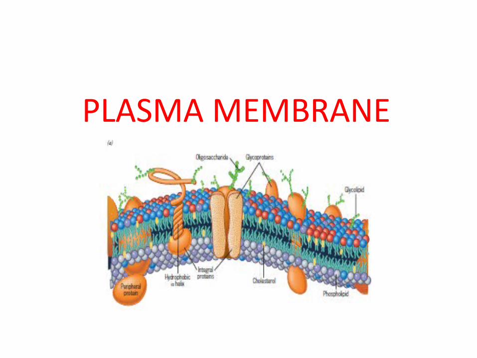
PLASMA MEMBRANE


THREE CLASSES OF MEMBRANE LIPIDS


• Plasma Membrane
• A lipid/protein/carbohydrate complex, providing a barrier and containing transport and signaling systems.
Fluid Mosaic Model of the Plasma Membrane
Carbohydrate chain
Glycoprotein
Intrinsic Protein
PhospholipidsNon-polar hydrophobic fatty acid

Membranes: timeline of discovery

When clear electron micrographs of membranes became available, they appeared to show support for Davson–Danielli’s model, showing a three-layered structure.
2nd cell membrane
This was taken to be the phospholipid bilayer (light) surrounded by two layers of protein (dark).
1st cell membrane
intracellular space (blue)
1 light layer = phospholipid bilayer
2 dark layers: protein
Evidence for the Davson–Danielli model

Later, it was discovered that the light layer represented the phospholipid tails and the dark layers represented the phospholipid heads.
2nd cell membrane
1st cell membrane
intracellular space (blue)
1 light layer = phospholipid tails
2 dark layers: phospholipid heads
Evidence for the Davson–Danielli model

By the end of the 1960s, new evidence cast doubts on the viability of the Davson–Danielli model.
The amount and type of membrane proteins vary greatly between different cells.
It was unclear how the proteins in the model would permit the membrane to change shape without bonds being broken.
Membrane proteins are largely hydrophobic and therefore should not be found where the model positioned them: in the aqueous cytoplasm and extracellular environment.
Problems with the Davson–Danielli model

Evidence from freeze-fracturing
E-face: looking up at outer layer of membrane
This revealed a smooth surface with small bumps sticking out. These were later identified as proteins.
In 1966, biologist Daniel Branton used freeze-fracturing to split cell membranes between the two lipid layers, revealing a 3D view of the surface texture.
P-face: looking down on inner layer of membrane

The fluid mosaic model
This model suggested that proteins are found within, not outside, the phospholipid bilayer.
The freeze-fracture images of cell membranes were further evidence against the Davson–Danielli model.
They led to the development of the fluid mosaic model, proposed by Jonathan Singer and Garth Nicholson in 1972.
E-face
P-face protein

Side view
Surface view
Biochemical Composition of the Plasma Membrane

Side view
Biochemical Composition of the Plasma Membrane
The main components are protein and phospholipid:
ProteinPhospholipid
4.6

Membrane Structure and Function

Membrane Function
• Membranes organize the chemical activities of cells.
• The outer plasma membrane – forms a boundary between a living cell and its
surroundings– Exhibits selective permeability
• Controls traffic of molecules in and out

Membrane Function
• Internal membranes provide structural order for metabolism
• Form the cell's organelles• Compartmentalize chemical reactions

Fluid Mosaic Model of the PM
• A membrane is a mosaic– Proteins and other molecules are embedded in a
framework of phospholipids
• A membrane is fluid– Most protein and phospholipid molecules can
move laterally

Membrane Structure
Phospholipids are the major structural component of membranes.
Phospholipid

Membrane Structure
All membranes are phospholipid bilayers with embedded proteins.
Label the:
Hydrophilic heads
Hydrophobic tails
Phospholipid Bilayer

• Embedded in the bilayer are proteins– Most of the membrane’s functions are
accomplished by the embedded proteins. • Integral proteins span the membrane• Peripheral proteins are on one side or the other of the
membrane

Plasma Membrane Components
• Glycoproteins and glycolipids are proteins/lipids with short chain carbohydrates attached on the extracellular side of the membrane.

Fig. 5-1a
Cholesterol
Glycoprotein
Glycolipid
Carbohydrate ofglycoprotein
Phospholipid
Microfilamentsof cytoskeleton
Integrin

Types of Membrane Proteins
1. Cell-cell recognition proteins2. Integrins3. Intercellular junction proteins4. Enzymes 5. Signal transduction proteins
• Aka - Receptor proteins
1. Transport proteins– Passive and active

• Cell-cell recognition proteins - identify type of cell and identify a cell as “self” versus foreign– Most are glycoproteins • Carbohydrate chains vary between species, individuals,
and even between cell types in a given individual.• Glycolipids also play a role in cell recognition

• Integrins are a type of integral protein– The cytoskeleton attaches to integrins on the
cytoplasmic side of the membrane– Integrins strengthen the membrane
• Intercellular junction proteins - help like cells stick together to form tissues

• Many membrane proteins are enzymes– This is especially important on the membranes of
organelles.

• Signal transduction (receptor) proteins bind hormones and other substances on the outside of the cell.– Binding triggers a change inside the cell.• Called signal transduction• Example: The binding of insulin to insulin receptors
causes the cell to put glucose transport proteins into the membrane.

Fig. 5-1c
Messenger molecule
Activatedmolecule
Receptor

Transport Proteins
• Passive Transport Proteins – allow water soluble substances (small polar
molecules and ions) to pass through the membrane without any energy cost
• Active Transport Proteins– The cell expends energy to transport water
soluble substances against their concentration gradient

Transport of Substances Across the Plasma Membrane (PM)
1. Passive Transport – (Simple) Diffusion (5.3)– Facilitated diffusion (5.6)– Osmosis (5.4, 5.5)
1. Active Transport (5.8)
2. Bulk Flow (5.9)– Endocytosis– Exocytosis

Passive Transport
• In passive transport substances cross the membrane by diffusion– Diffusion - net movement of substances from an
area of high concentration to low concentration• no energy required


Blood group antigens


INTEGRAL MEMBRANE PROTEINS
Integral membrane proteins typically contains one or more transmembrane helices

Peripheral proteins
Peripheral proteins are non covalently bonded to the polar head groups of the lipid bilayer

Lipid anchored proteins
Lipid –anchored proteins are covalently bonded to a lipid group


Various classes of proteins are associated with the lipid bilayer

Freeze fracture : A technique for investigating the cell membrane structure

Freeze- fracture technique


Integral proteins resides in the plasma membrane

Glycoporin a integral protein with a single transmembrane domain

Various amino acid residues within trans membrane helices

Transition temparature

LIPID RAFTS – GPI ANCHORED PROTEINS

The possible movements of phospholipids in a membrane

PATTERN OF MOVEMENT OF INTEGRAL PROTEINS


Plasma membrane functions

RBCS

Plasma membrane of the human erythrocyte

The dynamic properties of the plasma membrane


![Plasma Membrane [7.2] Goals: Understand the concept of homeostasis in relation to the plasma membrane Demonstrate and understand how the plasma membrane.](https://static.fdocuments.net/doc/165x107/5697c01d1a28abf838cd0a9a/plasma-membrane-72-goals-understand-the-concept-of-homeostasis-in-relation.jpg)