
Plasma fractionation: conventional and chromatographic ... · Plasma fractionation: conventional...
27
Plasma fractionation: conventional and chromatographic methods for albumin purification Plazma Fraksiyonlanması: Albumin Saflaştırılması için Geleneksel ve Kromatografik Yöntemler Review A. Denizli / Hacettepe J. Biol. & Chem., 2011, 39 (4), 315–341 Adil Denizli Department of Chemistry, Biochemistry Division, Hacettepe University, Ankara, Turkey ÖZET P lazma ayırma (fraksiyonlama) 60 yıl önce Cohn ve çalışma arkadaşları tarafından geliştirilmiş olan büyük ölçekli protein saflaştırılmasında ilk basamaktır. Son yılların tedavi amaçlı kullanılan önemli proteinleri insan serum albumini (HSA), immunoglobulin G (IgG), pıhtılaşma faktörü VIII ve proteaz inhibitörleridir. HSA plazmada en çok bulunan proteindir ve IgG ile tüm plazma proteinlerinin %80’ini oluşturur. Elde edilen verilere göre albumin plazma protein ürünlerinin yaklaşık %50’sini oluşturmaktadır. HSA saflaştırılması genel olarak, tıbbi kullanım için nispeten yüksek saflık gerektiren hipoproteinemi ve ciddi kan kaybı tedavilerinde gerekli olmaktadır. HSA ayrıca, şok, yanma, hipoalbuminemi, cerrahi, travma, kardiyopulmoner bypass, akut solunum yetmezliği, hemodiyaliz, akut nefroz, hiperbilirübinemi, akut karaciğer yetmezliği, karında sıvı birikmesi ve akut peritonit, pankreatit, mediastinit ve ekstansif selülitde proteince zengin sıvıların ayrılması gibi tedavi amaçlı kullanılmaktadır. Anahtar Kelimeler Albumin, plazma ayırma, affinite kromatografisi, plazma proteinleri ABSTRACT P lasma fractionation is the first process for large scale protein purification developed about 60 years ago by Cohn and co-workers. Currently therapeutically important proteins are human serum albumin (HSA), immunoglobulin G (IgG), coagulation factor VIII and protease inhibitors. HSA is one of the most abundant proteins in plasma and, together with IgGs, constitutes 80% of all plasma proteins. According to the data, HSA represented approximately 50% of all sales of therapeutic plasma protein products. The purification of HSA is generally required for the treatment of hypoproteinemia and heavy loss of blood, requires relatively high purity for medical use. HSA also used for therapeutic purposes such as shock, burns, hypoalbuminemia, surgery, trauma, cardiopulmonary bypass, the acute respiratory distress syndrome, hemodialysis, acute nephrosis, hyperbilirubinemia, acute liver failure, acites, and sequestration of protein-rich fluids in acute peritonitis, pancreatitis, mediastinitis and extensive cellulites. Key Words Albumin, plasma fractionation, affinity chromatography, plasma proteins Article History: Received June 16, 2011; Revised July 12, 2011; Accepted November 17, 2011; Avaliable Online: December 02, 2011. Correspondence to: Adil Denizli, Department of Chemistry, Biochemistry Division, Hacettepe University, Ankara, Turkey Tel: +90 312 297 67 76 Fax: +90 297 79 40 E-Mail: [email protected]
Transcript of Plasma fractionation: conventional and chromatographic ... · Plasma fractionation: conventional...
Plasma fractionation: conventional and chromatographic methods for albumin purification
Plazma Fraksiyonlanması: Albumin Saflaştırılması için Geleneksel ve Kromatografik Yöntemler
Review
A. Denizli / Hacettepe J. Biol. & Chem., 2011, 39 (4), 315–341
Adil Denizli Department of Chemistry, Biochemistry Division, Hacettepe University, Ankara, Turkey
ÖZ E T
Plazma ayırma (fraksiyonlama) 60 yıl önce Cohn ve çalışma arkadaşları tarafından geliştirilmiş olan büyük ölçekli protein saflaştırılmasında ilk basamaktır. Son yılların tedavi amaçlı kullanılan önemli proteinleri
insan serum albumini (HSA), immunoglobulin G (IgG), pıhtılaşma faktörü VIII ve proteaz inhibitörleridir. HSA plazmada en çok bulunan proteindir ve IgG ile tüm plazma proteinlerinin %80’ini oluşturur. Elde edilen verilere göre albumin plazma protein ürünlerinin yaklaşık %50’sini oluşturmaktadır. HSA saflaştırılması genel olarak, tıbbi kullanım için nispeten yüksek saflık gerektiren hipoproteinemi ve ciddi kan kaybı tedavilerinde gerekli olmaktadır. HSA ayrıca, şok, yanma, hipoalbuminemi, cerrahi, travma, kardiyopulmoner bypass, akut solunum yetmezliği, hemodiyaliz, akut nefroz, hiperbilirübinemi, akut karaciğer yetmezliği, karında sıvı birikmesi ve akut peritonit, pankreatit, mediastinit ve ekstansif selülitde proteince zengin sıvıların ayrılması gibi tedavi amaçlı kullanılmaktadır.
Anahtar KelimelerAlbumin, plazma ayırma, affinite kromatografisi, plazma proteinleri
A B S T R AC T
Plasma fractionation is the first process for large scale protein purification developed about 60 years ago by Cohn and co-workers. Currently therapeutically important proteins are human serum albumin (HSA),
immunoglobulin G (IgG), coagulation factor VIII and protease inhibitors. HSA is one of the most abundant proteins in plasma and, together with IgGs, constitutes 80% of all plasma proteins. According to the data, HSA represented approximately 50% of all sales of therapeutic plasma protein products. The purification of HSA is generally required for the treatment of hypoproteinemia and heavy loss of blood, requires relatively high purity for medical use. HSA also used for therapeutic purposes such as shock, burns, hypoalbuminemia, surgery, trauma, cardiopulmonary bypass, the acute respiratory distress syndrome, hemodialysis, acute nephrosis, hyperbilirubinemia, acute liver failure, acites, and sequestration of protein-rich fluids in acute peritonitis, pancreatitis, mediastinitis and extensive cellulites.
Key WordsAlbumin, plasma fractionation, affinity chromatography, plasma proteins
Article History: Received June 16, 2011; Revised July 12, 2011; Accepted November 17, 2011; Avaliable Online: December 02, 2011.
Correspondence to: Adil Denizli, Department of Chemistry, Biochemistry Division, Hacettepe University, Ankara, Turkey
Tel: +90 312 297 67 76 Fax: +90 297 79 40 E-Mail: [email protected]
A. Denizli / Hacettepe J. Biol. & Chem., 2011, 39 (4), 315–341316
INTRODUCTION
In 1839, Professor Ancell recognized the properties of a protein vital to many functions
in the body. He wrote in Lancet, “albumen is doubtless one of the most important of the animal proximate principles; it is found not only in the serum of the blood but in lymph, chyle, in the exhilations from surfaces, in the fluid of cellular tissue, in the aqueous and vitrous humus of the eye, in many other animal fluids-albumin acts sometimes as an acid and a base and its powers of maintaining its combinations is so great albumin proportions are pretty nearly the same in all higher animals, the levels are nearly the same in the sexes and betweeen the ages of 20 and 80”. Thus nearly 170 years ago a very accurate description of the properties of serum albumin was available [1].
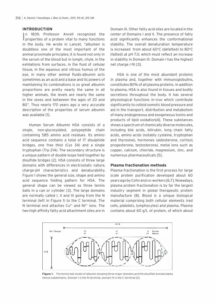
Human Serum Albumin HSA consists of a single, non-glycosylated, polypeptide chain containing 585 amino acid residues. Its amino-acid sequence contains a total of 17 disulphide bridges, one free thiol (Cys 34) and a single tryptophan (Trp 214). The secondary structure is a unique pattern of double loops held together by disulfide bridges [2]. HSA consists of three large domains with differences in electrostatic nature, charge-pH characteristics and denaturability. Figure 1 shows the general size, shape and amino acid sequence folding pattern for HSA. The general shape can be viewed as three tennis balls in a can or cylinder [3]. The large domains are normally called I, II and III going from the N terminal (left in Figure 1) to the C terminal. The N terminal end attaches Cu2+ and Ni2+ ions. The two high affinity fatty acid attachment sites are in
Domain III. Other fatty acid sites are located in the center of Domains I and II. The presence of fatty acid significantly enhances the conformational stability. The overall denaturation temperature is increased from about 60oC (defatted) to 80oC (fatted) at pH 7.0, which must reflect an increase in stability in Domain III. Domain I has the highest net charge (-9) [3].
HSA is one of the most abundant proteins in plasma and, together with immunoglobulins, constitutes 80% of all plasma proteins. In addition to plasma, HSA is also found in tissues and bodily secretions throughout the body. It has several physiological functions in-vivo which contribute significantly to colloid osmotic blood pressure and aid in the transport, distribution and metabolism of many endogeneous and exogeneous toxins and products of lipid oxidation[4]. These substances shows a spectrum of chemically diverse molecules, including bile acids, bilirubin, long chain fatty acids, amino acids (notably cysteine, tryptophan and thyrosine), hormones (aldosterone, cortisol, progesterone, testosterone), metal ions such as copper, calcium, chloride, magnesium, zinc, and numerous pharmaceuticals [5].
Plasma fractionation methodsPlasma fractionation is the first process for large scale protein purification developed about 60 years ago by Cohn and co-workers [6,7]. Nowadays, plasma protein fractionation is by far the largest industry segment in global therapeutic protein manufacture [8]. Blood is a unique biological material comprising both cellular elements (red cells, platelets, lymphocytes) and plasma. Plasma contains about 60 g/L of protein, of which about
Figure 1. The tennis ball model of albumin showing three major domains and the disulfide-bonded alpha
helical subdomains. Domain I is the N-terminal, domain III is the C terminal [3].
A. Denizli / Hacettepe J. Biol. & Chem., 2011, 39 (4), 315–341 317
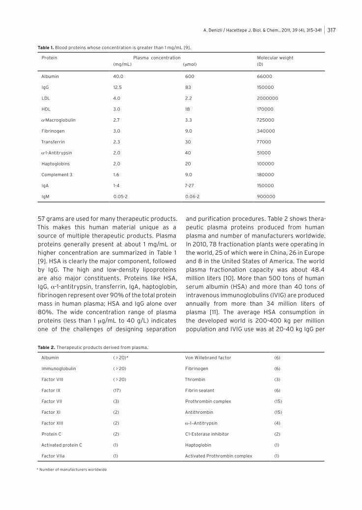
57 grams are used for many therapeutic products. This makes this human material unique as a source of multiple therapeutic products. Plasma proteins generally present at about 1 mg/mL or higher concentration are summarized in Table 1 [9]. HSA is clearly the major component, followed by IgG. The high and low-density lipoproteins are also major constituents. Proteins like HSA, IgG, a-1-antitrypsin, transferrin, IgA, haptoglobin, fibrinogen represent over 90% of the total protein mass in human plasma; HSA and IgG alone over 80%. The wide concentration range of plasma proteins (less than 1 mg/mL to 40 g/L) indicates one of the challenges of designing separation
and purification procedures. Table 2 shows thera-peutic plasma proteins produced from human plasma and number of manufacturers worldwide. In 2010, 78 fractionation plants were operating in the world, 25 of which were in China, 26 in Europe and 8 in the United States of America. The world plasma fractionation capacity was about 48.4 million liters [10]. More than 500 tons of human serum albumin (HSA) and more than 40 tons of intravenous immunoglobulins (IVIG) are produced annually from more than 34 million liters of plasma [11]. The average HSA consumption in the developed world is 200-400 kg per million population and IVIG use was at 20-40 kg IgG per
Table 1. Blood proteins whose concentration is greater than 1 mg/mL [9].
Protein Plasma concentration
(mg/mL) (mmol)
Molecular weight
(D)
Albumin 40.0 600 66000
IgG 12.5 83 150000
LDL 4.0 2.2 2000000
HDL 3.0 18 170000
a-Macroglobulin 2.7 3.3 725000
Fibrinogen 3.0 9.0 340000
Transferrin 2.3 30 77000
a-1-Antitrypsin 2.0 40 51000
Haptoglobins 2.0 20 100000
Complement 3 1.6 9.0 180000
IgA 1-4 7-27 150000
IgM 0.05-2 0.06-2 900000
Table 2. Therapeutic products derived from plasma.
Albumin ( > 20)* Von Willebrand factor (6)
Immunoglobulin ( > 20) Fibrinogen (6)
Factor VIII ( > 20) Thrombin (3)
Factor IX (17) Fibrin sealant (6)
Factor VII (3) Prothrombin complex (15)
Factor XI (2) Antithrombin (15)
Factor XIII (2) a-1-Antitrypsin (4)
Protein C (2) C1-Esterase inhibitor (2)
Activated protein C (1) Haptoglobin (1)
Factor VIIa (1) Activated Prothrombin complex (1)
* Number of manufacturers worldwide
A. Denizli / Hacettepe J. Biol. & Chem., 2011, 39 (4), 315–341318
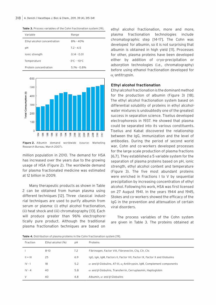
million population in 2010. The demand for HSA has increased over the years due to the growing usage of HSA (Figure 2). The worldwide demand for plasma fractionated medicine was estimated at 12 billion in 2009.
Many therapeutic products as shown in Table 2 can be obtained from human plasma using different techniques [12]. Three classical indust-rial techniques are used to purify albumin from serum or plasma: (i) ethyl alcohol fractionation, (ii) heat shock and (iii) chromatography [13]. Each will produce greater than 96% electrophore-tically pure product. Although the traditional plasma fractionation techniques are based on
ethyl alcohol fractionation, more and more, plasma fractionation technologies include chromatographic step [14-17]. The Cohn was developed for albumin, so it is not surprising that albumin is obtained in high yield [11]. Processes for other, plasma proteins have been developed either by addition of cryo-precipitation or adsorption technologies (i.e., chromatography) before using ethanol fractionation developed for a
1-antitrypsin.
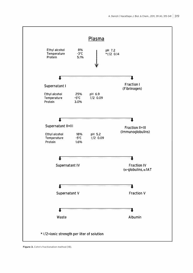
Ethyl alcohol fractionationEthyl alcohol fractionation is the dominant method for the production of albumin (Figure 3) [18]. The ethyl alcohol fractionation system based on differential solubility of proteins in ethyl alcohol-water mixtures is undoubtedly one of the greatest success in separation science. Tiselius developed electrophoresis in 1937. He showed that plasma could be separated into its various constituents. Tiselius and Kabat discovered the relationship between the IgG, immunization and the level of antibodies. During the period of second world war, Cohn and co-workers developed processes for the large scale production of plasma fractions [6,7]. They established a 5 variable system for the separation of plasma proteins based on pH, ionic strength, ethyl alcohol content and temperature (Figure 3). The five most abundant proteins were enriched in Fractions I to V by sequential precipitation by increasing concentration of ethyl alcohol. Following his work, HSA was first licensed on 27 August 1941. In the years 1944 and 1945, Stokes and co-workers showed the efficacy of the IgG in the prevention and attenuation of certain viral disorders.
The process variables of the Cohn system are given in Table 3. The proteins obtained at
Table 3. Process variables of the Cohn fractionation system [19].
Variable Range
Ethyl alcohol concentration 8% - 40%
pH 7.2 - 4.5
Ionic strength 0.14 - 0.01
Temperature -3oC - -10oC
Protein concentration 5.1% - 0.8%
Table 4. Distribution of plasma proteins in the Cohn fractionation system [19].
Fraction Ethyl alcohol (%) pH Proteins
I 8-10 7.2 Fibrinogen, Factor VIII, Fibronectin, C1q, C1r, C1s
II + III 25 6.9 IgG, IgA, IgM, Factors II, Factor VII, Factor IX, Factor X and Globulins
IV - 1 18 5.2 a- and b-Globulins, AT-III, a1-Antitrypsin, IgM, Complement components
IV - 4 40 5.8 a- and b-Globulins, Transferrin, Cerruplasmin, Haptoglobin
V 40 4.8 Albumin, a- and b-Globulins
Figure 2. Albumin demand worldwide (source: Marketing
Research Bureau, March 2007).
A. Denizli / Hacettepe J. Biol. & Chem., 2011, 39 (4), 315–341 319
Figure 3. Cohn’s fractionation method [18].
A. Denizli / Hacettepe J. Biol. & Chem., 2011, 39 (4), 315–341320
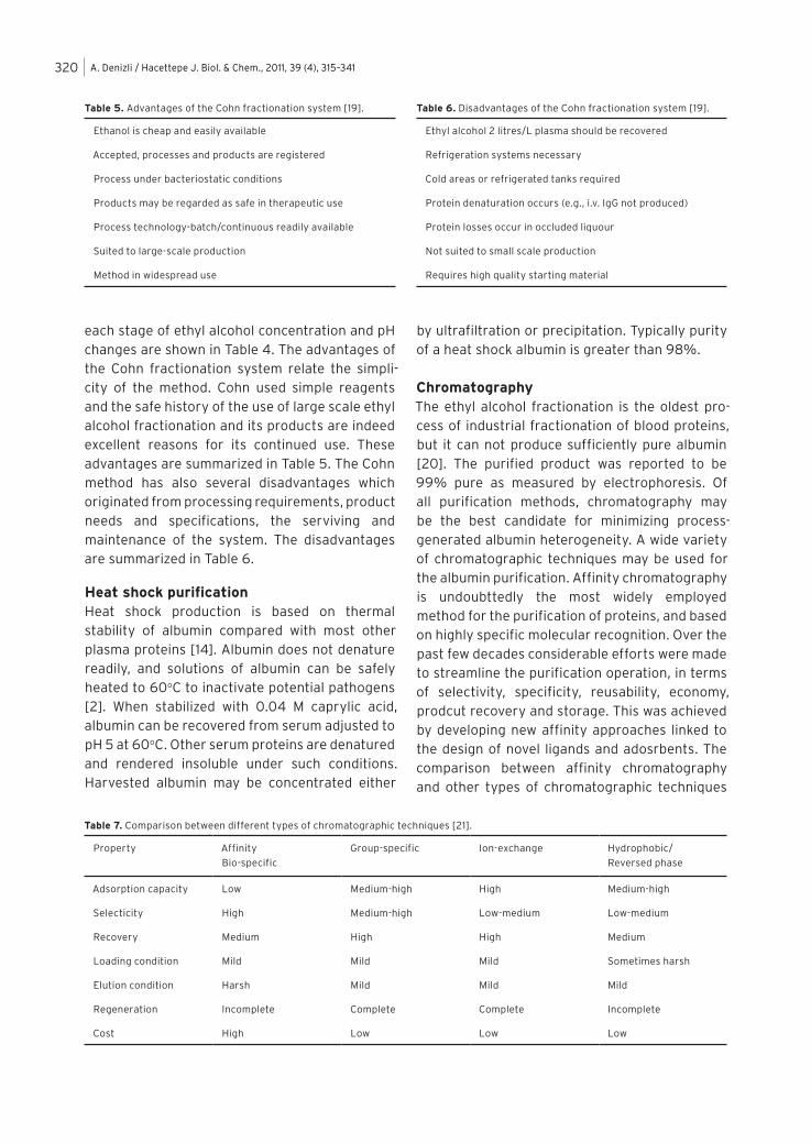
each stage of ethyl alcohol concentration and pH changes are shown in Table 4. The advantages of the Cohn fractionation system relate the simpli-city of the method. Cohn used simple reagents and the safe history of the use of large scale ethyl alcohol fractionation and its products are indeed excellent reasons for its continued use. These advantages are summarized in Table 5. The Cohn method has also several disadvantages which originated from processing requirements, product needs and specifications, the serviving and maintenance of the system. The disadvantages are summarized in Table 6.
Heat shock purificationHeat shock production is based on thermal stability of albumin compared with most other plasma proteins [14]. Albumin does not denature readily, and solutions of albumin can be safely heated to 60oC to inactivate potential pathogens [2]. When stabilized with 0.04 M caprylic acid, albumin can be recovered from serum adjusted to pH 5 at 60oC. Other serum proteins are denatured and rendered insoluble under such conditions. Harvested albumin may be concentrated either
by ultrafiltration or precipitation. Typically purity of a heat shock albumin is greater than 98%.
ChromatographyThe ethyl alcohol fractionation is the oldest pro-cess of industrial fractionation of blood proteins, but it can not produce sufficiently pure albumin [20]. The purified product was reported to be 99% pure as measured by electrophoresis. Of all purification methods, chromatography may be the best candidate for minimizing process-generated albumin heterogeneity. A wide variety of chromatographic techniques may be used for the albumin purification. Affinity chromatography is undoubttedly the most widely employed method for the purification of proteins, and based on highly specific molecular recognition. Over the past few decades considerable efforts were made to streamline the purification operation, in terms of selectivity, specificity, reusability, economy, prodcut recovery and storage. This was achieved by developing new affinity approaches linked to the design of novel ligands and adosrbents. The comparison between affinity chromatography and other types of chromatographic techniques
Table 7. Comparison between different types of chromatographic techniques [21].
Property Affinity
Bio-specific
Group-specific Ion-exchange Hydrophobic/
Reversed phase
Adsorption capacity Low Medium-high High Medium-high
Selecticity High Medium-high Low-medium Low-medium
Recovery Medium High High Medium
Loading condition Mild Mild Mild Sometimes harsh
Elution condition Harsh Mild Mild Mild
Regeneration Incomplete Complete Complete Incomplete
Cost High Low Low Low
Table 5. Advantages of the Cohn fractionation system [19].
Ethanol is cheap and easily available
Accepted, processes and products are registered
Process under bacteriostatic conditions
Products may be regarded as safe in therapeutic use
Process technology-batch/continuous readily available
Suited to large-scale production
Method in widespread use
Table 6. Disadvantages of the Cohn fractionation system [19].
Ethyl alcohol 2 litres/L plasma should be recovered
Refrigeration systems necessary
Cold areas or refrigerated tanks required
Protein denaturation occurs (e.g., i.v. IgG not produced)
Protein losses occur in occluded liquour
Not suited to small scale production
Requires high quality starting material
A. Denizli / Hacettepe J. Biol. & Chem., 2011, 39 (4), 315–341 321
are listed in Table 7. For the cost problem it is generally difficult to reduce the high cost for bio-specific affinity technique, and subsequently, cheaper group-specific affinity methods become a good alternative.
The Cohn method permits the fractionation of large volumes of plasma per batch at low cost compared to other methods. On the other hand, the quality of the product obtained by liquid chromatography is superior [15,16]. In order to obtain advantages from both methods, Tanaka et al developed a technologically attractive integrated technique for the albumin purification from plasma using alcohol fractionation followed by the chromatographic method [22].
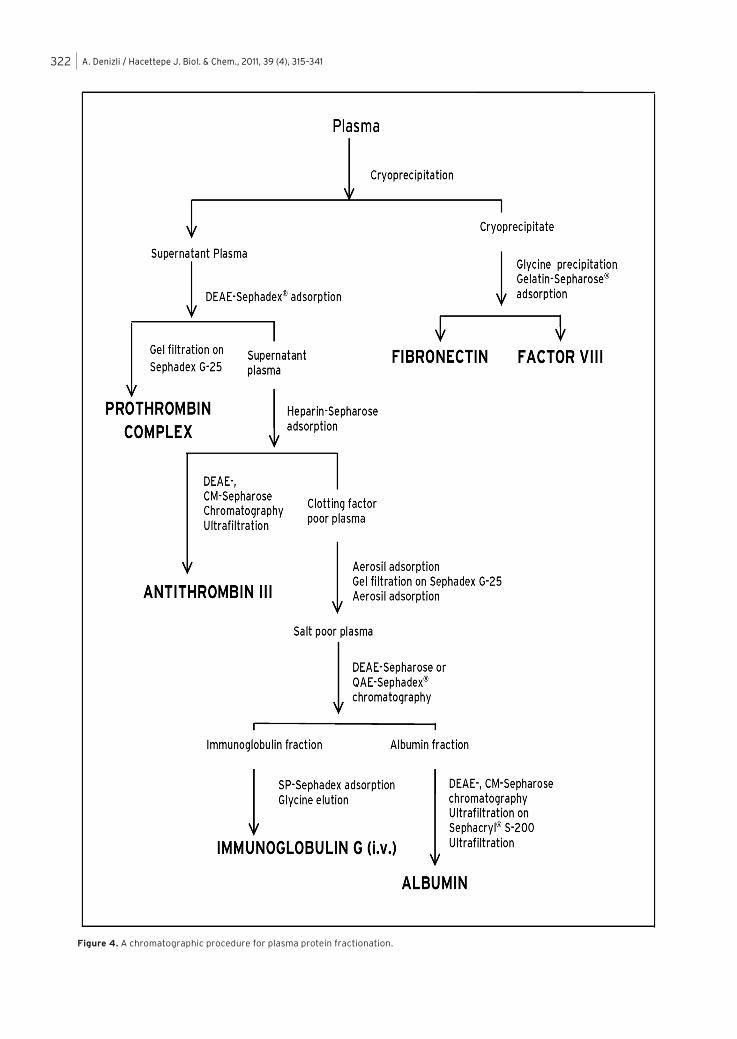
Curling described albumin purification from plasma by sequential anion, cation and gel-filtration chromatographic procedures [23]. A chromatog-raphic procedure for plasma fractionation is shown in Figure 4.
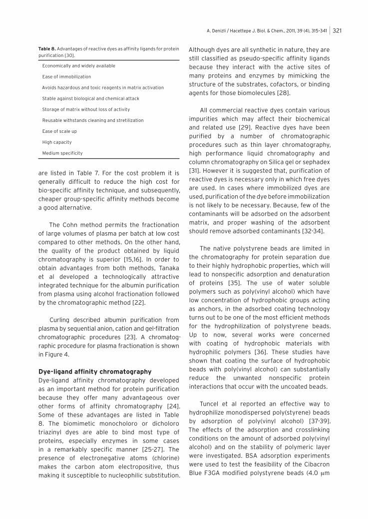
Dye-ligand affinity chromatographyDye-ligand affinity chromatography developed as an important method for protein purification because they offer many advantageous over other forms of affinity chromatography [24]. Some of these advantages are listed in Table 8. The biomimetic monocholoro or dicholoro triazinyl dyes are able to bind most type of proteins, especially enzymes in some cases in a remarkably specific manner [25-27]. The presence of electronegative atoms (chlorine) makes the carbon atom electropositive, thus making it susceptible to nucleophilic substitution.
Although dyes are all synthetic in nature, they are still classified as pseudo-specific affinity ligands because they interact with the active sites of many proteins and enzymes by mimicking the structure of the substrates, cofactors, or binding agents for those biomolecules [28].
All commercial reactive dyes contain various impurities which may affect their biochemical and related use [29]. Reactive dyes have been purified by a number of chromatographic procedures such as thin layer chromatography, high performance liquid chromatography and column chromatography on Silica gel or sephadex [31]. However it is suggested that, purification of reactive dyes is necessary only in which free dyes are used. In cases where immobilized dyes are used, purification of the dye before immobilization is not likely to be necessary. Because, few of the contaminants will be adsorbed on the adsorbent matrix, and proper washing of the adsorbent should remove adsorbed contaminants [32-34].
The native polystyrene beads are limited in the chromatography for protein separation due to their highly hydrophobic properties, which will lead to nonspecific adsorption and denaturation of proteins [35]. The use of water soluble polymers such as poly(vinyl alcohol) which have low concentration of hydrophobic groups acting as anchors, in the adsorbed coating technology turns out to be one of the most efficient methods for the hydrophilization of polystyrene beads. Up to now, several works were concerned with coating of hydrophobic materials with hydrophilic polymers [36]. These studies have shown that coating the surface of hydrophobic beads with poly(vinyl alcohol) can substantially reduce the unwanted nonspecific protein interactions that occur with the uncoated beads.
Tuncel et al reported an effective way to hydrophilize monodispersed poly(styrene) beads by adsorption of poly(vinyl alcohol) [37-39]. The effects of the adsorption and crosslinking conditions on the amount of adsorbed poly(vinyl alcohol) and on the stability of polymeric layer were investigated. BSA adsorption experiments were used to test the feasibility of the Cibacron Blue F3GA modified polystyrene beads (4.0 mm
Table 8. Advantages of reactive dyes as affinity ligands for protein
purification [30].
Economically and widely available
Ease of immobilization
Avoids hazardous and toxic reagents in matrix activation
Stable against biological and chemical attack
Storage of matrix without loss of activity
Reusable withstands cleaning and stretilization
Ease of scale up
High capacity
Medium specificity
A. Denizli / Hacettepe J. Biol. & Chem., 2011, 39 (4), 315–341322
Figure 4. A chromatographic procedure for plasma protein fractionation.
A. Denizli / Hacettepe J. Biol. & Chem., 2011, 39 (4), 315–341 323
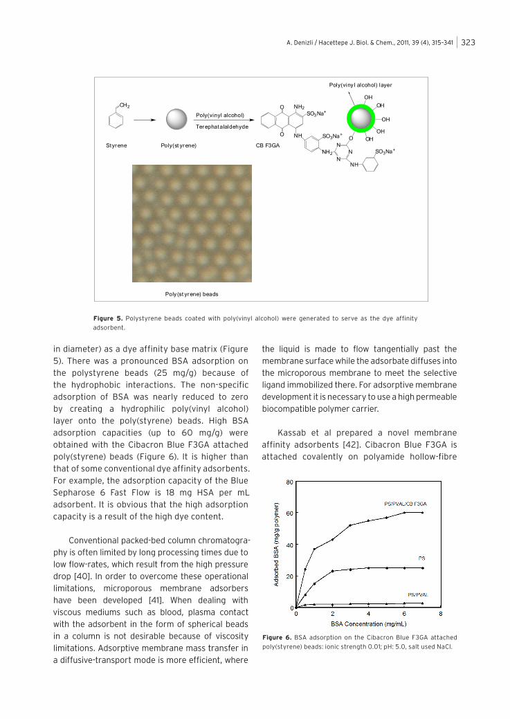
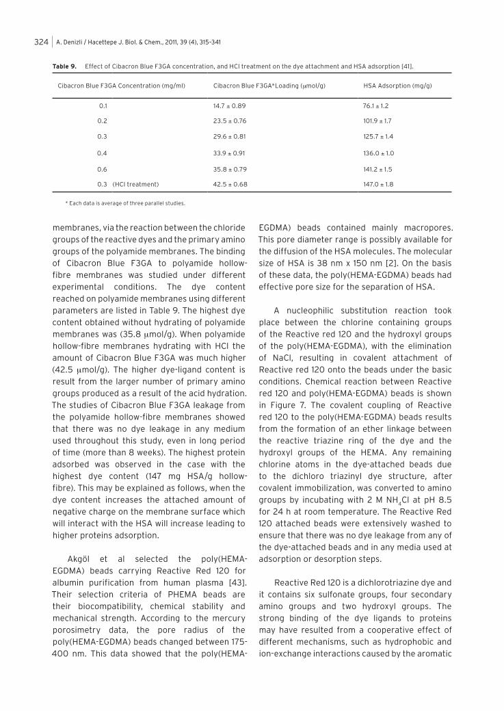
in diameter) as a dye affinity base matrix (Figure 5). There was a pronounced BSA adsorption on the polystyrene beads (25 mg/g) because of the hydrophobic interactions. The non-specific adsorption of BSA was nearly reduced to zero by creating a hydrophilic poly(vinyl alcohol) layer onto the poly(styrene) beads. High BSA adsorption capacities (up to 60 mg/g) were obtained with the Cibacron Blue F3GA attached poly(styrene) beads (Figure 6). It is higher than that of some conventional dye affinity adsorbents. For example, the adsorption capacity of the Blue Sepharose 6 Fast Flow is 18 mg HSA per mL adsorbent. It is obvious that the high adsorption capacity is a result of the high dye content.
Conventional packed-bed column chromatogra-phy is often limited by long processing times due to low flow-rates, which result from the high pressure drop [40]. In order to overcome these operational limitations, microporous membrane adsorbers have been developed [41]. When dealing with viscous mediums such as blood, plasma contact with the adsorbent in the form of spherical beads in a column is not desirable because of viscosity limitations. Adsorptive membrane mass transfer in a diffusive-transport mode is more efficient, where
the liquid is made to flow tangentially past the membrane surface while the adsorbate diffuses into the microporous membrane to meet the selective ligand immobilized there. For adsorptive membrane development it is necessary to use a high permeable biocompatible polymer carrier.
Kassab et al prepared a novel membrane affinity adsorbents [42]. Cibacron Blue F3GA is attached covalently on polyamide hollow-fibre
Figure 5. Polystyrene beads coated with poly(vinyl alcohol) were generated to serve as the dye affinity
adsorbent.
Figure 6. BSA adsorption on the Cibacron Blue F3GA attached
poly(styrene) beads: ionic strength 0.01; pH: 5.0, salt used NaCl.
Poly(vinyl alcohol)
Terephatalaldehyde
OH
OH
O
O
NH2
NH
NH2N
NN
NH
SO3Na+
SO3Na+
SO3Na+
OH
Poly(viny l alcohol) layer
Styrene Poly(st yrene)
OH
OH
O
CH2
CB F3GA
Poly(st yrene) beads
A. Denizli / Hacettepe J. Biol. & Chem., 2011, 39 (4), 315–341324
membranes, via the reaction between the chloride groups of the reactive dyes and the primary amino groups of the polyamide membranes. The binding of Cibacron Blue F3GA to polyamide hollow-fibre membranes was studied under different experimental conditions. The dye content reached on polyamide membranes using different parameters are listed in Table 9. The highest dye content obtained without hydrating of polyamide membranes was (35.8 mmol/g). When polyamide hollow-fibre membranes hydrating with HCl the amount of Cibacron Blue F3GA was much higher (42.5 mmol/g). The higher dye-ligand content is result from the larger number of primary amino groups produced as a result of the acid hydration. The studies of Cibacron Blue F3GA leakage from the polyamide hollow-fibre membranes showed that there was no dye leakage in any medium used throughout this study, even in long period of time (more than 8 weeks). The highest protein adsorbed was observed in the case with the highest dye content (147 mg HSA/g hollow-fibre). This may be explained as follows, when the dye content increases the attached amount of negative charge on the membrane surface which will interact with the HSA will increase leading to higher proteins adsorption.
Akgöl et al selected the poly(HEMA-EGDMA) beads carrying Reactive Red 120 for albumin purification from human plasma [43]. Their selection criteria of PHEMA beads are their biocompatibility, chemical stability and mechanical strength. According to the mercury porosimetry data, the pore radius of the poly(HEMA-EGDMA) beads changed between 175-400 nm. This data showed that the poly(HEMA-
EGDMA) beads contained mainly macropores. This pore diameter range is possibly available for the diffusion of the HSA molecules. The molecular size of HSA is 38 nm x 150 nm [2]. On the basis of these data, the poly(HEMA-EGDMA) beads had effective pore size for the separation of HSA.
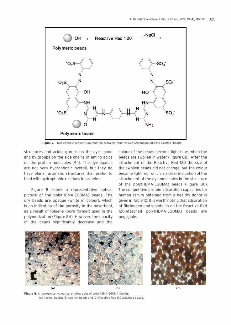
A nucleophilic substitution reaction took place between the chlorine containing groups of the Reactive red 120 and the hydroxyl groups of the poly(HEMA-EGDMA), with the elimination of NaCl, resulting in covalent attachment of Reactive red 120 onto the beads under the basic conditions. Chemical reaction between Reactive red 120 and poly(HEMA-EGDMA) beads is shown in Figure 7. The covalent coupling of Reactive red 120 to the poly(HEMA-EGDMA) beads results from the formation of an ether linkage between the reactive triazine ring of the dye and the hydroxyl groups of the HEMA. Any remaining chlorine atoms in the dye-attached beads due to the dichloro triazinyl dye structure, after covalent immobilization, was converted to amino groups by incubating with 2 M NH
4Cl at pH 8.5
for 24 h at room temperature. The Reactive Red 120 attached beads were extensively washed to ensure that there was no dye leakage from any of the dye-attached beads and in any media used at adsorption or desorption steps.
Reactive Red 120 is a dichlorotriazine dye and it contains six sulfonate groups, four secondary amino groups and two hydroxyl groups. The strong binding of the dye ligands to proteins may have resulted from a cooperative effect of different mechanisms, such as hydrophobic and ion-exchange interactions caused by the aromatic
Table 9. Effect of Cibacron Blue F3GA concentration, and HCl treatment on the dye attachment and HSA adsorption [41].
Cibacron Blue F3GA Concentration (mg/ml) Cibacron Blue F3GA*Loading (mmol/g) HSA Adsorption (mg/g)
0.1 14.7 ± 0.89 76.1 ± 1.2
0.2 23.5 ± 0.76 101.9 ± 1.7
0.3 29.6 ± 0.81 125.7 ± 1.4
0.4 33.9 ± 0.91 136.0 ± 1.0
0.6 35.8 ± 0.79 141.2 ± 1.5
0.3 (HCI treatment) 42.5 ± 0.68 147.0 ± 1.8
* Each data is average of three parallel studies.
A. Denizli / Hacettepe J. Biol. & Chem., 2011, 39 (4), 315–341 325
structures and acidic groups on the dye ligand and by groups on the side chains of amino acids on the protein molecules [44]. The dye ligands are not very hydrophobic overall, but they do have planar aromatic structures that prefer to bind with hydrophobic residues in proteins.
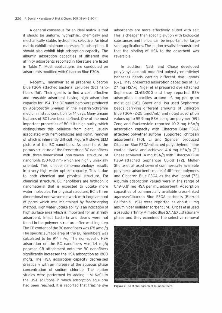
Figure 8 shows a representative optical picture of the poly(HEMA-EGDMA) beads. The dry beads are opaque (white in colour), which is an indication of the porosity in the adsorbent, as a result of toluene (pore former) used in the polymerization (Figure 8A). However, the opacity of the beads significantly decrease and the
colour of the beads become light blue, when the beads are swollen in water (Figure 8B). After the attachment of the Reactive Red 120 the size of the swollen beads did not change, but the colour became light red, which is a clear indication of the attachment of the dye molecules in the structure of the poly(HEMA-EGDMA) beads (Figure 8C). The competitive protein adsorption capacities for human serum obtained from a healthy donor is given in Table 10. It is worth noting that adsorption of fibrinogen and g-globulin on the Reactive Red 120-attached poly(HEMA-EGDMA) beads are negligible.
Figure 7. Nucleophilic substitution reaction between Reactive Red 120 and poly(HEMA-EGDMA) beads.
Figure 8. A representative optical photographs of poly(HEMA-EGDMA) beads;
(A) normal beads; (B) swollen beads and (C) Reactive Red 120 attached beads.
A. Denizli / Hacettepe J. Biol. & Chem., 2011, 39 (4), 315–341326
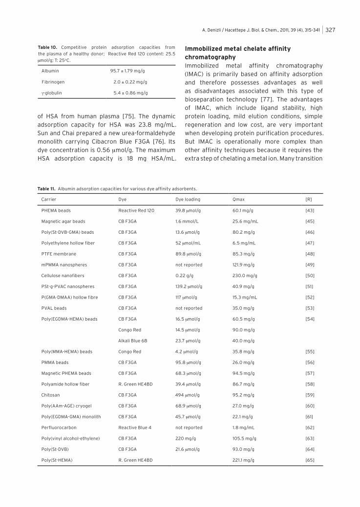
A general consensus for an ideal matrix is that it should be uniform, hydrophilic, chemically and mechanically stable, hydrophilic, selective. An ideal matrix exhibit minimum non-specific adsorption. It should also exhibit high adsorption capacity. The albumin adsorption capacities of different dye affinity adsorbents reported in literature are listed in Table 11. Most applications are conducted on adsorbents modified with Cibacron Blue F3GA.
Recently, Tamahkar et al prepared Cibacron Blue F3GA attached bacterial cellulose (BC) nano-fibers [66]. Their goal is to find a cost effective and reusable adsorbent having high adsorption capacity for HSA. The BC nanofibers were produced by Acetobacter xylinum in the Hestrin-Schramm medium in static condition for 14 days. Many unique features of BC have been defined. One of the most important properties of BC is its high purity, which distinguishes this cellulose from plant, usually associated with hemicelluloses and lignin, removal of which is inherently difficult. Figure 9 shows SEM picture of the BC nanofibers. As seen here, the porous structure of the freeze-dried BC nanofibers with three-dimensional non-woven structure of nanofibrils (50-100 nm) which are highly uniaxially oriented. This unique nano-morphology results in a very high water uptake capacity. This is due to both chemical and physical structure. For chemical structure, BC nanofibers are hydrophilic nanomaterial that is expected to uptake more water molecules. For physical structure, BC is three dimensional non-woven network with large amount of pores which was maintained by freeze-drying method. High water uptake ability is an indication of high surface area which is important for an affinity adsorbent. Intact bacteria and debris were not found in the polymer structure after washing step. The CB content of the BC nanofibers was 178 mmol/g. The specific surface area of the BC nanofibers was calculated to be 914 m2/g. The non-specific HSA adsorption on the BC nanofibers was 1.4 mg/g polymer. CB attachment onto the BC nanofibers significantly increased the HSA adsorption as 1800 mg/g. The HSA adsorption capacity decrea-sed drastically with an increase of the aqueous phase concentration of sodium chloride. The elution studies were performed by adding 1 M NaCl to the HSA solutions in which adsorption equilibria had been reached. It is reported that triazine dye
adsorbents are more effectively eluted with salt. This is cheaper than specific elution with biological substances and hence, can be important for large-scale applications. The elution results demonstrated that the binding of HSA to the adsorbent was reversible.
In addition, Nash and Chase developed poly(vinyl alcohol) modified poly(styrene-divinyl benzene) beads carring different dye ligands [67]. They presented adsorption capacities of 11.7-27 mg HSA/g. Nigel et al prepared dye-attached Sepharose CL-6B-200 and they reported BSA adsorption capacities around 1-3 mg per gram moist gel [68]. Boyer and Hsu used Sepharose beads carrying different amounts of Cibacron Blue F3GA (2-25 mmol/mL) and noted adsorption values up to 55.9 mg BSA per gram polymer [69]. Zeng and Ruckenstein reported 10.2 mg HSA/g adsorption capacity with Cibacron Blue F3GA-attached-polyether-sulfone supported chitosan adsorbents [70]. Li and Spencer produced Cibacron Blue F3GA-attached polyethylene imine coated titania and achieved 4.4 mg HSA/g [71]. Chase achieved 14 mg BSA/g with Cibacron Blue F3GA-attached Sepharose CL-6B [72]. Muller-Shulte et al used several commercially available polymeric adsorbents made of different polymers, and Cibacron Blue F3GA as the dye-ligand [73]. Albumin adsorption values were in the range of 0.19-0.81 mg HSA per mL adsorbent. Adsorption capacities of commercially available cross-linked agarose/Cibacron Blue F3GA sorbents (Bio-rad, California, USA) were reported as about 11 mg albumin per milliliter sorbent [74]. Urbas et al used a pseudo-affinity Mimetic Blue SA A6XL stationary phase and they examined the selective removal
Figure 9. SEM photograph of BC nanofibers.
A. Denizli / Hacettepe J. Biol. & Chem., 2011, 39 (4), 315–341 327
of HSA from human plasma [75]. The dynamic adsorption capacity for HSA was 23.8 mg/mL. Sun and Chai prepared a new urea-formaldehyde monolith carrying Cibacron Blue F3GA [76]. Its dye concentration is 0.56 mmol/g. The maximum HSA adsorption capacity is 18 mg HSA/mL.
Immobilized metal chelate affinity chromatographyImmobilized metal affinity chromatography (IMAC) is primarily based on affinity adsorption and therefore possesses advantages as well as disadvantages associated with this type of bioseparation technology [77]. The advantages of IMAC, which include ligand stability, high protein loading, mild elution conditions, simple regeneration and low cost, are very important when developing protein purification procedures. But IMAC is operationally more complex than other affinity techniques because it requires the extra step of chelating a metal ion. Many transition
Table 11. Albumin adsorption capacities for various dye affinity adsorbents.
Carrier Dye Dye loading Qmax [R]
PHEMA beads Reactive Red 120 39.8 mmol/g 60.1 mg/g [43]
Magnetic agar beads CB F3GA 1.6 mmol/L 25.6 mg/mL [45]
Poly(St-DVB-GMA) beads CB F3GA 13.6 mmol/g 80.2 mg/g [46]
Polyethylene hollow fiber CB F3GA 52 mmol/mL 6.5 mg/mL [47]
PTFE membrane CB F3GA 89.8 mmol/g 85.3 mg/g [48]
mPMMA nanospheres CB F3GA not reported 121.9 mg/g [49]
Cellulose nanofibers CB F3GA 0.22 g/g 230.0 mg/g [50]
PSt-g-PVAC nanospheres CB F3GA 139.2 mmol/g 40.9 mg/g [51]
P(GMA-DMAA) hollow fibre CB F3GA 117 mmol/g 15.3 mg/mL [52]
PVAL beads CB F3GA not reported 35.0 mg/g [53]
Poly(EGDMA-HEMA) beads CB F3GA 16.5 mmol/g 60.5 mg/g [54]
Congo Red 14.5 mmol/g 90.0 mg/g
Alkali Blue 6B 23.7 mmol/g 40.0 mg/g
Poly(MMA-HEMA) beads Congo Red 4.2 mmol/g 35.8 mg/g [55]
PMMA beads CB F3GA 95.8 mmol/g 26.0 mg/g [56]
Magnetic PHEMA beads CB F3GA 68.3 mmol/g 94.5 mg/g [57]
Polyamide hollow fiber R. Green HE4BD 39.4 mmol/g 86.7 mg/g [58]
Chitosan CB F3GA 494 mmol/g 95.2 mg/g [59]
Poly(AAm-AGE) cryogel CB F3GA 68.9 mmol/g 27.0 mg/g [60]
Poly(EGDMA-GMA) monolith CB F3GA 45.7 mmol/g 22.1 mg/g [61]
Perfluorocarbon Reactive Blue 4 not reported 1.8 mg/mL [62]
Poly(vinyl alcohol-ethylene) CB F3GA 220 mg/g 105.5 mg/g [63]
Poly(St-DVB) CB F3GA 21.6 mmol/g 93.0 mg/g [64]
Poly(St-HEMA) R. Green HE4BD 221.1 mg/g [65]
Table 10. Competitive protein adsorption capacities from
the plasma of a healthy donor; Reactive Red 120 content: 25.5
mmol/g; T: 25oC.
Albumin 95.7 ± 1.79 mg/g
Fibrinogen 2.0 ± 0.22 mg/g
g-globulin 5.4 ± 0.86 mg/g
A. Denizli / Hacettepe J. Biol. & Chem., 2011, 39 (4), 315–341328
metals can form stable complexes with electron-rich compounds (aromatic molecules and olefins) and may coordinate molecules containing oxygen, nitrogen and sulphur by ion dipole interactions [78]. Metal-chelating ligands are first-row transition metal ions (Zn2+, Ni2+, Cu2+ and Fe3+) incorporated by iminodiacetic acid, nitrilotriacetic acid, amino salicylic acid and carboxymethylated aminoacids [79]. The choice of the metal chelating ligands is rather critical and is a complicating part in the design of the affinity adsorbent [80]. The second major problem is that of slow
“release” of these covalently bonded chelators off the adsorbent. “Release” is a general problem encountered in any affinity chromatography [81]. The use of toxic chemicals in activating of adsorbent and the danger of traces of metal-chelating ligand release into the protein product have always been a mattern of concern [82].
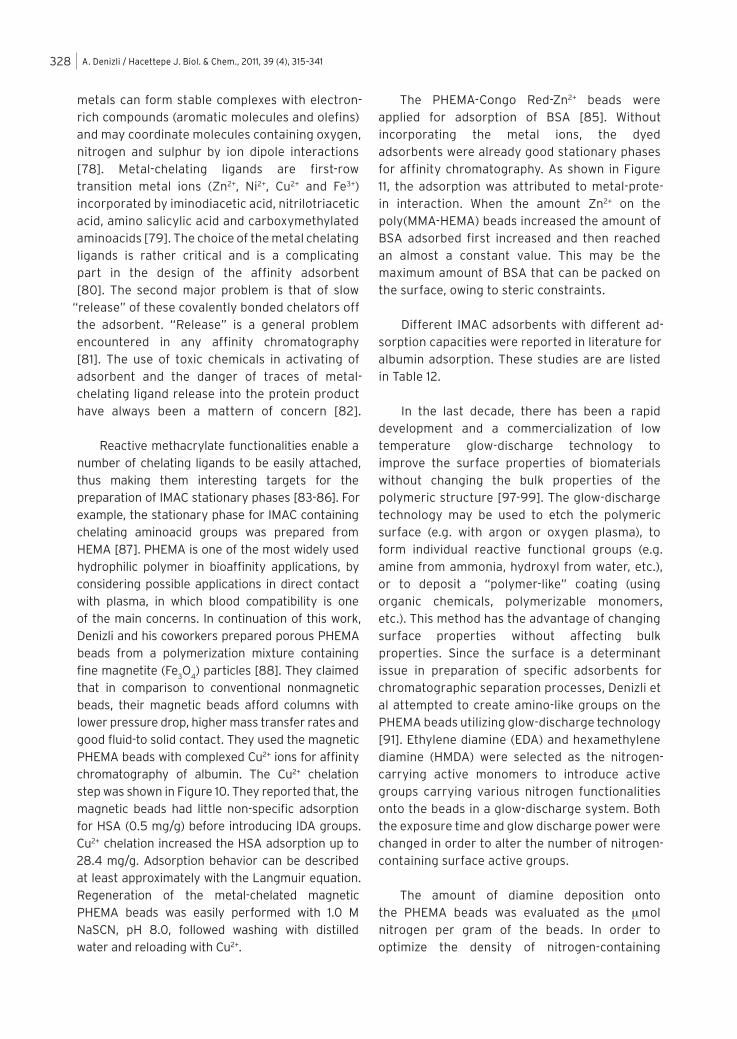
Reactive methacrylate functionalities enable a number of chelating ligands to be easily attached, thus making them interesting targets for the preparation of IMAC stationary phases [83-86]. For example, the stationary phase for IMAC containing chelating aminoacid groups was prepared from HEMA [87]. PHEMA is one of the most widely used hydrophilic polymer in bioaffinity applications, by considering possible applications in direct contact with plasma, in which blood compatibility is one of the main concerns. In continuation of this work, Denizli and his coworkers prepared porous PHEMA beads from a polymerization mixture containing fine magnetite (Fe
3O
4) particles [88]. They claimed
that in comparison to conventional nonmagnetic beads, their magnetic beads afford columns with lower pressure drop, higher mass transfer rates and good fluid-to solid contact. They used the magnetic PHEMA beads with complexed Cu2+ ions for affinity chromatography of albumin. The Cu2+ chelation step was shown in Figure 10. They reported that, the magnetic beads had little non-specific adsorption for HSA (0.5 mg/g) before introducing IDA groups. Cu2+ chelation increased the HSA adsorption up to 28.4 mg/g. Adsorption behavior can be described at least approximately with the Langmuir equation. Regeneration of the metal-chelated magnetic PHEMA beads was easily performed with 1.0 M NaSCN, pH 8.0, followed washing with distilled water and reloading with Cu2+.
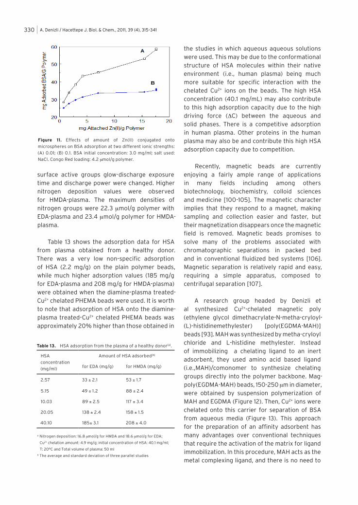
The PHEMA-Congo Red-Zn2+ beads were applied for adsorption of BSA [85]. Without incorporating the metal ions, the dyed adsorbents were already good stationary phases for affinity chromatography. As shown in Figure 11, the adsorption was attributed to metal-prote-in interaction. When the amount Zn2+ on the poly(MMA-HEMA) beads increased the amount of BSA adsorbed first increased and then reached an almost a constant value. This may be the maximum amount of BSA that can be packed on the surface, owing to steric constraints.
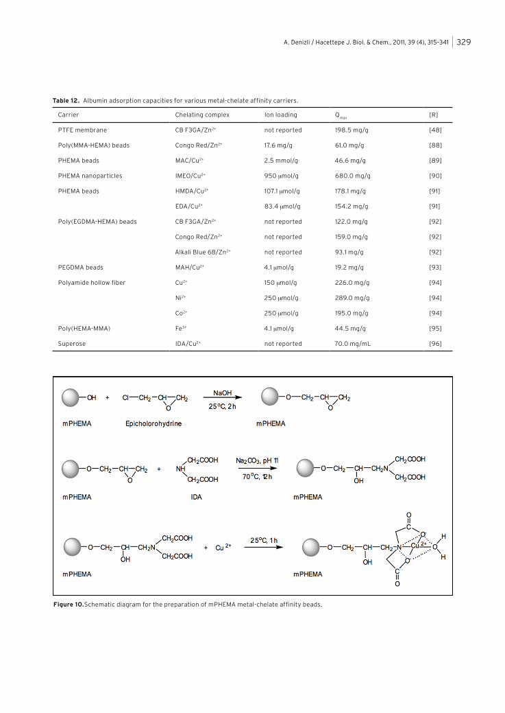
Different IMAC adsorbents with different ad-sorption capacities were reported in literature for albumin adsorption. These studies are are listed in Table 12.
In the last decade, there has been a rapid development and a commercialization of low temperature glow-discharge technology to improve the surface properties of biomaterials without changing the bulk properties of the polymeric structure [97-99]. The glow-discharge technology may be used to etch the polymeric surface (e.g. with argon or oxygen plasma), to form individual reactive functional groups (e.g. amine from ammonia, hydroxyl from water, etc.), or to deposit a “polymer-like” coating (using organic chemicals, polymerizable monomers, etc.). This method has the advantage of changing surface properties without affecting bulk properties. Since the surface is a determinant issue in preparation of specific adsorbents for chromatographic separation processes, Denizli et al attempted to create amino-like groups on the PHEMA beads utilizing glow-discharge technology [91]. Ethylene diamine (EDA) and hexamethylene diamine (HMDA) were selected as the nitrogen-carrying active monomers to introduce active groups carrying various nitrogen functionalities onto the beads in a glow-discharge system. Both the exposure time and glow discharge power were changed in order to alter the number of nitrogen-containing surface active groups.
The amount of diamine deposition onto the PHEMA beads was evaluated as the mmol nitrogen per gram of the beads. In order to optimize the density of nitrogen-containing
A. Denizli / Hacettepe J. Biol. & Chem., 2011, 39 (4), 315–341 329
Table 12. Albumin adsorption capacities for various metal-chelate affinity carriers.
Carrier Chelating complex Ion loading Qmax
[R]
PTFE membrane CB F3GA/Zn2+ not reported 198.5 mg/g [48]
Poly(MMA-HEMA) beads Congo Red/Zn2+ 17.6 mg/g 61.0 mg/g [88]
PHEMA beads MAC/Cu2+ 2.5 mmol/g 46.6 mg/g [89]
PHEMA nanoparticles IMEO/Cu2+ 950 mmol/g 680.0 mg/g [90]
PHEMA beads HMDA/Cu2+ 107.1 mmol/g 178.1 mg/g [91]
EDA/Cu2+ 83.4 mmol/g 154.2 mg/g [91]
Poly(EGDMA-HEMA) beads CB F3GA/Zn2+ not reported 122.0 mg/g [92]
Congo Red/Zn2+ not reported 159.0 mg/g [92]
Alkali Blue 6B/Zn2+ not reported 93.1 mg/g [92]
PEGDMA beads MAH/Cu2+ 4.1 mmol/g 19.2 mg/g [93]
Polyamide hollow fiber Cu2+ 150 mmol/g 226.0 mg/g [94]
Ni2+ 250 mmol/g 289.0 mg/g [94]
Co2+ 250 mmol/g 195.0 mg/g [94]
Poly(HEMA-MMA) Fe3+ 4.1 mmol/g 44.5 mg/g [95]
Superose IDA/Cu2+ not reported 70.0 mg/mL [96]
Figure 10.Schematic diagram for the preparation of mPHEMA metal-chelate affinity beads.
A. Denizli / Hacettepe J. Biol. & Chem., 2011, 39 (4), 315–341330
surface active groups glow-discharge exposure time and discharge power were changed. Higher nitrogen deposition values were observed for HMDA-plasma. The maximum densities of nitrogen groups were 22.3 mmol/g polymer with EDA-plasma and 23.4 mmol/g polymer for HMDA-plasma.
Table 13 shows the adsorption data for HSA from plasma obtained from a healthy donor. There was a very low non-specific adsorption of HSA (2.2 mg/g) on the plain polymer beads, while much higher adsorption values (185 mg/g for EDA-plasma and 208 mg/g for HMDA-plasma) were obtained when the diamine-plasma treated-Cu2+ chelated PHEMA beads were used. It is worth to note that adsorption of HSA onto the diamine-plasma treated-Cu2+ chelated PHEMA beads was approximately 20% higher than those obtained in
the studies in which aqueous aqueous solutions were used. This may be due to the conformational structure of HSA molecules within their native environment (i.e., human plasma) being much more suitable for specific interaction with the chelated Cu2+ ions on the beads. The high HSA concentration (40.1 mg/mL) may also contribute to this high adsorption capacity due to the high driving force (DC) between the aqueous and solid phases. There is a competitive adsorption in human plasma. Other proteins in the human plasma may also be and contribute this high HSA adsorption capacity due to competition.
Recently, magnetic beads are currently enjoying a fairly ample range of applications in many fields including among others biotechnology, biochemistry, colloid sciences and medicine [100-105]. The magnetic character implies that they respond to a magnet, making sampling and collection easier and faster, but their magnetization disappears once the magnetic field is removed. Magnetic beads promises to solve many of the problems associated with chromatographic separations in packed bed and in conventional fluidized bed systems [106]. Magnetic separation is relatively rapid and easy, requiring a simple apparatus, composed to centrifugal separation [107].
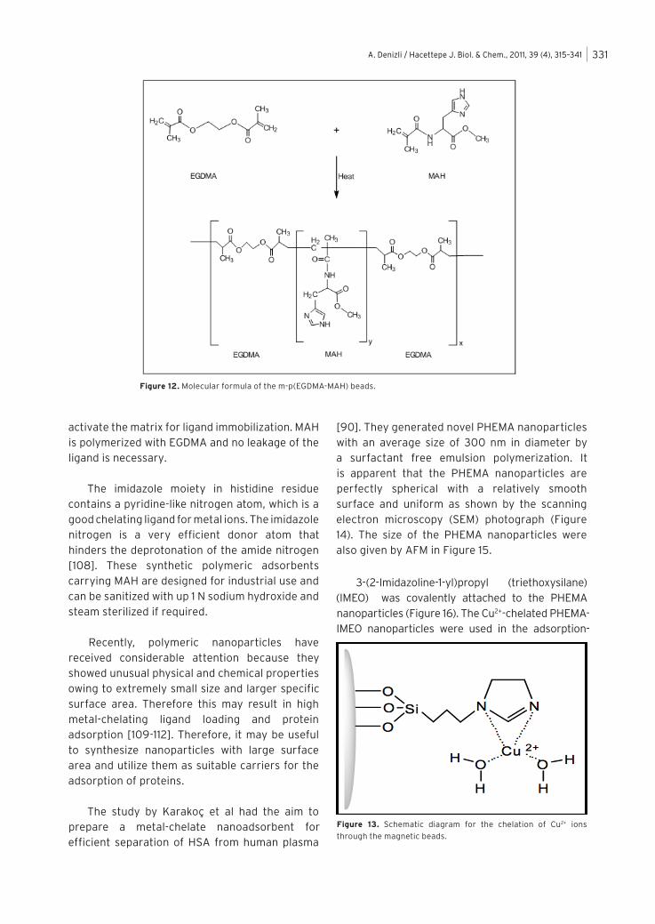
A research group headed by Denizli et al synthesized Cu2+-chelated magnetic poly (ethylene glycol dimethacrylate-N-metha-cryloyl-(L)-histidinemethylester) [poly(EGDMA-MAH)] beads [93]. MAH was synthesized by metha-cryloyl chloride and L-histidine methylester. Instead of immobilizing a chelating ligand to an inert adsorbent, they used amino acid based ligand (i.e.,MAH)/comonomer to synthesize chelating groups directly into the polymer backbone. Mag-poly(EGDMA-MAH) beads, 150-250 mm in diameter, were obtained by suspension polymerization of MAH and EGDMA (Figure 12). Then, Cu2+ ions were chelated onto this carrier for separation of BSA from aqueous media (Figure 13). This approach for the preparation of an affinity adsorbent has many advantages over conventional techniques that require the activation of the matrix for ligand immobilization. In this procedure, MAH acts as the metal complexing ligand, and there is no need to
Table 13. HSA adsorption from the plasma of a healthy donor[a].
HSA
concentration
(mg/ml)
Amount of HSA adsorbed[b]
for EDA (mg/g) for HMDA (mg/g)
2.57 33 ± 2.1 53 ± 1.7
5.15 49 ± 1.2 88 ± 2.4
10.03 89 ± 2.5 117 ± 3.4
20.05 138 ± 2.4 158 ± 1.5
40.10 185± 3.1 208 ± 4.0
a Nitrogen deposition: 16.8 mmol/g for HMDA and 18.6 mmol/g for EDA;
Cu2+ chelation amount: 4.9 mg/g; initial concentration of HSA: 40.1 mg/ml;
T: 20°C and Total volume of plasma: 50 mlb The average and standard deviation of three parallel studies
Figure 11. Effects of amount of Zn(II) conjugated onto
microspheres on BSA adsorption at two different ionic strengths:
(A) 0.01; (B) 0.1. BSA initial concentration: 3.0 mg/ml; salt used:
NaCl. Congo Red loading: 4.2 mmol/g polymer.
A. Denizli / Hacettepe J. Biol. & Chem., 2011, 39 (4), 315–341 331
activate the matrix for ligand immobilization. MAH is polymerized with EGDMA and no leakage of the ligand is necessary.
The imidazole moiety in histidine residue contains a pyridine-like nitrogen atom, which is a good chelating ligand for metal ions. The imidazole nitrogen is a very efficient donor atom that hinders the deprotonation of the amide nitrogen [108]. These synthetic polymeric adsorbents carrying MAH are designed for industrial use and can be sanitized with up 1 N sodium hydroxide and steam sterilized if required.
Recently, polymeric nanoparticles have received considerable attention because they showed unusual physical and chemical properties owing to extremely small size and larger specific surface area. Therefore this may result in high metal-chelating ligand loading and protein adsorption [109-112]. Therefore, it may be useful to synthesize nanoparticles with large surface area and utilize them as suitable carriers for the adsorption of proteins.
The study by Karakoç et al had the aim to prepare a metal-chelate nanoadsorbent for efficient separation of HSA from human plasma
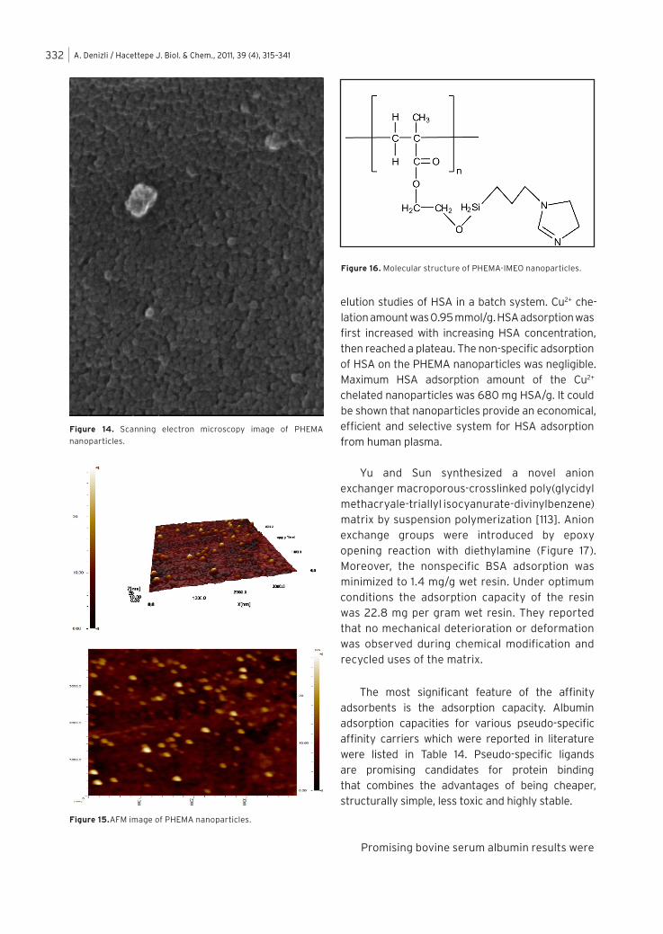
[90]. They generated novel PHEMA nanoparticles with an average size of 300 nm in diameter by a surfactant free emulsion polymerization. It is apparent that the PHEMA nanoparticles are perfectly spherical with a relatively smooth surface and uniform as shown by the scanning electron microscopy (SEM) photograph (Figure 14). The size of the PHEMA nanoparticles were also given by AFM in Figure 15.
3-(2-Imidazoline-1-yl)propyl (triethoxysilane)
(IMEO) was covalently attached to the PHEMA nanoparticles (Figure 16). The Cu2+-chelated PHEMA-IMEO nanoparticles were used in the adsorption-
Figure 12. Molecular formula of the m-p(EGDMA-MAH) beads.
Figure 13. Schematic diagram for the chelation of Cu2+ ions
through the magnetic beads.
A. Denizli / Hacettepe J. Biol. & Chem., 2011, 39 (4), 315–341332
elution studies of HSA in a batch system. Cu2+ che-lation amount was 0.95 mmol/g. HSA adsorption was first increased with increasing HSA concentration, then reached a plateau. The non-specific adsorption of HSA on the PHEMA nanoparticles was negligible. Maximum HSA adsorption amount of the Cu2+ chelated nanoparticles was 680 mg HSA/g. It could be shown that nanoparticles provide an economical, efficient and selective system for HSA adsorption from human plasma.
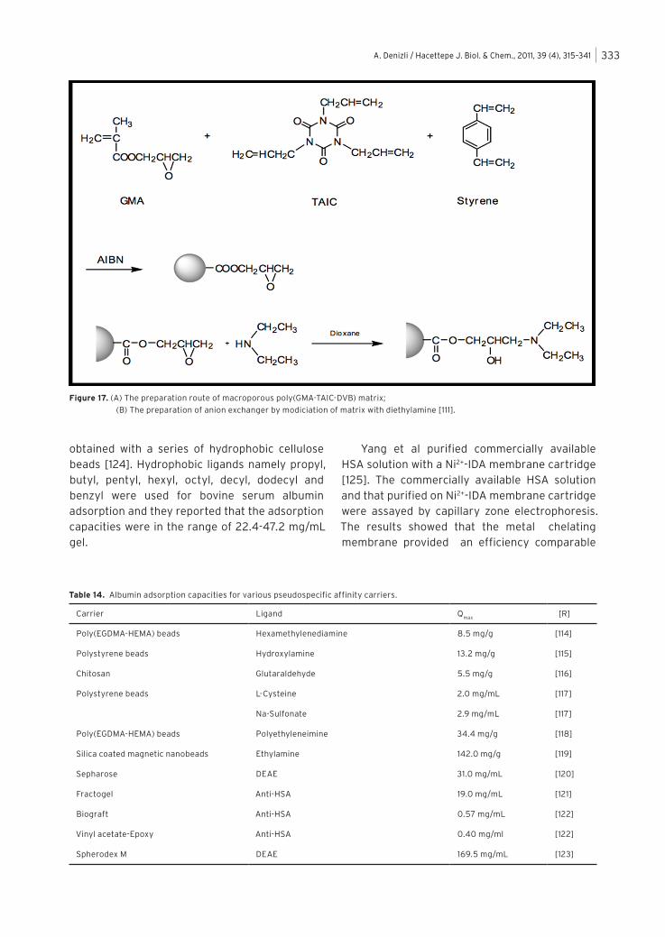
Yu and Sun synthesized a novel anion exchanger macroporous-crosslinked poly(glycidyl methacryale-triallyl isocyanurate-divinylbenzene) matrix by suspension polymerization [113]. Anion exchange groups were introduced by epoxy opening reaction with diethylamine (Figure 17). Moreover, the nonspecific BSA adsorption was minimized to 1.4 mg/g wet resin. Under optimum conditions the adsorption capacity of the resin was 22.8 mg per gram wet resin. They reported that no mechanical deterioration or deformation was observed during chemical modification and recycled uses of the matrix.
The most significant feature of the affinity
adsorbents is the adsorption capacity. Albumin adsorption capacities for various pseudo-specific affinity carriers which were reported in literature were listed in Table 14. Pseudo-specific ligands are promising candidates for protein binding that combines the advantages of being cheaper, structurally simple, less toxic and highly stable.
Promising bovine serum albumin results were
Figure 15.AFM image of PHEMA nanoparticles.
Figure 14. Scanning electron microscopy image of PHEMA
nanoparticles.
Figure 16. Molecular structure of PHEMA-IMEO nanoparticles.
A. Denizli / Hacettepe J. Biol. & Chem., 2011, 39 (4), 315–341 333
obtained with a series of hydrophobic cellulose beads [124]. Hydrophobic ligands namely propyl, butyl, pentyl, hexyl, octyl, decyl, dodecyl and benzyl were used for bovine serum albumin adsorption and they reported that the adsorption capacities were in the range of 22.4-47.2 mg/mL gel.
Yang et al purified commercially available HSA solution with a Ni2+-IDA membrane cartridge [125]. The commercially available HSA solution and that purified on Ni2+-IDA membrane cartridge were assayed by capillary zone electrophoresis. The results showed that the metal chelating membrane provided an efficiency comparable
Figure 17. (A) The preparation route of macroporous poly(GMA-TAIC-DVB) matrix;
(B) The preparation of anion exchanger by modiciation of matrix with diethylamine [111].
Table 14. Albumin adsorption capacities for various pseudospecific affinity carriers.
Carrier Ligand Qmax
[R]
Poly(EGDMA-HEMA) beads Hexamethylenediamine 8.5 mg/g [114]
Polystyrene beads Hydroxylamine 13.2 mg/g [115]
Chitosan Glutaraldehyde 5.5 mg/g [116]
Polystyrene beads L-Cysteine 2.0 mg/mL [117]
Na-Sulfonate 2.9 mg/mL [117]
Poly(EGDMA-HEMA) beads Polyethyleneimine 34.4 mg/g [118]
Silica coated magnetic nanobeads Ethylamine 142.0 mg/g [119]
Sepharose DEAE 31.0 mg/mL [120]
Fractogel Anti-HSA 19.0 mg/mL [121]
Biograft Anti-HSA 0.57 mg/mL [122]
Vinyl acetate-Epoxy Anti-HSA 0.40 mg/ml [122]
Spherodex M DEAE 169.5 mg/mL [123]
A. Denizli / Hacettepe J. Biol. & Chem., 2011, 39 (4), 315–341334
with agarose bead based metal affinity chromatography for HSA purification. The membrane chromatography exhibited a four to five times faster performance than the packed column.
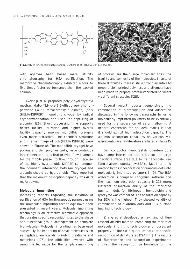
Avcıbaşı et al prepared poly(2-hydroxyethyl methacrylate-[N,N-bis(2,6-diisopropylphenyl)-perylene-3,4,9,10-tetracarboxylic diimide] [poly (HEMA-DIPPER)] monolithic cryogel by radical cryopolymerization and used for capturing of albumin [126]. Short processing time supports better facility utilization and higher overall facility capacity making monolithic cryogels even more attractive. The chemical structure and internal image of poly(HEMA-DIPPER) were shown in Figure 18. The monolithic cryogel have porous and thin polymer walls, large continous interconnected pores that provide flow channels for the mobile phase to flow through. Because of the highly hydrophobic DIPPER comonomer, the dominant interaction between cryogel and albumin should be hydrophobic. They reported that the maximum adsorption capacity was 40.9 mg/g polymer.
Molecular ImprintingIncreasing reports regarding the isolation or purification of HSA for therapeutic purpose using the molecular imprinting technology have been presented in recent years. Molecular imprinting technology is an attractive biomimetic approach that creates specific recognition sites to the shape and functional group arrangement to template biomolecules. Molecular imprinting has been used succesfully for imprinting of small molecules such as peptides, aminoacids, bilirubin, creatinine and metal-ions [127]. The difficulties involved with using the technique for the template-imprinting
of proteins are their large molecular sizes, the fragility and comlexity of the molecules. In spite of these difficulties, there is still a strong incentive to prepare bioimprinted polymers and attempts have been made to prepare protein-imprinted polymers via different strategies [128].
Several recent reports demonstrate the combination of biorecognition and adsorption discussed in the following paragraphs by using molecularly imprinted polymers to be eventually used for the separation of serum albumin. A general consensus for an ideal matrix is that it should exhibit high adsorption capacity. The albumin adsorption capacities on various MIP adsorbents given in literature are listed in Table 15.
Semiconductor nanocrystals quantum dots have some interesting properties such as large specific surface area due to its nanoscale size. Tang et al developed a new BSA surface imprinting method by the incorporation of quantum dots into molecularly imprinted polymers [140]. The BSA adsorption is complied Langmuir isotherm and the maximum adsorption capacity is 226 mg/g. Different adsorption ability of the imprinted quantum dots for fibrinogen, hemoglobin and lysozyme was compared. The adsorption capacity for BSA is the highest. They showed validity of combination of quantum dots and BSA surface imprinting technology.
Zhang et al developed a new kind of fluo-rescent affinity material combining the merits of molecular imprinting technology and fluorescent property of the CdTe quantum dots for specific recognition of denaturated BSA [141]. The results of fluorescence and adsorption experiments showed the recognition performance of the
C
COO
CH2
CH2
OH
C N
O
O
N
O
O
C
COO
CH2
CH2
OH
C
n
Figure 18. (A)Chemical structure and (B) SEM image of P(HEMA-DIPPER) cryogel.

A. Denizli / Hacettepe J. Biol. & Chem., 2011, 39 (4), 315–341 335
receptors toward the template BSA molecule. The maximum BSA adsorption capacity is 37.4 mg/g. They reported that the artificial receptor could be repeated many times with minimal adsorption capacity loss.
Bonini et al reported the synthesis of poly-aminophenylboronic acid imprinted beads for the recognition of HSA [132]. Aminophenylboronic acid possesses several functional groups including hydroxyl, secondary amine and an aromatic ring, which can interact with different aminoacids present in proteins. These multiple interaction points can sometimes result in high non-specific adsorption. Their study includes first exploiting silica beads as supports for the HSA immobilization, followed by the polymerization of the homopolymeric material onto the modified beads and finally the cleavage of the protein-silica covalent bonds, resulting in leaving the imprinted nanocavities freely accessible for protein binding.
The difference between the imprinted beads and the control beads correlated with the quantity of available binding sites. The resulting imprinted beads were selective for HSA and the binding capacity was 1.4 mg/g for the imprinted beads and 0.9 mg/g for control.
Gai et al synthesized BSA surface imprinted magnetic polymer based on atomic transfer radical polymerization method in the presence of common monomer (N-isopropylacrylamide) with the assistant of basic functional monomer [N-[3-(dimethylamino-propyl]-methacrylamide), which provides a achievable attempt for imprinting larger proteins with mild reaction conditions [143]. The BSA-imprinted polymer exhibited higher adsorption capacity (273.6 mg/g) and selectivity to BSA over the non-imprinted polymer. They also reported that competitive adsorption tests indicated the BSA-imprinted polymer had better selectivity and recognition properties to BSA in the mixture. The BSA imprinted polymer also showed selectivity to BSA for bovine serum.
Li et al attemted to present a new strategy for the preparation of spherical imprinted polymer for protein recognition [144]. They prepared high density cross-linked chitosan microsphere using emulsion-cross-linking method, constituted of the supporting core. The supporting chitosan beads were spherical with an average size of about 20 mm. The dense and slightly coarse beads possessed a porous structure. The BET surface area was 4.25 m2/g and average pore diameter was 8.7 nm. The surface imprinted adsorbent offered a fast kinetics for template re-adsorption and could be reused. Compared with the imprinted material with non-imprinted polymer possessed higher adsorption capacity (15.5 mg/g) towards BSA.
Hua et al showed Scatchard plots of BSA rebinding to molecularly imprinted and non-imprinted disks [145]. They found that the imprinted disk exhibited two binding sites, one with high and one with low binding affinity while the non-imprinted disk only had one binding site with low affinity. The maximum amount of adsorbed protein is around 200 mg/g polymer.
Table 15. Albumin adsorption capacities for various MIPs.
Carrier Qmax
[R]
Poly(MMA-EGDMA)
nanoparticles55.1 mg/g [129]
Poly(acrylamide) gel beads 5.5 mg/g [130]
Chitosan-Poly(acrylamide) gel 34.5 mg/g [131]
Poly(aminophenylboronic acid)
beads1.4 mg.g [132]
Poly(TBA-AAm-MA) 59.0 mg/g [133]
Poly(acrylamide) gel beads 3.5 mg/g [134]
Chitosan 6.8 mg/mL [135]
Poly(MMA-EGDMA)
nanoparticles26.4 mg/g [136]
Poly(dimethylaminopropyl)-
methacrylamide190 mg/g [137]
Polyacrylamide nanowires 25.1 mg/g [138]
Hybrid silica monolith 9.1 mg/g [139]
A. Denizli / Hacettepe J. Biol. & Chem., 2011, 39 (4), 315–341336
Zhang et al synthesized a novel protein molecularly imprinted membrane on the surface of multi-walled carbon nanotubes through a surface molecular imprinting technique by using BSA [146]. They used acrylamide as the functional monomer and and N,N’-methylenebisacrylamide as the croslinker. Maximum adsorption capacity was 5.53 mg/g. The BSA imprinted membrane exhibited excellent recognition and slective ability to BSA when compared with the non-imprinted membrane. They noted that the protein imprinted membrane had higher adsorpion capacities for BSA than hemoglobin, pepsin and horseradish peroxidase.
REFERENCES
1. H. Ancell, Course of lectures on the physiology
and pathology of the blood and other animal fluids,
Lancet, 1 (1839) 222 (from the Am. J. Dig. Dis., 14
(1969) 711-744.
2. M.A. Rothschild, M. Oratz, S.S. Schreiber, Serum
Albumin, Hepatology, 2 (1988) 385-401.
3. J.D. Andrade, V. Hlady, A.P. Wei, C.G. Gölander, A
domain approach to the adsorption of complex
proteins: preliminary analysis and application to
albumin, Croatica Chemica Acta, 63 (1990) 527-538.
4. X.M. He and D.C. Carter, Atomic structure and
chemistry of human serum albumin, Nature, 358
(1992) 209.
5. T. Peters, Serum Albumin, Adv. Protein Chem., 37
(1985) 161-245.
6. E.J. Cohn, L.E. Strong, W.L. Hughes, D.J. Mulford,
J.N. Ashworth, M. Melin, H.L. Taylor, Preparation
and properties of serum and plasma proteins: a
system for the preparation into fractions of protein
and lipoprotein components of biological tissues and
fluids, J. Am. Chem. Soc., 68 (1946) 459-475.
7. E. J. Cohn, F. R. N. Gurd, D. M. Surgenor, B. A. Barnes,
R.K. Brown, Deoaux, G., et.al., A system for the
separation of the components of human blood, J. Am.
Chem. Soc., 72 (1950) 465-474.
8. J. Curling, Integrating new technology into blood
plasma fractionation, BioPharm., September 2002,
16-25.
9. J.D. Andrade, V. Hlady, Plasma protein adsorption:
the big twelve, Ann. N.Y. Acad. Sci., p. 158-172,
1990.
10. P. Robert, Worldwide supply and demand of plasma
and plasma derived medicines, J. Blood and Cancer,
3 (2011) 111-120.
11. J. Curling, C. Bryant, The plasma fractionation
industry: new opportunities to move forward,
Bioprocess Int., March 2005, 18-27.
12. T. Burnouf, M. Radosevich, Affinity chromatography
in the industrial purification of plasma proteins for
therapeutic use, J. Biochem. Biophys. Methods, 49
(2001) 575-586.
13. J. Curling (ed) Methods of Plasma Protein
Fractionation, Academic Press, London, 1980.
14. T. Burnouf, Integration of chromatography with
traditional plasma protein fractionation methods,
Bioseparation, 1 (1991) 383-396.
15. T. Burnouf, H. A. Goubran, M. Radosevich,
Application of bioaffinity technology in therapeu-
tic extracorporeal plasmapheresis and large scale
fractionation of human plasma, J. Chromatogr., 715
(1998) 715-765.
16. F.J. Mannuzza, J.G. Montalto, Is bovine albumin
complex too complex to be just a commodity,
BioProcess Int., April 2010, 42-48.
17. J. Curling, J.H. Berglöf, L.O. Lindquits, S. Eriksson,
A chromatographic procedure for the purification of
human plasma albumin, Vox. Sanguinis, 33 (1977)
97-107.
18. A. Buchacher, G. Iberer, Purification of intraveneous
immunoglobulin G from human plasma-aspects of
yield and virus safety, Biotechnol. J., 1 (2006) 148-
163.
19. J. Curling, Current practice and future possibilities in
plasma protein fractionation, Plenary Lecture Notes,
The Joint Meeting of the 19th Congress of the Int.
Soc. Haematol. and 17th Congress of the Int. Soc.
Blood Transf., Budapest, August 1-7, 1982.
20. J.F. Stotz, C. Rivat, C. Geschier, P. Colosett, F. Streiff,
Chromatography purification of a high purity human
plasmatic albumin for clinical or biological uses,
Swiss Biotech., 8 (1990) 7-10.
21. G.S. Chaga, Twenty five years of immobilized metal
ion affinity chromatography: past, present and
future, J. Biochem. Biophys. Methods, 49 (2001) 313.
22. K. Tanaka, E.M. Shigueoka, E. Sawatani, G.A. Dias, F.
Arashiro, T.C.X.B. Campos, H.C. Nakao, Purification
of human albumin by the combination of the method
of Cohn with liquid chromatography, Braz. J. Med.
Biol. Res., 31 (1998) 1383-1388.
23. J.H. Berglöf, S. Eriksson, J.M. Curling, Chroma-
tographic preparation and in vitro properties of
albumin from human plasma, J. Appl. Biochem., 5
(1983) 282-292.
24. S. Zhang, Y. Sun, Further studies on the contribution
of electrostatic and hydrophobic interactions to
protein adsorption on dye-ligand adsorbents,
Biotechnol. Bioeng., 75 (2001) 710-717.

A. Denizli / Hacettepe J. Biol. & Chem., 2011, 39 (4), 315–341 337
25. A. Denizli, E. Pişkin, Dye-ligand affinity systems, J.
Biochem. Biophys. Methods, 49 (2001) 391-416.
26. A. Denizli, G. Köktürk, H. Yavuz, E. Pişkin, albumin
adsorption from aqueous solutions and human
plasma in a packed-bed column with Cibacron
Blue F3GA-Zn(II) attached poly(EGDMA-HEMA)
microbeads, React. Functl. Polym., 40 (1999) 195.
27. N.E. Katsos, N.E. Labrou, Y.D. Clonis, Interaction of
L-glutamate oxidase with triazine dyes: selection of
ligands for affinity chromatography, J. Chromatogr.
B., 807 (2004) 277-285.
28. X. Dong, Y. Sun, Agar based magnetic affinity support
for protein adsorption, Biotechnol. Prog., 1 ( 2 0 0 1 )
738-743.
29. P.M. Boyer, J.T. Hsu, Protein purification by dye-
ligand chromatography, Advances in Biochemical
Engineering, 49 (1993) 1-44.
30. D. Hangghi, P. Carr, Analytical evaluation of the
purity of commercial preparations of Cibacron Blue
F3GA and related dyes, Anal. Biochem., 149 (1985)
91-104.
31. J. Sereikate, Z. Bumeliene, V.A. Bumelis, Bovine
serum albumin-dye binding, Acta Chromatographica,
15 (2005) 298-306.
32. R.A. Billington, J. Bak, A.M. Coscolla, M. Debidda,
A.A. Genazzani, Triazine dyes are agonists of the
NAADP receptor, British J. Pharmacol., 142 (2004)
1231-1246.
33. Y.D. Clonis, A. Atkinson, C.J. Burton, C.R. Lowe (eds)
Reactive Dyes in Protein and Enzyme Technology,
Macmillan, Basingstoke (1987).
34. C.R. Lowe, in: Topics in Enzyme and Fermantation
Technology, A. Wiseman (ed), p. 78, Ellis Horwood,
Chichester (1984).
35. J.B. Qu, .Q. Zhou, W. Wei, Z.G. Su, G.H. Ma, An
effective way to hydrophilize gigaporous polystyrene
microspheres as rapid chromatographic separation
media for proteins, Langmuir, 24 (2008) 13646-
13652.
36. D.A. Barrett, M.S. Hartshornem M.A. Hussain, P.N.
Shaw, M.C. Davies, Resistance to nonspecific protein
adsorption by PVAL thin films adsorbed to a PS
support matrix studied using SPR, Anal. Chem., 73
(2001) 5232-5239.
37. A. Tuncel A. Denizli, D. Purvis, C.R. Lowe, E. Pişkin,
Cibacron Blue-F3GA attached monosize PVAL-
coated PS microspheres for specific albumin
adsorption, J. Chromatogr., 634 (1993) 161-168.
38. E. Pişkin, A. Tuncel, A. Denizli, H. Ayhan, Monosize
microbeads based on polystyrene and their modified
forms for some selected medical and biological
applications, J. Biomater. Sci. Polym. Ed., 5 (1994)
451- 471.
39. E. Pişkin, A. Tuncel, A, Denizli, E. Denkbaş, X.
Kaitian, H. Ayhan, H. Çiçek, Nondegradable and
biodegradable polymeric particles: preparation
and some selected biomedical applications, in:
Diagnostic Biosensor Polymers, A. Usmani,
N. Akmal, Eds., American Chemical Society
(ACS) Symposium Book Series 556, pp. 222-237,
Washington, DC, 1994.
40. A. Denizli, G. Köktürk, H. Yavuz, E. Pişkin, Dye
ligand column chromatography: albumin adsorp-
tion from aqueous media and human plasma with
poly(EGDMA-HEMA) microbeads, J. Appl. Polym. Sci.,
74 (1999) 2803-2810.
41. J. Ghosh, Protein separation using membrane
chromatography: opportunities and challenges, J.
Chromatogr. A., 952 (2002) 13-27.
42. A. Kassab, H. Yavuz, M. Odabaşı, A. Denizli, Human
serum albumin chromatography by cibacron blue
F3GA-derived microporous polyamide hollow fibre
affinity membranes, J. Chromatogr. B, 746 (2000)
123-132.
43. S. Akgöl, N. Tüzmen, A. Denizli, Porous dye affinity
beads for albumin separation from human plasma, J.
Appl. Polym. Sci., 105 (2007) 1251-1260.
44. S.B. McLoughlin, C.R. Lowe, Applications of triazinyl
dyes in protein purification, Rev. Prog. Coloration, 18
(1988) 16-28.
45. X.D. Tong, Y. Sun, Agar based magnetic affinity
support for protein adsorption, Biotechnol. Prog., 17
(2001) 738-743.
46. Z.Y. Ma, Y.P. Guan, H.Z. Liu, Affinity adsorption of
albumin on Cibacron blue F3GA-coupled non-porous
micrometer-sized magnetic polymer microspheres,
React. Functl. Polym., 66 (2006) 618-624.
47. F.J. Wolman, M. Graselli, E.E. Smolko, O. Cascone,
Preparation and characterisation of cibacron
blue F3GA poly(ethylene) hollowfibre membranes,
Biotechnol. Lett., 22 (2000) 1407-1411.
48. J. Gu, Z. Lei, Y. Qizhi, Novel method for human
serum albumin adsorption/separation from
aqueous solutions and human plasma with Cibacron
Blue F3GA-Zn(II) attached microporous affinity
membranous capillaries, J. Membr. Sci., 287 (2007)
271-279.
49. F. Qu, Y. Guan, Z. Ma, Q. Zhang, Synthesis of Cibacron
Blue F3GA-coupled magnetic PMMA nanospheres
and their use for protein affinity separation, Polym.
Int., 58 (2009) 888-892.
50. M. Miyauchi, J. Miao, T.J. Simmons, J.S. Dordick, R.J.
Linhardt, Flexible electrospun fibers as an af f in i ty
material for the separation of bovine serum albumin,
J. Chromatogr. Sep. Techniq., 2 (2011) 1-6.
A. Denizli / Hacettepe J. Biol. & Chem., 2011, 39 (4), 315–341338
51. H.Y. Shen, W.B. Xiong, J. Yang, Y. Gong, X.Y. Liu, M.O.
Chen, Studies of human serum albumin adsorption
on cibacron blue F3GA functional microspheres, J.
Functl. Mater., 40 (2009) 1005-1008.
52. F.J. Wolman, E.E. Smolko, O. Cascone, M. Grasselli,
Improved hollow-fibre membranes for dye- af f in i ty
chromatography, J. Sep. Sci., 28 (2005) 45-51.
53. A. Denizli, A. Tuncel, A. Kozluca, K. Ecevit, E. Pişkin,
Cibacron Blue F3GA attached poly(vinyl alcohol)
particles for specific albumin adsorption, Sep. Sci.
Technol., 32 (1997) 1003-1015.
54. A. Denizli, A. Kozluca, B. Salih, E. Pişkin, Comparison
of albumin binding capacities of three different
reactive dye derivatized poly(EGDMA-HEMA) beads,
J. Biomater. Sci. Polym. Ed., 8 (1997) 411-420.
55. A. Denizli, G. Köktürk, B. Salih, A. Kozluca, E.
Pişkin, Congo Red and Zn(II) derivatized monosize
poly(MMA-HEMA) microspheres as specific sorbent
in metal chelate affinity of albumin, J. Appl. Polym.
Sci., 63 (1997) 27-33.
56. H. Yavuz, E. Duru, Ö. Genç, A. Denizli, Cibacron Blue
F3GA incorporated poly(methyl methacrylate) beads
for albumin adsorption in batch system, Colloids
Surfaces A, 223 (2003) 185-193.
57. M. Odabaşı, A. Denizli, Cibacron Blue F3GA-attached
magnetic PHEMA beads for human serum albumin
adsorption, Polym. Int., 53 (2004) 332-338.
58. H. Yavuz, A. Denizli, Dye affinity hollow fibers for
albumin purification, Macromol. Biosci., 4 (2004)
84-91.
59. J. Zhang, Z. Zhang, Y. Song, H. Cai, Bovine serum
albumin (BSA) adsorption with cibacron blue F3GA
attached chitosan microspheres, React. Functl.
Polym., 66 (2006) 916-923.
60. N. Demiryas, N. Tüzmen, I.Y. Galaev, E. Pişkin,
A. Denizli, Poly(acrylamide-allyl glycidyl ether)
cryogel as a novel stationary phase in dye-affinity
chromatography, J. Appl. Polym. Sci., 105 (2007)
1808-1816.
61. L. Uzun, H. Yavuz, R. Say, A. Ersöz, A. Denizli,
Poly(ethylene dimethacrylate-glycidyl methacrylate)
monolith as stationary phase in dye-affinity
chromatography, Ind. Eng. Chem. Res., 43 (2004)
6507- 6513.
62. G.E. McCreath, H.A. Chase, D.R. Purvis, C.R. Lowe,
Novel afinity separations based on perfluorocarbon
emulsions, J. Chromatogr., 629 (1993) 201-213.
63. J. Zhu, J. Yang, G. Sun, Cibacron Blue F3GA
functionalized poly(vinyl alcohol-co-ethylene)
nanofibrous membranes as high effivient affinity ad-
sorption materials, J. Membr. Sci., 385 (2011) 269-
276.
64. S.T. Camlı, S. Şenel, A. Tuncel, Cibacron Blue F3GA-
attached uniform and macroporous poly(St-DVB)
particles for specific albumin adsorption, J. Biomater.
Sci. Polym. Ed., 8 (1999) 875-889.
65. L. Uzun, M. Odabaşı, Y. Arıca, A. Denizli, Poly(St-
DVB) monodisperse microspheres as specific sorbent
in dye affinity adsorption of albumin, Sep. Sci.
Technol., 39 (2004) 2401-2418.
66. E. Tamahkar, C. Babaç, T. Kutsal, E. Pişkin, A.
Denizli, A., Bacterial cellulose nanofibers for albumin
depletion from human serum, Process Biochem., 45
(2010) 1713-1719.
67. D.C. Nash, H.A. Chase, odification of postyrenic
matrices for the purification of proteins, J.
Chromatogr. A, 776 (1997) 55-63.
68. M. Nigel, R. Lindner, R. Jeffcoat, C.R. Lowe, Design
and applications of biomimetic antraquinone dyes, J.
Chromatogr., 473 (1989) 227-240.
69. P.M. Boyer, J.T. Hsu, Effects of ligand concentration
on protein adsorption in dye-ligand adsorbents,
Chem. Eng. Sci., 47 (1992) 241-251.
70. X. Zeng, E. Ruckenstein, Supported chitosan dye
affinity membranes and their protein adsorption, J.
Membr. Sci., 117 (1996) 271-278.
71. Y. Li , H.G. Spencer, in: Polymers of Biological and
Biomedical Significance, W. Shalaby eds., ACS,
Washington, DC, 1994, pp. 297-305.
72. H.A. Chase, Prediction of the performance of
preparative affinity chromatography, J. Chromatogr.,
297 (1984) 179-202.
73. D. Muller-Schulte, S. Manjini, M.A. Vijayalakshmi,
Comparative affinity chromatographic studies using
novel grafted polyamide and poly(vinyl alcohol)
media, J. Chromatogr., 539 (1991) 307-314.
74. Bio-Rad, Life Science Research Product Catalog
(1995).
75. L. Urbas, P. Brne, B. Gabor, M. Barut, M. Strlic, T.C.
Petric, A. Strancar, Depletion of high abundance
proteins from human plasma using a combination
of an affinity and pseudo-affinity column, J.
Chromatogr. A., 1216 (2009) 2689-2694.
76. X. Sun, Z. Chai, Urea-formaldehyde resin monolith as
a new packing material for affinity chromatography,
J. Chromatogr.A., 943 (2002) 209-218.
77. R. Gutierrez, E.M. Martin del Valle, M.A. Galan,
Immobilized metal ion affinity chromatography:
status and trends, Sep. Purif. Rev., 36 (2007) 71-111.
78. S.Y. Suen, Y.C. Liu, C.S. Chang, Exploiting immobilized
metal affinity membranes for the isolation or
purification of therapeutically relevant species, J.
Chromatogr. A., 797 (2003) 305-319.
A. Denizli / Hacettepe J. Biol. & Chem., 2011, 39 (4), 315–341 339
79. J. Porath, J. Carlsson, I. Olsson and G. Belfrage,
Metal-chelate affinity chromatography, a new
approach to protein fractionation, Nature, 258
(1975) 598-599.
80. L. Andersson, E. Sulkowski, J. Porath, Immobilized
metal ion affinity chromatography of serum albumins,
Bioseparation, 2 (1991) 15-22.
81. R.D. Johnson, R.J. Todd, F.H. Arnold, J.
Chromatogr. A., Multipoint binding in metal affinity
chromatography II: effect of pH and imidazole of
chromatographic retention of engineered histidine
containing cytochromes, 725 (1996) 225-235.
82. S. Jain, M.N. Gupta, An integrated process for
separation of major and minor proteins from goat
serum, Appl. Biochem. Biotechnol., 125 (2005) 53-
62.
83. B. Garipcan, N. Bereli, S. Patır, Y. Arıca, A. Denizli,
Synthesis of poly(hydroxyethyl methacrylate-
methacryloamidoalanine) membranes and iIts
utilization as a affinity sorbent for lysozyme
sdsorption, Macromol. Biosci., 1 (2001) 332-340.
84. A. Denizli, B. Salih, E. Pişkin, Congo red and Cu(II)
carrying poly(EGDMA-HEMA) microbeads as
specific sorbents: albumin adsorption/desorption, J.
Chromatogr. A, 731 (1996) 57-63.
85. A. Denizli, G. Köktürk, B. Salih, A. Kozluca, E.
Pişkin, Congo Red and Zn(II) derivatized monosize
poly(MMA-HEMA) microspheres as specific sorbent
in metal chelate affinity of albumin, J. Appl. Polym.
Sci., 63 (1997) 27-33.
86. A. Denizli, B. Salih, S. Şenel, M.Y. Arıca, New metal
chelate sorbent for albumin adsorption: Cibacron
Blue F3GA-Zn(II) attached microporous PHEMA
membranes, J. Appl. Polym. Sci., 68 (1998) 657-
664.
87. M. Odabaşı, B. Garipcan, A. Denizli, Preparation of
a novel metal-chelate affinity beads for albumin
isolation from human plasma, J. Appl. Polym. Sci.,
90 (2003) 2840-2847.
88. M. Odabaşı, L. Uzun, A. Denizli, Porous magnetic
chelator support for albumin adsorption by
immobilized metal affinity separation, J. Appl. Polym.
Sci., 93 (2004) 2501-2510.
89. B. Garipcan, M. Andaç, L. Uzun, A. Denizli,
Methacryloylamidocysteine fuctionalized PHEMA
beads and its design as a metal-chelate affinity
support for human serum albumin adsorption, React.
Functl. Polym., 59 (2004) 119-128.
90. V. Karakoç, E. Yılmaz, D. Türkmen, N. Öztürk, S.
Akgöl, A. Denizli, Selective separation of human
serum albumin with copper(II) chelated PHEMA based
nanoparticles, Int. J. Biol. Macromol., 45 (2009)
188-199.
91. A. Denizli, F. Denizli, E. Pişkin, Diamine-plasma
treated and Cu(II)-incorporated PHEMA microbeads
for albumin adsorption, J. Biomater. Sci., Polym. Ed.,
10 (1999) 305-318.
92. A. Denizli, B. Salih, E. Pişkin, Comparison of metal
chelate affinity sorption of BSA onto dye/Zn(II)
derived poly(EGDMA-HEMA) beads, J. Appl. Polym.
Sci., 65 (1997) 2085.
93. S. Akgöl, D. Türkmen, A. Denizli, Cu(II)-incorporated,
histidine-containing, magnetic-metal-complexing
beads as specific sorbents for the metal chelate
affinity of albumin, J. Appl. Polym. Sci., 93 (2004)
2669-2677.
94. L. Uzun, A. Denizli, Metal-chelated polyamide hollow
fibres for human serum albumin separation, J. Appl.
Polym. Sci., 86 (2002) 3346-3354.
95. A. Denizli, H. Yavuz, Y. Arıca, Monosize and non-
porous p(HEMA-MMA) microparticles designed as
dye-and metal-chelate affinity sorbents, Colloids
Surfaces A., 174 (2000) 307-317.
96. A.V, Patwardhan, M.M. Ataai, Site accessibility and
the pH dependence of the saturation capacity of a
highly cross-linked matrix IMAC of BSA on chelating
Superose, J. Chromatogr. A., 767 (1997) 11-23.
97. H. Yasuda, (ed.) (1985), Plasma polymerization,
Academic Press Inc., Orlando.
98. R. d’Agostino, (ed.) (1991) Plasma deposition,
treatment and etching of polymers, Academic Press,
New York.
99. A. Denizli, B. Salih and E. Pişkin, in: Plasma
Processing of Polymers, R. d’Agostino, (ed), pp. 423-
433, Kluwer Academic Publ., 1997.
100. I. Safarik, M. Safarikova, Magnetic techniques for the
isolation and purification of proteins and peptides,
Biomagn. Res. Technol., 2 (2004) 1-17.
101. I. Safarik, M. Safarikova, Use of magnetic techniques
for for the isolation of cells, J. Chromatogr. B., 722
(1999) 33-53.
102. M. Odabaşı, A. Denizli, Cibacron Blue F3GA
incorporated magnetic PHEMA beads for lysozyme
adsorption, J. Appl. Polym. Sci., 93 (2004) 719.
103. Z.M. Saiyed, S.D. Telang, C.N. Ramchand, Application
of magnetic techniques in the field of drug discovery
and biomedicine, Biomagn. Res. Technol., 1 (2003)
1-8.
104. B. Xue, Y. Sun, Protein adsorption equilibra and
kinetics to a poly(vinyl alcohol)-based magnetic
affinity support, J. Chromatogr. A., 921 (2001) 109-
119.
105. Z.Y. Ma, Y.P. Guan, X.Q. Liu, H.Z. Liu, Synthesis of
magnetic chelator for high-capacity immobilized
metal affinity adsorption of protein by cerium
initiated graft polymerization, Langmuir, 21 (2005)
6987-6994.
A. Denizli / Hacettepe J. Biol. & Chem., 2011, 39 (4), 315–341340
106. M. Safarikova, I. Roy, M. N. Gupta, I. Safarik, Magnetic
alginate microparticles for purification a-amylases,
J. Biotechnol., 105 (2003) 255-260.
107. W.S. Xing, W. Sun, Y. Zhou, Preparation of Cu(II)/
NTA-derivatized branch polyglycerol magnetic
nanoparticles for protein adsorption, J. Nanopart.
Res., 12 (2010) 2467-2472.
108. X. Bi, K.L. Yang, Complexation of copper ions with
histidine containing tripeptides immobilized on solid
surfaces, Langmuir, 23 (2007) 11067-11073.
109. A. Poma, A.P.F. Turner, S.A. Piletsky, Advances
in the manufacture of MIP nanoparticles, Trends
Biotechnol., 28 (2010) 629-637.
110. D. Türkmen, N. Öztürk, S. Akgöl, A. Elkak, A. Denizli,
Phenylalanine containing hydrophobic nanospheres
for antibody purification, Biotechnol. Prog., 2 (2008)
1297-1303.
111. A. Luchini, C. Longo, V. Espina, E.F. Petricoin,
L.A. Liotta, Nanoparticle technology: addressing
the fundamental roadblocks to protein biomarker
discovery, J. Mater. Chem., 19 (2009) 5071-5077.
112. D. Türkmen, N. Öztürk, S. Akgöl, S., A. Denizli,
High capacity removal of Hg(II) ions by PHEMA
nanoparticles, in: Environanotechnology, M. Fan,
C.P. Huang, R.B. Slimane, I.G. Wright, A.E. Bland, Z.I.
Wang, eds., Elsevier B.V., The Netherland, 2010.
113. Y. Yu, Y. Sun, Macroporous Poly(GMA-TAIC-DVB)
matrix as an anion-exchange resin for protein
adsorption, J. Chromatogr. A., 855 (1999) 129-136.
114. C.G. Gomez, M.C. Strumia, Synthesis and modification
of supports with an alkylamine and their use in
albumin adsorption, J. Polym. Sci. Part A: Polym.
Chem., 46 (2008) 2557-2566.
115. X. Hou, B. Liu, X. Deng, B. Zhang, J. Yan,
Monodisperse polystyrene microspheres by
dispersion copolymerization of styrene and other
vinyl comonomers: characteriation and protein
adsorption properties, J. Biomed. Mater. Res., 83A
(2007) 280-289.
116. M.A. Torres, M.M. Beppu, C.C. Santana, Charac-
terization of chemically modifed chitosan micro-
spheres as adsorbents using albumin and lysozyme,
Braz. J. Chem. Eng., 24 (2007) 2-15.
117. N. Belattar, T. Mekhalif, Adsorption of human serum
albumin on to synthesized dye-like polystyrene gel
beads, Mater. Sci. Eng., 24 (2004) 507-511.
118. N.P. Gonzalez, M.C. Strumia, C.I.A. Igarzabal,
Macroporous bead modification with polyethylene-
imines of different molecular weights as polycationic
ligands, J. Appl. Polym. Sci., 16 (2010) 2857-2865.
119. X. He, Y. Chen, L. Wang, P. Wu, P. Gong, H. Huo,
Selective separation of proteins with pH dependent
magnetic nanoadsorbents, Nanotechnolohy, 18
(2007) 1-6.
120. K.H. Gebauer, J. Thömmes, M.R. Kula, Plasma protein
fractionation with advanced membrane adsorbents,
Biotechnol. Bioeng., 54 (1997) 181-189.
121. A. Pingali, B. McGuinness, H. Keshishian, J.F. Wu, L.
Varady, F. Regbier, Peptides as affinity surfaces for
protein purification, J. Mol. Recogn., 9 (1996) 426-
432.
122. D. Müller-Schulte, Novel media for chromatography
and immobilization using a radiation grafting
technique, J. Chromatogr., 510 (1990) 115-121.
123. W.D. Chen, X.D. Tong, X.Y. DOng, Y. Sun, Expanded
bed adsorption of protein with Spherodex M,
Biotechnol. Prog., 19 (2003) 880.
124. H.F. Boden, K. Pommerening, M. Becker, et.al., Bead
cellulose derivatives as supports for immobilization
and chromatographic purification of proteins, J.
Chromatogr., 552 (1991) 389.
125. L. Yang, L.Y. Jia, H.F. Zou, Immobilized Ni2+-IDA
metal chelating affinty membrane chromatography
for HSA purification, Sheng Wu Bao, 16 (2000) 74-
77.
126. N. Avcıbaşı, M. Uygun, M.E. Çorman, S. Akgöl, A,
Denizli, Application of supermacroporous monolithic
hydrophobic cryogel in capturing of albumin, Appl.
Biochem. Biotechnol., 162 (2010) 2232-2243.
127. N. Bereli, M. Andac, G. Baydemir, R. Say, I.Y. Galaev,
A. Denizli, Protein recognition via ion-coordinated
molecularly imprinted supermacroporous cryogels,
J. Chromatogr. A, 1190 (2008) 18.
128. N. Bereli, Y. Saylan, L. Uzun, R. Say, A. Denizli,
L-Histidine imprinted supermacroporous cryogels
for protein recognition, Sep. Purif. Technol., 82
(2011) 28-35.
129. C.J. Tan, H.G. Chua, K.H. Ker, Y.Q. Tong, Proparation
of BSA surface imprinted submicrometer particles
with magnetic suscepribility through core-shell
miniemulsion polymerization, Anal. Chem., 80
(2008) 683.
130. X. Pang, G. Cheng, S. Lu, E. Tang, Synthesis
of polyacrylamide gel beads with electrostatic
functional groups for the molecular imprinting of
BSA, Anal. Bional. Chem., 384 (2006) 225-230.
131. G. Fu, J. Zhao, H. Yu, K. Liu, B. He, BSA imprinted
polymer gels preparaed by graft copolymerization
of acrylamide on chitosam, React. Functl. Polym., 67
(2007) 442-450.
132. F. Bonini, S. Piletsky, A.P.F. Turner, A. Speghini, A.
Bossi, Surface imprinted beads for the recognition
of human serum albumin, Biosens. Bioelectron., 22
(2007) 2322-2328.
133. G. Demirel, G. Özçetin, E. Turan, T. Çaykara,
pH-temperature sensitive imprinted ionic
poly(butylacryl-amide-acryalmide-maleic acid)
hydrogels for BSA, Macromol. Biosci., 5 (2005)
1032-1037.
A. Denizli / Hacettepe J. Biol. & Chem., 2011, 39 (4), 315–341 341
134. X. Pang, G. Cheng, Y. Zhang, S. Lu, Soft-wet
polyacrylamide gel beads with the imprinting of
bovine serum albumin, React. Functl. Polym.,
66(2006) 1182-1188.
135. F. Li, X.M. Li, S.S. Zhang, One-pot preparation
of silica supported hybrid IMAC adsorbent with
macroporous surface based on surface imprinting
coating technique combined with polysaccharide
incorporated sol-gel process, J. Chromatogr. A.,
1129 (2006) 223-230.
136. C.J. Tan, S. Wangrangsimakul, R. Bai, Y.W. Tong,
Chem. Mater., Defining the interactions between
proteins and surfactants for nanoparticle surface
imprinting through miniemulsion polymerization,
Chem. Mater., 20 (2008) 118-127.
137. Z.D. Hua, Z.Y. Chen, Y.Z. Li, M.P. Zhao,
Thermosensitive and salt-sensitive molecularly im-
printed hydrogel for bovine serum albumin, Langmuir,
24 (2008) 5773-5780.
138. Y. Li, H.H. Yang, Q.H. You, Z.X. Zhuang, X.R. Wang,
Protein recognition via surface molecularly imprinted
polymer nanowires, Anal. Chem., 78 (2006) 317-320.
139. Z. Lin, F. Yang, X. He, X. Zhao, Y. Zhang, Preparation
and evaluation of a macroporous molecularly
imprinted silica column for recognition of proteins by
HPLC, J. Chromatogr. A., 1216 (2009( 8612-8622.
140. P.P. Tang, J.B. Cai, Q.D. Su, Synthesis and adsorption
study of BSA surface imprinted polymer on CdS
quantum dost, Chin. J. Chem. Phys., 23 (2010) 195-
200.
141. W. Zhang, X.W. He, Y. Chen, W.Y. Li, Y.K. Zhang, MIP
anchored on the surface of denaturated BSAmodified
CdTe quantum dots as fluorescent artificial
receptor for recognition of target protein, Biosens.
Bioelectron., 2011, in press.
142. N. Bereli, Y. Saylan, L. Uzun, R. Say, A. Denizli,
L-Histidine imprinted supermacroporous cryogels
for protein recognition, Sep. Purif. Technol., 82
(2011) 28-35, 2011.
143. Q.Q. Gai, F. Qu, T. Zhang, Y.K. Zhang, The preparation
of BSA surface imprinted superparamagnetic polymer
with the assistance of basic functional monomer and
its application for protein separation, J. Chromatogr.
A., 1218 (2011) 3489-3495.
144. F. Li, J. Li, S.S. Zhang, Molecularly imprinted polymer
grafted on polysaccharide microsphere surface by
the sol-gel process for protein recognition, Talanta,
74 (2008) 1247-1255.
145. Z. Hua, Z. Chen, Y. Li, M. Zhao, Thermosensitive
and salt sensitive molecularly imprinted hydrogel for
bovine serum albumin, Langmuir, 24 (2008) 5773-
5780.
146. M. Zhang, J. Huang, P. Yu, X. Chen, Preparation
and characteristics of protein molecularly imprinted
membranes on the surface of multiwalled carbon
nanotubes, Talanta, 81 (2010) 162-166.


![t e o m ics& Journal of Saha et al., J Proteomics ... · [7]; and multidimensional chromatographic fractionation [3,4,6]. But all these methods are expensive, laborious and time-consuming,](https://static.fdocuments.net/doc/165x107/5f0f60d17e708231d443dcef/t-e-o-m-ics-journal-of-saha-et-al-j-proteomics-7-and-multidimensional.jpg)


