Plant viruses as biotemplates by faisal
29
PLANT VIRUSES AS A BIOTEMPLATE FOR NANOTECHNOLOGY Course Teacher Dr. T. GANAPATHY Student MOHAMMED FAISAL P
-
Upload
mohammed-faisal-peeran -
Category
Education
-
view
21 -
download
0
Transcript of Plant viruses as biotemplates by faisal

PLANT VIRUSES AS A BIOTEMPLATE FOR NANOTECHNOLOGY
Course TeacherDr. T. GANAPATHY
StudentMOHAMMED FAISAL P

Nano Definitions
• Design, engineer, manufacture, or …• Control a process
– at the nanoscale dimension• Atom by atom precise manipulation• Functionalize and monetize properties at the nanoscale
dimension• self-assembly
Scale and Dimension
• Quantum scale dimension– Small things– Short times– Small numbers– Low probabilities
• Heisenberg and Plank got it rightThings happen differently at nanoscale
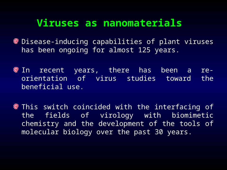
Viruses as nanomaterials Disease-inducing capabilities of plant viruses has been ongoing for almost 125 years.
In recent years, there has been a re-orientation of virus studies toward the beneficial use.
This switch coincided with the interfacing of the fields of virology with biomimetic chemistry and the development of the tools of molecular biology over the past 30 years.

From the chemist’s point of view, viruses are captivating for the following reasons
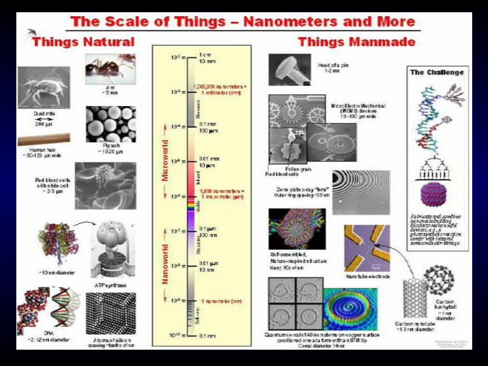
Their size range, from approximately 10 nm to more than a micron, is unique for organic structures characterized at atomic resolution
They can be found in a variety of distinct shapes and with a variety of properties (such as varying sensitivities to pH, salt concentration, and temperature)
They represent the ultimate examples of self-assembly and polyvalence.

Self Assembled Nanostructures?

They have large surface areas, which allow for the display of many copies of the same molecule or many different molecules, without concerns of steric crowding
They can be made in quantity
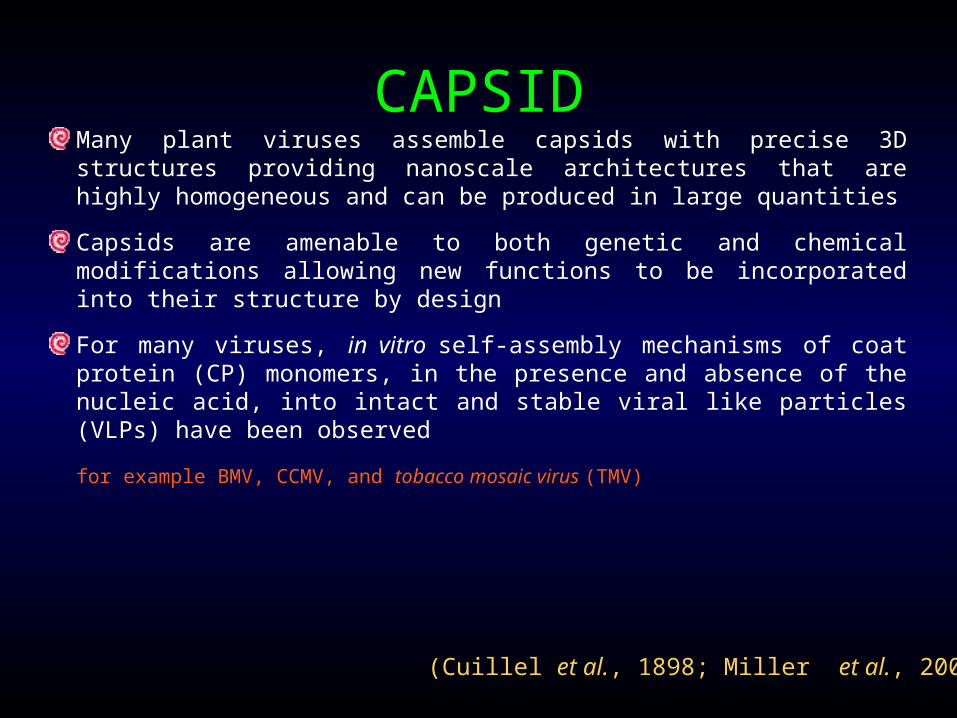
CAPSIDMany plant viruses assemble capsids with precise 3D structures providing nanoscale architectures that are highly homogeneous and can be produced in large quantities
Capsids are amenable to both genetic and chemical modifications allowing new functions to be incorporated into their structure by design
For many viruses, in vitro self-assembly mechanisms of coat protein (CP) monomers, in the presence and absence of the nucleic acid, into intact and stable viral like particles (VLPs) have been observed
for example BMV, CCMV, and tobacco mosaic virus (TMV)
(Cuillel et al., 1898; Miller et al., 2007)

Nano-Bio-Info
Nano
Bio Info
Concept by Robert Cormia
Nano-Bio
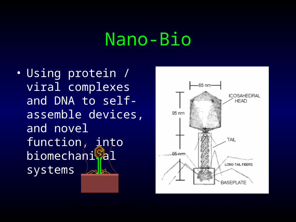
• Using protein / viral complexes and DNA to self-assemble devices, and novel function, into biomechanical systems
Earth’s early nanostructures ~ 2 billion years ago

The three capsid surfaces, the interior surface, the exterior surface, or the interface between coat protein subunits, can be independently functionalized to produce multi- functional biotemplates.
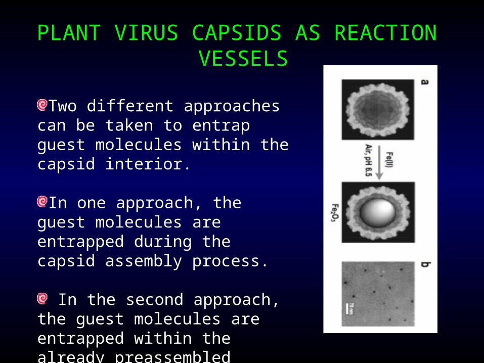
PLANT VIRUS CAPSIDS AS REACTION VESSELS
Two different approaches can be taken to entrap guest molecules within the capsid interior.
In one approach, the guest molecules are entrapped during the capsid assembly process.
In the second approach, the guest molecules are entrapped within the already preassembled capsid architectures.
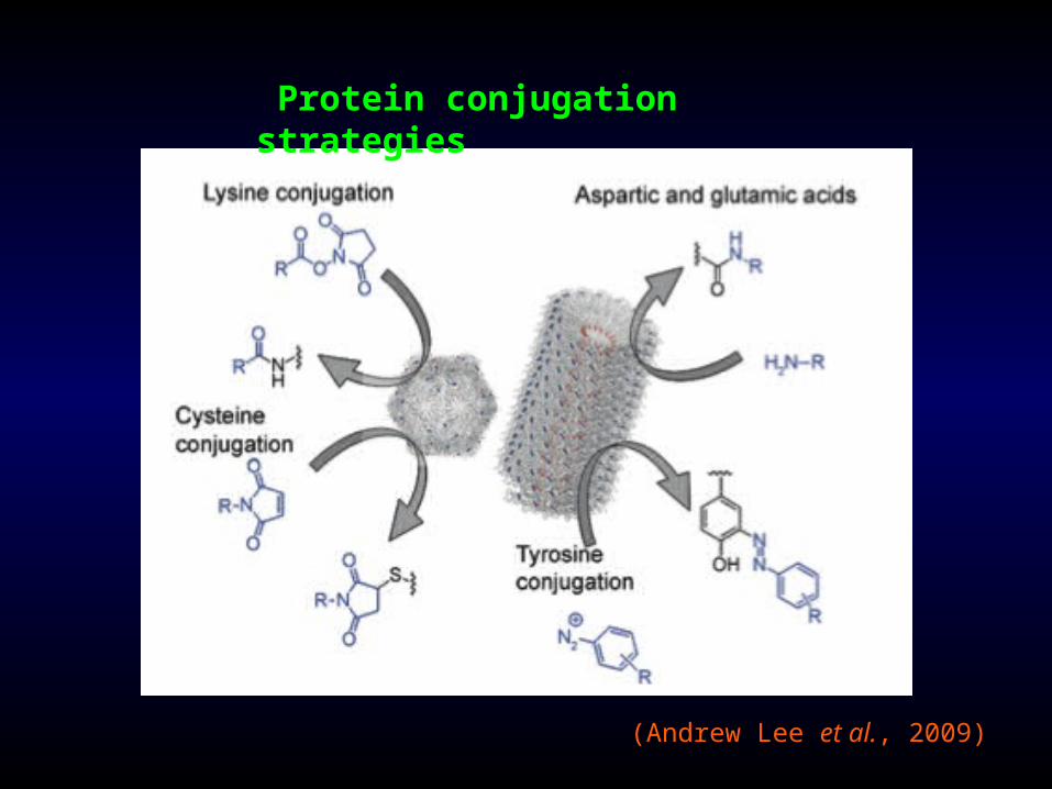
Protein conjugation strategies
(Andrew Lee et al., 2009)
Certain plant viruses and nanotechnology

Plant virus capsids used as biotemplates for nanomaterials and their application in biomedicine
Viruses Nanomaterials Reference
Brome mosaic virus (BMV)
Au nano particles Chen et al., 2006
CdSi/ZnS semiconductor
Endo et al., 2007
Iron oxide nanoparticles Haung et al., 2007
Carnation mottle virus (CarMV)
2D/3D Patterning/array formation
Lvov et al., 1997
Cowpea chlorotic mottle virus (CCMV)
Polymer loading/encapsidation
Chang et al., 2008
2D/3D Patterning/array formation
Klem et al., 2003
Enzyme nanoreactor Comellas-Aragone et al., 2003
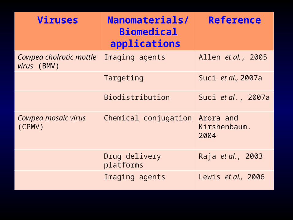
Viruses Nanomaterials/ Biomedical
applications
Reference
Cowpea cholrotic mottle virus (BMV)
Imaging agents Allen et al., 2005
Targeting Suci et al., 2007a
Biodistribution Suci et al., 2007a
Cowpea mosaic virus (CPMV)
Chemical conjugation Arora and Kirshenbaum. 2004
Drug delivery platforms Raja et al., 2003
Imaging agents Lewis et al., 2006

Viruses Nanomaterials/ Biomedical
applications
Reference
Tobacco mosaic virus (TMV)
Surface modifications Miller et al., 2007
Liquid crystals Fowler et al., 2003
Nanowires/mineralization Bittner, 2005.
Patterning/arrays
Dujardin et al., 2003
Turnip yellow mosaic virus (TYMV)
Fluorescent labeling/sensor development
Barnhill et al., 2007
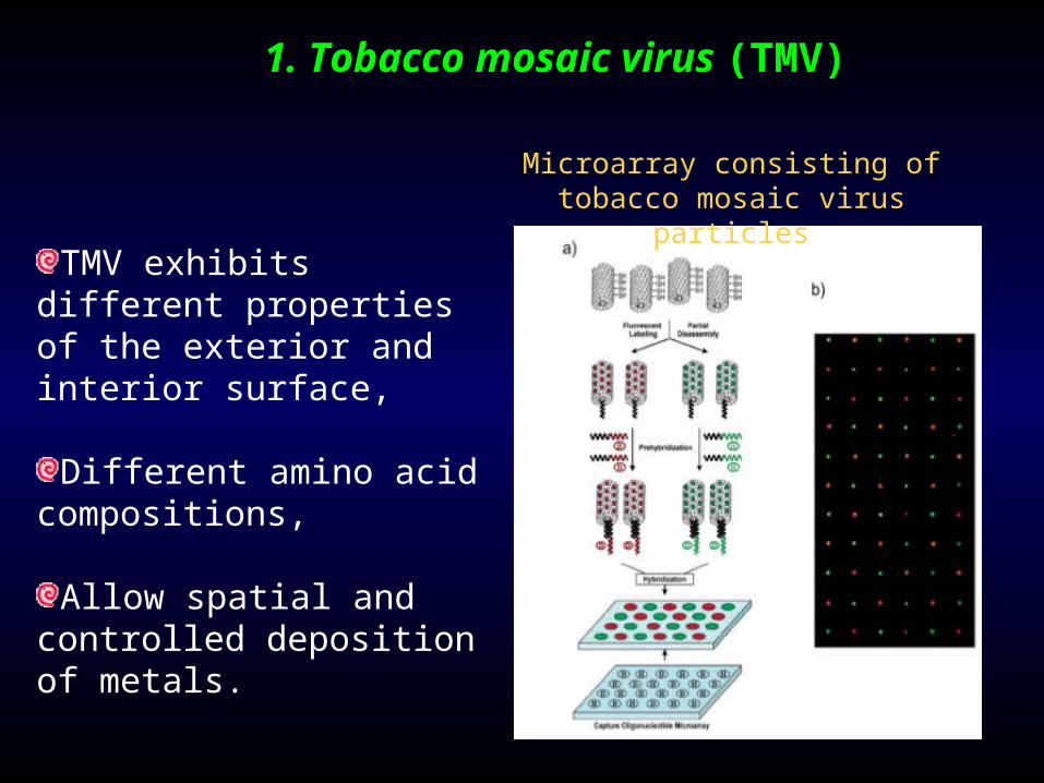
1. Tobacco mosaic virus (TMV)
Microarray consisting of tobacco mosaic virus particles
TMV exhibits different properties of the exterior and interior surface,
Different amino acid compositions,
Allow spatial and controlled deposition of metals.

Deena Awad, 2010
Deposition of TMV in an aligned coating.

Deena Awad, 2010
Stepwise preparation of nanowires from TMV-coated substrate. a) Model of a TMV-coated sub- strate plate, which is then placed in b) a glutaraldehyde solution to crosslink the TMV fibers. The resulting complex is then placed into a c) solution containing gold nanoparticles for conjugation onto the cros- slinked TMV. Finally, the crosslinked, conjugated TMV in placed in a silver ion-hyrdoquinone solution for enhancement of the TMV nanowires.
TMV nanowires
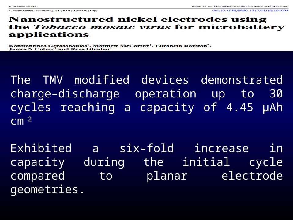
The TMV modified devices demonstrated charge–discharge operation up to 30 cycles reaching a capacity of 4.45 μAh cm−2
Exhibited a six-fold increase in capacity during the initial cycle compared to planar electrode geometries.
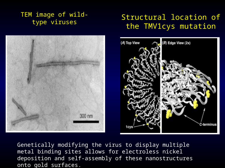
Genetically modifying the virus to display multiple metal binding sites allows for electroless nickel deposition and self-assembly of these nanostructures onto gold surfaces.
Structural location of the TMV1cys mutation
TEM image of wild-type viruses

Schematic of the microbattery layers.
Schematic representation of the TMV assembly and nickel coating process: the TMV binds on the gold surface (step 1), it is activated with a palladium catalyst (step 2) and it is finally coated with nickel (step 3).
TMV assembly and nickel coating process

Initial discharges of cells with and without TMV coating
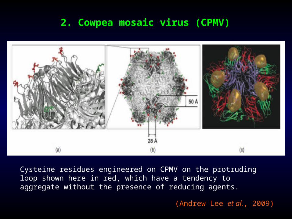
2. Cowpea mosaic virus (CPMV)
Cysteine residues engineered on CPMV on the protruding loop shown here in red, which have a tendency to aggregate without the presence of reducing agents.
(Andrew Lee et al., 2009)

Multifunctional bionanoparticles for in vivo imaging and drug delivery
CPMV for drug delivery. Therapeutics is loaded in the capsid interior and homing domains (Int 8) are attached for the targeting of cancer
cells. Once the virus particles are attached to the cells, they will be endocytosed and the drugs will be released once the virus particles
are degraded in the cytosol
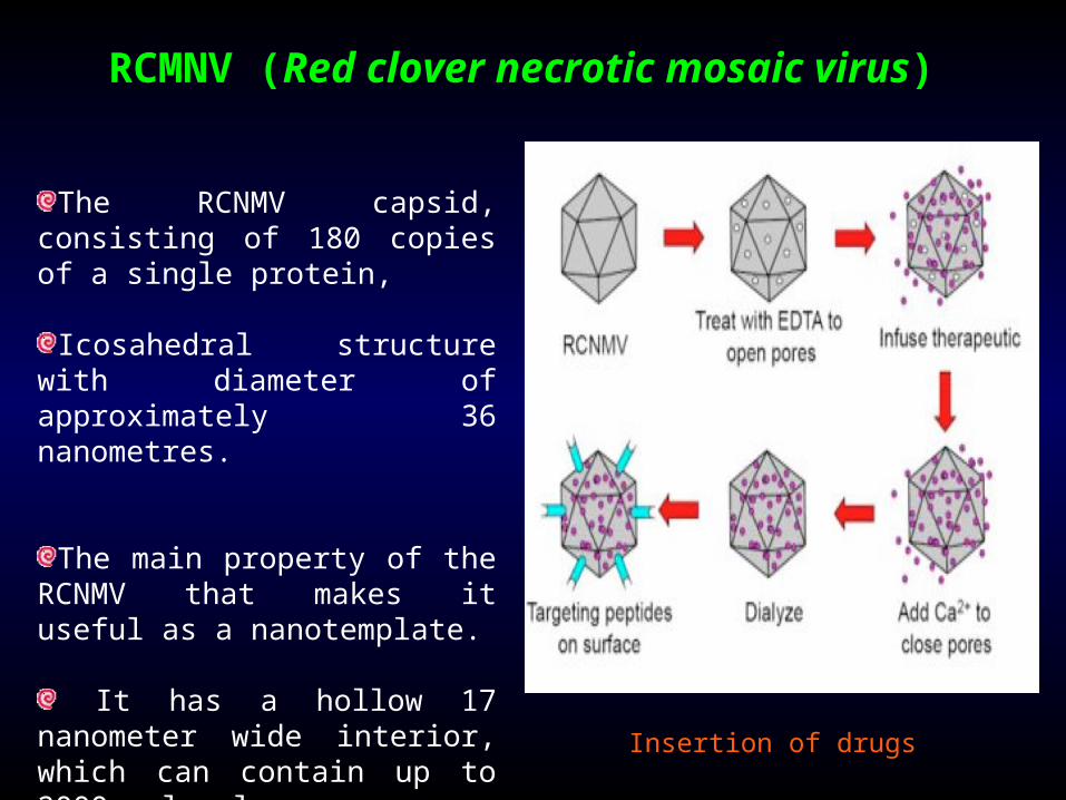
RCMNV (Red clover necrotic mosaic virus)
The RCNMV capsid, consisting of 180 copies of a single protein,
Icosahedral structure with diameter of approximately 36 nanometres.
The main property of the RCNMV that makes it useful as a nanotemplate.
It has a hollow 17 nanometer wide interior, which can contain up to 2000 molecules
Insertion of drugs
Drug-delivery system
RCNMV is to be used in a drug-delivery system within the human body
controlled by placing small proteins on the surface of the capsid
signal-peptides which are attracted to specific predetermined cells
The human blood vessels contain concentrations of calcium high enough for automatic sealing of the capsid.
This leads to emission of the content of the capsid as soon as it has entered the desired cell.
Such a target specific delivery of drugs will greatly reduce side effects of all kinds of chemotherapy.