PLANT AND ANIMAL TISSUE 10 APRIL 2013...Animal Tissue Animal cells with the same structure and...
11
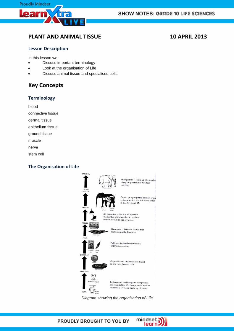
PLANT AND ANIMAL TISSUE 10 APRIL 2013 Lesson Description In this lesson we: Discuss important terminology Look at the organisation of Life Discuss animal tissue and specialised cells Key Concepts Terminology blood connective tissue dermal tissue epithelium tissue ground tissue muscle nerve stem cell The Organisation of Life Diagram showing the organisation of Life
Transcript of PLANT AND ANIMAL TISSUE 10 APRIL 2013...Animal Tissue Animal cells with the same structure and...
PLANT AND ANIMAL TISSUE 10 APRIL 2013
Lesson Description
In this lesson we:
Discuss important terminology
Look at the organisation of Life
Discuss animal tissue and specialised cells
Key Concepts
Terminology
blood
connective tissue
dermal tissue
epithelium tissue
ground tissue
muscle
nerve
stem cell
The Organisation of Life
Diagram showing the organisation of Life
Animal Tissue
Animal cells with the same structure and function are organised into tissues.
Embryonic Tissue
Embryonic tissue can be divided into two kinds of stem cells:
Embryonic stem cells- tissue in an embryo that produces all other tissue during growth
Adult stem cells- tissue in adult that produces new tissue cells to replace old and damaged
ones.
Structure and Function of Embryonic Tissue Structure Function
Embryonic stem cells Produces cells for specialised tissues
Adult Stem cells Replaces old and damaged cells and tissues
Large nucleus
Cellular extensions of cytoplasm
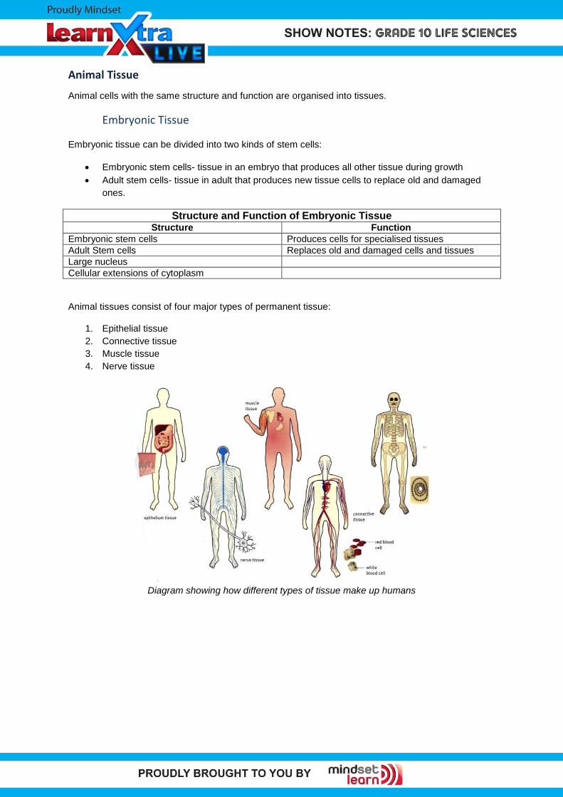
Animal tissues consist of four major types of permanent tissue:
1. Epithelial tissue
2. Connective tissue
3. Muscle tissue
4. Nerve tissue
Diagram showing how different types of tissue make up humans
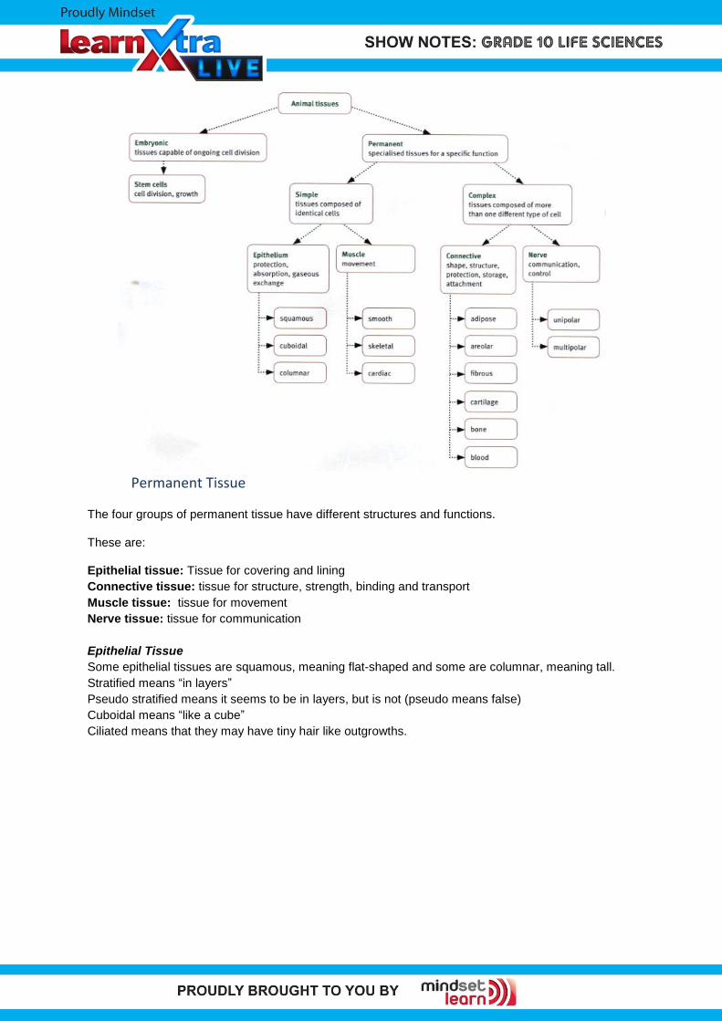
Permanent Tissue
The four groups of permanent tissue have different structures and functions.
These are:
Epithelial tissue: Tissue for covering and lining
Connective tissue: tissue for structure, strength, binding and transport
Muscle tissue: tissue for movement
Nerve tissue: tissue for communication
Epithelial Tissue
Some epithelial tissues are squamous, meaning flat-shaped and some are columnar, meaning tall.
Stratified means “in layers”
Pseudo stratified means it seems to be in layers, but is not (pseudo means false)
Cuboidal means “like a cube”
Ciliated means that they may have tiny hair like outgrowths.
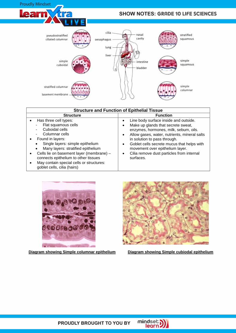
Structure and Function of Epithelial Tissue Structure Function
Has three cell types: - Flat squamous cells - Cuboidal cells - Columnar cells
Found in layers:
Single layers: simple epithelium
Many layers: stratified epithelium
Cells lie on basement layer (membrane) – connects epithelium to other tissues
May contain special cells or structures: goblet cells, cilia (hairs)
Line body surface inside and outside.
Make up glands that secrete sweat, enzymes, hormones, milk, sebum, oils.
Allow gases, water, nutrients, mineral salts in solution to pass through.
Goblet cells secrete mucus that helps with movement over epithelium layer.
Cilia remove dust particles from internal surfaces.
Diagram showing Simple columnar epithelium Diagram showing Simple cubiodal epithelium
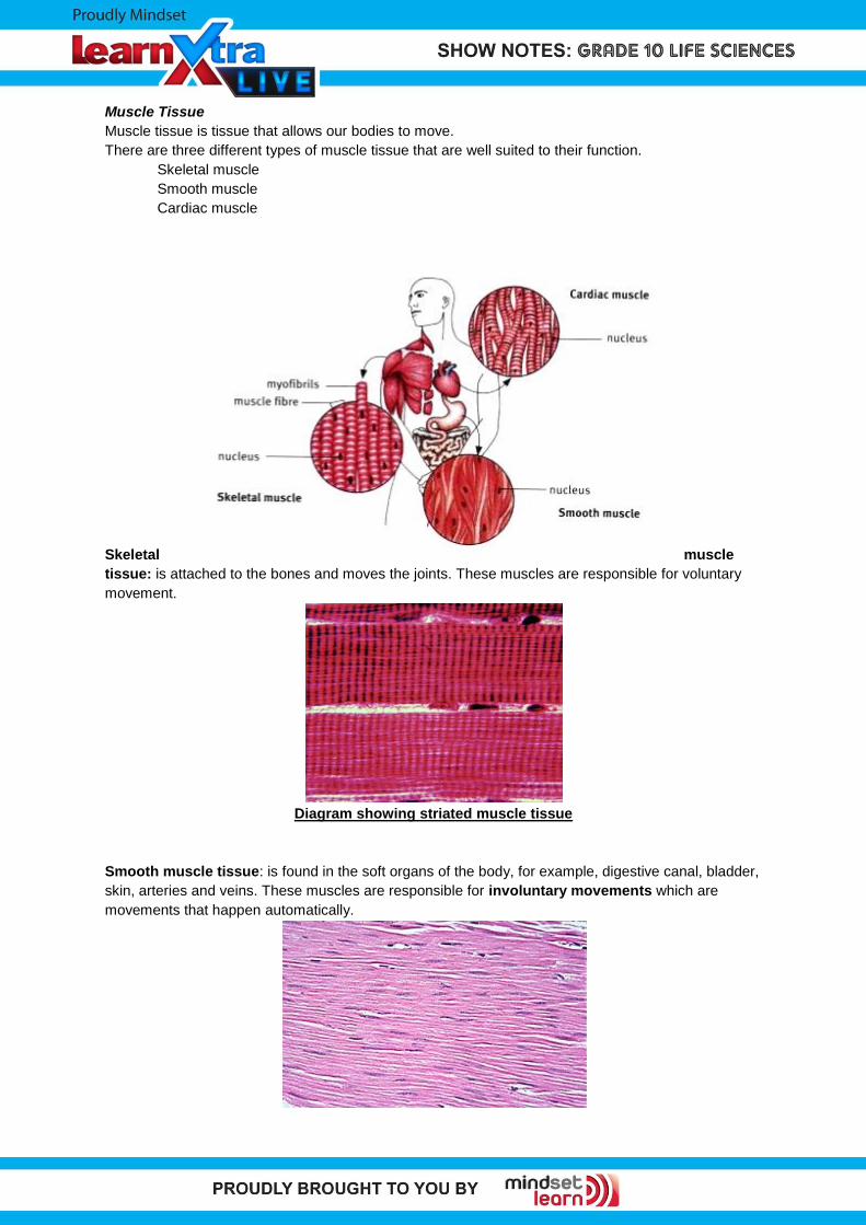
Muscle Tissue
Muscle tissue is tissue that allows our bodies to move.
There are three different types of muscle tissue that are well suited to their function.
Skeletal muscle
Smooth muscle
Cardiac muscle
Skeletal muscle
tissue: is attached to the bones and moves the joints. These muscles are responsible for voluntary
movement.
Diagram showing striated muscle tissue
Smooth muscle tissue: is found in the soft organs of the body, for example, digestive canal, bladder,
skin, arteries and veins. These muscles are responsible for involuntary movements which are
movements that happen automatically.
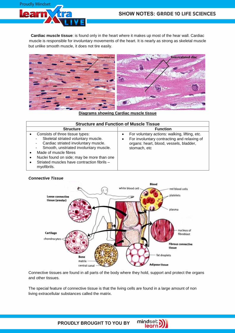
Cardiac muscle tissue: is found only in the heart where it makes up most of the hear wall. Cardiac
muscle is responsible for involuntary movements of the heart. It is nearly as strong as skeletal muscle
but unlike smooth muscle, it does not tire easily.
Diagrams showing Cardiac muscle tissue
Structure and Function of Muscle Tissue Structure Function
Consists of three tissue types: - Skeletal striated voluntary muscle. - Cardiac striated involuntary muscle. - Smooth, unstriated involuntary muscle.
Made of muscle fibres
Nuclei found on side; may be more than one
Striated muscles have contraction fibrils – myofibrils.
For voluntary actions: walking, lifting, etc.
For involuntary contracting and relaxing of organs: heart, blood, vessels, bladder, stomach, etc
Connective Tissue
Connective tissues are found in all parts of the body where they hold, support and protect the organs
and other tissues.
The special feature of connective tissue is that the living cells are found in a large amount of non
living extracellular substances called the matrix.
The matrix makes each kind of connective tissue different and gives it a different texture.
We can divide connective tissue into four groups:
Connective tissue with a jelly like matrix
Connective tissue with a rubbery matrix
Connective tissue with a rigid matrix
Connective tissue with a liquid matrix
3.1. Connective tissue with a jelly like matrix
This tissue can be divide into two groups.
Loose or areolar connective tissue.
Dense connective tissue
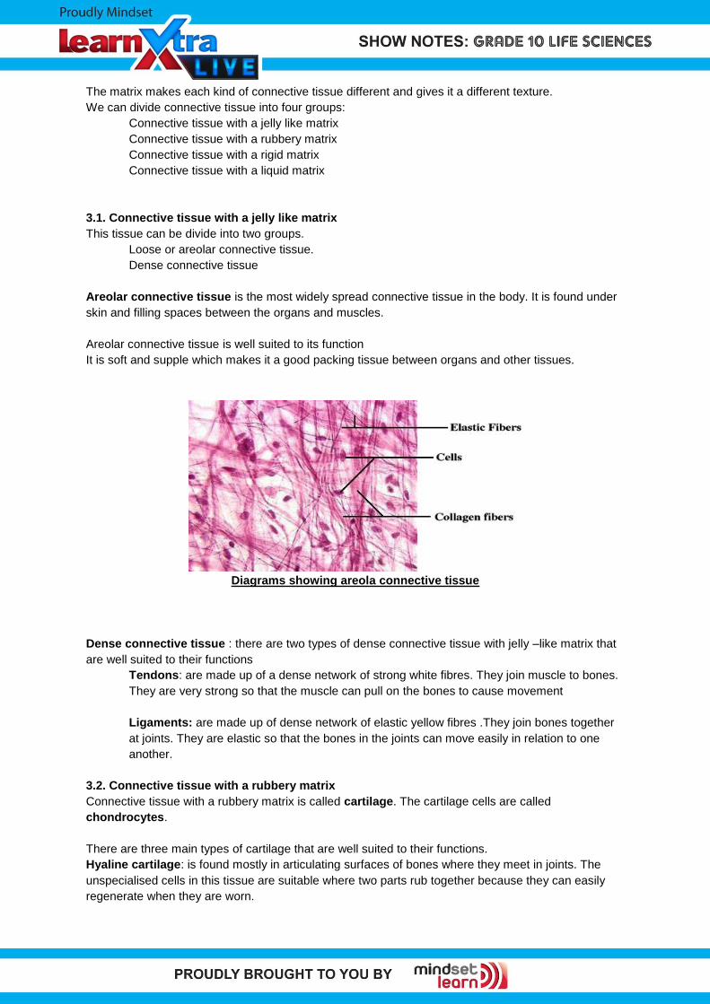
Areolar connective tissue is the most widely spread connective tissue in the body. It is found under
skin and filling spaces between the organs and muscles.
Areolar connective tissue is well suited to its function
It is soft and supple which makes it a good packing tissue between organs and other tissues.
Diagrams showing areola connective tissue
Dense connective tissue : there are two types of dense connective tissue with jelly –like matrix that
are well suited to their functions
Tendons: are made up of a dense network of strong white fibres. They join muscle to bones.
They are very strong so that the muscle can pull on the bones to cause movement
Ligaments: are made up of dense network of elastic yellow fibres .They join bones together
at joints. They are elastic so that the bones in the joints can move easily in relation to one
another.
3.2. Connective tissue with a rubbery matrix
Connective tissue with a rubbery matrix is called cartilage. The cartilage cells are called
chondrocytes.
There are three main types of cartilage that are well suited to their functions.
Hyaline cartilage: is found mostly in articulating surfaces of bones where they meet in joints. The
unspecialised cells in this tissue are suitable where two parts rub together because they can easily
regenerate when they are worn.
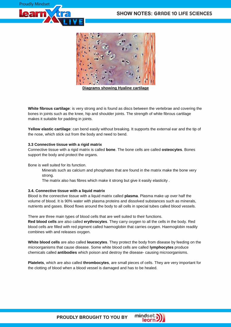
Diagrams showing Hyaline cartilage
White fibrous cartilage: is very strong and is found as discs between the vertebrae and covering the
bones in joints such as the knee, hip and shoulder joints. The strength of white fibrous cartilage
makes it suitable for padding in joints.
Yellow elastic cartilage: can bend easily without breaking. It supports the external ear and the tip of
the nose, which stick out from the body and need to bend.
3.3 Connective tissue with a rigid matrix
Connective tissue with a rigid matrix is called bone. The bone cells are called osteocytes. Bones
support the body and protect the organs.
Bone is well suited for its function.
Minerals such as calcium and phosphates that are found in the matrix make the bone very
strong.
The matrix also has fibres which make it strong but give it easily elasticity .
3.4. Connective tissue with a liquid matrix
Blood is the connective tissue with a liquid matrix called plasma. Plasma make up over half the
volume of blood. It is 90% water with plasma proteins and dissolved substances such as minerals,
nutrients and gases. Blood flows around the body to all cells in special tubes called blood vessels.
There are three main types of blood cells that are well suited to their functions.
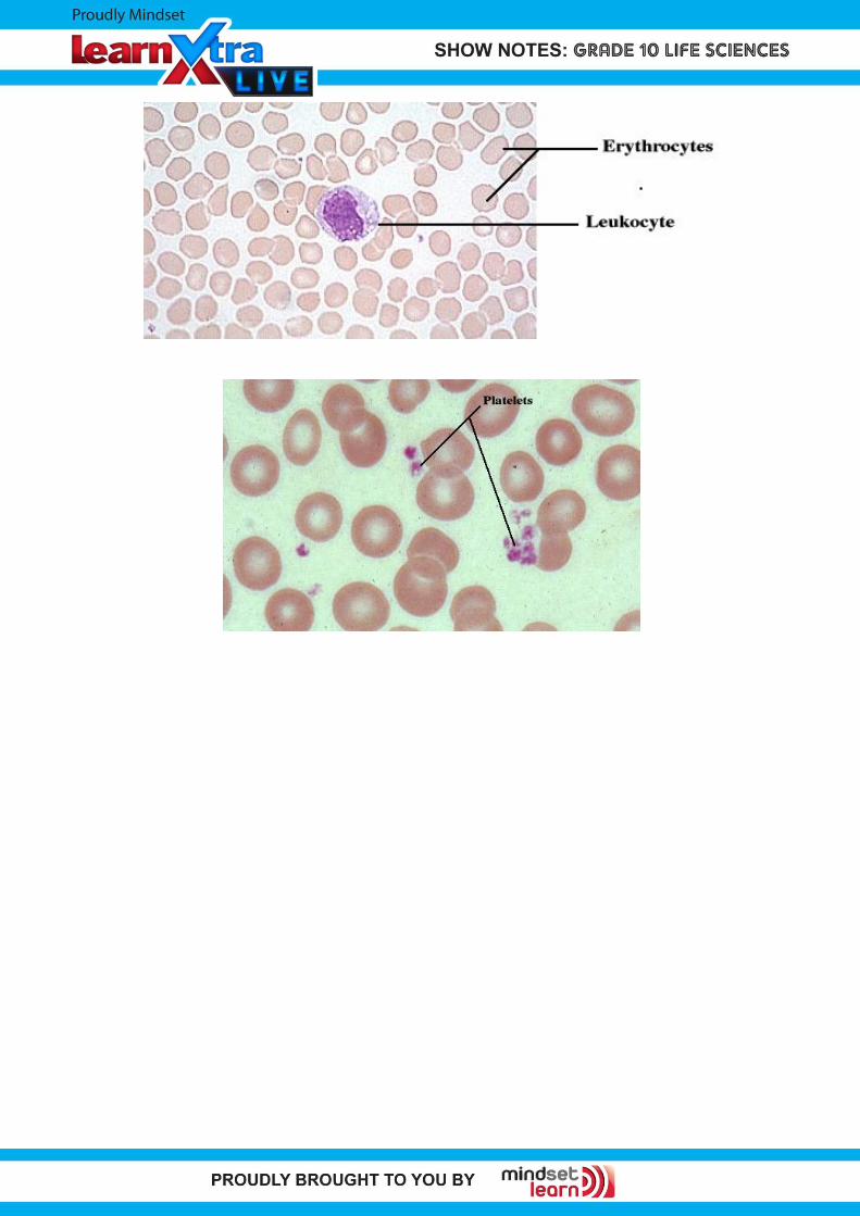
Red blood cells are also called erythrocytes. They carry oxygen to all the cells in the body. Red
blood cells are filled with red pigment called haemoglobin that carries oxygen. Haemoglobin readily
combines with and releases oxygen.
White blood cells are also called leucocytes. They protect the body from disease by feeding on the
microorganisms that cause disease. Some white blood cells are called lymphocytes produce
chemicals called antibodies which poison and destroy the disease- causing microorganisms.
Platelets, which are also called thrombocytes, are small pieces of cells. They are very important for
the clotting of blood when a blood vessel is damaged and has to be healed.
Nerve Tissue
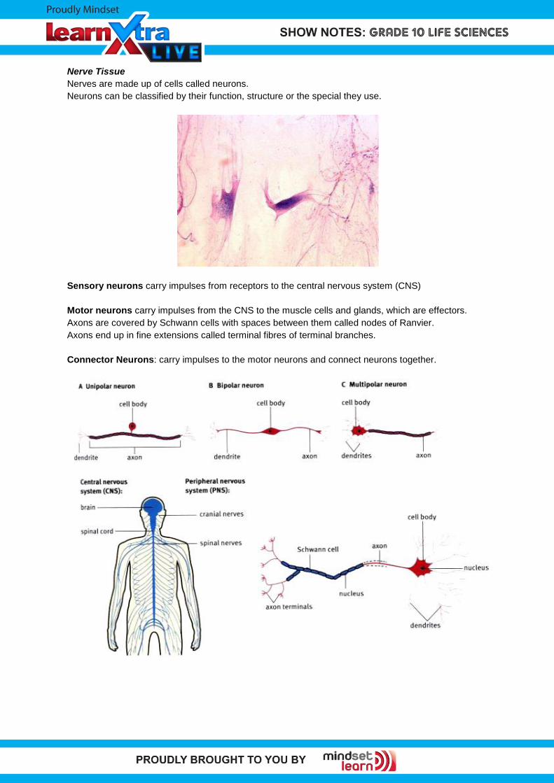
Nerves are made up of cells called neurons.
Neurons can be classified by their function, structure or the special they use.
Sensory neurons carry impulses from receptors to the central nervous system (CNS)
Motor neurons carry impulses from the CNS to the muscle cells and glands, which are effectors.
Axons are covered by Schwann cells with spaces between them called nodes of Ranvier.
Axons end up in fine extensions called terminal fibres of terminal branches.
Connector Neurons: carry impulses to the motor neurons and connect neurons together.
Questions
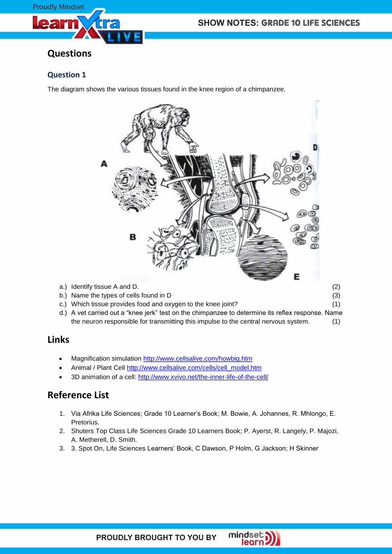
Question 1
The diagram shows the various tissues found in the knee region of a chimpanzee.
a.) Identify tissue A and D. (2)
b.) Name the types of cells found in D (3)
c.) Which tissue provides food and oxygen to the knee joint? (1)
d.) A vet carried out a “knee jerk” test on the chimpanzee to determine its reflex response. Name
the neuron responsible for transmitting this impulse to the central nervous system. (1)
Links
Magnification simulation http://www.cellsalive.com/howbig.htm
Animal / Plant Cell http://www.cellsalive.com/cells/cell_model.htm
3D animation of a cell: http://www.xvivo.net/the-inner-life-of-the-cell/
Reference List
1. Via Afrika Life Sciences; Grade 10 Learner’s Book; M. Bowie, A. Johannes, R. Mhlongo, E.
Pretorius.
2. Shuters Top Class Life Sciences Grade 10 Learners Book; P. Ayerst, R. Langely, P. Majozi,
A. Metherell, D. Smith.
3. 3. Spot On, Life Sciences Learners’ Book, C Dawson, P Holm, G Jackson; H Skinner