
PLANES AND DIRECTION OF THE BODY GENERAL · PDF fileplanes and direction of the body general...
47
PLANES AND DIRECTION OF THE BODY GENERAL OSTEOLOGY SKELETON OF THE SPINE AND THORAX 1. Lecture, DENTISTRY AUTUMN 2015 Lecturer: RNDr. MICHAELA RAČANSKÁ, Ph.D.
Transcript of PLANES AND DIRECTION OF THE BODY GENERAL · PDF fileplanes and direction of the body general...

PLANES AND DIRECTION OF THE BODYGENERAL OSTEOLOGY
SKELETON OF THE SPINE AND THORAX
1. Lecture, DENTISTRY AUTUMN 2015
Lecturer: RNDr. MICHAELA RAČANSKÁ, Ph.D.

ANATOMY 1 Dentistry- autumn 2015
Date Lectures Seminars
1. 23. 10. Planes and direction of the body. General osteology Skeleton of the spine and thorax
Planes and direction of the body. X-Ray - anatomy Skeleton of the spine and the thorax
2. 30. 9. Skeleton of the upper extremity Skeleton of the upper extremity
3. 7. 10. Skeleton of the lower extremity Skeleton of the lower extremity
4. 14. 10. Neurocranium Neurocranium
5. 21. 10. Splanchnocranium Splanchnocranium
6. 28. 10. Cavities of the skull Cavities of the skull
7. 4. 11. General arthrology
Joints of the spine and thorax
General arthrology
Joints of the spine and thorax
8. 11. 11. Joints of the skull and upper extremity Joints of the skull and upper extremity
9. 18. 11. Joints of the lower extremity. Pelvis Joints of the lower extremity. The pelvis
10. 25. 11. General myology. Muscles and fasciae of the head. Control examination (osteology, arthrology)
11. 2. 12. The muscles of the neck, thorax, back
12. 9. 12. Muscles of the upper limb
13. 16. 12. Muscles of the lower limb
14. 6. 1. Muscles of the abdomen Muscles – overview
Syllabus Lectures: We 13,20-15,00 Seminars: We 15,40- 17,20 MUDr. Roman Kopáčik

Where you can study from?
Liebgott, Bernard. The anatomical basis of dentistry. 3rd ed. Mosby, ISBN 0-323-06807-3 PÁČ, Libor, Ladislava HORÁČKOVÁ a Hana NECHUTOVÁ. Anatomy of human locomotor system. 1. vyd. Brno: Masarykova univerzita Brno, 2010. 119 s. ISBN 978-80-210-5258-1.
Atlas of human anatomy. Edited by Johannes Sobotta - Reinhard Putz - Reinhard Pabst - Renate Putz. 13th English ed., 21st Germa. Philadelphia: Lippincott Williams & Wilkins, 2001. 404 s. ISBN 0-7817-3174-7.
NETTER, Frank H. Atlas of human anatomy. 4th ed. Philadelphia: Saunders Elsevier, 2006. 548 color. ISBN 1-4160-3385-8.

Anatomical nomenclature
• Anatomy is the basis of the language of medicine. Students learn a new language consisting of at least 4500 words. International.
• Many anatomical terms are derived from Latin and Greek.
• To describe the relationship of one structure to another, the anatomical nomenclature should be used.
• To be understood you must express yourself clearly, using the official terms in the correct way.
• 1. Andreas Vesálius, founder of the modern anatomy, 16. century.
• 2. Basiliensia Nomina Anatomica, B. N. A.,
1895
• 3. Ienaiensia Nomina Anatomica, I. N. A.,
1935
• 4. Parisiensia Nomina Anatomica, P. N. A.,
1955 accepted 1960, last corrections - 1985 (5640 terms)
Terminologia Anatomica – International Anatomical terminology (FCAT) 1998

The first word is name of described formation, next adjectives specificate it
and in the end there is a name of formation where the describedformation is located.
Examples:
Collum (neck) radii (of radius)
Collum (a neck) anatomicum (anatomical) humeri (of humerus)Collum (a neck) chirurgicum (surgical) humeri (of humerus)Tuberculum (a tubercle, a bulge) majus (big) humeri (of humerus)Spina (a thorn) iliaca (iliac) anterior (fore) superior (upper) ossis coxae (of coxal bone)Epicondylus medialis humeriEpicondylus medialis femoris
Anatomical nomenclature

Orientation on the body
Anatomical positionstandard erect position Not a military position!
x

PLANES – 3 anatomical planes or sections
Sagittal plane (median), Midsagittalvertical plane - Right and leftAcc. to sagittal axis
Transverse plane (horizontal, axial, cross sections)Vertical plane - Superior and inferior(acc. to transversal axis)
Frontal plane (coronal)Anterior and posterior(acc. to longitudinal axis)

cranialis caudalis
superior inferior
ventralis dorsalis
anterior posterior
medialis lateralis
medianus medius (intermedius)
dexter sinister
superficilais profundus
internus externus
Directions on the body

Directions at the limbs
PROXIMALIS DISTALISRADIALIS (lateralis)ULNARIS (medialis)PALMARISDORSALIS PLANTARISFIBULARIS (lateralis)TIBIALIS (medialis)

Marking of bones -positive and negative relief
• Sulcus – a groove
• Incisura – a notch
• Canalis – a canal
• Fossa – a pit, hollow
• Fovea – a pit, hollow
• Foramen – an opening, orifice, gap
• Groove – a furrow
• Processus – a projection, prominence
• Spina – a thorn
• Tuberculum – a tubercle
• Tuber – a torus
• Tuberositas – a tuberosity, large rounded eminence
Internus – internalExternus – externalSuperficialis – superficialProfundus – deep
Caput – a headCapitulum – a small headCollum, cervix – a neck
Os, ossis, ossa – a bone, bonesArticulus – a jointFacies – a facet, surface
NEGATIVE
POSITIVE

Fossa x fovea
Fovea capitis femoris
Fossa iliaca
Fossa trochantericaFossa olecrani
Source of the pictures: Atlas der Anatomie des Menschen/Sobotta. Putz,R., und Pabst,R. 20. Auflage. München:Urban & Schwarzenberg, 1993

Caput x condylus
Caput humeri
Condylus medialis
et lateralis
et epicondylus med. et lat. femoris
Epicondylus med. et lat. humeri
Caput tali
Caput femoris
Source of the pictures: Atlas der Anatomie des Menschen/Sobotta. Putz,R., und Pabst,R. 20. Auflage. München:Urban & Schwarzenberg, 1993

Incisura x foramen
Incisura scapulae
Foramen obturatum
Source of the pictures: Atlas der Anatomie des Menschen/Sobotta. Putz,R., und Pabst,R. 20. Auflage. München:Urban & Schwarzenberg, 1993

General osteology
The skeleton is composed of a living, dynamic, rigid, connective tissue that formsbones and cartilages In total 214 (incl. sessamoid bones), it varies Cartilage at the places where flexibility is important, or covers articulation surfaces
FUNCTION OF SKELETAL SYSTEM Support Protection of vital organs Together with muscles a mechanism for movement Storage of calcium (99% of body´s calcium is stored in bone) and other salts A source of blood cells (Bone marrow in the central cavity, hemopoetic (blood- forming) cells

Basic structure of bones
• Bone as a conective tissue consists of :
bone cells (osteocytes)
Ground substance+ collagenous fibrils form - osteoid (ossein) – organic material
Different sallts – hardness and strenght – anorganic material
A salt free or decalcified bone is pliable
• in young 52% of organic component
• In elderly 40 %

2) spongy (trabecular or cancellous) bone
A less dense trabeculated network of bone spicules making up the substance of most bones, surrounding an inner marrow cavity,
TYPES OF BONE ACCORDING TO THE STRUCTURE
1) compact boneA relatively solid mass of bone
Commonly seen as a superficial layerof bone, that provides strenght

BONE STRUCTURE PeriosteumExternal fibrousInternal cambious layer(osteoblasts, Sharpey´s fibers, remodelation
Substantia compacta
Substantia spongiosaBone architecture, trajectories
EndosteumBone reconstruction, it is not possible to peel it off
Cavitas medullaris- (bone marrow)
medulla ossium rubramedulla ossium flavamedulla ossium gelatinosa

1 – periosteum
2 – Sharpey´s fibers
3 – vessels in a periosteum
4 – endosteum
5 – a vessel from periosteum passes through Volkmann´s canal
to vessels of Havers´s systems
Periosteum (periost)covers almost all parts of the bone (not at the joint
surfaces
it contains many blood and lymph vessels and nerves.
A bone from which the periosteum
has been removed will die.
Periosteum consists of:
a) a fibrous layer (external)
b) a cambious layer (the site of osteoblasts –
built up bone and help of healing – fractures)

1 – Haversian lamellae
2 – intersticial lamellae
3 – superficial lamellae
4 – lamellae of spongy bone
H – Haversian system of lamellae, osteon
1 – osteocyte
2 – lacuna
3 – canaliculus osseus
4 – Haversian canal of osteon
5 – concentric lamellae of osteon
6 – superficial lamellae
Lamellar bone tissue

BLOOD SUPPLY
• Nutrient arteries (one or more, through thediaphyssis)
• Periosteal arteries (supply the compact bone)
• Metaphysiel arteries
• Epiphyseal arteries
• Apophyseal arteries

BONE DEVELOPMENT (ossification)
a. nutricia
aa. epiphysariae
aa. epiphysariae
1) Intramembranous formation
Flat bones
Direct calcium deposition
into mesenchymal model of the bone
2) Endochondral formation
Long bones, irregular bones
Calcium deposition into a cartilagineous
model of the bone
a) perichondral
originates in diaphysis
b) enchondral
in cartilage near epiphyses

Growing of the epifyseal cartilage followed ba the osification of both epiphysis and diaphysis
as the background of growing into the lenght
To the thickness growth the bone thanks to the periostal cells of the cambial (inner) layer!
Bone growthGrowth plate = epiphyseal diskis necessary for growth in length, forms a layer betweenthe epiphysis and the diaphysis.
The part of diaphysis adjacentto the epiphysial disk is calledmetaphysis.

Classification of bones accoring to the shape
Ossa longa (long bones) Ossa brevia (short bones) Ossa plana (flat bones)
Proximal end
Distal end
Body (corpus)
Compact bone=corticalis
Spongy bonebone architecture
Lamina externa
Lamina interna
diploe

Ossa sesamoidea (sesamoid bones) – in tendons of some muscles
Ossa pneumatica (pneumatised)– paranasal sinuses
Ossa irregularia (irregular)
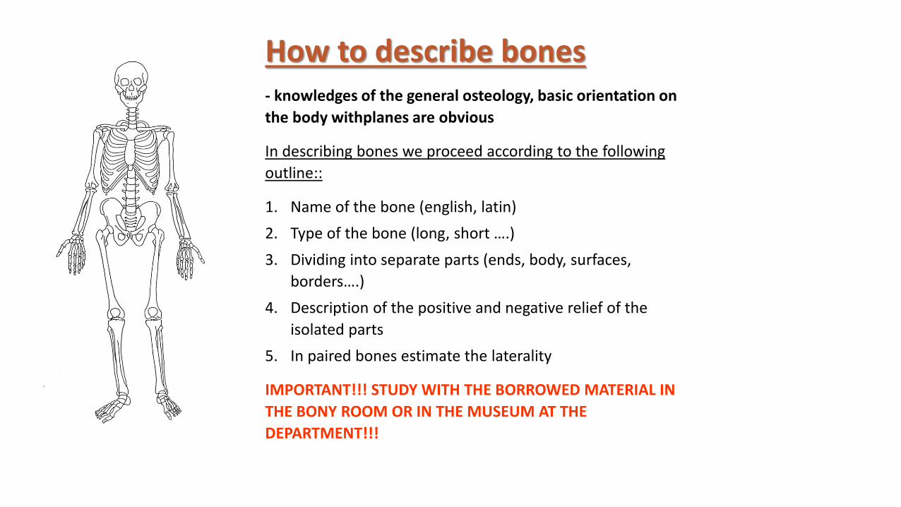
How to describe bones- knowledges of the general osteology, basic orientation on
the body withplanes are obvious
In describing bones we proceed according to the following
outline::
1. Name of the bone (english, latin)
2. Type of the bone (long, short ….)
3. Dividing into separate parts (ends, body, surfaces,
borders….)
4. Description of the positive and negative relief of the
isolated parts
5. In paired bones estimate the laterality
IMPORTANT!!! STUDY WITH THE BORROWED MATERIAL IN
THE BONY ROOM OR IN THE MUSEUM AT THE
DEPARTMENT!!!

AXIAL SKELETON
Bones of the skull
Vertebral column (spine)
Ribs
Sternum
APPENDICULAR SKELETON
Bones of the limbs
Pectoral girdle
Pelvic girdle
Central line of the body (80)
Attach the limbs to the body´s axis (134)

Thorax
Columna vertebralis (vertebral column, spine 26)
Costae (ribs, 24)
Sternum (breast bone)

COLUMNA VERTEBRALIS (vertebral column)
33-34, usually 24 free vertebras
7 vertebrae cervicales (C) cervical vertebra
12 vertebrae thoracicae (Th) thoracic vertebra
5 vertebrae lumbales (L) lumbar vertebra
5 vertebrae sacrales – os sacrum (sacral bone)
4–5 vertebrae coccygeae – os coccygis (coccyx)

Costa
Processus transversus
Corpus vertebrae
Processus articulares
Processus spinosus
Arcus vertebrae
DEVELOPMENT OF VERTEBRAS

Corpus vertebrae(facies terminalis superior et inferior)
Pediculus arcus vertebrae
Arcus vertebrae
Foramen vertebrale(canalis vertebralis)
Incisura vertebralis superior et inferior
Foramen intervertebrale
Processus vertebrales4x processus articulares
(processus articularis superior et inferior - dexter et sinister)2x processus transversus (dexter et sinister) 1x processus spinosus
General features of all vertebras

Processus vertebrales
Processus articulares (4)
processus articularis superior - dexter et sinisterprocessus articularis inferior - dexter et sinister
Processus transversus dexter et sinister (2)
Processus spinosus (1)

Vertebrae cervicales C1 – C7 (Cervical vertebras)
Foramen processus transversi !!!!!!
Sulcus nervi spinalis
Tuberculum anterius et posterius processus
transversi
Uncus corporis vertebrae
Procesus articulares
Bifurcations of the spinous processes (C2 – C6)
C6 - tuberculum caroticum
C3 – the smallest body
C7 – vertebra prominens

Arcus anterior et posterior atlantis
fovea dentis
tuberculum anterius et posterius atlantis
foramen vertebrale
Massae laterales
facies/fovea articularis superior et inferior
sulcus arteriae vertebralis
processus transversi
Corpus vertebrae
Dens axis
facies articularis ant. et post. dentis
apex dentis
C1 - Atlas
C2 - Axis

fovea costalis (dextra et sinistra)
fovea costalis processus transversi
processus articulares
Vertebrae thoracicae Th1 – Th12 (thoracic vertebras)
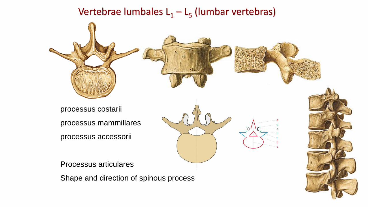
processus costarii
processus mammillares
processus accessorii
Processus articulares
Shape and direction of spinous process
Vertebrae lumbales L1 – L5 (lumbar vertebras)

Lumbar punction - between L3 – L4
Cerebral liquor

Vertebrae sacrales, os sacrum (sacral bone)
basis – facies terminalis superior
apex – facies terminalis inferior
facies pelvina
lineae transversales
foramina sacralia pelvina
promontorium
canalis sacralis – hiatus canalis sacraliscornua sacralia

Os sacrumFacies dorsalis
crista sacralis mediana
cristae sacrales intermediae
cristae sacrales laterales
foramina sacralia dorsalia
tuberositas sacralis
Partes laterales
facies auriculares
Canalis sacralis
hiatus canalis sacraliscornua sacralia

basis – facies terminalis superior
cornua ossis coccygis
apex
Vertebrae coccygeae, os coccygis (coccyx)(Co1 – Co 4-5)

Costa, rib (12)
Costae verae (1.-7.)
Costae spuriae (8.-10.)
Costae fluctuantes (11., 12.)
Cervical rib
Lumbar rib (near to the kidneys)

Os costae
Cartilago costae
Caput facies articularis(2. - 10. rib - crista capitis costae)
Collumtuberculum costaefacies articularis tuberculi costae
Corpusangulus costaecrista costaesulcus costae

tuberculum musculi scaleni anterioris
sulcus arteriae subclaviae
tuberculum musculi scaleni medii
tuberositas musculi scaleni posterioris
tuberositas musculi serrati anterioris
Costa prima
Costa secunda
11. and 12. ribs – tuberculum costae and sulcus costae are missing!!!

Cervical rib

Manubrium sterni
incisura clavicularis
incisura jugularis
incisurae costales 1.,2.
Angulus sterni
Corpus sterni
incisurae costales (3.-7. žebro)
Processus xiphoideus
Sternum (breast bone)

Sternebrae
Hollow in the body of the breast bone

2nd intercostal space
Sternal punction
heart
diaphragma
Breast bone
column

Any questions?
The pictures used in this lectures were taken from following sources:• Atlas der Anatomie des Menschen/Sobotta. Putz,R., und Pabst,R. 20. Auflage.
München:Urban & Schwarzenberg, 1993• Netter: Interactive Atlas of Human Anatomy.• Naňka, Elišková: Přehled anatomie. Galén, Praha 2009.• Čihák: Anatomie I, II, III.• Drake et al: Gray´s Anatomy for Students. 2010• Own archiv of the lecturer


