Placental Extravillous Cytotrophoblasts Persistently Express Class I ...
9
JOURNAL OF VIROLOGY, Aug. 2003, p. 8187–8195 Vol. 77, No. 15 0022-538X/03/$08.000 DOI: 10.1128/JVI.77.15.8187–8195.2003 Copyright © 2003, American Society for Microbiology. All Rights Reserved. Placental Extravillous Cytotrophoblasts Persistently Express Class I Major Histocompatibility Complex Molecules after Human Cytomegalovirus Infection Masakazu Terauchi, 1 Hideki Koi, 1 Chikako Hayano, 1 Noriko Toyama-Sorimachi, 2 Hajime Karasuyama, 2 Yuji Yamanashi, 3 Takeshi Aso, 1 and Masaki Shirakata 3 * Department of Comprehensive Reproductive Medicine, 1 and Department of Immune Regulation, 2 Graduate School, and Department of Cell Regulation, Medical Research Institute, 3 Tokyo Medical and Dental University, Bunkyo, Tokyo 113-8510, Japan Received 19 February 2003/Accepted 5 May 2003 Human cytomegalovirus (HCMV) downregulates the class I major histocompatibility complexes (MHCs), HLA-A and -B, in infected fibroblasts to escape from antigen-specific cytotoxic T lymphocytes. The HCMV genes responsible for the downregulation of MHCs are US2, US3, US6, and US11, which encode type I membrane proteins working at the endoplasmic reticulum (ER). However, it is largely unknown whether HCMV downregulates the class I MHC molecules in placental extravillous cytotrophoblasts (EVT), which express HLA-C, -E, and -G to protect a semiallogenic fetus from maternal natural killer (NK) cells at the fetomaternal interface. Here, we report that differentiated EVT prepared from human first-trimester chorionic villi persistently express class I MHC molecules upon HCMV infection. When these US proteins were expressed in uninfected EVT, they were localized at the ER in the entire cytoplasm. However, subsequent HCMV infection resulted in dissociation of these US proteins from the ER, which relocated toward the cell membrane. In fibroblasts, these US proteins were localized at the ER before and after HCMV infection. These results suggest that the US gene products are not integrated into ER of HCMV-infected EVT and fail to downregulate class I MHC molecules. Human cytomegalovirus (HCMV) is the most common viral agent of intrauterine infection, affecting 1% of all newborns. Five percent of infants with symptomatic congenital HCMV infection die, and most of the survivors as well as 10% of those who are asymptomatic at birth have neurological sequelae, including mental retardation and hearing loss. Maternal vire- mia and transplacental virus transmission following primary infection or reactivation of the virus are possible causes of congenital HCMV infection. When HCMV infects fibroblasts, it downregulates the sur- face expression of class I major histocompatibility complexes (MHCs), HLA-A and -B, to protect the infected cells from attacks by antigen-specific cytotoxic T lymphocytes (4, 44, 48). HCMV US2, US3, US6, and US11 genes are responsible for the HCMV-induced downregulation of the MHCs. US6 inhib- its TAP-mediated peptide translocation to endoplasmic retic- ulum (ER) and subsequent peptide loading of MHC molecules (3, 16, 29). US3 prevents transport of assembled MHC-antigen complexes to the cell surface and causes an accumulation of the complexes at the ER (2, 20). Furthermore, US2 and US11 gene products mediate the rapid dislocation of MHC mole- cules from the ER to the cytoplasm, where they are degraded by proteasomes (21, 46). However, they show the difference in their abilities to degrade MHC molecules in dendritic cells, suggesting that HCMV has adapted itself to divergent host cell types with its multiple immunoevasive strategies (39). Placenta as the fetomaternal interface facilitates a vast amount of trafficking of the resources required for fetal devel- opment, and HCMV is thought to take this pathway to invade the fetus. Placental villi are either floating in the intervillous space filled with maternal blood or are anchored to the basal plate facing the maternal tissue called decidua, which lines the pregnant uterus (6). A villous surface is constituted of two layers of trophoblasts: the villous cytotrophoblast in the inner layer and the villous syncytiotrophoblast in the outer layer facing the intervillous space. Another type of trophoblast re- siding outside the villi is called extravillous cytotrophoblasts (EVT) (10). EVT differentiate from the stem cells in cell col- umns of anchoring villi to proliferative and then to invasive phenotypes. They migrate into the uterine interstitium and the maternal vasculature and construct a perfusion system in early pregnancy. Trophoblasts including EVT do not express HLA-A and HLA-B molecules to protect the semiallogeneic embryo from rejection of the maternal immune system (13). However, this might make trophoblasts susceptible to natural killer (NK)-mediated cell lysis. To prevent attack by maternal CD16 CD56 bright NK cells abundant in decidua (10), EVT express the nonclassical class I molecule HLA-G (28, 32, 40). HLA-G was first identified in EVT and then found in other types of cells, including monocytes, thymic epithelial cells, and tumor cells (7, 37, 45, 49). Unlike HLA-A and -B, the major function of HLA-G is to protect cells from NK lysis by acti- vating an inhibitory receptor, KIR2DL4 (33, 38). In addition to HLA-G, EVT express HLA-C and -E, which also act as ligands * Corresponding author. Mailing address: Department of Cell Reg- ulation, Medical Research Institute, Tokyo Medical and Dental Uni- versity, Yushima 1-5-45, Bunkyo, Tokyo 113-8510, Japan. Phone: (81) 3-5803-5815. Fax: (81) 3-5803-0241. E-mail: shirakata.creg@mri .tmd.ac.jp. 8187 on March 11, 2018 by guest http://jvi.asm.org/ Downloaded from
Transcript of Placental Extravillous Cytotrophoblasts Persistently Express Class I ...

JOURNAL OF VIROLOGY, Aug. 2003, p. 8187–8195 Vol. 77, No. 150022-538X/03/$08.00�0 DOI: 10.1128/JVI.77.15.8187–8195.2003Copyright © 2003, American Society for Microbiology. All Rights Reserved.
Placental Extravillous Cytotrophoblasts Persistently Express Class IMajor Histocompatibility Complex Molecules after Human
Cytomegalovirus InfectionMasakazu Terauchi,1 Hideki Koi,1 Chikako Hayano,1 Noriko Toyama-Sorimachi,2
Hajime Karasuyama,2 Yuji Yamanashi,3 Takeshi Aso,1 and Masaki Shirakata3*Department of Comprehensive Reproductive Medicine,1 and Department of Immune Regulation,2 Graduate School,
and Department of Cell Regulation, Medical Research Institute,3 Tokyo Medical and Dental University,Bunkyo, Tokyo 113-8510, Japan
Received 19 February 2003/Accepted 5 May 2003
Human cytomegalovirus (HCMV) downregulates the class I major histocompatibility complexes (MHCs),HLA-A and -B, in infected fibroblasts to escape from antigen-specific cytotoxic T lymphocytes. The HCMVgenes responsible for the downregulation of MHCs are US2, US3, US6, and US11, which encode type Imembrane proteins working at the endoplasmic reticulum (ER). However, it is largely unknown whetherHCMV downregulates the class I MHC molecules in placental extravillous cytotrophoblasts (EVT), whichexpress HLA-C, -E, and -G to protect a semiallogenic fetus from maternal natural killer (NK) cells at thefetomaternal interface. Here, we report that differentiated EVT prepared from human first-trimester chorionicvilli persistently express class I MHC molecules upon HCMV infection. When these US proteins were expressedin uninfected EVT, they were localized at the ER in the entire cytoplasm. However, subsequent HCMV infectionresulted in dissociation of these US proteins from the ER, which relocated toward the cell membrane. Infibroblasts, these US proteins were localized at the ER before and after HCMV infection. These results suggestthat the US gene products are not integrated into ER of HCMV-infected EVT and fail to downregulate classI MHC molecules.
Human cytomegalovirus (HCMV) is the most common viralagent of intrauterine infection, affecting 1% of all newborns.Five percent of infants with symptomatic congenital HCMVinfection die, and most of the survivors as well as 10% of thosewho are asymptomatic at birth have neurological sequelae,including mental retardation and hearing loss. Maternal vire-mia and transplacental virus transmission following primaryinfection or reactivation of the virus are possible causes ofcongenital HCMV infection.
When HCMV infects fibroblasts, it downregulates the sur-face expression of class I major histocompatibility complexes(MHCs), HLA-A and -B, to protect the infected cells fromattacks by antigen-specific cytotoxic T lymphocytes (4, 44, 48).HCMV US2, US3, US6, and US11 genes are responsible forthe HCMV-induced downregulation of the MHCs. US6 inhib-its TAP-mediated peptide translocation to endoplasmic retic-ulum (ER) and subsequent peptide loading of MHC molecules(3, 16, 29). US3 prevents transport of assembled MHC-antigencomplexes to the cell surface and causes an accumulation ofthe complexes at the ER (2, 20). Furthermore, US2 and US11gene products mediate the rapid dislocation of MHC mole-cules from the ER to the cytoplasm, where they are degradedby proteasomes (21, 46). However, they show the difference intheir abilities to degrade MHC molecules in dendritic cells,
suggesting that HCMV has adapted itself to divergent host celltypes with its multiple immunoevasive strategies (39).
Placenta as the fetomaternal interface facilitates a vastamount of trafficking of the resources required for fetal devel-opment, and HCMV is thought to take this pathway to invadethe fetus. Placental villi are either floating in the intervillousspace filled with maternal blood or are anchored to the basalplate facing the maternal tissue called decidua, which lines thepregnant uterus (6). A villous surface is constituted of twolayers of trophoblasts: the villous cytotrophoblast in the innerlayer and the villous syncytiotrophoblast in the outer layerfacing the intervillous space. Another type of trophoblast re-siding outside the villi is called extravillous cytotrophoblasts(EVT) (10). EVT differentiate from the stem cells in cell col-umns of anchoring villi to proliferative and then to invasivephenotypes. They migrate into the uterine interstitium and thematernal vasculature and construct a perfusion system in earlypregnancy. Trophoblasts including EVT do not expressHLA-A and HLA-B molecules to protect the semiallogeneicembryo from rejection of the maternal immune system (13).However, this might make trophoblasts susceptible to naturalkiller (NK)-mediated cell lysis. To prevent attack by maternalCD16� CD56bright NK cells abundant in decidua (10), EVTexpress the nonclassical class I molecule HLA-G (28, 32, 40).HLA-G was first identified in EVT and then found in othertypes of cells, including monocytes, thymic epithelial cells, andtumor cells (7, 37, 45, 49). Unlike HLA-A and -B, the majorfunction of HLA-G is to protect cells from NK lysis by acti-vating an inhibitory receptor, KIR2DL4 (33, 38). In addition toHLA-G, EVT express HLA-C and -E, which also act as ligands
* Corresponding author. Mailing address: Department of Cell Reg-ulation, Medical Research Institute, Tokyo Medical and Dental Uni-versity, Yushima 1-5-45, Bunkyo, Tokyo 113-8510, Japan. Phone: (81)3-5803-5815. Fax: (81) 3-5803-0241. E-mail: [email protected].
8187
on March 11, 2018 by guest
http://jvi.asm.org/
Dow
nloaded from
for inhibitory NK cell receptors, KIR2DL and CD94/NKG2A,to downregulate the cytotoxicity of NK cells (5, 23, 24). Theexpression of class I MHC molecules differs among subgroupsof trophoblast cells. Neither villous cytotrophoblasts nor vil-lous syncytiotrophoblasts express any class I MHC molecules(17, 18).
In placental development, EVT invade the endometriumand uterine spiral arteries. Because inefficient invasion of EVTresults in infertility, miscarriage, and preeclampsia (34), it hasbeen a great concern whether HCMV infects EVT. Earlierstudies (12, 14, 15) showed that trophoblasts are permissive forHCMV infection. Recently, it was demonstrated that HCMVefficiently infects first-trimester villous cytotrophoblast in vitroand in utero (8). These results indicate that the placentaltrophoblasts form a major pathway of HCMV transmissionfrom mother to fetus. Previous studies also suggested that classI MHC expression in cytotrophoblast may be downregulatedby HCMV infection. Overexpression of US3 and US6, butneither US2 nor US11, can induce downregulation of HLA-Cand -G in the trophoblast-derived choriocarcinoma cell line,JEG-3 (22, 42). However, JEG-3 is nonpermissive for HCMV,and the effects of viral infection are yet unknown. Anotherstudy using indirect immunofluorescence microscopy alsoshowed that HCMV infection reduces HLA-G expression infirst-trimester trophoblasts prepared with collagenase, hyal-uronidase and trypsin treatment (8), but the surface expressionof class I MHC molecules has not been analyzed yet. Thus, itis still unknown whether HCMV infection downregulates classI MHC expression in fully differentiated EVT.
In this study, we report that differentiated EVT, which wereprepared from first-trimester placental explants, persistentlyexpress class I MHC molecules after HCMV infection.HCMV-infected EVT expressed US gene products, US2, US3,US6, and US11, but class I MHC molecules were expressed onthe cell surface at a level similar to that observed in the unin-fected cells. When US2, US3, US6, or US11 was overexpressedin uninfected EVT, each US protein was localized at the ERthroughout the cytoplasm but was largely dissociated from theER after HCMV infection. In contrast, in human embryoniclung fibroblasts (HEL), these US proteins were colocalizedwith the ER after HCMV infection, indicating their tight as-sociation. These results suggest that the US gene products doesnot function in HCMV-infected EVT.
MATERIALS AND METHODS
Cells. Informed consent was obtained before tissue collections, and the studywas approved by the local ethics committee. EVT were isolated and propagatedfrom human first-trimester placentas of women seeking elective termination at 6to 11 weeks of gestation (11, 47). Chorionic villous fragments were excised withcurved scissors and cultured in Dulbecco’s modified Eagle’s medium (DMEM)supplemented with 10% fetal bovine serum (FBS), 1 mM sodium pyruvate,penicillin (50 U/ml), and streptomycin (50 �g/ml) under 5% CO2 at 37°C.Adherent explants were cultured for 1 to 2 weeks to allow cells to grow out of thevilli. These migrant cells were then replated for propagation. HEL were obtainedfrom the American Type Culture Collection (Manassas, Va.) and maintained inthe same medium.
HCMV infection. HCMV (Towne) was adsorbed by HEL and EVT in DMEMcontaining 2% FBS for 2 h. These cells were washed five times with serum-freeDMEM and then cultured in the medium containing 2% FBS for 1 to 7 days. Fortitration of HCMV, serial dilutions of culture medium of HCMV-infected cellswere used to inoculate subconfluent HEL cultures in eight-well culture slides
(Becton Dickinson). These cells were cultured for 1 week and then examined forexpression of UL112-113 using M23 monoclonal antibody (MAb) (19).
Indirect immunofluorescence microscopy. Cells cultured in eight-well slideswere fixed with 4% paraformaldehyde in phosphate-buffered saline for 20 min atroom temperature. Cells were reacted with primary antibodies in phosphate-buffered saline containing 3% bovine serum albumin for 1 h, and then withfluorescein isothiocyanate-, rhodamine- or Cy3-conjugated secondary antibodiesfor 1 h. Cells were examined using an Axiophot 2 (Zeiss) fluorescence micro-scope equipped with a Fujix HC-300 (Fuji) digital camera and an LSM510(Zeiss) confocal laser scanning microscope.
Flow cytometry. After trypsinization, cells were incubated with anti-class IMHC MAb (1:100) or an isotype control for 30 min at 4°C and with a fluoresceinisothiocyanate-conjugated F(ab�)2 fraction of goat anti-mouse IgG (1:200) for 30min at 4°C and then were analyzed with the flow cytometer FACScalibur (BectonDickinson).
Antibodies. Antibodies used in this study were anti-IE1/2 MAb 810 (Chemi-con), anti-UL112-113 MAb M23 (9), anti-pp65 MAb 1-I-11 (ViroGen), anti-classI MHC MAb W6/32 (Dako), anti-FLAG MAb M2 (Stratagene), anticalnexinrabbit polyclonal antibody SPA-860 (StressGen), anticalreticulin goat polyclonalantibody C-17 (Santa Cruz), anticytokeratin MAb MNF116 (Dako), antivimentinMAb V9 (Nichirei), anti-integrin �1 MAb 1973 (Chemicon), anti-integrin �1MAb 1951 (Chemicon), anti-integrin �6 MAb CBL 458 (Cymbus), and anti-integrin �4 MAb 2058 (Chemicon).
Transient expression of US proteins. To construct expression plasmids for theUS2, US3, US6 and US11 proteins tagged with FLAG epitope at the C-terminalend, the coding regions were amplified by PCR and cloned into pBluescript IIKS(�) (Stratagene) containing a FLAG sequence. The US-FLAG cDNAs werethen cloned into an expression plasmid, pME18S. These expression plasmids (0.2�g) were introduced into the cells plated on eight-well culture slides with Fu-Gene 6 (Roche). The efficiency of the plasmid transfection to EVT and HEL inthis experiment was about 1%.
RT-PCR. Total RNA (0.2 �g) of cultured cells was examined with the one-stepreverse transcription (RT)-PCR kit (Invitrogen) according to the manufacturer’sprotocol. The primer sets used for RT-PCR were as follows: 5�-TCGTTAAAGTGGAACGTG-3� (US2 sense, the nucleotide coordinates in HCMV AD169:193416 to 193433), 5�-ACTATTGTCCAGGCCACA-3� (US2 antisense, 193598to 193615), 5�-CTTACATGGACAGACTGC-3� (US3 sense, 194353 to 194370),5�-GCTGAAGGTACCAGTTGA-3� (US3 antisense, 194554 to 194571), 5�-GCACAGACCCGTTTGTTA-3� (US6 sense, 195534 to 195551), 5�-TAGCCGACGGACTCGTTG-3� (US6 antisense, 195703 to 195720), 5�-CCTGCCACCAATGCCAAA-3� (US11 sense, 200104 to 200121), and 5�-AAAATGTCGGTGCAGCCA-3� (US11 antisense, 200316 to 200333). The primer set for �-actin mRNAwas purchased from TAKARA. The RT reaction was performed at 50°C for 30min. PCR was repeated for 30 cycles of 15 s at 94°C, 30 s at 55°C, and 1 min at72°C with a thermal cycler GeneAmp PCR System 9700 (Applied Biosystems).
RESULTS
Differentiated EVT were permissive to the complete replica-tive cycle of HCMV. Cytotrophoblasts have been preparedfrom chorionic villi by enzymatic digestion using collagenase,hyaluronidase, and trypsin (8, 9, 31). However, this cytotro-phoblast preparation could contain a mixture of cells at variousstages of differentiation, which is initiated from the stem cellsin cell columns and completed at the differentiated invasiveEVT (Fig. 1). To prepare a substantial amount of first-trimes-ter EVT with the invasive phenotype, we explanted fragmentsof human first-trimester chorionic villi and collected cellsgrowing out of the tissues (Fig. 2A and B) according to themethod described previously (11, 47) and used for examinationof adenovirus and herpes simplex virus infection (26, 27). Iso-lated cells (�95%) showed a phenotype of differentiated EVT:expression of class I MHC molecules, cytokeratin, and inte-grins �1 and �1 (Fig. 2C) and the ability to invade through thereconstituted basement membrane, Matrigel (data not shown).We also confirmed that they expressed neither a marker offibroblasts, vimentin, nor those of proliferative cytotropho-blasts, such as integrins �6 and �4 (Fig. 2C).
8188 TERAUCHI ET AL. J. VIROL.
on March 11, 2018 by guest
http://jvi.asm.org/
Dow
nloaded from

When EVT were infected with HCMV at a multiplicity ofinfection (MOI) of 5, most of the cells became rounded at 24 hpostinfection (hpi) (Fig. 3A). At 72 hpi, these cells spreadagain but did not recover the original morphology of unin-fected cells. The expression of major immediate-early proteins,IE1 and IE2, was detected in nuclei at 24 hpi, and the stainingpattern at 72 hpi showed large and irregular structures (Fig.3B). Early gene products, UL112-113 proteins, were found atseveral spots in nuclei at 24 hpi and had diffused throughoutthe nuclei at 72 hpi. An early-late gene product, pp65, was alsofound in nuclei at 24 hpi and then moved to the cytoplasm at72 hpi. The subcellular localization of these viral gene productswas similar to those reported for HCMV-infected fibroblasts(1, 41). More than 80% of EVT expressed IE1 or IE2 proteinat 24 hpi, while UL112-113- and pp65-positive cells accountedfor about 60 and 20% of the population, respectively (Fig. 3C).All EVT expressed pp65 by 72 hpi. As a control, HEL wereinfected with HCMV at the same MOI. The percentages ofIE1/IE2-, UL112-113-, and pp65-expressing HEL at 24 hpiwere 100, 100, and 40%, respectively. We performed a similarinfection experiment at an MOI of 1. At 24 hpi, the percent-ages of EVT positive for IE and UL112-113 were about 20percentile points less than those of HEL. About 60% of EVTexpressed pp65 at 72 hpi when most of HEL had alreadyexpressed it (Fig. 3C).
We next examined whether HCMV-infected EVT producedinfectious virions. EVT were infected at an MOI of 5 and theculture medium was collected for the titration of releasedvirions after 1 to 7 days of incubation (Fig. 3D). In 3 days afterinfection, HCMV-infected EVT did not produce a detectableamount of virions. A substantial amount of infectious virions
was first detected in the culture medium at 4 days postinfection(dpi), and then the amount gradually accumulated. A similarexperiment using HEL was performed in parallel. Infectiousvirions released from HEL were first detected as early as 2 dpi.
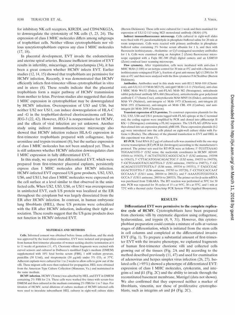
HCMV infection did not downregulate surface expression ofclass I MHC molecules in EVT. EVT express the class I MHCmolecules HLA-C, -E, and -G but not HLA-A and -B (13). Itwas previously shown by an indirect immunofluorescencemethod that HCMV infection suppresses expression ofHLA-G in cultured first-trimester villous cytotrophoblasts (8).We first examined the expression of class I MHC molecules inHCMV-infected EVT by a similar method. EVT were infectedat an MOI of 5, and their expression of the class I MHCmolecules was examined using the MAb W6/32 that recognizesHLA-A, -B, -C, -E, and -G. Use of a relatively high MOIensured that most cells (�95%) were infected with HCMV(Fig. 3C). As shown in Fig. 4A, HCMV-infected EVT ex-pressed the class I MHC molecules at a level similar to thatobserved in uninfected cells. Then, we examined the surfaceexpression of the class I MHC molecules (HLA-C, -E, and -G)by flow cytometry and found that HCMV infection did notaffect their expression at 72 hpi (Fig. 4B). As a control exper-iment, we examined HCMV-infected HEL and confirmed thatHCMV infection completely suppressed the surface expressionof the class I MHC molecules (HLA-A and -B) at 72 hpi asreported previously (4, 44, 48). We also examined class I MHCexpression at later stages of infection, and found that theexpression was maintained 7 days after HCMV infection (Fig.4C).
HCMV-infected EVT expressed US2, US3, US6, and US11mRNA. HCMV US2, US3, US6, and US11 genes are respon-
FIG. 1. Schematic view of an anchoring chorionic villus in earlypregnancy. The EVT express HLA-C, -E, and -G along the differen-tiation process from the proliferative phenotype in the cell column(CC) to the invasive phenotype in the basal plate (BP). Abbreviations:CP, chorionic plate; DC, decidual cell; FA, fetal artery; FB, fibroblast;FV, fetal vein; HC, Hofbauer cell (placental macrophage); IVS, inter-villous space; LGL: large granular lymphocyte (NK cell); UPA, utero-placental artery; UPV, uteroplacental vein; vCTB, villous cytotropho-blast; vSTB, villous syncytiotrophoblast.
FIG. 2. HCMV infection of the differentiated EVT. (A) Explant ofa first-trimester chorionic villus; (B) isolated EVT; (C) immunocyto-chemical analysis of EVT. Isolated EVT were stained with specificantibodies indicated in the figure and detected by peroxidase-conju-gated secondary antibodies and DAB. Scale bars: 10 �m (A) and 5 �m(B and C).
VOL. 77, 2003 CLASS I MHC ON HCMV-INFECTED EVT 8189
on March 11, 2018 by guest
http://jvi.asm.org/
Dow
nloaded from
sible for the downregulation of class I MHC molecules infibroblasts. Because the surface expression of class I MHCmolecules was not downregulated in HCMV-infected EVT, weexamined the expression of these US genes by RT-PCR. Total
FIG. 3. Viral gene expression and production of infectious virionsin HCMV-infected EVT. (A) EVT infected with HCMV at an MOI of5. Scale bar; 5 �m. (B) Expression of HCMV genes in infected EVT.EVT were infected with HCMV at an MOI of 5, and the expression ofimmediate-early (IE1/IE2), early (UL112-113), and early-late (pp65)proteins was examined by indirect immunofluorescence microscopy
FIG. 4. Expression of class I MHC molecules in HCMV-infectedEVT. (A) Indirect immunofluorescence analysis. Expression of class IMHC molecules in HCMV-infected EVT. EVT were infected withHCMV at an MOI of 5 and examined at 72 hpi using the MAb W6/32,which reacts with HLA-A, -B, -C, -E, and -G. (B) Flow cytometricanalysis of the surface expression of class I MHC molecules at 72 hpi.EVT express HLA-C, -E, and -G. HEL express HLA-A and -B. Theseclass I MHC molecules of HCMV-infected cells (thick line) and mock-infected cells (thin line) were detected using the MAb W6/32 or iso-type control (dotted line). (C) Expression of class I MHC molecules inHCMV-infected EVT on later days. The flow cytometry of EVT wasperformed for 7 days after infection. Shaded curve, W6/32 antibody;open curve, isotype control antibody.
(C) Expression of IE1/IE2, UL112-113, and pp65 in HCMV-infectedEVT and HEL. EVT and HEL were infected with HCMV at an MOIof 1 or 5 and were examined by the indirect immunofluorescencemethod at 24, 48, and 72 hpi. (D) Production of infectious virions inEVT and HEL. The single-step growth curve of HCMV in EVT andHEL after infection of HCMV at an MOI of 5.
8190 TERAUCHI ET AL. J. VIROL.
on March 11, 2018 by guest
http://jvi.asm.org/
Dow
nloaded from
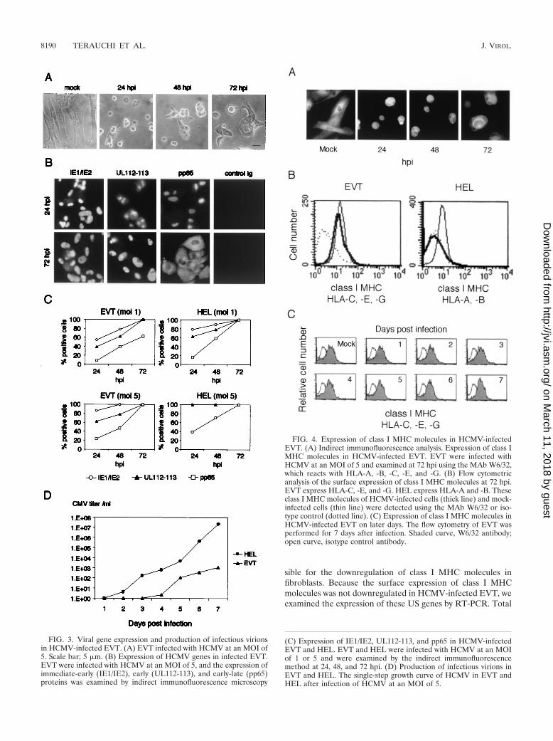
RNA was prepared from mock- or HCMV-infected EVT at 1,2, and 5 dpi. US2, US3, US6, and US11 mRNA were detectedin HCMV-infected EVT as early as 1 dpi (data not shown) andtheir expression was maintained by 5 dpi (Fig. 5). There wereno differences in the expression of these US gene mRNAs inHCMV-infected EVT and HEL.
ER relocated toward the cell membrane in HCMV-infectedEVT. US2, US3, US6, and US11 were previously shown tofunction at the ER (2, 3, 16, 46). ER, the polygonal lattice ofinterconnected membrane tubules and cisternae, associateswith microtubules and behaves dynamically in living cells (25,30). Because the ER structure in HCMV-infected cells waslargely unknown, we examined the subcellular localization ofER in HCMV-infected EVT by using a MAb against calnexin,an ER-resident chaperone (Fig. 6A). In uninfected EVT, theER was distributed in the entire cytoplasm, and the ER net-work was well developed. HCMV infection, surprisingly, in-duced a relocation of the ER toward the cell membrane at 24hpi in all infected cells we examined (100%). We also noticedthat ER tubules, which were abundant in uninfected cells,mostly disappeared in the HCMV-infected cells. We also ex-amined the cells using the antibody against calreticulin locatedin the ER lumen and confirmed that HCMV infection inducedthe relocation of ER (Fig. 6A). As a control experiment, weexamined the ER structure of HCMV-infected HEL andfound that it did not relocate toward either the cell membraneor the nucleus. Relocation of the ER was not a fixation artifactbecause it was observed by examining unfixed living EVT usingan ER-specific dye, ER-Tracker (data not shown). These re-sults indicate that the EVT ER and the HEL ER behavedifferently in response to HCMV infection.
US2, US3, US6, and US11 gene products were displacedfrom ER in HCMV-infected EVT. US2, US3, US6, and US11gene products are type I membrane proteins whose integrationwith the ER membrane is essential for their functions. Toknow the localization of these US proteins in EVT, we ex-pressed the FLAG-tagged versions, US2F, US3F, US6F, andUS11F, and examined their subcellular localization using theanti-FLAG peptide antibody. The ER of these transfected cellswas also detected with the anticalnexin antibody to examinetheir colocalization with US proteins. As shown in Fig. 6B,
US2F, US3F, US6F, and US11F were located at the ER in theentire cytoplasm of most transfected cells, and the structure ofthe ER showed no differences from that of the vector-trans-fected EVT. US3F induced small aggregates of ER at theperinuclear domain, but most of the protein was localized atthe ER network. Furthermore, ectopic expression of these USgenes did not induce the ER relocation, which was observedafter HCMV infection. We also examined the expression ofcytokeratin in these transfected cells and confirmed that theUS-expressing cells were EVT. These results indicated that theUS gene products associated with the ER but did not affecttheir subcellular localization. We next examined the localiza-tion of the US proteins in HCMV-infected EVT (Fig. 6C). Atfirst we introduced the expression plasmid for US2F, US3F,US6F, and US11F into EVT and then infected these cells withHCMV at 24 h after the transfection. The localization of theER and US gene products in these cells was examined at 24hpi. As described above, HCMV infection induced a relocationof the ER toward the cell membrane, but significant amountsof US2F, US3F, US6F, and US11F proteins were detected inthe middle of the cytoplasm where the ER was mostly absent.Different localization of the US proteins and the ER wasevident in most of the plasmid-transfected cells (73 to 83%).We also examined the localization of the ER of these cellsusing anticalreticulin antibody and confirmed that the ER wasabsent in the middle of the cytoplasm (Fig. 6D). These resultsindicated that these US gene products were displaced from theER in HCMV-infected EVT.
US2, US3, US6, and US11 proteins were colocalized with ERin HCMV-infected HEL. As a control experiment, we ex-pressed US2F, US3F, US6F, and US11F and examined theirlocalization in HEL, in which class I MHC is known to bedownregulated by HCMV infection. In the uninfected HEL,US2F, US3F, and US11F were colocalized with the ER in theperinuclear domain (Fig. 7A). Overexpression of these USproteins resulted in the aggregation of ER at the perinuclearregion because it was not observed in HCMV-infected HEL(Fig. 6A). US6F also colocalized with the ER, but it did notinduce aggregation. We then examined the localization of theFLAG-tagged US proteins in the plasmid-transfected HELafter subsequent HCMV infection (Fig. 7B). In contrast toEVT, these US proteins were colocalized with the ER in mostof the HCMV-infected HEL (77 to 86%), indicating that theywere tightly associated with or integrated into the ER mem-brane.
DISCUSSION
In early pregnancy, differentiated EVT invade the basalplate of a placenta to replace maternal spiral arteries andprotect the embryo from maternal immune cells in the decidua(10). For this specialized purpose, EVT express class I MHCHLA-C, -E, and -G, which protect them from attack by NKcells, the principal component of decidual leukocytes. In thisstudy we demonstrated that EVT are resistant to the HCMV-mediated downregulation of these class I MHC molecules (Fig.4).
We showed that EVT continued to express class I MHCmolecules after HCMV infection. However, this result doesnot imply that the HLA-C, -E, and -G molecules themselves
FIG. 5. Expression of US2, US3, US6, and US11 mRNA inHCMV-infected EVT and HEL. Total RNA (0.2 �g) was preparedfrom mock- or HCMV-infected (MOI � 5) cells and examined byRT-PCR.
VOL. 77, 2003 CLASS I MHC ON HCMV-INFECTED EVT 8191
on March 11, 2018 by guest
http://jvi.asm.org/
Dow
nloaded from

8192 TERAUCHI ET AL. J. VIROL.
on March 11, 2018 by guest
http://jvi.asm.org/
Dow
nloaded from

are resistant to the HCMV-mediated downregulation, becausethe surface expression of HLA-C and -G was suppressed byUS3 and US6 in JEG-3 cells (22). The difference between HELand EVT in expression levels of US2, US3, US6, and US11mRNA was too small to account for the resistance of EVT tothe downregulation of class I MHC (Fig. 5). It is thus likely thatthese US proteins are expressed but do not function in EVT.US6 inhibits the TAP-mediated peptide translocation to ER(3, 16, 29), and US3 prevents the transport of assembledMHC-antigen complexes (2, 20). US2 and US11 mediate arapid dislocation of MHC molecules from the ER to the cyto-plasm (21, 46). All these US products are type I transmem-brane glycoproteins, their integration into the ER membranebeing essential for their functions. In this study, we found thatthese US gene products were largely displaced from ER in
HCMV-infected EVT (Fig. 6C and D). This indicates that theproducts do not effectively associate with or integrate into theER, thus allowing the surface expression of class I MHC mol-ecules.
In a previous study suggesting downregulation of HLA-G inHCMV-infected cytotrophoblasts, cells were prepared with en-zymatic digestion of chorionic villi (8). We examined differen-tiated EVT that had grown out of villous explants by immu-nofluorescence microscopy and flow cytometer analysis andfound that HCMV infection did not reduce expression of classI MHC molecules (Fig. 4). It has been widely accepted thatEVT differentiate through several stages after leaving the cellcolumns (10). The EVT located on and near the basal laminashow a proliferative phenotype, and those left there graduallybecome invasive. It is likely that the cytotrophoblasts prepared
FIG. 7. Subcellular localization of ER in HCMV-infected HEL. (A) Localization of FLAG-tagged US proteins in HEL was examined. HELwere transfected with the expression plasmids for FLAG-tagged US proteins, i.e., US2F, US3F, US6F, and US11F, and FLAG-tagged proteinswere detected at 24 h after transfection. (B) Localization of FLAG-tagged US proteins in HCMV-infected HEL was examined at 24 hpi. HEL weretransfected with the expression plasmids for FLAG-tagged US proteins as described for panel A and then infected with HCMV at an MOI of 5at 24 h after transfection. The localization of FLAG-tagged proteins and the ER was detected using anti-FLAG antibody and anticalnexin antibody.These cells were examined using a confocal laser scanning microscope. DIC; differential interference contrast microscopy. Scale bar, 10 mm.
FIG. 6. Subcellular localization of ER in HCMV-infected EVT. (A) The localization of ER in EVT and HEL was examined using anticalnexinantibody and anticalreticulin antibody. DNA was stained with Hoechst 33258. Cells were infected at an MOI of 5 and examined at 24 hpi.(B) Localization of FLAG-tagged US proteins in EVT was examined. EVT were transfected with the expression plasmids for FLAG-tagged USproteins, US2F, US3F, US6F, and US11F. The localization of FLAG-tagged proteins was detected at 24 h after transfection using anti-FLAGantibody. The localization of the ER in the same cells was examined with anticalnexin antibody. (C) Localization of FLAG-tagged US proteinsand ER (calnexin) in HCMV-infected EVT was examined. EVT were transfected with the expression plasmids for FLAG-tagged US proteins andthen infected with HCMV at an MOI of 5 at 24 h after transfection. The localization of FLAG-tagged proteins was detected at 24 hpi usinganti-FLAG antibody. The localization of the ER in the same cells was examined with anticalnexin antibody. (D) Localization of FLAG-tagged USproteins and ER (calreticulin) in HCMV-infected EVT. EVT were transfected with the expression plasmids and then infected with HCMV asdescribed in Fig. 6C. The localization of FLAG-tagged proteins and the ER were detected at 24 hpi using anti-FLAG antibody and anticalreticulinantibody. All cells were examined using a confocal laser scanning microscope. DIC, differential interference contrast microscopy. Scale bar, 10 �m.
VOL. 77, 2003 CLASS I MHC ON HCMV-INFECTED EVT 8193
on March 11, 2018 by guest
http://jvi.asm.org/
Dow
nloaded from

by enzymatic digestion include a wide spectrum of cells atvarious stages, to which the difference of MHC class I sensi-tivity may be attributed. We noticed that the morphology wasalso significantly different among these trophoblast cells. Thecytotrophoblasts and JEG-3 cells sensitive to the MHC down-regulation show a typical polygonal morphology of epithelialcells with a small cytoplasm and a large nucleus. In contrast,the differentiated EVT used in our study have a large andextended cytoplasm relative to the nucleus (Fig. 2 and 6A). Asubset of cytotrophoblasts, presumably with the proliferativephenotype, may be sensitive to the HCMV-mediated MHCdownregulation, but the differentiated EVT are not. Althoughwe demonstrated that the total amount of the class I MHCmolecules in EVT was not affected by HCMV infection, we didnot use specific antibodies against either HLA-C, -E, or -G inthis study. Therefore, it is possible that the ratio of HLA-C, -E,or -G molecules expressed in EVT may change after HCMVinfection.
The ER is known to associate directly with microtubules butnot with actin filaments (25, 30, 43). Disruption of microtu-bules by nocodazole induces retraction of the ER networktoward the cell center, indicating that the ER is delivered bymicrotubule-dependent machinery. Overexpression of theUS2, US3, or US11 gene in HEL induced an aggregation of theER (Fig. 7A) similar to that caused by microtubule disruption(43), suggesting that an excess of US proteins affects the in-teraction between the ER and microtubules. Interestingly,HCMV infection of EVT induced ER relocation toward thecell membrane in the opposite direction to that induced by theUS proteins in HEL (Fig. 6A). Microtubules are oriented withtheir minus ends anchored in the centrosome and their plusends toward the cell membrane, and they transport membranevesicles and organelles in either direction using motor proteinssuch as kinesin and dynein. Therefore, EVT ER may associatewith microtubules via molecular machinery different from thatused in fibroblasts.
The first-trimester EVT infected with HCMV face maternalNK cells, which dominate the leukocytes of decidua by 70%(10). If HCMV downregulates HLA-C, -E, and -G on EVT,the infected cells should be targets of decidual NK cells, andthis may trigger a full-scale response of the maternal immunesystem against the placenta. Therefore, the persistent expres-sion of these MHC molecules in HCMV-infected EVT mayhelp the virus to survive in the fetus and the placenta. Expres-sion of HLA-G was first detected in EVT (28) and then wasalso discovered in monocytes where HCMV established a la-tent and persistent infection (49). Interestingly, HLA-G is up-regulated during HCMV reactivation in macrophages differ-entiated from latently infected monocytes (35, 36). Theseresults suggest that HCMV may have adapted itself to survivein HLA-G-expressing cells, taking advantage of HLA-G toevade the host immune system.
ACKNOWLEDGMENTS
We are grateful to Kwangseog Ahn for US2, US3, US6, and US11cDNAs, to Kazuo Ichimiya for his kind cooperation, and to membersof T. Aso’s and Y. Yamanashi’s laboratories for discussions.
This work is supported by a grant-in-aid for scientific research of theJapan Society for the Promotion of Science (JSPS).
REFERENCES
1. Ahn, J.-H., W.-J. Jang, and G. S. Hayward. 1999. The human cytomegalo-virus IE2 and UL112-113 proteins accumulate in viral DNA replicationcompartments that initiate from the periphery of promyelocytic leukemiaprotein-associated nuclear bodies (PODs or ND10). J. Virol. 73:10458–10471.
2. Ahn, K., A. Angulo, P. Ghazal, P. A. Petersen, Y. Yang, and K. Fruh. 1996.Human cytomegalovirus inhibits antigen presentation by a sequential mul-tistep process. Proc. Natl. Acad. Sci. USA 93:10990–10995.
3. Ahn, K., A. Gruhler, B. Galocha, T. R. Jones, E. J. H. J. Wiertz, H. L. Ploegh,P. A. Petersen, Y. Yang, and K. Fruh. 1997. The ER-luminal domain of theHCMV glycoprotein US6 inhibits peptide translocation by TAP. Immunity6:613–621.
4. Beersma, M. F. C., M. J. E. Bijlmakers, and H. L. Ploegh. 1993. Humancytomegalovirus down-regulates HLA class I expression by reducing thestability of class I H chains. J. Immunol. 151:4455–4464.
5. Borrego, F., M. Ulbrecht, E. H. Weiss, J. E. Coligan, and A. G. Brooks. 1998.Recognition of human histocompatibility leukocyte antigen (HLA)-E com-plexed with HLA class I signal sequence-derived peptides by CD94/NKG2confers protection from natural killer cell-mediated lysis. J. Exp. Med. 187:813–818.
6. Castellucci, M., and P. Kaufmann. 1999. Basic structure of the villous trees,p. 50–115. In K. Benirschke and P. Kaufmann (ed.), Pathology of the humanplacenta, 4th ed. Springer-Verlag, New York, N.Y.
7. Crisa, L., M. T. McMaster, J. K. Ishii, S. J. Fisher, and D. R. Salomon. 1997.Identification of a thymic epithelial cell subset sharing expression of the classIb HLA-G molecule with fetal trophoblasts. J. Exp. Med. 186:289–298.
8. Fisher, S., O. Genbacev, E. Maidji, and L. Pereira. 2000. Human cytomeg-alovirus infection of placental cytotrophoblasts in vitro and in utero: impli-cations for transmission and pathogenesis. J. Virol. 74:6808–6820.
9. Fisher, S. J., T.-Y. Cui, L. Zhang, L. Hartman, K. Grahl, Z. Guo-Yang, J.Tarpey, and C. H. Damsky. 1989. Adhesive and degradative properties ofhuman placental cytotrophoblast cells in vitro. J. Cell Biol. 109:891–902.
10. Frank, H. G., and P. Kaufmann. 1999. Nonvillous parts and trophoblastinvasion, p. 171–272. In K. Benirschke and P. Kaufmann (ed.), Pathology ofthe human placenta, 4th ed. Springer-Verlag, New York, N.Y.
11. Graham, C. H., T. S. Hawley, R. G. Hawley, J. R. MacDougall, R. S. Kerbel,N. Khoo, and P. K. Lala. 1993. Establishment and characterization of firsttrimester human trophoblast cells with extended lifespan. Exp. Cell Res.206:204–211.
12. Halwachs-Baumann, G., M. Wilders-Truschnig, G. Desoye, T. Hahn, L.Kiesel, K. Klingel, P. Rieger, G. Jahn, and C. Singer. 1998. Human tropho-blast cells are permissive to the complete replicative cycle of human cyto-megalovirus. J. Virol. 72:7598–7602.
13. Hammer, A., H. Hutter, and G. Dohr. 1997. HLA class I expression on thematerno-fetal interface. Am. J. Reprod. Immunol. 38:150–157.
14. Hemmings, D. G., R. Kilani, C. Nykiforuk, J. Preiksaitis, and L. J. Guilbert.1998. Permissive cytomegalovirus infection of primary villous term and firsttrimester trophoblasts. J. Virol. 72:4970–4979.
15. Hemmings, D. G., and L. J. Guilbert. 2002. Polarized release of humancytomegalovirus from placental trophoblasts. J. Virol. 76:6710–6717.
16. Hengel, H., J. Koopmann, T. Flohr, W. Muranyi, E. Goulmy, G. J. Ham-merling, U. H. Koszinowski, and F. Momburg. 1997. A viral ER-residentglycoprotein inactivates the MHC-encoded peptide transporter. Immunity6:623–632.
17. Hunt, J. S., J. L. Fishback, G. Chumbley, and Y. W. Loke. 1990. Identifica-tion of class I MHC mRNA in human first trimester trophoblast cells by insitu hybridization. J. Immunol. 144:4420–4425.
18. Hunt, J. S., B.-L. Hsi, C. R. King, and J. L. Fishback. 1991. Detection of classI MHC mRNA in subpopulations of first trimester cytotrophoblast cells by insitu hybridization. J. Reprod. Immunol. 19:315–323.
19. Iwayama, S., T. Yamamoto, T. Furuya, R. Kobayashi, K. Ikuta, and K. Hirai.1994. Intracellular localization and DNA-binding activity of a class of viralearly phosphoproteins in human fibroblasts infected with human cytomega-lovirus (Towne strain). J. Gen. Virol. 75:3309–3318.
20. Jones, T. R., E. J. H. J. Wiertz, L. Sun, K. N. Fish, J. A. Nelson, and H. L.Ploegh. 1996. Human cytomegalovirus US3 impairs transport and matura-tion of major histocompatibility complex. Proc. Natl. Acad. Sci. USA 93:11327–11333.
21. Jones, T. R., and L. Sun. 1997. Human cytomegalovirus US2 destabilizesmajor histocompatibility complex class I heavy chains. J. Virol. 71:2970–2979.
22. Jun, Y., E. Kim, M. Jin, H. C. Sung, H. Han, D. E. Geraghty, and K. Ahn.2000. Human cytomegalovirus gene products US3 and US6 down-regulatetrophoblast class I MHC molecules. J. Immunol. 164:805–811.
23. King, A., C. Boocock, A. M. Sharkey, L. Gardner, A. Beretta, A. G. Siccardi,and Y. W. Loke. 1996. Evidence for the expression of HLA A-C class ImRNA and protein by human first trimester trophoblast. J. Immunol. 156:2068–2076.
24. King, A., D. S. J. Allan, M. Brown, S. J. Powis, S. Joseph, S. Verma, S. E.Hiby, A. J. McMichael, Y. W. Loke, and V. M. Braud. 2000. HLA-E is
8194 TERAUCHI ET AL. J. VIROL.
on March 11, 2018 by guest
http://jvi.asm.org/
Dow
nloaded from

expressed on trophoblast and interacts with CD94/NKG2 receptors on de-cidual NK cells. Eur. J. Immunol. 30:1623–1631.
25. Klopfenstein, D. R. C., F. Keppeler, and H.-P. Hauri. 1998. A novel directinteraction of endoplasmic reticulum with microtubules. EMBO J. 17:6168–6177.
26. Koi, H., J. Zhang, A. Makrigiannakis, S. Getsios, C. D. MacCalman, G. S.Kopf, J. F. Strauss III, and S. Parry. 2001. Differential expression of thecoxsackievirus and adenovirus receptor regulates adenovirus infection of theplacenta. Biol. Reprod. 64:1001–1009.
27. Koi, H., J. Zhang, A. Makrigiannakis, S. Getsios, C. D. MacCalman, J. F.Strauss III, and S. Parry. 2002. Syncytiotrophoblast is a barrier to maternal-fetal transmission of herpes simplex virus. Biol. Reprod. 67:1572–1579.
28. Kovats, S., E. K. Main, C. Librach, M. Stubblebine, S. J. Fisher, and R.DeMars. 1990. A class I antigen, HLA-G, expressed in human trophoblasts.Science 248:220–223.
29. Kyritsis, C., S. Gorbulev, S. Hutschenreiter, K. Pawlitschko, R. Abele, and R.Tampe. 2001. Molecular mechanism and structural aspects of transporterassociated with antigen processing inhibition by the cytomegalovirus proteinUS6. J. Biol. Chem. 276:48031–48039.
30. Lee, C., and L. B. Chen. 1988. Dynamic behavior of endoplasmic reticulumin living cells. Cell 54:37–46.
31. Librach, C. L., Z. Werb, M. L. Fitzgerald, K. Chiu, N. M. Corwin, R. A.Esteves, D. Grobelny, R. Galardy, C. H. Damsky, and S. J. Fisher. 1991.92-kD type IV collagenase mediates invasion of human cytotrophoblasts.J. Cell Biol. 113:437–449.
32. McMaster, M. T., C. L. Librach, Y. Zhou, K.-H. Lim, M. J. Janatpour, R.DeMars, S. Kovats, C. Damsky, and S. J. Fisher. 1995. Human placentalHLA-G expression is restricted to differentiated cytotrophoblasts. J. Immu-nol. 154:3771–3778.
33. Munz, C., N. Holmes, A. King, Y. W. Loke, M. Colonna, H. Schild, and H.-G.Rammensee. 1997. Human histocompatibility leukocyte antigen (HLA)-Gmolecules inhibit NKAT3 expressing natural killer cells. J. Exp. Med. 185:385–391.
34. Norwitz, E. R., D. J. Schust, and S. J. Fisher. 2001. Implantation and thesurvival of early pregnancy. N. Engl. J. Med. 345:1400–1408.
35. Onno, M., G. Le Friec, C. Pangault, L. Amiot, V. Guilloux, B. Drenou, S.Caulet-Maugendre, P. Andre, and R. Fauchet. 2000. Modulation of HLA-Gantigens expression in myelomonocytic cells. Hum. Immunol. 61:1086–1094.
36. Onno, M., C. Pangault, G. Le Friec, V. Guilloux, P. Andre, and R. Fauchet.2000. Modulation of HLA-G antigens expression by human cytomegalovirus:specific induction in activated macrophages harboring human cytomegalovi-rus infection. J. Immunol. 164:6426–6434.
37. Paul, P., N. Rouas-Freiss, I. Khalil-Daher, P. Moreau, B. Riteau, F. A. LeGal, M. F. Avril, J. Dausset, J. G. Guillet, and E. D. Carosella. 1998. HLA-Gexpression in melanoma: a way for tumor cells to escape from immunosur-veillance. Proc. Natl. Acad. Sci. USA 95:4510–4515.
38. Ponte, M., C. Cantoni, R. Biassoni, A. Tradori-Cappai, G. Bentivoglio, C.
Vitale, S. Bertone, A. Moretta, L. Moretta, and M. C. Mingari. 1999. Inhib-itory receptors sensing HLA-G1 molecules in pregnancy: decidua-associatednatural killer cells express LIR-1 and CD94/NKG2A and acquire p49, anHLA-G1-specific receptor. Proc. Natl. Acad. Sci. USA 96:5674–5679.
39. Rehm, A., A. Engelsberg, D. Tortorella, I. J. Korner, I. Lehmann, H. L.Ploegh, and U. E. Hopken. 2002. Human cytomegalovirus gene products US2and US11 differ in their ability to attack major histocompatibility class Iheavy chains in dendritic cells. J. Virol. 76:5043–5050.
40. Rouas-Freiss, N., R. M.-B. Goncalves, C. Menier, J. Dausset, and E. D.Carosella. 1997. Direct evidence to support the role of HLA-G in protectingthe fetus from maternal uterine natural killer cytolysis. Proc. Natl. Acad. Sci.USA 94:11520–11525.
41. Sanchez, V., K. D. Greis, E. Sztul, and W. J. Britt. 2000. Accumulation ofvirion tegument and envelope proteins in a stable cytoplasmic compartmentduring human cytomegalovirus replication: characterization of a potentialsite of virus assembly. J. Virol. 74:975–986.
42. Schust, D. J., D. Tortorella, J. Seebach, C. Phan, and H. L. Ploegh. 1998.Trophoblast class I major histocompatibility complex (MHC) products areresistant to rapid degradation imposed by the human cytomegalovirus(HCMV) gene products US2 and US11. J. Exp. Med. 188:497–503.
43. Terasaki, M., L. B. Chen, and K. Fujiwara. 1986. Microtubules and theendoplasmic reticulum are highly interdependent structures. J. Cell Biol.103:1557–1568.
44. Warren, A. P., D. H. Ducroq, P. J. Lehner, and L. K. Borysiewicz. 1994.Human cytomegalovirus-infected cells have unstable assembly of major his-tocompatibility complex class I complexes and are resistant to lysis by cyto-toxic T lymphocytes. J. Virol. 68:2822–2829.
45. Wiendl, H., M. Mitsdoerffer, V. Hofmeister, J. Wischhusen, A. Bornemann,R. Meyermann, E. H. Weiss, A. Melms, and M. Weller. 2002. A functionalrole of HLA-G expression in human gliomas: an alternative strategy ofimmune escape. J. Immunol. 168:4772–4780.
46. Wiertz, E. J. H. J., T. R. Jones, L. Sun, M. Bogyo, H. J. Geuze, and H. L.Ploegh. 1996. The human cytomegalovirus US11 gene product dislocatesMHC class I heavy chains from the endoplasmic reticulum to the cytosol.Cell 84:769–779.
47. Yagel, S., R. F. Casper, W. Powell, R. S. Parhar, and P. K. Lala. 1989.Characterization of pure human first-trimester cytotrophoblast cells in long-term culture: growth pattern, markers, and hormone production. Am. J.Obstet. Gynecol. 160:938–945.
48. Yamashita, Y., K. Shimokata, S. Saga, S. Mizuno, T. Tsurumi, and Y.Nishiyama. 1994. Rapid degradation of the heavy chain of class I majorhistocompatibility complex antigens in the endoplasmic reticulum of humancytomegalovirus-infected cells. J. Virol. 68:7933–7943.
49. Yang, Y., W. Chu, D. E. Geraghty, and J. S. Hunt. 1996. Expression ofHLA-G in human mononuclear phagocytes and selective induction byIFN-. J. Immunol. 156:4224–4231.
VOL. 77, 2003 CLASS I MHC ON HCMV-INFECTED EVT 8195
on March 11, 2018 by guest
http://jvi.asm.org/
Dow
nloaded from