Placement & Management: JACKSON-PRATT CLOSED ACTIVE ... · JACKSON-PRATT DRAINS A Jackson-Pratt...
7
PRACTICE STEP BY STEP Peer Reviewed 52 TODAY’S VETERINARY PRACTICE | An Official Journal of the NAVC | November/December 2016 | tvpjournal.com Drains are placed in wounds to evacuate fluid that would otherwise accumulate due to dead space, inflammation, infection, and/or necrosis. Fluid accu- mulation in a wound can be detrimental because it: • Acts as a medium for bacteria • Separates tissues that need to heal together • Creates pressure that can cause pain and decrease local blood flow. 1 CONSIDERATIONS Drains do not replace debridement and lavage, and wounds that contain bacteria, foreign material, or tissues that might necrose are preferably left open and bandaged with moisture retentive dressings rather than being prematurely closed over a drain. 2 Drains should be used sparingly in neoplastic sites due to the potential of tumor cell tracking along the drain. If a drain must be placed after tumor resection, it should be exited close to the wound edge at a site that could be readily resected or radiated if clean margins were not obtained. 1,3 JACKSON-PRATT DRAINS A Jackson-Pratt drain (JPD) is an active suction drain consisting of a fenestrated, flexible, silicone tube connected by a nonfenestrated tube to a compressible reservoir (called a grenade due to its shape) (Figure 1, page 54). The fenestrated end of a JPD (which is placed in the wound bed) is available in a range of widths and may be flat or round. The fenestrations direct fluid into the nonfenestrated portion of the tubing and then into the grenade. The grenade has a one- way valve to prevent retrograde movement of the fluid. Common grenade sizes range from 100 mL to 400 mL. Placement & Management: JACKSON-PRATT CLOSED ACTIVE SUCTION DRAIN Bonnie Grambow Campbell, DVM, PhD, Diplomate ACVS Washington State University Learn More Read Moist Wound Healing: The New Standard of Care in the September/ October 2015 issue of Today’s Veterinary Practice, available at tvpjournal.com. Troubleshooting If the JPD will not hold suction, make sure: • The grenade is securely attached to the tube • The evacuation port is closed • There are no holes in the external tubing (eg, from patient chewing or from the needle used for the purse- string suture). If the incision itself is not airtight, add sutures or staples or cover it with a dressing +/- sterile ointment. The incision should become more airtight as the fibrin clot in the wound edge becomes more organized. A JPD can become obstructed with blood clots, fibrin, pus, or tissue, although due to the large number of holes, this is unusual. If there is an obstruction: • Make sure the bulldog clamp is open and the tube is not kinked. • Empty and then reactivate the grenade to maximize the amount of suction and determine if the improved suction dislodges the obstruction. A tube that remains obstructed may be flushed with sterile saline using sterile technique. This should only be done after carefully considering whether keeping the drain functional is more important than the potential risk for introducing contaminants into the wound when flushing. Drains that are not functioning should be removed, as their presence may increase the amount of wound fluid and risk for infection. 4,5
Transcript of Placement & Management: JACKSON-PRATT CLOSED ACTIVE ... · JACKSON-PRATT DRAINS A Jackson-Pratt...
PRACTICE STEP BY STEPPeer Reviewed
52 TODAY’S VETERINARY PRACTICE | An Official Journal of the NAVC | November/December 2016 | tvpjournal.com
Drains are placed in wounds to evacuate fluid that would otherwise accumulate due to dead space, inflammation, infection, and/or necrosis. Fluid accu-mulation in a wound can be detrimental because it: • Acts as a medium for bacteria• Separates tissues that need to heal together• Creates pressure that can cause pain and decrease
local blood flow.1
CONSIDERATIONSDrains do not replace debridement and lavage, and wounds that contain bacteria, foreign material, or tissues that might necrose are preferably left open and bandaged with moisture retentive dressings rather than being prematurely closed over a drain.2
Drains should be used sparingly in neoplastic sites due to the potential of tumor cell tracking along the drain. If a drain must be placed after
tumor resection, it should be exited close to the wound edge at a site that could be readily resected or radiated if clean margins were not obtained.1,3
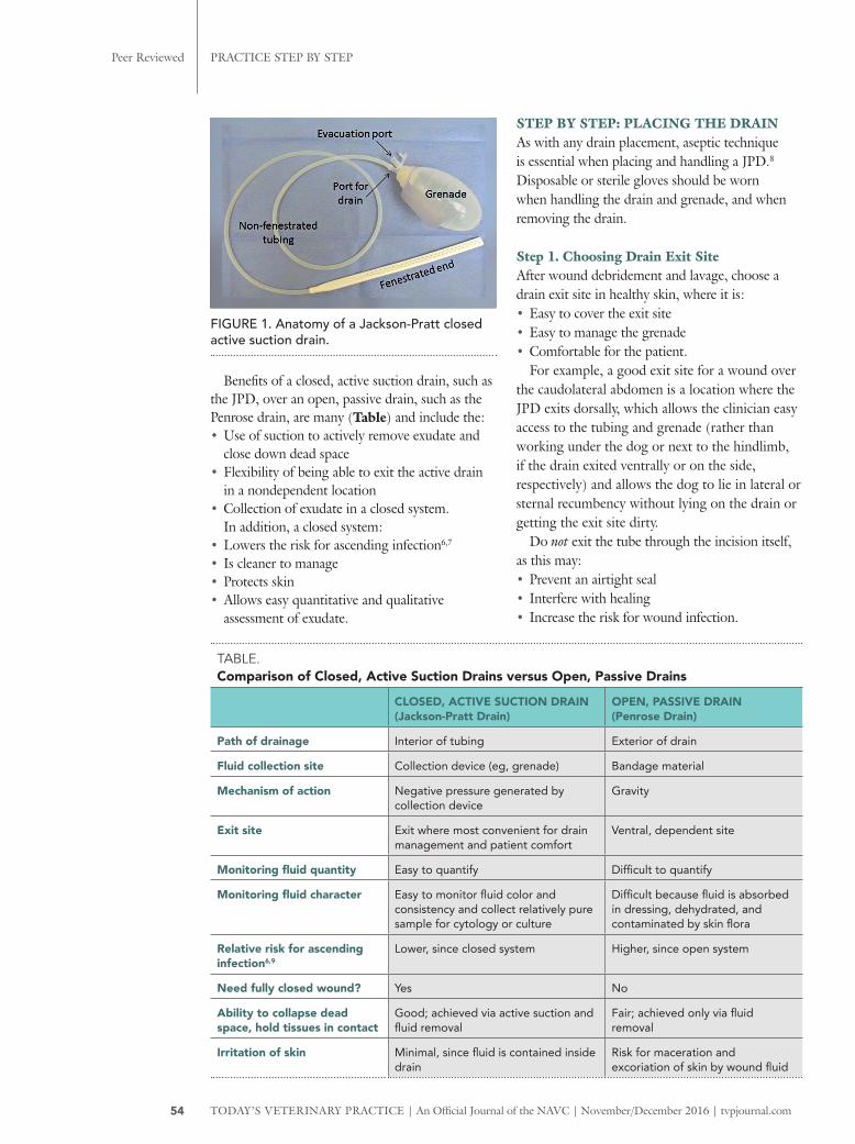
JACKSON-PRATT DRAINSA Jackson-Pratt drain (JPD) is an active suction drain consisting of a fenestrated, flexible, silicone tube connected by a nonfenestrated tube to a compressible reservoir (called a grenade due to its shape) (Figure 1, page 54).
The fenestrated end of a JPD (which is placed in the wound bed) is available in a range of widths and may be flat or round. The fenestrations direct fluid into the nonfenestrated portion of the tubing and then into the grenade. The grenade has a one-way valve to prevent retrograde movement of the fluid. Common grenade sizes range from 100 mL to 400 mL.
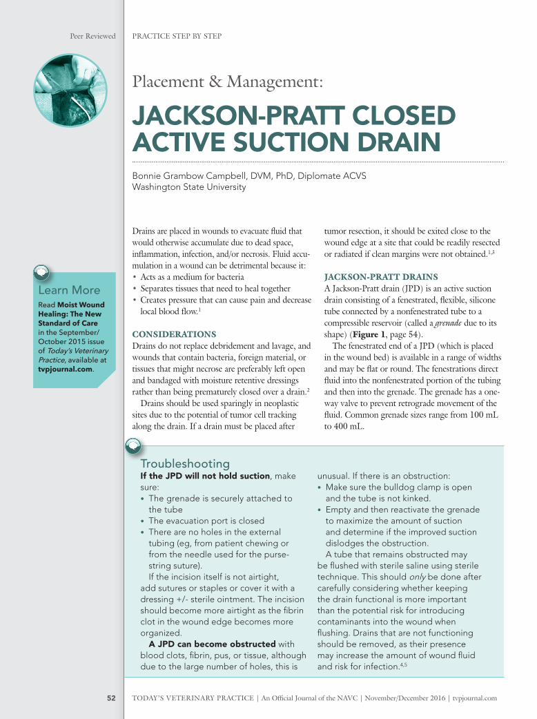
Placement & Management:
JACKSON-PRATT CLOSED ACTIVE SUCTION DRAINBonnie Grambow Campbell, DVM, PhD, Diplomate ACVS Washington State University
Learn MoreRead Moist Wound Healing: The New Standard of Care in the September/October 2015 issue of Today’s Veterinary Practice, available at tvpjournal.com.
Troubleshooting If the JPD will not hold suction, make sure:• The grenade is securely attached to
the tube• The evacuation port is closed• There are no holes in the external
tubing (eg, from patient chewing or from the needle used for the purse-string suture).If the incision itself is not airtight,
add sutures or staples or cover it with a dressing +/- sterile ointment. The incision should become more airtight as the fibrin clot in the wound edge becomes more organized.
A JPD can become obstructed with blood clots, fibrin, pus, or tissue, although due to the large number of holes, this is
unusual. If there is an obstruction:• Make sure the bulldog clamp is open
and the tube is not kinked.• Empty and then reactivate the grenade
to maximize the amount of suction and determine if the improved suction dislodges the obstruction.A tube that remains obstructed may
be flushed with sterile saline using sterile technique. This should only be done after carefully considering whether keeping the drain functional is more important than the potential risk for introducing contaminants into the wound when flushing. Drains that are not functioning should be removed, as their presence may increase the amount of wound fluid and risk for infection.4,5
PRACTICE STEP BY STEPPeer Reviewed
54 TODAY’S VETERINARY PRACTICE | An Official Journal of the NAVC | November/December 2016 | tvpjournal.com
Benefits of a closed, active suction drain, such as the JPD, over an open, passive drain, such as the Penrose drain, are many (Table) and include the:• Use of suction to actively remove exudate and
close down dead space• Flexibility of being able to exit the active drain
in a nondependent location• Collection of exudate in a closed system.
In addition, a closed system:• Lowers the risk for ascending infection6,7
• Is cleaner to manage• Protects skin• Allows easy quantitative and qualitative
assessment of exudate.
STEP BY STEP: PLACING THE DRAINAs with any drain placement, aseptic technique is essential when placing and handling a JPD.8 Disposable or sterile gloves should be worn when handling the drain and grenade, and when removing the drain.
Step 1. Choosing Drain Exit SiteAfter wound debridement and lavage, choose a drain exit site in healthy skin, where it is:• Easy to cover the exit site• Easy to manage the grenade• Comfortable for the patient.
For example, a good exit site for a wound over the caudolateral abdomen is a location where the JPD exits dorsally, which allows the clinician easy access to the tubing and grenade (rather than working under the dog or next to the hindlimb, if the drain exited ventrally or on the side, respectively) and allows the dog to lie in lateral or sternal recumbency without lying on the drain or getting the exit site dirty.
Do not exit the tube through the incision itself, as this may:• Prevent an airtight seal• Interfere with healing• Increase the risk for wound infection.
TABLE. Comparison of Closed, Active Suction Drains versus Open, Passive Drains
CLOSED, ACTIVE SUCTION DRAIN(Jackson-Pratt Drain)
OPEN, PASSIVE DRAIN (Penrose Drain)
Path of drainage Interior of tubing Exterior of drain
Fluid collection site Collection device (eg, grenade) Bandage material
Mechanism of action Negative pressure generated by collection device
Gravity
Exit site Exit where most convenient for drain management and patient comfort
Ventral, dependent site
Monitoring fluid quantity Easy to quantify Difficult to quantify
Monitoring fluid character Easy to monitor fluid color and consistency and collect relatively pure sample for cytology or culture
Difficult because fluid is absorbed in dressing, dehydrated, and contaminated by skin flora
Relative risk for ascending infection6,9
Lower, since closed system Higher, since open system
Need fully closed wound? Yes No
Ability to collapse dead space, hold tissues in contact
Good; achieved via active suction and fluid removal
Fair; achieved only via fluid removal
Irritation of skin Minimal, since fluid is contained inside drain
Risk for maceration and excoriation of skin by wound fluid
FIGURE 1. Anatomy of a Jackson-Pratt closed active suction drain.
PRACTICE STEP BY STEP Peer Reviewed
55tvpjournal.com | November/December 2016 | An Official Journal of the NAVC | TODAY’S VETERINARY PRACTICE
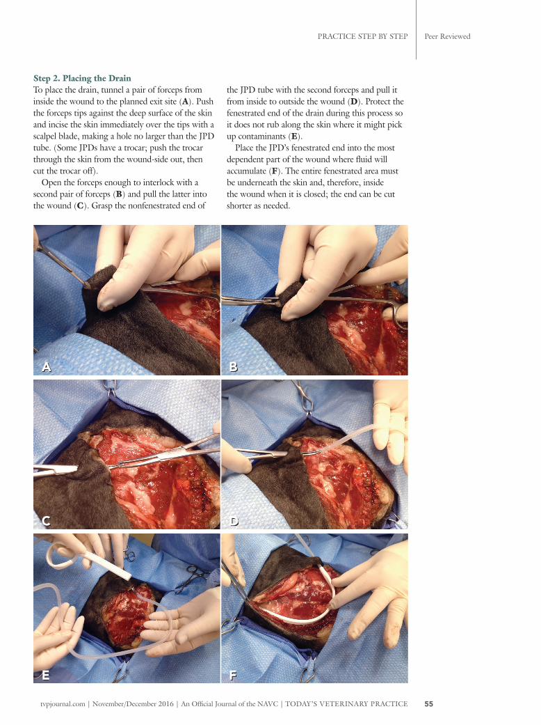
Step 2. Placing the Drain To place the drain, tunnel a pair of forceps from inside the wound to the planned exit site (A). Push the forceps tips against the deep surface of the skin and incise the skin immediately over the tips with a scalpel blade, making a hole no larger than the JPD tube. (Some JPDs have a trocar; push the trocar through the skin from the wound-side out, then cut the trocar off).
Open the forceps enough to interlock with a second pair of forceps (B) and pull the latter into the wound (C). Grasp the nonfenestrated end of
the JPD tube with the second forceps and pull it from inside to outside the wound (D). Protect the fenestrated end of the drain during this process so it does not rub along the skin where it might pick up contaminants (E).
Place the JPD’s fenestrated end into the most dependent part of the wound where fluid will accumulate (F). The entire fenestrated area must be underneath the skin and, therefore, inside the wound when it is closed; the end can be cut shorter as needed.
A B
C D
E F
PRACTICE STEP BY STEPPeer Reviewed
56 TODAY’S VETERINARY PRACTICE | An Official Journal of the NAVC | November/December 2016 | tvpjournal.com
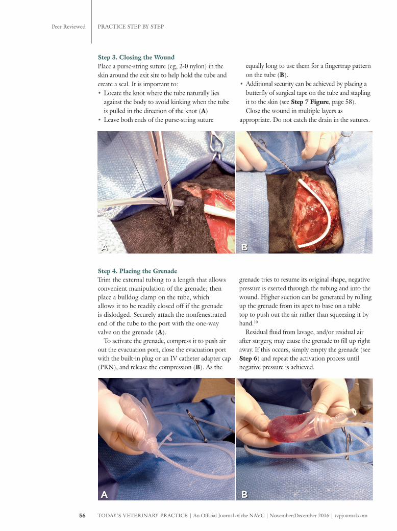
Step 3. Closing the Wound Place a purse-string suture (eg, 2-0 nylon) in the skin around the exit site to help hold the tube and create a seal. It is important to:• Locate the knot where the tube naturally lies
against the body to avoid kinking when the tube is pulled in the direction of the knot (A)
• Leave both ends of the purse-string suture
equally long to use them for a fingertrap pattern on the tube (B).
• Additional security can be achieved by placing a butterfly of surgical tape on the tube and stapling it to the skin (see Step 7 Figure, page 58).Close the wound in multiple layers as
appropriate. Do not catch the drain in the sutures.
A B
Step 4. Placing the Grenade Trim the external tubing to a length that allows convenient manipulation of the grenade; then place a bulldog clamp on the tube, which allows it to be readily closed off if the grenade is dislodged. Securely attach the nonfenestrated end of the tube to the port with the one-way valve on the grenade (A).
To activate the grenade, compress it to push air out the evacuation port, close the evacuation port with the built-in plug or an IV catheter adapter cap (PRN), and release the compression (B). As the
grenade tries to resume its original shape, negative pressure is exerted through the tubing and into the wound. Higher suction can be generated by rolling up the grenade from its apex to base on a table top to push out the air rather than squeezing it by hand.10
Residual fluid from lavage, and/or residual air after surgery, may cause the grenade to fill up right away. If this occurs, simply empty the grenade (see Step 6) and repeat the activation process until negative pressure is achieved.
A B
PRACTICE STEP BY STEP Peer Reviewed
57tvpjournal.com | November/December 2016 | An Official Journal of the NAVC | TODAY’S VETERINARY PRACTICE
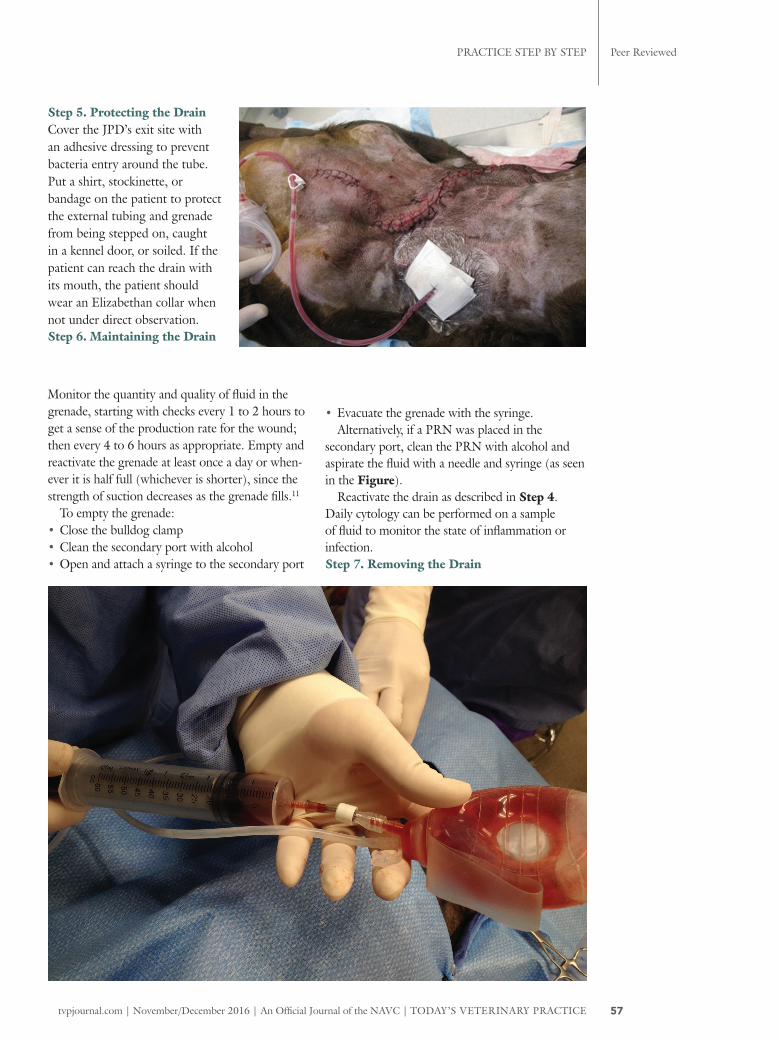
Step 5. Protecting the Drain Cover the JPD’s exit site with an adhesive dressing to prevent bacteria entry around the tube. Put a shirt, stockinette, or bandage on the patient to protect the external tubing and grenade from being stepped on, caught in a kennel door, or soiled. If the patient can reach the drain with its mouth, the patient should wear an Elizabethan collar when not under direct observation.Step 6. Maintaining the Drain
Monitor the quantity and quality of fluid in the grenade, starting with checks every 1 to 2 hours to get a sense of the production rate for the wound; then every 4 to 6 hours as appropriate. Empty and reactivate the grenade at least once a day or when-ever it is half full (whichever is shorter), since the strength of suction decreases as the grenade fills.11
To empty the grenade: • Close the bulldog clamp• Clean the secondary port with alcohol• Open and attach a syringe to the secondary port
• Evacuate the grenade with the syringe. Alternatively, if a PRN was placed in the
secondary port, clean the PRN with alcohol and aspirate the fluid with a needle and syringe (as seen in the Figure).
Reactivate the drain as described in Step 4. Daily cytology can be performed on a sample of fluid to monitor the state of inflammation or infection.Step 7. Removing the Drain
PRACTICE STEP BY STEPPeer Reviewed
58 TODAY’S VETERINARY PRACTICE | An Official Journal of the NAVC | November/December 2016 | tvpjournal.com
It is difficult to establish rules on when to remove a drain because fluid production varies with the wound’s etiology, dimensions, location, and degree of inflammation or infection.1,10 Because the drain itself stimulates some fluid production, the amount of drainage is not expected to reach zero.
When a wound is healing, the: • Fluid becomes serosanguineous• Volume decreases to a steady state• Cytology shows resolving inflammation and
improved cell health.All these signs indicate that it is time to remove
the drain. The risk for seroma formation was higher
in human and canine patients that were still producing relatively large amounts of wound fluid when the JPD was removed compared with patients in which the wound fluid production had steadily decreased.8,9
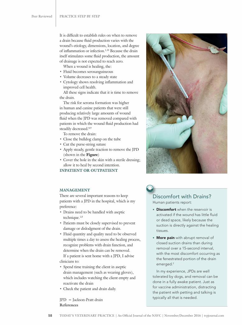
To remove the drain:• Close the bulldog clamp on the tube• Cut the purse-string suture• Apply steady, gentle traction to remove the JPD
(shown in the Figure) • Cover the hole in the skin with a sterile dressing;
allow it to heal by second intention.INPATIENT OR OUTPATIENT
Discomfort with Drains? Human patients report:
• Discomfort when the reservoir is activated if the wound has little fluid or dead space, likely because the suction is directly against the healing tissues.
• More pain with abrupt removal of closed suction drains than during removal over a 15-second interval, with the most discomfort occurring as the fenestrated portion of the drain emerged.2
In my experience, JPDs are well tolerated by dogs, and removal can be done in a fully awake patient. Just as for vaccine administration, distracting the patient with petting and talking is typically all that is needed.
MANAGEMENTThere are several important reasons to keep patients with a JPD in the hospital, which is my preference: • Drains need to be handled with aseptic
technique.1,8 • Patients must be closely supervised to prevent
damage or dislodgment of the drain. • Fluid quantity and quality need to be observed
multiple times a day to assess the healing process, recognize problems with drain function, and determine when the drain can be removed.If a patient is sent home with a JPD, I advise
clinicians to:• Spend time training the client in aseptic
drain management (such as wearing gloves), which includes watching the client empty and reactivate the drain
• Check the patient and drain daily.
JPD = Jackson-Pratt drainReferences
PRACTICE STEP BY STEP
59tvpjournal.com | November/December 2016
1. Campbell BG. Bandages and drains. In Tobias M, Johnson SA (eds): Veterinary Surgery Small Animal, 1st ed. St. Louis: Elsevier Saunders, 2012, pp 221-230.
2. Campbell BG. Moist wound healing: The new standard of care. Today’s Vet Pract 2015; 5(4):32-42.
3. Lascelles BDX. Strategic planning in oncological surgery. British Small Animal Veterinary Congress Proc, 2007, Belfast, Ireland.
4. Isik O, Kaya E, Dundar HZ, Sarkut P. Surgical site infection: Re-assessment of the risk factors. Chirurgia 2015; 110(5):457-461.
5. Bristow PC, Halfacree ZJ, Baines SJ. A retrospective study of the use of active suction wound drains in dogs and cats. J Sm Anim Prac 2015; 56(5):325-330.
6. Raves JJ, Slifkin M, Diamond DL. A bacteriologic study comparing closed suction and simple conduit drainage. Am J Surg 1984; 148(5):618-620.
7. Reiffel AJ, Barie PS, Spector JA. A multi-disciplinary review of the potential association between closed-suction drains and surgical site infection. Surg Infect 2013; 14(3):244-269.
8. Chim JH, Borsting EA, Thaller SR. Urban myths in plastic surgery: Postoperative management of surgical drains. Wounds 2016; 28(2):35-39.
9. Shaver SL, Hunt GB, Kidd SW. Evaluation of fluid production and seroma formation after placement of closed suction drains in clean subcutaneous surgical wounds of dogs: 77 cases (2005-2012). JAVMA 2014; 245(2):211-215.
10.Halfacree ZJ, Wilson AM, Baines SJ. Evaluation of in vitro performance of suction drains. Am J Vet Res 2009; 70(2):283-289.
11.Williams J, Toews D, Prince M. Survey of the use of suction drains in head and neck surgery and analysis of their biomechanical properties. J Otolaryngol 2003; 32(1):16-22.
BONNIE GRAMBOW CAMPBELLBonnie Grambow Campbell, DVM, PhD, Diplomate ACVS, is a clinical associate professor of small animal surgery at Washington State Uni-versity. Dr. Campbell received her DVM and PhD from Cornell Univer-sity and completed a small animal surgical residency at University of Wisconsin. She has received a number of teaching awards and speaks at continuing education programs nationally and internationally. Her special clinical interests include wound management and oncologic and reconstructive surgery.