Pituitary Self-priming Actions of Gonadotropin … Self-priming Actionsof Gonadotropin-releasing...
8
Pituitary Self-priming Actions of Gonadotropin-releasing Hormone Kinetics of Estradiol's Potentiating Effects on Gonadotropin-releasing Hormone-Facilitated Hormone and Follicle-stimulating Hormone Release in Healthy Postmenopausal Women I Luteinizing Johannes D. Veldhuis, William S. Evans, Alan D. Rogol, Lisa Kolp, Michael 0. Thomer, and Paul Stumpf* Departments of Internal Medicine, Pharmacology, Pediatrics, and Obstetrics and Gynecology, University of Virginia School ofMedicine, Charlottesville, Virginia 22908; and *Department of Obstetrics and Gynecology, Jersey Shore Medical Center, Neptune, New Jersey 07753 Abstract We examined the kinetically distinct characteristics of estradiol's effects upon pituitary luteinizing hormone (LH) and follicle- stimulating hormone (FSH) release in response to pulses of ex- ogenous gonadotropin-releasing hormone (GnRH) in healthy postmenopausal individuals. The putative self-priming actions of GnRH on LH and FSH release were tested by intravenous injections of equal paired doses of GnRH (10 jig) before and after 1, 5, 10, and 30 d of pure estradiol-17,6 delivery via an intravaginal silastic ring. Self-priming actions of GnRH, as de- fined by heightened gonadotropin release in response to the sec- ond pulse of GnRH compared with the first, were completely absent in the hypoestrogenemic state. However, estradiol ad- ministration unmasked GnRH self-priming in a time-dependent fashion, with maximal expression after 5 and 10 d of steroid replacement, followed by attenuation by 30 d. Since estradiol's modulation of GnRH action was expressed differentially on LH and FSH release, we suggest that such facilitation of GnRH- stimulated pituitary LH and FSH release may provide an ad- ditional mechanism for dissociated secretion of gonadotropic hormones in health or disease. Introduction Under certain physiological conditions, repetitive stimulation of the anterior pituitary gland by serial pulses of gonadotropin- releasing hormone (GnRH)' results in marked potentiation of gonadotropin release (1-5). This ability of repetitive GnRH stimulation to facilitate pituitary responsiveness has been referred to as the "self-priming" action of this hypothalamic decapeptide (1). Studies in a variety of experimental animals have suggested that such amplifying effects of serial GnRH stimulation may be critically important to the genesis of the preovulatory surge-like release of gonadotropic hormones during the final stages of fol- licular development (1-8). Investigations in ovariectomized rodents subjected to various regimens of sex-steroid hormone replacement have implicated estradiol as one critical determinant of the facilitative effects of Address correspondence to Dr. Veldhuis. Receivedfor publication 14 November 1985 and in revisedform 21 January 1986. 1. Abbreviations used in this paper: FSH, follicle-stimulating hormone; GnRH, gonadotropin-releasing hormone; LH, luteinizing hormone. repetitive GnRH stimulation on pituitary responsiveness in vivo and in vitro (8-16). Similarly, short-term administration of es- trogen to postmenopausal women is accompanied by altered pituitary responsiveness to exogenously infused GnRH (17, 18). Such alterations include either inhibition or facilitation of GnRH actions (17, 18). Studies in the follicular phase of the human menstrual cycle further suggest that injected estradiol and/or one or more events associated with follicular maturation can result in either diminished or enhanced pituitary responses to exogenous GnRH stimuli (19-24). However, the ability to relate such alterations in pituitary responsiveness explicitly to estradiol is limited in gonadally intact individuals. In addition, available data have not yet delineated: (a) the kinetics of estrogen's elic- itation of self-priming actions of GnRH; (b) the extent to which acute responses to single GnRH injections and GnRH self- priming represent temporally distinct events; and (c) the differ- ential self-priming effects of GnRH on LH and FSH release. In the present study, we have used a model of physiological estradiol replacement and paired exogenous GnRH pulses to test for precise temporal correlations between circulating estradiol levels, basal gonadotropin concentrations, and the GnRH-fa- cilitated release of LH and FSH. To obviate the confounding influences of unstable serum estradiol concentrations that result after oral or intramuscular estrogen dosing, we have used an estradiol-impregnated silastic ring placed intravaginally. The latter mode of steroid-hormone delivery results in the rapid at- tainment of steady state serum estradiol concentrations com- mensurate with those of the mid-to-late follicular phase of the normal menstrual cycle. This paradigm has permitted us to elu- cidate kinetically distinct characteristics of estradiol's effects on pituitary luteinizing hormone (LH) and follicle-stimulating hor- mone (FSH) release in response to exogenous GnRH pulses in previously hypoestrogenemic postmenopausal women. Methods Vaginal rings. Estradiol-containing silastic rings were prepared exactly as described earlier, with a dose of 400 mg of estradiol-17,7 impregnated in each ring (25). Subjects. Healthy spontaneously postmenopausal women were stud- ied after provision of written informed consent, approved by the Human Investigation Committee of the University of Virginia School of Medicine. The subjects who participated in this study ranged in age from 55 to 63 yr (mean, 58±2 yr, n = eight women) and were 3-9 yr postmenopausal. Each volunteer underwent a detailed history and physical examination, with the documentation of normal hepatic, renal, and hematologic func- tion, biochemical euthyroidism, and postmenopausal concentrations of gonadotropic hormones. At least 5 wk before study, women were with- drawn from any drugs, including estrogen or sex-steroid hormone treat- ments. Blood sampling protocols. Sampling was conducted in the Clinical Research Center of the University of Virginia by withdrawing 2.5 ml Estradiol Modulates Gonadotropin-releasing Hormone Self-priming 1849 J. Clin. Invest. © The American Society for Clinical Investigation, Inc. 0021-9738/86/06/1849/08 $ 1.00 Volume 77, June 1986, 1849-1856
Transcript of Pituitary Self-priming Actions of Gonadotropin … Self-priming Actionsof Gonadotropin-releasing...
Pituitary Self-priming Actions of Gonadotropin-releasing HormoneKinetics of Estradiol's Potentiating Effects on Gonadotropin-releasing Hormone-FacilitatedHormone and Follicle-stimulating Hormone Release in Healthy Postmenopausal Women
I Luteinizing
Johannes D. Veldhuis, William S. Evans, Alan D. Rogol, Lisa Kolp, Michael 0. Thomer, and Paul Stumpf*Departments of Internal Medicine, Pharmacology, Pediatrics, and Obstetrics and Gynecology, University of Virginia School of Medicine,Charlottesville, Virginia 22908; and *Department of Obstetrics and Gynecology, Jersey Shore Medical Center, Neptune, NewJersey 07753
Abstract
Weexamined the kinetically distinct characteristics of estradiol'seffects upon pituitary luteinizing hormone (LH) and follicle-stimulating hormone (FSH) release in response to pulses of ex-ogenous gonadotropin-releasing hormone (GnRH) in healthypostmenopausal individuals. The putative self-priming actionsof GnRHon LH and FSH release were tested by intravenousinjections of equal paired doses of GnRH(10 jig) before andafter 1, 5, 10, and 30 d of pure estradiol-17,6 delivery via anintravaginal silastic ring. Self-priming actions of GnRH, as de-fined by heightened gonadotropin release in response to the sec-ond pulse of GnRHcompared with the first, were completelyabsent in the hypoestrogenemic state. However, estradiol ad-ministration unmasked GnRHself-priming in a time-dependentfashion, with maximal expression after 5 and 10 d of steroidreplacement, followed by attenuation by 30 d. Since estradiol'smodulation of GnRHaction was expressed differentially on LHand FSH release, we suggest that such facilitation of GnRH-stimulated pituitary LH and FSH release may provide an ad-ditional mechanism for dissociated secretion of gonadotropichormones in health or disease.
Introduction
Under certain physiological conditions, repetitive stimulationof the anterior pituitary gland by serial pulses of gonadotropin-releasing hormone (GnRH)' results in marked potentiation ofgonadotropin release (1-5). This ability of repetitive GnRHstimulation to facilitate pituitary responsiveness has been referredto as the "self-priming" action of this hypothalamic decapeptide(1). Studies in a variety of experimental animals have suggestedthat such amplifying effects of serial GnRHstimulation may becritically important to the genesis of the preovulatory surge-likerelease of gonadotropic hormones during the final stages of fol-licular development (1-8).
Investigations in ovariectomized rodents subjected to variousregimens of sex-steroid hormone replacement have implicatedestradiol as one critical determinant of the facilitative effects of
Address correspondence to Dr. Veldhuis.Receivedfor publication 14 November 1985 and in revisedform 21
January 1986.
1. Abbreviations used in this paper: FSH, follicle-stimulating hormone;GnRH, gonadotropin-releasing hormone; LH, luteinizing hormone.
repetitive GnRHstimulation on pituitary responsiveness in vivoand in vitro (8-16). Similarly, short-term administration of es-trogen to postmenopausal women is accompanied by alteredpituitary responsiveness to exogenously infused GnRH(17, 18).Such alterations include either inhibition or facilitation of GnRHactions (17, 18). Studies in the follicular phase of the humanmenstrual cycle further suggest that injected estradiol and/orone or more events associated with follicular maturation canresult in either diminished or enhanced pituitary responses toexogenous GnRHstimuli (19-24). However, the ability to relatesuch alterations in pituitary responsiveness explicitly to estradiolis limited in gonadally intact individuals. In addition, availabledata have not yet delineated: (a) the kinetics of estrogen's elic-itation of self-priming actions of GnRH; (b) the extent to whichacute responses to single GnRH injections and GnRHself-priming represent temporally distinct events; and (c) the differ-ential self-priming effects of GnRHon LH and FSH release.
In the present study, we have used a model of physiologicalestradiol replacement and paired exogenous GnRHpulses totest for precise temporal correlations between circulating estradiollevels, basal gonadotropin concentrations, and the GnRH-fa-cilitated release of LH and FSH. To obviate the confoundinginfluences of unstable serum estradiol concentrations that resultafter oral or intramuscular estrogen dosing, we have used anestradiol-impregnated silastic ring placed intravaginally. Thelatter mode of steroid-hormone delivery results in the rapid at-tainment of steady state serum estradiol concentrations com-mensurate with those of the mid-to-late follicular phase of thenormal menstrual cycle. This paradigm has permitted us to elu-cidate kinetically distinct characteristics of estradiol's effects onpituitary luteinizing hormone (LH) and follicle-stimulating hor-mone (FSH) release in response to exogenous GnRHpulses inpreviously hypoestrogenemic postmenopausal women.
Methods
Vaginal rings. Estradiol-containing silastic rings were prepared exactlyas described earlier, with a dose of 400 mgof estradiol-17,7 impregnatedin each ring (25).
Subjects. Healthy spontaneously postmenopausal womenwere stud-ied after provision of written informed consent, approved by the HumanInvestigation Committee of the University of Virginia School of Medicine.The subjects who participated in this study ranged in age from 55 to 63yr (mean, 58±2 yr, n = eight women) and were 3-9 yr postmenopausal.Each volunteer underwent a detailed history and physical examination,with the documentation of normal hepatic, renal, and hematologic func-tion, biochemical euthyroidism, and postmenopausal concentrations ofgonadotropic hormones. At least 5 wk before study, womenwere with-drawn from any drugs, including estrogen or sex-steroid hormone treat-ments.
Blood sampling protocols. Sampling was conducted in the ClinicalResearch Center of the University of Virginia by withdrawing 2.5 ml
Estradiol Modulates Gonadotropin-releasing Hormone Self-priming 1849
J. Clin. Invest.© The American Society for Clinical Investigation, Inc.0021-9738/86/06/1849/08 $ 1.00Volume 77, June 1986, 1849-1856
blood at 15-min intervals for 6 h beginning at 0800 h. After 2 h of basalsampling, a dose of 10 ug GnRHwas administered intravenously bybolus injection. After two more hours, a second dose of 10 ug GnRHwas given similarly. The first dose was used to appraise acute pituitaryresponsiveness. The paired pulses of GnRHwere used to test for theemergence and/or disappearance of self-priming actions of GnRH. Self-priming was defined as an increase in pituitary responsiveness to thesecond dose of GnRHcompared with the first (see below: data analysis).
Sampling was performed basally (no estrogen treatment), and ondays 1, 5, 10, and 30 after initiation of estradiol replacement.
Assays. Serum concentrations of LH and FSH were assayed in du-plicate with a dual-label radioimmunoassay (RIA) kit (Clinetics Cor-poration, Tusten, CA). The sensitivities for LH and FSHwere 1.8 mIU/ml and 1.4 mIU/ml, respectively. Samples were diluted 1:2 or 1:4 to fallwithin the least variable region of the displacement curve, where theintraassay coefficients of variation averaged 6.3-8.5%, and interassayvariability was 9-14% (LH) and 5-13% (FSH). The cross-reactions ofLH and FSHwith alpha subunit in this assay were <10%. Serum estadiollevels were quantitated by specific RIA after celite chromatography (26).
Data analyses. Data are expressed as means±SEMfor the group ofeight volunteers. Significant overall treatment effects were sought byanalysis of variance with the Newman-Keul's procedure to test for in-dividually significant effects (27). Where indicated, specific a priori com-parisons of mean, incremental, or absolute peak (maximal) gonadotropinconcentrations were made by paired two-tailed Student's t testing withBonferonni's correction (27). Fractional (percentage) increases in go-nadotropin concentrations for the first and second GnRH-stimulatedgonadotropin peaks were compared by the Wilcoxon signed rankstest (27).
To assess the possible contribution of multiple parameters to themagnitude of the gonadotropin response to the second pulse of GnRH,multivariate analysis was employed. Multiple linear regression was per-formed using the mean peak 2 gonadotropin level as the dependent vari-able, and the following individual independent variables: (a) the meanbasal serum gonadotropin concentration over the 2 h preceding GnRHadministration; (b) the mean gonadotropin response to the first GnRHpulse; and (c) the simultaneous serum estradiol concentration. In thisstatistical model, individually significant correlations as well as partialand multiple linear correlations were sought. This permitted us to evaluatethe most significant contributors to the amplitude of the gonadotropinresponse to the second GnRHpulse at various times after (or before)estradiol administration.
Results
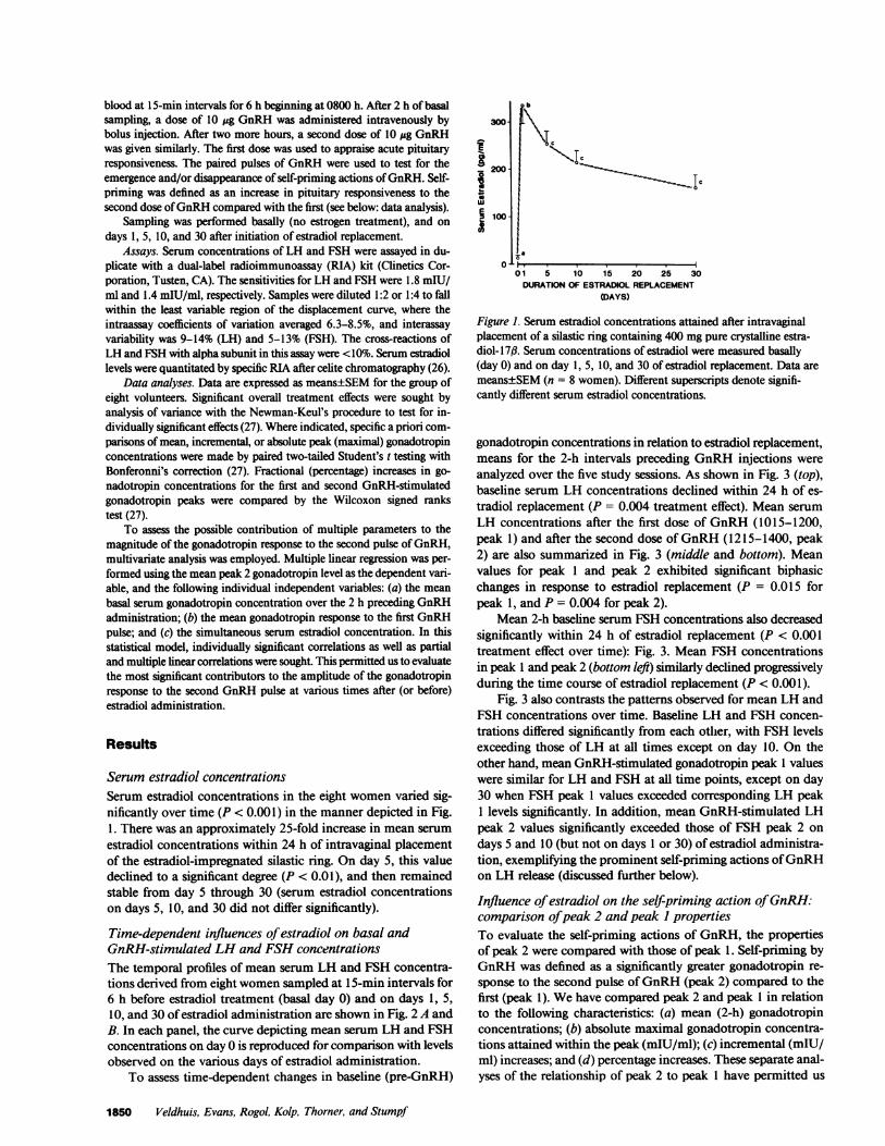
Serum estradiol concentrationsSerum estradiol concentrations in the eight women varied sig-nificantly over time (P < 0.001) in the manner depicted in Fig.1. There was an approximately 25-fold increase in mean serumestradiol concentrations within 24 h of intravaginal placementof the estradiol-impregnated silastic ring. On day 5, this valuedeclined to a significant degree (P < 0.01), and then remainedstable from day 5 through 30 (serum estradiol concentrationson days 5, 10, and 30 did not differ significantly).
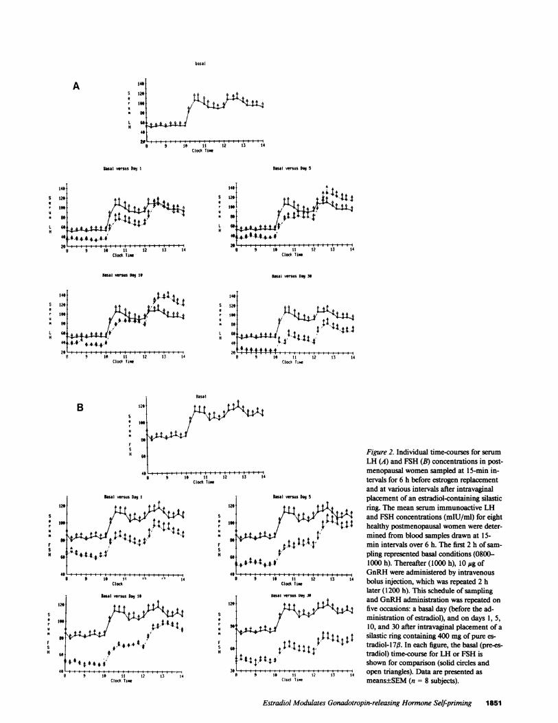
Time-dependent influences of estradiol on basal andGnRH-stimulated LH and FSHconcentrationsThe temporal profiles of mean serum LH and FSH concentra-tions derived from eight womensampled at 15-min intervals for6 h before estradiol treatment (basal day 0) and on days 1, 5,10, and 30 of estradiol administration are shown in Fig. 2 A andB. In each panel, the curve depicting mean serum LH and FSHconcentrations on day 0 is reproduced for comparison with levelsobserved on the various days of estradiol administration.
To assess time-dependent changes in baseline (pre-GnRH)
iuJ;U1
01 5 10 15 20 25 30DURATIONOF ESTRADIOL REPLACEMENT
(DAYS)
Figure 1. Serum estradiol concentrations attained after intravaginalplacement of a silastic ring containing 400 mgpure crystalline estra-diol- 1 7#. Serum concentrations of estradiol were measured basally(day 0) and on day 1, 5, 10, and 30 of estradiol replacement. Data aremeans±SEM(n = 8 women). Different superscripts denote signifi-cantly different serum estradiol concentrations.
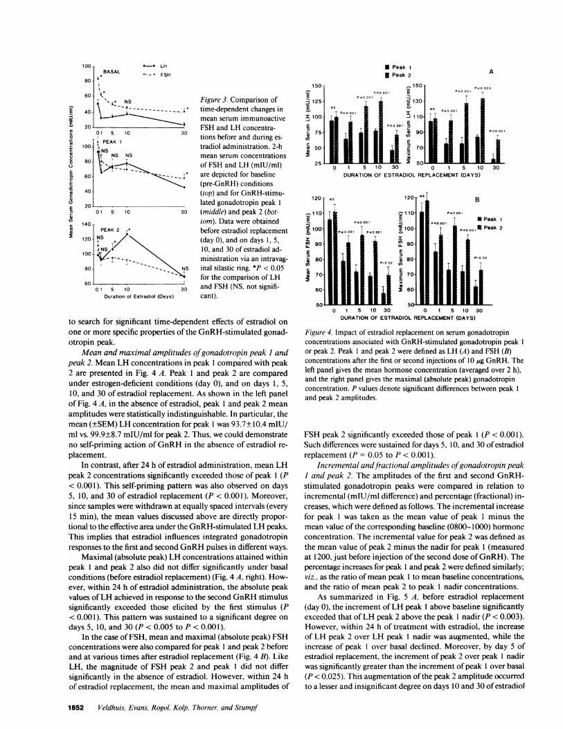
gonadotropin concentrations in relation to estradiol replacement,means for the 2-h intervals preceding GnRHinjections wereanalyzed over the five study sessions. As shown in Fig. 3 (top),baseline serum LH concentrations declined within 24 h of es-tradiol replacement (P = 0.004 treatment effect). Mean serumLH concentrations after the first dose of GnRH(1015-1200,peak 1) and after the second dose of GnRH(1215-1400, peak2) are also summarized in Fig. 3 (middle and bottom). Meanvalues for peak 1 and peak 2 exhibited significant biphasicchanges in response to estradiol replacement (P = 0.015 forpeak 1, and P = 0.004 for peak 2).
Mean 2-h baseline serum FSHconcentrations also decreasedsignificantly within 24 h of estradiol replacement (P < 0.001treatment effect over time): Fig. 3. Mean FSH concentrationsin peak 1 and peak 2 (bottom left) similarly declined progressivelyduring the time course of estradiol replacement (P < 0.001).
Fig. 3 also contrasts the patterns observed for mean LH andFSH concentrations over time. Baseline LH and FSH concen-trations differed significantly from each other, with FSH levelsexceeding those of LH at all times except on day 10. On theother hand, mean GnRH-stimulated gonadotropin peak 1 valueswere similar for LH and FSH at all time points, except on day30 when FSH peak 1 values exceeded corresponding LH peak1 levels significantly. In addition, mean GnRH-stimulated LHpeak 2 values significantly exceeded those of FSH peak 2 ondays 5 and 10 (but not on days 1 or 30) of estradiol administra-tion, exemplifying the prominent self-priming actions of GnRHon LH release (discussed further below).
Influence of estradiol on the self-priming action of GnRH:comparison of peak 2 and peak I propertiesTo evaluate the self-priming actions of GnRH, the propertiesof peak 2 were compared with those of peak 1. Self-priming byGnRHwas defined as a significantly greater gonadotropin re-sponse to the second pulse of GnRH(peak 2) compared to thefirst (peak 1). Wehave compared peak 2 and peak 1 in relationto the following characteristics: (a) mean (2-h) gonadotropinconcentrations; (b) absolute maximal gonadotropin concentra-tions attained within the peak (mIU/ml); (c) incremental (mIU/ml) increases; and (d) percentage increases. These separate anal-yses of the relationship of peak 2 to peak 1 have permitted us
1850 Veldhuis, Evans, Rogol, Kolp, Thorner, and Stumpf
b3usa
49
i29
6904.
8 9 1' I I 12 13 14Clod. Time
Basal versus Day 5
14.'S 129r 1t
L 6H
4.
140
8 9 '1'0 *11* 12 13 14Clocd Tam
l veus D 19
.4* 4. -,.4.4.-a,. ' ..,.
8 9 1i 11 12 13 14Clode Tim
A~~~~~.t4~ ~ ~
9.13
9 1.t i 2 13 14Clodc Time
Basal versus Day 39
1t
S 129
u 9
L 69 I'1it4, I' .428
B 9 14 11 12 13 14Clock Tame
B 129
1
H 60
41
Basal
IL.g
e 9 1i t1 12Clock Taim
Busal versus Dao I
. *4IJ,.''*+t,t t1 S'
S
ru
S
8 9 ie 1 ' ,"l 14clock
S
H
Figure 2. Individual time-courses for serumLH (A) and FSH (B) concentrations in post-menopausal womensampled at 15-min in-
13 14 tervals for 6 h before estrogen replacementand at various intervals after intravaginal
BI vMus D9 5 placement of an estradiol-containing silastic129 t t t ffi ring. The mean serum immunoactive LH
199 u> Y ; g: and FSH concentrations (mIU/ml) for eight/NA t tt healthy postmenopausal womenwere deter-99 t mined from blood samples drawn at 15-
min intervals over 6 h. The first 2 h of sam-9, ; pling represented basal conditions (0800-
+ 0 S.4.t-t IOM1000 h). Thereafter (1000 h), 10 Ag of4C ! | 1§ &"@ |GnRH were administered by intravenous
Clocd Timw bolus injection, which was repeated 2 hlater (1200 h). This schedule of sampling
12a v and GnRHadministration was repeated onfive occasions: a basal day (before the ad-ministration of estradiol), and on days 1, 5,
9.' 10, and 30 after intravaginal placement of asilastic ring containing 400 mg of pure es-
69 : X t t l l:tradiol- 1 7,. In each figure, the basal (pre-es-tradiol) time-course for LH or FSH is
14tt | t4 S shown for comparison (solid circles and3 9 19 11 1? 13 14 open triangles). Data are presented asClol Tame means±SEM(n = 8 subjects).
Estradiol Modulates Gonadotropin-releasing Hormone Self-priming 1851
Ai
u'H
&Bsal versus Dag 1
14
5 129
r 19u
L 6CH
4
49,
129
S
C 199ru
of
H ,
41
129S
ua 199
SH t
8
100- 0*- LHBASAL *- -* FSH
80-
60-'
I ,,j NS
40- \ _ - .
20-01 5 10 30
. PEAK 1100-
NS NS80-
60-
40-
20-01 5 10 30
140PEAK 2
120- NSI \
100- '''''''' \
80--- NS
6001 5 10 30
Duration of Estradiol (Days)
* Peak 1
X Peak 2
Figure 3. Comparison oftime-dependent changes inmean serum immunoactiveFSH and LH concentra-tions before and during es-
tradiol administration. 2-hmean serum concentrationsof FSH and LH (mIU/ml)are depicted for baseline(pre-GnRH) conditions(top) and for GnRH-stimu-lated gonadotropin peak 1
(middle) and peak 2 (bot-tom). Data were obtainedbefore estradiol replacement(day 0), and on days 1, 5,10, and 30 of estradiol ad-ministration via an intravag-inal silastic ring. *P < 0.05for the comparison of LHand FSH (NS, not signifi-cant).
A
150 _150- P<0_1 P<0 00 E P< 001oE P<0.000
o125 1 2130
ERNS OSE<TI R E (
~P<0,, 000I1ilOE ~~~~~~P<0000
50-70
0 1 5 1 0 30 0 1 5 1 0 30
DURATIONOF ESTRADIOL REPLACEMENT(DAYS)
to search for significant time-dependent effects of estradiol on
one or more specific properties of the GnRH-stimulated gonad-otropin peak.
Mean and maximal amplitudes of gonadotropin peak I andpeak 2. Mean LH concentrations in peak 1 compared with peak2 are presented in Fig. 4 A. Peak 1 and peak 2 are comparedunder estrogen-deficient conditions (day 0), and on days 1, 5,10, and 30 of estradiol replacement. As shown in the left panelof Fig. 4 A, in the absence of estradiol, peak 1 and peak 2 mean
amplitudes were statistically indistinguishable. In particular, themean (±SEM) LH concentration for peak 1 was 93.7±10.4 mIU/ml vs. 99.9±8.7 mIU/ml for peak 2. Thus, we could demonstrateno self-priming action of GnRHin the absence of estradiol re-
placement.In contrast, after 24 h of estradiol administration, mean LH
peak 2 concentrations significantly exceeded those of peak 1 (P< 0.001). This self-priming pattern was also observed on days5, 10, and 30 of estradiol replacement (P < 0.001). Moreover,since samples were withdrawn at equally spaced intervals (every15 min), the mean values discussed above are directly propor-
tional to the effective area under the GnRH-stimulated LH peaks.This implies that estradiol influences integrated gonadotropinresponses to the first and second GnRHpulses in different ways.
Maximal (absolute peak) LH concentrations attained withinpeak 1 and peak 2 also did not differ significantly under basalconditions (before estradiol replacement) (Fig. 4 A, right). How-ever, within 24 h of estradiol administration, the absolute peakvalues of LH achieved in response to the second GnRHstimulussignificantly exceeded those elicited by the first stimulus (P< 0.001). This pattern was sustained to a significant degree on
days 5, 10, and 30 (P < 0.005 to P < 0.001).In the case of FSH, mean and maximal (absolute peak) FSH
concentrations were also compared for peak 1 and peak 2 beforeand at various times after estradiol replacement (Fig. 4 B). LikeLH, the magnitude of FSH peak 2 and peak 1 did not differsignificantly in the absence of estradiol. However, within 24 hof estradiol replacement, the mean and maximal amplitudes of
0 1 5 10 30 0 1 5 10 30DURATIONOF ESTRADIOL REPLACEMENT(DAYS)
Figure 4. Impact of estradiol replacement on serum gonadotropinconcentrations associated with GnRH-stimulated gonadotropin peak 1
or peak 2. Peak 1 and peak 2 were defined as LH (A) and FSH (B)concentrations after the first or second injections of 0,g GnRH. Theleft panel gives the mean hormone concentration (averaged over 2 h),and the right panel gives the maximal (absolute peak) gonadotropinconcentration. P values denote significant differences between peak 1
and peak 2 amplitudes.
FSH peak 2 significantly exceeded those of peak 1 (P < 0.001).Such differences were sustained for days 5, 10, and 30 of estradiolreplacement (P = 0.05 to P < 0.001).
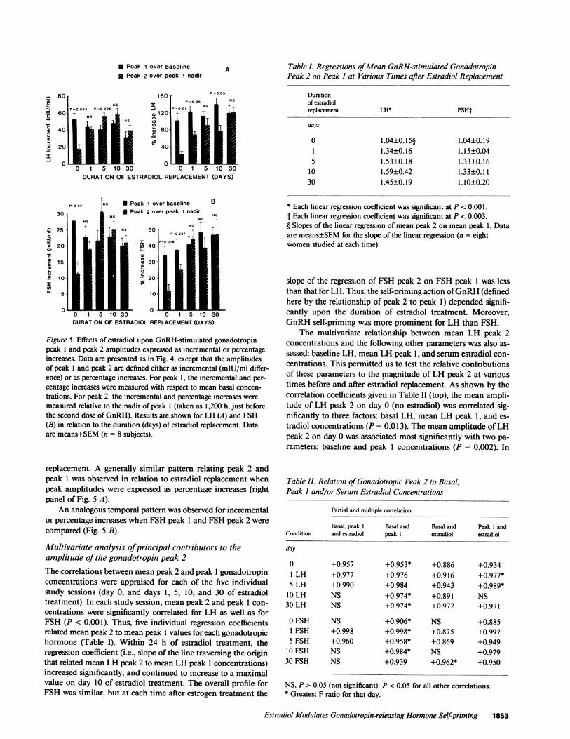
Incremental andJfractional amplitudes of gonadotropin peakI and peak 2. The amplitudes of the first and second GnRH-stimulated gonadotropin peaks were compared in relation toincremental (mIU/ml difference) and percentage (fractional) in-creases, which were defined as follows. The incremental increasefor peak 1 was taken as the mean value of peak 1 minus themean value of the corresponding baseline (0800-1000) hormoneconcentration. The incremental value for peak 2 was defined as
the mean value of peak 2 minus the nadir for peak 1 (measuredat 1200, just before injection of the second dose of GnRH). Thepercentage increases for peak 1 and peak 2 were defined similarly;viz., as the ratio of mean peak 1 to mean baseline concentrations,and the ratio of mean peak 2 to peak 1 nadir concentrations.
As summarized in Fig. 5 A, before estradiol replacement(day 0), the increment of LH peak 1 above baseline significantlyexceeded that of LH peak 2 above the peak 1 nadir (P < 0.003).However, within 24 h of treatment with estradiol, the increaseof LH peak 2 over LH peak 1 nadir was augmented, while theincrease of peak 1 over basal declined. Moreover, by day 5 ofestradiol replacement, the increment of peak 2 over peak 1 nadirwas significantly greater than the increment of peak 1 over basal(P < 0.025). This augmentation of the peak 2 amplitude occurredto a lesser and insignificant degree on days 10 and 30 of estradiol
1852 Veldhuis, Evans, Rogol, Kolp, Thorner, and Stumpf
E0
U)c
0
coI
cx00
c0
E
c
a
* Peak 1 over baseline
X Peak 2 over peak 1 nadirA
160 P.0.05NS N'°S T NS
003 PO0O05OT ]P 0.05
NS
01200NS co
h~~~aR 40
1 5 10 30 0 1 5 10 30DURATIONOF ESTRADIOL REPLACEMENT(DAYS)
* Peak 1 over baseline
v Peak 2 over peak 1 nadir
B
- 25E
E 20
150
u
.' 10
coU.
0 1 5 10 30 0 1 5 10 30DURATIONOF ESTRADIOL REPLACEMENT(DAYS)
Figure 5. Effects of estradiol upon GnRH-stimulated gonadotropinpeak 1 and peak 2 amplitudes expressed as incremental or percentageincreases. Data are presented as in Fig. 4, except that the amplitudesof peak 1 and peak 2 are defined either as incremental (mIU/ml differ-ence) or as percentage increases. For peak 1, the incremental and per-centage increases were measured with respect to mean basal concen-
trations. For peak 2, the incremental and percentage increases were
measured relative to the nadir of peak 1 (taken as 1,200 h, just beforethe second dose of GnRH). Results are shown for LH (A) and FSH(B) in relation to the duration (days) of estradiol replacement. Dataare means+SEM(n = 8 subjects).
replacement. A generally similar pattern relating peak 2 andpeak 1 was observed in relation to estradiol replacement whenpeak amplitudes were expressed as percentage increases (rightpanel of Fig. 5 A).
An analogous temporal pattern was observed for incrementalor percentage increases when FSHpeak 1 and FSHpeak 2 were
compared (Fig. 5 B).
Multivariate analysis of principal contributors to theamplitude of the gonadotropin peak 2The correlations between mean peak 2 and peak 1 gonadotropinconcentrations were appraised for each of the five individualstudy sessions (day 0, and days 1, 5, 10, and 30 of estradioltreatment). In each study session, mean peak 2 and peak 1 con-
centrations were significantly correlated for LH as well as forFSH (P < 0.001). Thus, five individual regression coefficientsrelated mean peak 2 to mean peak I values for each gonadotropichormone (Table I). Within 24 h of estradiol treatment, theregression coefficient (i.e., slope of the line traversing the originthat related mean LH peak 2 to mean LH peak 1 concentrations)increased significantly, and continued to increase to a maximalvalue on day 10 of estradiol treatment. The overall profile forFSH was similar, but at each time after estrogen treatment the
Table I. Regressions of Mean GnRH-stimulated GonadotropinPeak 2 on Peak I at Various Times after Estradiol Replacement
Durationof estradiolreplacement LH* FSHt
days
0 1.04±0.15§ 1.04±0.191 1.34±0.16 1.15±0.045 1.53±0.18 1.33±0.16
10 1.59±0.42 1.33±0.1130 1.45±0.19 1.10±0.20
* Each linear regression coefficient was significant at P < 0.001.t Each linear regression coefficient was significant at P < 0.003.§ Slopes of the linear regression of mean peak 2 on mean peak 1. Dataare means±SEMfor the slope of the linear regression (n = eightwomen studied at each time).
slope of the regression of FSH peak 2 on FSH peak 1 was lessthan that for LH. Thus, the self-priming action of GnRH(definedhere by the relationship of peak 2 to peak 1) depended signifi-cantly upon the duration of estradiol treatment. Moreover,GnRHself-priming was more prominent for LH than FSH.
The multivariate relationship between mean LH peak 2concentrations and the following other parameters was also as-sessed: baseline LH, meanLH peak 1, and serum estradiol con-centrations. This permitted us to test the relative contributionsof these parameters to the magnitude of LH peak 2 at varioustimes before and after estradiol replacement. As shown by thecorrelation coefficients given in Table II (top), the mean ampli-tude of LH peak 2 on day 0 (no estradiol) was correlated sig-nificantly to three factors: basal LH, mean LH peak 1, and es-tradiol concentrations (P = 0.013). The mean amplitude of LHpeak 2 on day 0 was associated most significantly with two pa-rameters: baseline and peak 1 concentrations (P = 0.002). In
Table II. Relation of Gonadotropic Peak 2 to Basal,Peak I and/or Serum Estradiol Concentrations
Partial and multiple correlation
Basal, peak I Basal and Basal and Peak I andCondition and estradiol peak I estradiol estradiol
day
0 +0.957 +0.953* +0.886 +0.9341 LH +0.977 +0.976 +0.916 +0.977*5 LH +0.990 +0.984 +0.943 +0.989*
10 LH NS +0.974* +0.891 NS30 LH NS +0.974* +0.972 +0.971
0 FSH NS +0.906* NS +0.8851 FSH +0.998 +0.998* +0.875 +0.9975 FSH +0.960 +0.958* +0.869 +0.949
10 FSH NS +0.984* NS +0.97930 FSH NS +0.939 +0.962* +0.950
NS, P> 0.05 (not significant): P < 0.05 for all other correlations.* Greatest F ratio for that day.
Estradiol Modulates Gonadotropin-releasing Hormone Self-priming 1853
E
aE
c
I-J
contrast, after 1 and 5 d of estradiol administration, the ampli-tude of LH peak 2 was correlated most significantly to meanLH peak 1 and serum estradiol concentrations (P < 0.001). Ondays 10 and 30 of estradiol administration, peak 2 was describedbest by its relationship to baseline and LH peak 1 concentrations(P = 0.003). Thus, the statistical correlates of the mean amplitudeof LH peak 2 varied in a distinctive manner over time, withpeak 1 amplitude and serum estradiol concentrations being mostinfluential on days 1 and 5, while baseline and peak 1 concen-trations were most contributory on days 0, 10, and 30 of estrogenadministration.
The preceding overall pattern for LH differed from that ob-served in the case of FSH (bottom, Table II), since basal FSHand mean FSHpeak 1 concentrations provided the best statisticalcorrelates of the amplitude of FSH peak 2 for days 0, 1, 5, and10 after estradiol (P = 0.014 to P< 0.001). By day 30 of estradiol,baseline FSHand the serum estradiol concentrations representedthe most significant correlates of mean FSHpeak 2 levels. Theseanalyses indicate that the correlates of LH and FSH responsive-ness to paired GnRHpulses are distinctively influenced by therelative and time-dependent contributions of baseline gonado-tropin concentrations, mean peak 1 gonadotropin concentra-tions, and concurrent estradiol levels.
Influence of estradiol on total incremental gonadotropinrelease in response to paired exogenous GnRHpulsesThe time-dependent effects of estradiol on total GnRH-promotedgonadotropin release were estimated by determining the sum ofpeak 1 and peak 2 increments over basal before and at varioustimes after estradiol administration. Such estimates of total in-cremental LH and FSH release are given in Table III for days0, 1, 5, 10, and 30 of study. For both LH and FSH, total incre-mental gonadotropin release was relatively reduced on day 30.In contrast, on day 10 of estradiol treatment, the total incre-mental value for GnRH-stimulated LH release was significantlygreater than that on day 0 (pre-estrogen) or day 30. Similarly,for FSH, the sum of the peak 1 and peak 2 increments on days5 and 10 significantly exceeded that on day 30. Thus, estimatedtotal LH and FSHrelease in response to exogenous paired GnRHpulses varies significantly in relation to the duration of estradiolreplacement.
Table III. Time-dependent Influence of Estradiol on TotalIncremental Gonadotropin Release in Response to PairedPulses of Exogenous GnRh
Duration ofestradiol treatment Total LH increments Total FSH increments
days mIU/mI* mIU/mI*
0 109±14 64±8.61 114±16 58±7.75 133±14 72±7.9§
10 145±10t 68±6.6§30 88±15 52±5.4
* Data are means±SEM(n = eight subjects) for the total incrementalamplitudes (mIU/ml) of GnRH-stimulated gonadotropin peaks 1 and2 at the indicated times before and after estradiol administration.* P = 0.044 vs. day 0 or day 30.§ P = 0.029 vs. day 30.
Discussion
Our results clearly show that estradiol differentially regulates theresponsiveness of anterior pituitary LH and FSH release to ex-ogenous GnRHpulses, and that these actions of estradiol arecritically time-dependent. Thus, in the estrogen-deprived state,LH reponses to the first and second of paired GnRHpulses didnot differ significantly. However, within 24 h of estradiol ad-ministration, the mean and maximal values of LH elicited bythe second GnRHstimulus significantly exceeded those evokedby the first stimulus. This pattern of increased LH release inresponse to the second, compared with the first, pulse of releasingfactor exemplifies the self-priming action of GnRH(1-5).
Our appraisal of the detailed time-course of estradiol's in-fluence on this self-potentiating action of GnRHindicates thedynamic nature of estrogen action, which is characterized bythe emergence of maximal GnRHself-priming within 5-10 dof increased circulating estradiol concentrations, followed by anattenuation of GnRHself-priming after 30 d of continuous es-tradiol exposure. Moreover, increased GnRH-stimulated LHrelease after the second pulse of releasing factor was accompaniedby parallel changes in total gonadotropin release in response toboth GnRHpulses, indicating that self-priming did not simplyreflect redistribution of LH release from a diminishing peak 1to an expanding peak 2. Of additional interest, the biphasic pat-tern of emergence and subsequent attenuation of GnRHself-priming occurred despite unchanging serum estradiol concen-trations over days 5-30 of estrogen administration.
The intravaginal route of estradiol delivery via polysiloxanevehicle results in uniform and selective elevation of serum es-tradiol concentrations into the normal physiological range char-acteristic of the mid- to late follicular phase, with a lesser risein serum estrone concentrations (approximately twofold in-crease) (28). In response to this estrogenic milieu, serum freetestosterone concentrations decline significantly within 5 d andreturn to baseline by day 10 (28). These steroid hormone changessuggest that the self-priming actions of GnRHobserved on day5 of estradiol replacement might reflect the combined impactof an increase in circulating estradiol concentrations as well asa diminution in androgen negative-feedback effects associatedwith declining serum free testosterone concentration. Althoughthe present data do not permit us to distinguish between thesetwo possibilities unambiguously, the influence of decreasedserum free testosterone levels appears to be relatively minimal,since prominent self-priming actions of GnRHwere also ob-served on day 10 of estradiol administration, when plasma con-centrations of free testosterone, total testosterone, dehydro-epiandrosterone sulfate, and androstenedione are no differentfrom basal (before estradiol administration) (28). Thus, thedominant steroidal correlate of GnRHself-priming is estradiolper se. As such, these results may be pertinent to understandingestradiol's regulation of gonadotropin secretory patterns. How-ever, the present model may not necessarily apply in all its detailsto younger, normally menstruating women.
The ability of estradiol to amplify pituitary responsivenessto paired exogenous pulses of GnRHin a time-dependent man-ner could be observed whether GnRH-stimulated gonadotropinpeaks were appraised as mean, maximal, incremental, fractional,or total increases above baseline. Although estrogen does notseem to influence the metabolic clearance of GnRHper se (17),the effects of estradiol on GnRH-stimulated LH and FSHreleasecould be modified by estrogen-associated alterations in rates of
1854 Veldhuis, Evans, Rogol, Kolp, Thomer, and Stumpf
gonadotropin metabolic clearance (29, 30). However, estrogen'ssuppression of mean plasma gonadotropin concentrations wouldactually tend to accelerate LH clearance, since the metabolicclearance of LH increases at lower serum hormone concentra-tions (31). This increase in the rate of gonadotropin removalfrom the circulation would actually render the detection ofGnRHself-priming more difficult. Moreover, our analysis ofincremental and fractional increases in peak 2 relative to thenadir of peak 1, which provides the least favorable conditionsfor detecting self-priming effects of GnRH, still reveals prefer-ential augmentation of GnRH-stimulated gonadotropin peak 2over peak 1. Such self-priming was maximal on day 5 of sustainedestrogen administration. Thus, these different but complemen-tary analyses suggest that estradiol amplifies pituitary respon-siveness to paired exogenous pulses of GnRH, and that suchamplifying actions of estradiol emerge in a distinct time-depen-dent fashion in previously hypoestrogenemic postmenopausalwomen.
Estradiol administration potentiated both GnRH-stimulatedLH and FSH release. In comparing the individual time coursesof the self-priming actions of GnRHon LH and FSHsecretion,we observed a consistently more prominent facilitative effect ofestradiol on GnRH-stimulated LH than FSH release indepen-dently of how the data were expressed. However, the overalltemporal profile of estradiol's potentiation of GnRHaction wasanalogous for LH and FSH, with maximal GnRHself-primingof gonadotropin release observed after 5 and 10 d of estradiolreplacement.
The statistical correlates of GnRHself-priming varied overtime and were also distinguishable for LH and FSH. In relationto maximal GnRHself-priming of LH release (day 5), the am-plitude of GnRH-stimulated peak 2 could be accounted for pre-dominantly by the amplitude of corresponding LH peak 1 andthe simultaneous serum estradiol concentration. In contrast, forFSH, the predominant predictors of peak 2 amplitude were basaland peak 1 FSH concentrations. Such multivariate analyses in-dicated that the individual correlates of GnRH-stimulated LHand FSH release in response to estradiol administration weretemporally distinguishable for the two gonadotropic hormones,at least in postmenopausal individuals. This may suggest thatdifferent pituitary mechanisms operate to regulate GnRHself-priming of LH and FSH. However, independently of the precisemechanisms proposed, our observations document significantdifferences in the relative self-priming actions of GnRHon LHand FSH release. Wesuggest that such differences may providean additional mechanism for dissociated release of gonadotropichormones under conditions of health or disease.
Acknowledgments
Wethank Chris McNett for her skillful preparation of the manuscript;Paula P. Azimi for the artwork; National Hormone and Pituitary Programfor the provision of purified human LH; the Clinetics Corporation forits grant-in-aid in support of the gonadotropin assay kits; E. ElizabethTaylor and Rebecca Weaver for technical support; Sandra Jackson andthe expert nursing staff at the Clinical Research Center at the Universityof Virginia; and Mr. David Boyd for valued assistance with Clinfo.
This work was supported in part by National Institutes of Healthgrant No. RR00847 to the Clinical Research Center of the Universityof Virginia, by RCDA1 K04 HD00634 (JDV), HD13197 (MOT), HD0439 (WSE), Diabetes and Research Training Center grant No. 5 P60AM22125-05, and National Institutes of Health-supported Clinfo DataReduction Systems.
References
1. Aiyer, M. S., S. A. Chiappa, and G. Fink. 1974. A priming effectof luteinizing hormone releasing factor on the anterior pituitary glandin the female rat. J. Endocrinol. 62:573-588.
2. Castro-Vaszquez, A., and S. M. McCann. 1975. Cyclic variationsin the increased responsiveness of the pituitary to luteinizing hormone-releasing hormone (LHRH) induced by LHRH. Endocrinology. 97:13-18.
3. Fink, G., S. A. Chiappa, and M. S. Aiyer. 1976. Priming effect ofluteinizing hormone releasing factor elicited by preoptic stimulation andby intravenous infusion and multiple injections of the synthetic deca-peptide. J. Endocrinol. 69:359-372.
4. Pickering, A. J. M. C., and G. Fink. 1976. Priming effect of lu-teinizing hormone releasing factor in vitro and in vivo evidence consistentwith its dependence upon protein and RNAsynthesis. J. Endocrinol.69:373-379.
5. Pickering, A. J. M. C., and G. Fink. 1977. Priming effect of lu-teinizing hormone releasing factor with respect to release of follicle-stim-ulating hormone in vitro and in vivo. J. Endocrinol. 75:155-159.
6. Waring, D. W., and J. L. Turgeon. 1980. Luteinizing hormone-releasing hormone-induced luteinizing hormone secretion in vitro: Cyclicchanges in responsiveness and self-priming. Endocrinology. 106:1430-1436.
7. Gordon, J. H., and S. Reichlin. 1974. Changes in pituitary re-sponsiveness to luteinizing hormone-releasing factor during the rat estrouscycle. Endocrinology. 94:974-978.
8. Speight, A., R. Popkin, A. G. Watts, and G. Fink. 1981. Oestradiol-17,B increases pituitary responsiveness by a mechanism that involves therelease and the priming effect of luteinizing hormone releasing factor. J.Endocrinol. 88:301-308.
9. Vilchez-Martinez, J. A., A. Arimura, L. Debeljuk, and A. V. Schally.1974. Biphasic effect of estradiol benzoate on the pituitary responsivenessof LHRH. Endocrinology. 94:1300-1303.
10. Libertun, C., R. Orias, and S. M. McCann. Biphasic effect ofestrogen on the sensitivity of the pituitary to LH-RH (LRF). Endocri-nology. 94:1094-1100.
11. Drouin, J., L. Lagace, and F. Labrie. 1976. Estradiol-inducedincrease of the LH responsiveness to LH releasing hormone (LHRH) inrat anterior pituitary cells in culture. Endocrinology. 99:1477-1481.
12. Goodman, R. L., and E. Knobil. 1981. The sites of action ofovarian steroids in the regulation of LH secretion. Neuroendocrinology.32:57-63.
13. Clayton, R. N., A. Solano, A. Garcia-Vela, M. L. Dufau, andK. J. Catt. 1980. Regulation of pituitary receptors for gonadotropin-releasing hormone during the rat estrous cycle. Endocrinology. 107:699-706.
14. Higuchi, T., and M. Kawakami. 1982. Luteinizing hormone re-sponses to repeated injections of luteinizing hormone releasing hormonein the rat during the oestrous cycle and after ovariectomy with or withoutoestrogen treatment. J. Endocrinol. 93:161-168.
15. Padmanabhan, V., K. Leung, and E. M. Convey. 1982. Ovariansteroids modulate the self-priming effect of luteinizing hormone-releasinghormone on bovine pituitary cells in vitro. Endocrinology. 110:717-721.
16. Evans, W. S., D. R. Uskavitch, D. L. Kaiser, P. Hellmann, J. L.Borges, and M. 0. Thorner. 1984. The self-priming effect of gonadotropin-releasing hormone on luteinizing hormone release: Observations usingrat anterior pituitary fragments and dispersed cells continuously perifuedin parallel. Endocrinology. 114:861-867.
17. Keye, W. R., and R. B. Jaffe. 1974. Modulation of pituitarygonadotropin response to gonadotropin-releasing hormone by estradiol.J. Clin. Endocrinol. Metab. 38:805-810.
18. Lasley, B. L., C. F. Wang, and S. S. C. Yen. 1975. The effects ofestrogen and progesterone on the functional capacity of the gonadotrophs.J. Clin. Endocrinol. Metab. 41:820-826.
19. Yen, S. S. C., G. Vandenberg, R. Rebar, and Y. Ehara. 1972.Variation in pituitary responsiveness to synthetic LRF during differentphases of the menstrual cycle. J. Clin. Endocrinol. Metab. 35:931-934.
Estradiol Modulates Gonadotropin-releasing Hormone Self-priming 1855
20. Shaw, R. W., W. R. Butt, D. R. London, and J. C. Marshall.1974. Variation in response to synthetic luteinizing hormone-releasinghormone (LHRH) at different phases of the same menstrual cycle innormal women. J. Obstet. Gynaecol. Br. Commonw. 81:632-639.
21. Jaffe, R. B., and W. R. Keye, Jr. 1974. Estrdiol augmentationof pituitary responsiveness to gonadotropin-releasing hormone in women.J. Clin. Endocrinol. Metab. 39:850-855.
22. Keye, W. R., Jr., and R. B. Jaffe. 1975. Strength-duration char-acteristics of estrogen effects on gonadotropin response to gonadotropinreleasing hormone in women. I. Effects of varying duration of estradioladministration. J. Clin. Endocrinol. Metab. 41:1003-1008.
23. Young, J. R., and R. B. Jaffe. 1976. Strength-duration charac-teristics of estrogen effects on gonadotropin-releasing hormone in women.II. Effects of varying concentrations of estradiol. J. Clin. Endocrinol.Metab. 42:432-440.
24. Shaw, R. W., W. R. Butt, and D. R. London. 1975. The effectof oestrogen pretreatment on subsequent response to luteinizing hormonereleasing hormone in normal women. Clin. Endocrinol. 4:297-304.
25. Stumpf, P. G., J. Maruca, R. J. Santen, and L. M. Demers. 1982.Development of a vaginal ring for achieving physiologic levels of 17B-estradiol in hypoestrogenic women. J. Clin. Endocrinol. Metab. 54:208-210.
26. Samojlik, E., J. D. Veldhuis, S. A. Wells, and R. J. Santen. 1980.Preservation of androgen secretion during estrogen suppression withaminoglutethimide in the treatment of metastatic breast carcinoma. J.Clin. Invest. 65:602-612.
27. Winer, B. J. 1971. Statistical Principles in Experimental Design.McGraw-Hill, Inc., NewYork. 196.
28. Veldhuis, J. D., E. Samojlik, W. S. Evans, A. D. Rogol, C. E.Ridgeway, W. F. Crowley, L. Kolp, E. Checinska, M. A. Kirschner,M. 0. Thorner, and P. Stumpf. 1986. Endocrine impact of pure estradiolreplacement in postmenopausal women: alterations in anterior pituitaryhormone release and circulating sex steroid hormone concentrations.Am. J. Obstet. Gynecol. In press.
29. Kohler, P. O., G. T. Ross, and W. D. Odell. 1968. Metabolicclearance and production rates of human luteinizing hormone in pre-and post-menopausal women. J. Clin. Invest. 47:38-47.
30. Pepperell, R. J., D. M. de Kretser, and H. G. Burger. 1975. Studieson the metabolic clearance rate and production rate of human luteinizinghormone and on the initial half-time of its subunits in man. J. Clin.Invest. 56:118-126.
31. Veldhuis, J. D., F. Fraioli, A. D. Rogol, and M. L. Dufau. 1986.Metabolic clearance of biologically active luteinizing hormone in man.J. Clin. Invest. 77:1122-1128.
1856 Veldhuis, Evans, Rogol, Kolp, Thorner, and Stumpf