Pigmented Lesions of the Oral Cavity: Review, …cda-adc.ca/jcda/vol-70/issue-10/682.pdf · the...
9
Journal of the Canadian Dental Association 682 November 2004, Vol. 70, No. 10 C L I N I CA L P R AC T I C E P igmented lesions are commonly found in the mouth. Such lesions represent a variety of clinical entities, ranging from physiologic changes (e.g., racial pigmentation) to manifestations of systemic illnesses (e.g., Addison’s disease) and malignant neoplasms (e.g., melanoma and Kaposi’s sarcoma). Therefore, an understanding of the causes of mucosal pigmentation and appropriate evaluation of the patient are essential. Oral pigmentation may be exogenous or endogenous in origin. Exogenous pigmentation is commonly due to foreign-body implantation in the oral mucosa. Endogenous pigments include melanin, hemoglobin, hemosiderin and carotene. Melanin is produced by melanocytes in the basal layer of the epithelium and is transferred to adjacent keratinocytes via membrane-bound organelles called melanosomes. Melanin is also synthesized by nevus cells, which are derived from the neural crest and are found in the skin and mucosa. Pigmented lesions caused by increased melanin deposition may be brown, blue, grey or black, depending on the amount and location of melanin in the tissues. 1 Differential Diagnosis of Oral Pigmented Lesions Evaluation of a patient presenting with a pigmented lesion should include a full medical and dental history, extraoral and intraoral examinations, and laboratory tests. 2 The history should include the onset and duration of the lesion, the presence of associated skin hyperpigmentation, the presence of systemic signs and symptoms (e.g., malaise, fatigue, weight loss), use of prescription and nonprescrip- tion medications, and smoking habits. Pigmented lesions on the face, perioral skin and lips should be noted. The number, distribution, size, shape and colour of intraoral pigmented lesions should be assessed. In general, benign pigmented lesions show regular borders and are small, symmetric and uniform in colour. They may be either flat or slightly elevated. In contrast, irregular borders, colour variation, and surface ulceration suggest malignancy. Clinical tests such as diascopy and radiography and labo- ratory investigations such as blood tests can be used to confirm a clinical impression and reach a definitive diagno- sis. However, because it is not always possible to distinguish between a benign pigmented lesion and an early melanoma on the basis of clinical features alone, biopsy is usually recommended for focal oral pigmented lesions that cannot be explained by local factors. In this paper, we present an algorithm to guide the assessment of pigmented lesions of the oral cavity on the basis of history, clinical examination and laboratory investigations (Fig. 1). The algorithm is based on the typical or predominant clinical presentation of Pigmented Lesions of the Oral Cavity: Review, Differential Diagnosis, and Case Presentations • Adel Kauzman, BDS, DMD, MSc • • Marisa Pavone, DDS • • Nick Blanas, BSc, DDS, FRCD(C) • • Grace Bradley, DDS, MSc, FRCD(C) • Abstract Pigmented lesions are commonly found in the mouth. Such lesions represent a variety of clinical entities, ranging from physiologic changes to manifestations of systemic illnesses and malignant neoplasms. Evaluation of a patient presenting with a pigmented lesion should include a full medical and dental history, extraoral and intraoral examinations and, in some cases, biopsy and laboratory investigations. In this paper, an algorithm is proposed for the assessment of pigmented lesions of the oral cavity, and 3 patients with such lesions are described. MeSH Key Words: diagnosis, differential; mouth mucosa/pathology; pigmentation disorders/diagnosis © J Can Dent Assoc 2004; 70(10):682–3 This article has been peer reviewed.
-
Upload
nguyendiep -
Category
Documents
-
view
220 -
download
0
Transcript of Pigmented Lesions of the Oral Cavity: Review, …cda-adc.ca/jcda/vol-70/issue-10/682.pdf · the...

Journal of the Canadian Dental Association682 November 2004, Vol. 70, No. 10
C L I N I C A L P R A C T I C E
Pigmented lesions are commonly found in the mouth.Such lesions represent a variety of clinical entities,ranging from physiologic changes (e.g., racial
pigmentation) to manifestations of systemic illnesses (e.g., Addison’s disease) and malignant neoplasms(e.g., melanoma and Kaposi’s sarcoma). Therefore, anunderstanding of the causes of mucosal pigmentation andappropriate evaluation of the patient are essential.
Oral pigmentation may be exogenous or endogenous inorigin. Exogenous pigmentation is commonly due toforeign-body implantation in the oral mucosa. Endogenouspigments include melanin, hemoglobin, hemosiderin andcarotene. Melanin is produced by melanocytes in the basallayer of the epithelium and is transferred to adjacentkeratinocytes via membrane-bound organelles calledmelanosomes. Melanin is also synthesized by nevus cells,which are derived from the neural crest and are found inthe skin and mucosa. Pigmented lesions caused byincreased melanin deposition may be brown, blue, grey orblack, depending on the amount and location of melaninin the tissues.1
Differential Diagnosis of Oral PigmentedLesions
Evaluation of a patient presenting with a pigmentedlesion should include a full medical and dental history,
extraoral and intraoral examinations, and laboratory tests.2
The history should include the onset and duration of thelesion, the presence of associated skin hyperpigmentation,the presence of systemic signs and symptoms (e.g., malaise,fatigue, weight loss), use of prescription and nonprescrip-tion medications, and smoking habits. Pigmented lesionson the face, perioral skin and lips should be noted. Thenumber, distribution, size, shape and colour of intraoralpigmented lesions should be assessed. In general, benignpigmented lesions show regular borders and are small,symmetric and uniform in colour. They may be either flator slightly elevated. In contrast, irregular borders, colourvariation, and surface ulceration suggest malignancy.
Clinical tests such as diascopy and radiography and labo-ratory investigations such as blood tests can be used toconfirm a clinical impression and reach a definitive diagno-sis. However, because it is not always possible to distinguishbetween a benign pigmented lesion and an early melanomaon the basis of clinical features alone, biopsy is usuallyrecommended for focal oral pigmented lesions that cannotbe explained by local factors. In this paper, we present analgorithm to guide the assessment of pigmented lesions ofthe oral cavity on the basis of history, clinical examinationand laboratory investigations (Fig. 1). The algorithm isbased on the typical or predominant clinical presentation of
Pigmented Lesions of the Oral Cavity: Review,Differential Diagnosis, and Case Presentations
• Adel Kauzman, BDS, DMD, MSc •• Marisa Pavone, DDS •
• Nick Blanas, BSc, DDS, FRCD(C) •• Grace Bradley, DDS, MSc, FRCD(C) •
A b s t r a c tPigmented lesions are commonly found in the mouth. Such lesions represent a variety of clinical entities, rangingfrom physiologic changes to manifestations of systemic illnesses and malignant neoplasms. Evaluation of a patientpresenting with a pigmented lesion should include a full medical and dental history, extraoral and intraoral examinations and, in some cases, biopsy and laboratory investigations. In this paper, an algorithm is proposed forthe assessment of pigmented lesions of the oral cavity, and 3 patients with such lesions are described.
MeSH Key Words: diagnosis, differential; mouth mucosa/pathology; pigmentation disorders/diagnosis
© J Can Dent Assoc 2004; 70(10):682–3This article has been peer reviewed.

November 2004, Vol. 70, No. 10 683Journal of the Canadian Dental Association
Pigmented Lesions of the Oral Cavity
the various lesions and should not be taken as absoluteindicator of diagnosis. Moreover, although differences incolour can help to differentiate among pigmented lesions,the interpretation of colour can be subjective and is influ-enced by the amount and location of the pigment withinthe mucosa.
Diffuse and Bilateral PigmentationPhysiologic (Racial) Pigmentation
Physiologic pigmentation, which is common in African,Asian and Mediterranean populations,3 is due to greatermelanocyte activity rather than a greater number ofmelanocytes. Physiologic pigmentation develops during the
first 2 decades of life but may not come to the patient’sattention until later. The colour ranges from light to darkbrown. The attached gingiva is the most common intraoralsite of such pigmentation, where it appears as a bilateral,well-demarcated, ribbon-like, dark brown band that usuallyspares the marginal gingiva1 (Fig. 2). Pigmentation of thebuccal mucosa, hard palate, lips and tongue may also beseen as brown patches with less well-defined borders. Thepigmentation is asymptomatic, and no treatment isrequired.
Peutz-Jeghers SyndromePeutz-Jeghers syndrome is a rare genetic disorder associ-
ated with mutation of the LKB1 gene on chromosome19.4,5 It is characterized by pigmented mucocutaneousmacules, intestinal hamartomatous polyposis and anincreased risk of cancer in many organs, including the smallintestine, colon, stomach, pancreas, breast and genitaltract.6 The melanotic spots of Peutz-Jeghers syndrome arecharacteristically small and multiple, and are very obviousaround the lips. Pigmented spots also occur inside themouth, in the mucosa of the nose, conjunctiva and rectum,and on the skin of the extremities.7 The melanotic spots donot require treatment and are not associated with increasedrisk of melanoma. However, the patient should be moni-tored for the development of internal malignancies.
Addison’s Disease Addison’s disease, or primary hypoadrenalism, is due to
progressive bilateral destruction of the adrenal cortex byautoimmune disease, infection or malignancy.8 The lack of adrenocortical hormones in the blood stimulates
Figure 1: An algorithm for evaluation of pigmented lesions of the oral cavity.
Pigmented lesions
Diffuse and bilateral Focal
Early
onset
Predominantly
adult onset
Red-blue-purple Blue–grey Brown
Blanching NonblanchingPhysiologic
pigmentation
Peutz-
Jeghers
syndromeDrug-induced
pigmentation
Postinflammatory
pigmentation
Smoker’s melanosis
Hemangioma
Varix
Thrombus
Hematoma
Amalgam
tattoo
Other foreign-
body tattoos
Blue nevus
Melanotic
macule
Pigmented
nevus
Melano-
acanthoma
Melanoma
With systemic
signs and
symptoms
No systemic
signs and
symptoms
Addison’s
disease
Heavy metal
pigmentation
Kaposi’s
sarcoma
Figure 2: Physiologic (racial) pigmentation in an African boypresenting as a well-demarcated dark brown band on the attachedgingiva. The marginal gingiva is unaffected.
Journal of the Canadian Dental Association683a November 2004, Vol. 70, No. 10
Kauzman, Pavone, Blanas, Bradley
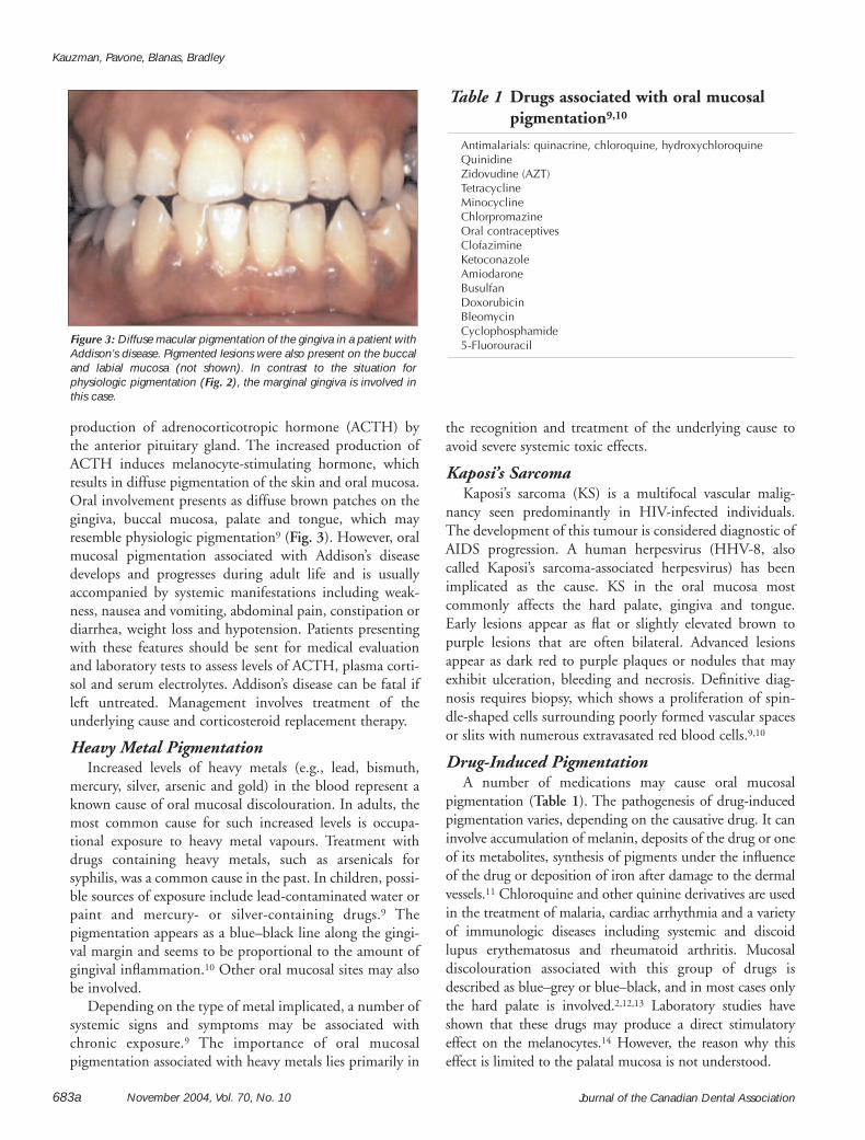
production of adrenocorticotropic hormone (ACTH) bythe anterior pituitary gland. The increased production ofACTH induces melanocyte-stimulating hormone, whichresults in diffuse pigmentation of the skin and oral mucosa.Oral involvement presents as diffuse brown patches on thegingiva, buccal mucosa, palate and tongue, which mayresemble physiologic pigmentation9 (Fig. 3). However, oralmucosal pigmentation associated with Addison’s diseasedevelops and progresses during adult life and is usuallyaccompanied by systemic manifestations including weak-ness, nausea and vomiting, abdominal pain, constipation ordiarrhea, weight loss and hypotension. Patients presentingwith these features should be sent for medical evaluationand laboratory tests to assess levels of ACTH, plasma corti-sol and serum electrolytes. Addison’s disease can be fatal ifleft untreated. Management involves treatment of theunderlying cause and corticosteroid replacement therapy.
Heavy Metal PigmentationIncreased levels of heavy metals (e.g., lead, bismuth,
mercury, silver, arsenic and gold) in the blood represent aknown cause of oral mucosal discolouration. In adults, themost common cause for such increased levels is occupa-tional exposure to heavy metal vapours. Treatment withdrugs containing heavy metals, such as arsenicals forsyphilis, was a common cause in the past. In children, possi-ble sources of exposure include lead-contaminated water orpaint and mercury- or silver-containing drugs.9 Thepigmentation appears as a blue–black line along the gingi-val margin and seems to be proportional to the amount ofgingival inflammation.10 Other oral mucosal sites may alsobe involved.
Depending on the type of metal implicated, a number ofsystemic signs and symptoms may be associated withchronic exposure.9 The importance of oral mucosalpigmentation associated with heavy metals lies primarily in
the recognition and treatment of the underlying cause toavoid severe systemic toxic effects.
Kaposi’s SarcomaKaposi’s sarcoma (KS) is a multifocal vascular malig-
nancy seen predominantly in HIV-infected individuals.The development of this tumour is considered diagnostic ofAIDS progression. A human herpesvirus (HHV-8, alsocalled Kaposi’s sarcoma-associated herpesvirus) has beenimplicated as the cause. KS in the oral mucosa mostcommonly affects the hard palate, gingiva and tongue.Early lesions appear as flat or slightly elevated brown topurple lesions that are often bilateral. Advanced lesionsappear as dark red to purple plaques or nodules that mayexhibit ulceration, bleeding and necrosis. Definitive diag-nosis requires biopsy, which shows a proliferation of spin-dle-shaped cells surrounding poorly formed vascular spacesor slits with numerous extravasated red blood cells.9,10
Drug-Induced PigmentationA number of medications may cause oral mucosal
pigmentation (Table 1). The pathogenesis of drug-inducedpigmentation varies, depending on the causative drug. It caninvolve accumulation of melanin, deposits of the drug or oneof its metabolites, synthesis of pigments under the influenceof the drug or deposition of iron after damage to the dermalvessels.11 Chloroquine and other quinine derivatives are usedin the treatment of malaria, cardiac arrhythmia and a varietyof immunologic diseases including systemic and discoidlupus erythematosus and rheumatoid arthritis. Mucosaldiscolouration associated with this group of drugs isdescribed as blue–grey or blue–black, and in most cases onlythe hard palate is involved.2,12,13 Laboratory studies haveshown that these drugs may produce a direct stimulatoryeffect on the melanocytes.14 However, the reason why thiseffect is limited to the palatal mucosa is not understood.
Table 1 Drugs associated with oral mucosalpigmentation9,10
Antimalarials: quinacrine, chloroquine, hydroxychloroquineQuinidineZidovudine (AZT) TetracyclineMinocyclineChlorpromazineOral contraceptivesClofazimineKetoconazoleAmiodaroneBusulfanDoxorubicinBleomycinCyclophosphamide5-Fluorouracil Figure 3: Diffuse macular pigmentation of the gingiva in a patient with
Addison’s disease. Pigmented lesions were also present on the buccaland labial mucosa (not shown). In contrast to the situation forphysiologic pigmentation (Fig. 2), the marginal gingiva is involved inthis case.
November 2004, Vol. 70, No. 10 683bJournal of the Canadian Dental Association
Pigmented Lesions of the Oral Cavity
Minocycline is a synthetic tetracycline used in the long-term treatment of refractory acne vulgaris. It can causepigmentation of the alveolar bone, which can be seenthrough the thin overlying oral mucosa (especially themaxillary anterior alveolar mucosa) as a grey discoloura-tion.15 Minocycline has also been reported to causepigmentation of the tongue mucosa.16
Postinflammatory PigmentationLong-standing inflammatory mucosal diseases, particu-
larly lichen planus, can cause mucosal pigmentation.1 Thisis seen more frequently in dark-skinned individuals.Clinically, multiple brown–black pigmented areas are notedadjacent to reticular or erosive lesions of lichen planus. Thepathogenesis of postinflammatory pigmentation remainsunclear.17 Histologically, there is increased production ofmelanin by the melanocytes and accumulation of melanin-laden macrophages in the superficial connective tissue.
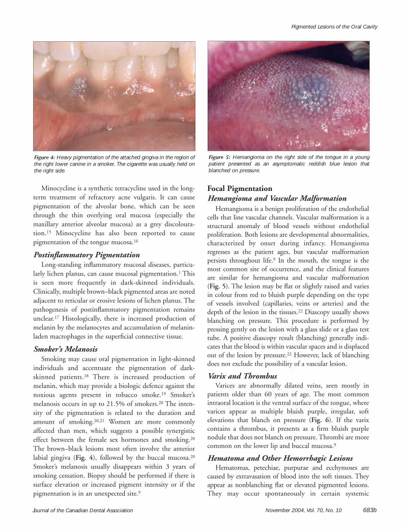
Smoker’s MelanosisSmoking may cause oral pigmentation in light-skinned
individuals and accentuate the pigmentation of dark-skinned patients.18 There is increased production ofmelanin, which may provide a biologic defence against thenoxious agents present in tobacco smoke.19 Smoker’smelanosis occurs in up to 21.5% of smokers.20 The inten-sity of the pigmentation is related to the duration andamount of smoking.20,21 Women are more commonlyaffected than men, which suggests a possible synergisticeffect between the female sex hormones and smoking.20
The brown–black lesions most often involve the anteriorlabial gingiva (Fig. 4), followed by the buccal mucosa.20
Smoker’s melanosis usually disappears within 3 years ofsmoking cessation. Biopsy should be performed if there issurface elevation or increased pigment intensity or if thepigmentation is in an unexpected site.9
Focal PigmentationHemangioma and Vascular Malformation
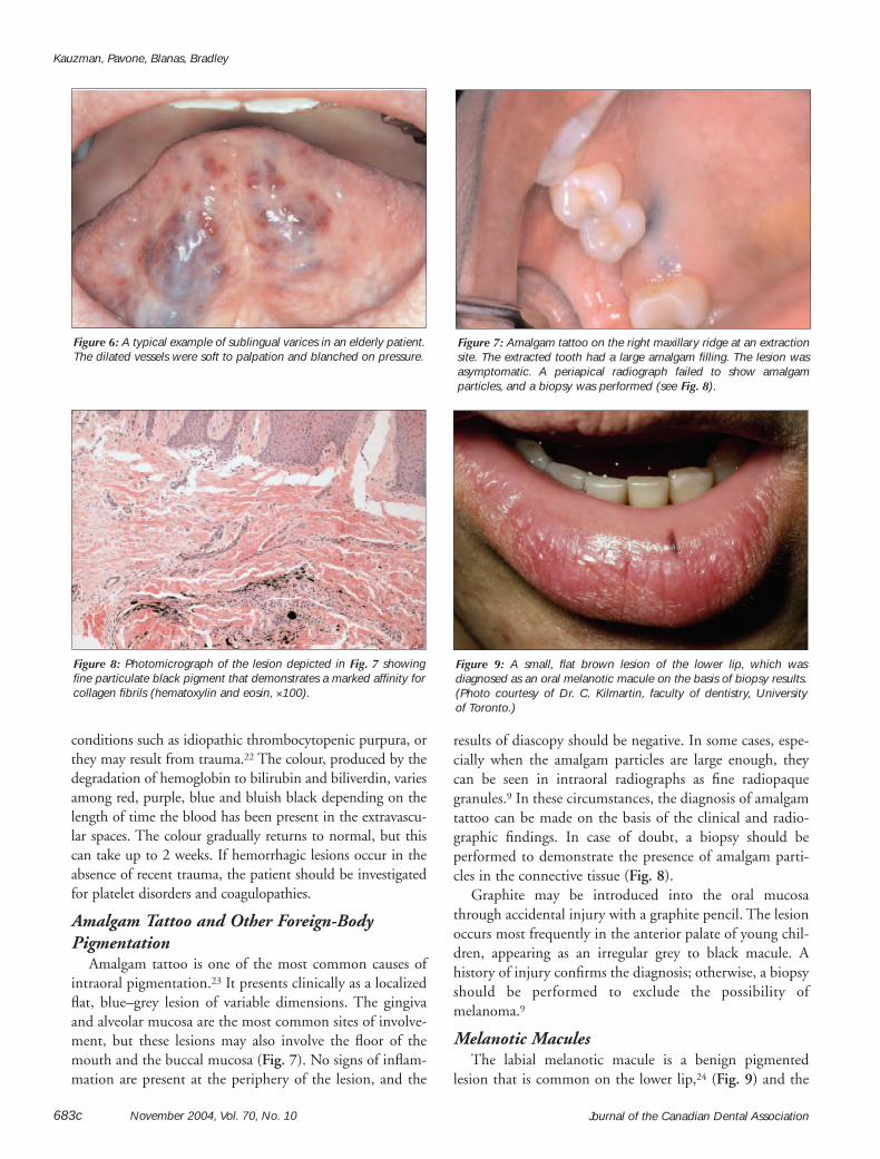
Hemangioma is a benign proliferation of the endothelialcells that line vascular channels. Vascular malformation is astructural anomaly of blood vessels without endothelialproliferation. Both lesions are developmental abnormalities,characterized by onset during infancy. Hemangiomaregresses as the patient ages, but vascular malformationpersists throughout life.9 In the mouth, the tongue is themost common site of occurrence, and the clinical featuresare similar for hemangioma and vascular malformation (Fig. 5). The lesion may be flat or slightly raised and variesin colour from red to bluish purple depending on the typeof vessels involved (capillaries, veins or arteries) and thedepth of the lesion in the tissues.22 Diascopy usually showsblanching on pressure. This procedure is performed bypressing gently on the lesion with a glass slide or a glass testtube. A positive diascopy result (blanching) generally indi-cates that the blood is within vascular spaces and is displacedout of the lesion by pressure.22 However, lack of blanchingdoes not exclude the possibility of a vascular lesion.
Varix and ThrombusVarices are abnormally dilated veins, seen mostly in
patients older than 60 years of age. The most commonintraoral location is the ventral surface of the tongue, wherevarices appear as multiple bluish purple, irregular, softelevations that blanch on pressure (Fig. 6). If the varixcontains a thrombus, it presents as a firm bluish purplenodule that does not blanch on pressure. Thrombi are morecommon on the lower lip and buccal mucosa.9
Hematoma and Other Hemorrhagic LesionsHematomas, petechiae, purpurae and ecchymoses are
caused by extravasation of blood into the soft tissues. Theyappear as nonblanching flat or elevated pigmented lesions.They may occur spontaneously in certain systemic
Figure 4: Heavy pigmentation of the attached gingiva in the region ofthe right lower canine in a smoker. The cigarette was usually held onthe right side.
Figure 5: Hemangioma on the right side of the tongue in a youngpatient presented as an asymptomatic reddish blue lesion thatblanched on pressure.
Journal of the Canadian Dental Association683c November 2004, Vol. 70, No. 10
Kauzman, Pavone, Blanas, Bradley
conditions such as idiopathic thrombocytopenic purpura, orthey may result from trauma.22 The colour, produced by thedegradation of hemoglobin to bilirubin and biliverdin, variesamong red, purple, blue and bluish black depending on thelength of time the blood has been present in the extravascu-lar spaces. The colour gradually returns to normal, but thiscan take up to 2 weeks. If hemorrhagic lesions occur in theabsence of recent trauma, the patient should be investigatedfor platelet disorders and coagulopathies.
Amalgam Tattoo and Other Foreign-BodyPigmentation
Amalgam tattoo is one of the most common causes ofintraoral pigmentation.23 It presents clinically as a localizedflat, blue–grey lesion of variable dimensions. The gingivaand alveolar mucosa are the most common sites of involve-ment, but these lesions may also involve the floor of themouth and the buccal mucosa (Fig. 7). No signs of inflam-mation are present at the periphery of the lesion, and the
results of diascopy should be negative. In some cases, espe-cially when the amalgam particles are large enough, theycan be seen in intraoral radiographs as fine radiopaquegranules.9 In these circumstances, the diagnosis of amalgamtattoo can be made on the basis of the clinical and radio-graphic findings. In case of doubt, a biopsy should beperformed to demonstrate the presence of amalgam parti-cles in the connective tissue (Fig. 8).
Graphite may be introduced into the oral mucosathrough accidental injury with a graphite pencil. The lesionoccurs most frequently in the anterior palate of young chil-dren, appearing as an irregular grey to black macule. Ahistory of injury confirms the diagnosis; otherwise, a biopsyshould be performed to exclude the possibility ofmelanoma.9
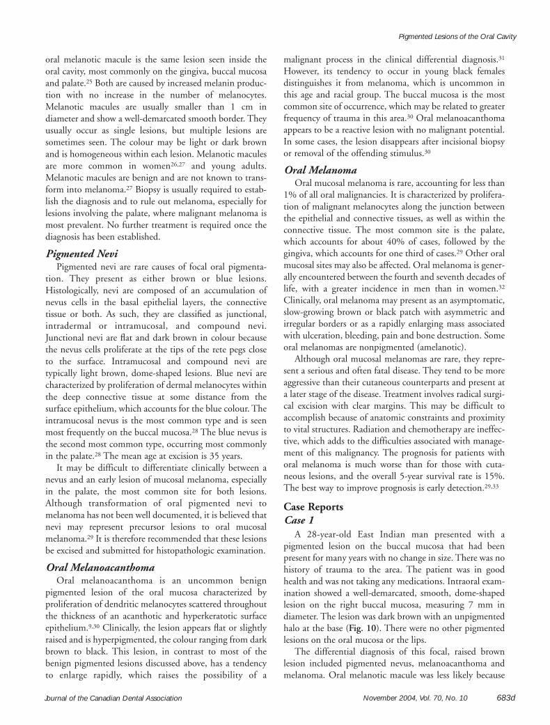
Melanotic MaculesThe labial melanotic macule is a benign pigmented
lesion that is common on the lower lip,24 (Fig. 9) and the
Figure 6: A typical example of sublingual varices in an elderly patient.The dilated vessels were soft to palpation and blanched on pressure.
Figure 7: Amalgam tattoo on the right maxillary ridge at an extractionsite. The extracted tooth had a large amalgam filling. The lesion wasasymptomatic. A periapical radiograph failed to show amalgamparticles, and a biopsy was performed (see Fig. 8).
Figure 8: Photomicrograph of the lesion depicted in Fig. 7 showingfine particulate black pigment that demonstrates a marked affinity forcollagen fibrils (hematoxylin and eosin, ×100).
Figure 9: A small, flat brown lesion of the lower lip, which wasdiagnosed as an oral melanotic macule on the basis of biopsy results.(Photo courtesy of Dr. C. Kilmartin, faculty of dentistry, University of Toronto.)
November 2004, Vol. 70, No. 10 683dJournal of the Canadian Dental Association
Pigmented Lesions of the Oral Cavity
oral melanotic macule is the same lesion seen inside the oral cavity, most commonly on the gingiva, buccal mucosaand palate.25 Both are caused by increased melanin produc-tion with no increase in the number of melanocytes.Melanotic macules are usually smaller than 1 cm in diameter and show a well-demarcated smooth border. Theyusually occur as single lesions, but multiple lesions aresometimes seen. The colour may be light or dark brownand is homogeneous within each lesion. Melanotic maculesare more common in women26,27 and young adults.Melanotic macules are benign and are not known to trans-form into melanoma.27 Biopsy is usually required to estab-lish the diagnosis and to rule out melanoma, especially forlesions involving the palate, where malignant melanoma ismost prevalent. No further treatment is required once thediagnosis has been established.
Pigmented NeviPigmented nevi are rare causes of focal oral pigmenta-
tion. They present as either brown or blue lesions.Histologically, nevi are composed of an accumulation ofnevus cells in the basal epithelial layers, the connectivetissue or both. As such, they are classified as junctional,intradermal or intramucosal, and compound nevi.Junctional nevi are flat and dark brown in colour becausethe nevus cells proliferate at the tips of the rete pegs closeto the surface. Intramucosal and compound nevi are typically light brown, dome-shaped lesions. Blue nevi arecharacterized by proliferation of dermal melanocytes withinthe deep connective tissue at some distance from thesurface epithelium, which accounts for the blue colour. Theintramucosal nevus is the most common type and is seenmost frequently on the buccal mucosa.28 The blue nevus isthe second most common type, occurring most commonlyin the palate.28 The mean age at excision is 35 years.
It may be difficult to differentiate clinically between anevus and an early lesion of mucosal melanoma, especiallyin the palate, the most common site for both lesions.Although transformation of oral pigmented nevi tomelanoma has not been well documented, it is believed thatnevi may represent precursor lesions to oral mucosalmelanoma.29 It is therefore recommended that these lesionsbe excised and submitted for histopathologic examination.
Oral Melanoacanthoma Oral melanoacanthoma is an uncommon benign
pigmented lesion of the oral mucosa characterized byproliferation of dendritic melanocytes scattered throughoutthe thickness of an acanthotic and hyperkeratotic surfaceepithelium.9,30 Clinically, the lesion appears flat or slightlyraised and is hyperpigmented, the colour ranging from darkbrown to black. This lesion, in contrast to most of thebenign pigmented lesions discussed above, has a tendencyto enlarge rapidly, which raises the possibility of a
malignant process in the clinical differential diagnosis.31
However, its tendency to occur in young black femalesdistinguishes it from melanoma, which is uncommon inthis age and racial group. The buccal mucosa is the mostcommon site of occurrence, which may be related to greaterfrequency of trauma in this area.30 Oral melanoacanthomaappears to be a reactive lesion with no malignant potential.In some cases, the lesion disappears after incisional biopsyor removal of the offending stimulus.30
Oral MelanomaOral mucosal melanoma is rare, accounting for less than
1% of all oral malignancies. It is characterized by prolifera-tion of malignant melanocytes along the junction betweenthe epithelial and connective tissues, as well as within theconnective tissue. The most common site is the palate,which accounts for about 40% of cases, followed by thegingiva, which accounts for one third of cases.29 Other oralmucosal sites may also be affected. Oral melanoma is gener-ally encountered between the fourth and seventh decades oflife, with a greater incidence in men than in women.32
Clinically, oral melanoma may present as an asymptomatic,slow-growing brown or black patch with asymmetric andirregular borders or as a rapidly enlarging mass associatedwith ulceration, bleeding, pain and bone destruction. Someoral melanomas are nonpigmented (amelanotic).
Although oral mucosal melanomas are rare, they repre-sent a serious and often fatal disease. They tend to be moreaggressive than their cutaneous counterparts and present ata later stage of the disease. Treatment involves radical surgi-cal excision with clear margins. This may be difficult toaccomplish because of anatomic constraints and proximityto vital structures. Radiation and chemotherapy are ineffec-tive, which adds to the difficulties associated with manage-ment of this malignancy. The prognosis for patients withoral melanoma is much worse than for those with cuta-neous lesions, and the overall 5-year survival rate is 15%.The best way to improve prognosis is early detection.29,33
Case ReportsCase 1
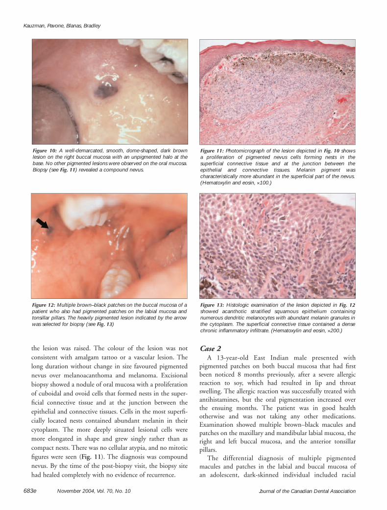
A 28-year-old East Indian man presented with apigmented lesion on the buccal mucosa that had beenpresent for many years with no change in size. There was nohistory of trauma to the area. The patient was in goodhealth and was not taking any medications. Intraoral exam-ination showed a well-demarcated, smooth, dome-shapedlesion on the right buccal mucosa, measuring 7 mm indiameter. The lesion was dark brown with an unpigmentedhalo at the base (Fig. 10). There were no other pigmentedlesions on the oral mucosa or the lips.
The differential diagnosis of this focal, raised brownlesion included pigmented nevus, melanoacanthoma andmelanoma. Oral melanotic macule was less likely because
Journal of the Canadian Dental Association683e November 2004, Vol. 70, No. 10
Kauzman, Pavone, Blanas, Bradley
the lesion was raised. The colour of the lesion was notconsistent with amalgam tattoo or a vascular lesion. Thelong duration without change in size favoured pigmentednevus over melanoacanthoma and melanoma. Excisionalbiopsy showed a nodule of oral mucosa with a proliferationof cuboidal and ovoid cells that formed nests in the super-ficial connective tissue and at the junction between theepithelial and connective tissues. Cells in the most superfi-cially located nests contained abundant melanin in theircytoplasm. The more deeply situated lesional cells weremore elongated in shape and grew singly rather than ascompact nests. There was no cellular atypia, and no mitoticfigures were seen (Fig. 11). The diagnosis was compoundnevus. By the time of the post-biopsy visit, the biopsy sitehad healed completely with no evidence of recurrence.
Case 2A 13-year-old East Indian male presented with
pigmented patches on both buccal mucosa that had firstbeen noticed 8 months previously, after a severe allergicreaction to soy, which had resulted in lip and throatswelling. The allergic reaction was successfully treated withantihistamines, but the oral pigmentation increased overthe ensuing months. The patient was in good health otherwise and was not taking any other medications.Examination showed multiple brown–black macules andpatches on the maxillary and mandibular labial mucosa, theright and left buccal mucosa, and the anterior tonsillarpillars.
The differential diagnosis of multiple pigmentedmacules and patches in the labial and buccal mucosa of an adolescent, dark-skinned individual included racial
Figure 10: A well-demarcated, smooth, dome-shaped, dark brownlesion on the right buccal mucosa with an unpigmented halo at thebase. No other pigmented lesions were observed on the oral mucosa.Biopsy (see Fig. 11) revealed a compound nevus.
Figure 11: Photomicrograph of the lesion depicted in Fig. 10 shows a proliferation of pigmented nevus cells forming nests in thesuperficial connective tissue and at the junction between theepithelial and connective tissues. Melanin pigment wascharacteristically more abundant in the superficial part of the nevus.(Hematoxylin and eosin, ×100.)
Figure 12: Multiple brown–black patches on the buccal mucosa of apatient who also had pigmented patches on the labial mucosa andtonsillar pillars. The heavily pigmented lesion indicated by the arrowwas selected for biopsy (see Fig. 13)
Figure 13: Histologic examination of the lesion depicted in Fig. 12showed acanthotic stratified squamous epithelium containingnumerous dendritic melanocytes with abundant melanin granules inthe cytoplasm. The superficial connective tissue contained a densechronic inflammatory infiltrate. (Hematoxylin and eosin, ×200.)

November 2004, Vol. 70, No. 10 683fJournal of the Canadian Dental Association
Pigmented Lesions of the Oral Cavity
(physiologic) pigmentation, Addison’s disease, postinflam-matory pigmentation and drug-induced pigmentation.Physiologic pigmentation was considered most likely,although the relatively rapid onset and the location of thepigmentation were unusual. The patient did not have signsand symptoms suggestive of Addison’s disease, was nottaking any medications that commonly cause oral pigmen-tation and did not have a chronic mucosal inflammatorydisease that would typically be associated with postinflam-matory pigmentation. Biopsy of a dark brown lesion on theright buccal mucosa (Fig. 12) showed oral mucosa coveredby acanthotic stratified squamous nonkeratinized epithe-lium and numerous dendritic melanocytes, with cytoplas-mic melanin throughout the epithelium (Fig. 13). Thesuperficial connective tissue contained a dense, chronicinflammatory infiltrate, composed predominantly ofplasma cells and eosinophils. There was no cellular atypia tosuggest melanoma. The histologic appearance was charac-teristic of melanoacanthoma. This patient’s clinical lesionswere unusually extensive for melanoacanthoma, althoughthe condition has been reported to present bilaterally and aspigmented areas several centimetres in diameter.30 Nofurther treatment was indicated, and the patient will befollowed clinically to monitor the lesions.
Case 3A 77-year-old Asian man complained of a loose left
maxillary molar. The tooth was heavily restored with astainless steel crown that had been repaired with an amal-gam filling. An irregular greyish black patch was present onthe buccal and distal gingiva, with smaller “satellite” lesionson the palatal aspect of the tooth.
The differential diagnosis was a large amalgam tattoo (orsimilar foreign-body implantation) and melanoma. Thetooth was extracted, and an incisional biopsy was takenfrom the gingiva. Once the tooth had been removed, it
became apparent that the pigmented lesion was larger thanhad previously been appreciated (Fig. 14). Histologic examination showed oral mucosa in which the laminapropria was infiltrated by pleomorphic cells, some withmelanin pigment in the cytoplasm (Fig. 15). Abnormalmelanin-containing cells were also present within the strat-ified squamous epithelium. The diagnosis was malignantmelanoma, and the patient was referred to the Head andNeck Oncology Clinic for treatment. C
Dr. Kauzman is associate professor, department of stomatology, faculty of dentistry, University ofMontreal, Montreal, Quebec.
Dr. Pavone is staff dentist, department of dentistry, Sunnybrook andWomen’s College Health Science Centre, University of Toronto,Toronto, Ontario.
Dr. Blanas is staff oral and maxillofacial surgeon,department of dentistry, Sunnybrook and Women’sCollege Health Science Centre, University of Toronto,Toronto, Ontario.
Dr. Bradley is associate professor, oral pathology and oral medicine,faculty of dentistry, and Sunnybrook and Women’s College HealthScience Centre, University of Toronto, Toronto, Ontario.Correspondence to: Dr. Grace Bradley, Faculty of Dentistry,University of Toronto, 124 Edward St., Toronto, ON M5G 1G6.E-mail: [email protected] authors have no declared financial interests.
References1. Eisen D. Disorders of pigmentation in the oral cavity. Clin Dermatol2000; 18(5):579–87.2. Kleinegger CL, Hammond HL, Finkelstein MW. Oral mucosal hyper-pigmentation secondary to antimalarial drug therapy. Oral Surg Oral MedOral Pathol Oral Radiol Endod 2000; 90(2):189–94.3. Gaeta GM, Satriano RA, Baroni A. Oral pigmented lesions.Clin Dermatol 2002; 20(3):286–8.
Figure 14: Photograph taken 1 week after extraction of a loose leftmaxillary molar shows an irregular greyish black patch on themaxillary alveolar ridge, distal and buccal to the extraction socket ofthe molar. Smaller satellite lesions were present on the palatalmucosa. An incisional biopsy was performed (see Fig. 15).
Figure 15: Histologic examination of the lesion depicted in Fig. 14showed malignant melanocytes invading the superficial connectivetissue and the overlying epithelium. Note the cellular pleomorphismand nuclear hyperchromatism. (Hematoxylin and eosin, ×125.)

Journal of the Canadian Dental Association683g November 2004, Vol. 70, No. 10
Kauzman, Pavone, Blanas, Bradley
4. Hemminki A, Markie D, Tomlinson I, Avizienyte E, Roth S, LoukolaA, and others. A serine/threonine kinase gene defective in Peutz-Jegherssyndrome. Nature 1998; 391(6663):184–7.5. Hemminki A, Tomlinson I, Markie D, Jarvinen H, Sistonen P,Bjorkqvist AM, and others. Localization of a susceptibility locus forPeutz-Jeghers syndrome to 19p using comparative genomic hybridizationand targeted linkage analysis. Nat Genet 1997; 15(1):87–90.6. Hemminki A. The molecular basis and clinical aspects of Peutz-Jegherssyndrome. Cell Mol Life Sci 1999; 55(5):735–50.7. McGrath DR, Spigelman AD. Preventive measures in Peutz-Jegherssyndrome. Fam Cancer 2001; 1(2):121–5.8. Kim HW. Generalized oral and cutaneous hyperpigmentation inAddison’s disease. Odontostomatol Trop 1988; 11(3):87–90.9. Neville BW, Damm DD, Allen CM, Bouquot JE, editors. Oral andmaxillofacial pathology. 2nd ed. Toronto (ON): W.B. SaundersCompany; 2002.10. Regezi JA, Sciubba JJ, Jordan RC, editors. Oral pathology. Clinicalpathologic correlations. 4th ed. Philadelphia: W.B. Saunders; 2003.11. Dereure O. Drug-induced skin pigmentation. Epidemiology, diagno-sis and treatment. Am J Clin Dermatol 2001; 2(4):253–62.12. Birek C, Main JH. Two cases of oral pigmentation associated withquinidine therapy. Oral Surg Oral Med Oral Pathol 1988; 66(1):59–61.13. McAllan LH, Adkins KF. Drug-induced palatal pigmentation.Aust Dent J 1986; 31(1):1–4.14. Savage NW, Barber MT, Adkins KF. Pigmentary changes in rat oralmucosa following antimalarial therapy. J Oral Pathol 1986; 15(9):468–71.15. Westbury LW, Najera A. Minocycline-induced intraoral pharmaco-genic pigmentation: case reports and review of the literature. J Periodontol1997; 68(1):84–91.16. Meyerson MA, Cohen PR, Hymes SR. Lingual hyperpigmentationassociated with minocycline therapy. Oral Surg Oral Med Oral Pathol OralRadiol Endod 1995; 79(2):180–4.17. Halder RM, Nootheti PK. Ethnic skin disorders overview. J Am AcadDermatol 2003; 48(6 Suppl):S143–8.18. Hedin CA, Axell T. Oral melanin pigmentation in 467 Thai andMalaysian people with special emphasis on smoker’s melanosis.J Oral Pathol Med 1991; 20(1):8–12.19. Hedin C, Pindborg JJ, Daftary DK, Mehta FS. Melanin depigmenta-tion of the palatal mucosa in reverse smokers: a preliminary study.J Oral Pathol Med 1992; 21(10):440–4.
20. Axell T, Hedin CA. Epidemiologic study of excessive oral melaninpigmentation with special reference to the influence of tobacco habits.Scand J Dent Res 1982; 90(6):434–42.21. Araki S, Murata K, Ushio K, Sakai R. Dose-response relationshipbetween tobacco consumption and melanin pigmentation in the attachedgingiva. Arch Environ Health 1983; 38(6):375–8.22. Carpenter WM, Rudd M. Focal, flat pigmentations of the oralmucosa: a clinical approach to the differential diagnosis. J Calif Dent Assoc2000; 28(12):949–54.23. Perusse R, Blackburn E. Differential diagnosis of pigmented lesions ofthe oral cavity. J Can Dent Assoc 1984; 50(10):783–7.24. Weathers DR, Corio RL, Crawford BE, Giansanti JS, Page LR.The labial melanotic macule. Oral Surg Oral Med Oral Pathol 1976;42(2):196–205.25. Page LR, Corio RL, Crawford BE, Giansanti JS, Weathers DR.The oral melanotic macule. Oral Surg Oral Med Oral Pathol 1977;44(2):219–26.26. Buchner A, Hansen LS. Melanotic macule of the oral mucosa. A clin-icopathologic study of 105 cases. Oral Surg Oral Med Oral Pathol 1979;48(3):244–9.27. Gupta G, Williams RE, Mackie RM. The labial melanotic macule: areview of 79 cases. Br J Dermatol 1997; 136(5):772–5.28. Buchner A, Hansen LS. Pigmented nevi of the oral mucosa: a clini-copathologic study of 36 new cases and review of 155 cases from the liter-ature. Part II: analysis of 191 cases. Oral Surg Oral Med Oral Pathol 1987;63(6):676–82.29. Hicks MJ, Flaitz CM. Oral mucosal melanoma: epidemiology andpathobiology. Oral Oncol 2000; 36(2):152–69.30. Goode RK, Crawford BE, Callihan MD, Neville BW. Oral melano-acanthoma. Review of the literature and report of ten cases. Oral SurgOral Med Oral Pathol 1983; 56(6):622–8.31. Whitt JC, Jennings DR, Arendt DM, Vinton JR. Rapidly expandingpigmented lesion of the buccal mucosa. J Am Dent Assoc 1988;117(5):620–2.32. Barker BF, Carpenter WM, Daniels TE, Kahn MA, Leider AS,Lozada-Nur F, and others. Oral mucosal melanomas: the WESTOP Banffworkshop proceedings. Western Society of Teachers of Oral Pathology.Oral Surg Oral Med Oral Pathol Oral Radiol Endod 1997; 83(6):672–9.33. Gorsky M, Epstein JB. Melanoma arising from the mucosal surfacesof the head and neck. Oral Surg Oral Med Oral Pathol Oral Radiol Endod1998; 86(6):715–9.