PIERCE PROTEIN RESEARCH PRODUCTS - Cultek · controlling their functions are ... is used to pull...
24
Cellular Proteomics The proteome is an organism’s entire complement of proteins, and the field of cellular proteomics is concerned not only with identifying all constituent proteins in an idealized static cell, but also with elucidating protein functions at CONTINUED ON PAGE 2 volume 12, issue 1 WHAT'S IN YOUR CELL? TOOLS FOR CELLULAR PROTEOMICS, PAGES 2-6 STRESS ON CELLS HIGH-CONTENT SCREENING SYSTEM SOLUTIONS, PAGES 7-8 LIGHT UP AND BE HAPPY BRIGHT PERFORMING FLUORESCENT DYES, PAGES 9-12 COOL PULLOUT POSTER! CELLULAR PROTEOMICS REFERENCE POSTER, BETWEEN PAGES 12-13 THE TARGET IS WIDER OUR LINE OF RNA INTERFERENCE/WESTERN BLOTTING TOOLS HAS GROWN, PAGES 13-14 OH SO SENSITIVE! DETECT A SPECIFIC PROTEIN DOWN TO THE PICOGRAM WITH NEW SCREENING SETS, PAGE 15 PROTECT YOUR PROTEINS THIS COCKTAIL IS A SINGLE SHOT OF PROTEIN PROTECTION, PAGES 16-17 BOUND BY SUCCESS NEW PROTEIN A/G PLUS BINDS ANTIBODIES MORE EFFICIENTLY, PAGES 18-19 CLEARLY BETTER WESTERNS! REVEAL YOUR TARGET PROTEIN ON YOUR WESTERN BLOT, PAGES 20-21 PLAY TAG YOUR WAY... READY-TO-USE KITS PURIFY HIS-TAGGED PROTEINS IN NEW FORMATS, PAGE 22 MINI GOES MAXI MINI DIALYSIS IN A NEW 20K MWCO OPTION, PAGE 23 PIERCE PROTEIN RESEARCH PRODUCTS SPECIAL ISSUE CELLULAR PROTEOMICS
-
Upload
truongcong -
Category
Documents
-
view
214 -
download
0
Transcript of PIERCE PROTEIN RESEARCH PRODUCTS - Cultek · controlling their functions are ... is used to pull...

Cellular ProteomicsThe proteome is an organism’s entire complement ofproteins, and the field of cellular proteomics is concerned notonly with identifying all constituent proteins in an idealizedstatic cell, but also with elucidating protein functions at
CONTINUED ON PAGE 2
volume 12, issue 1WHAT'S IN YOUR CELL?TOOLS FOR CELLULAR PROTEOMICS, PAGES 2-6
STRESS ON CELLSHIGH-CONTENT SCREENING SYSTEM SOLUTIONS, PAGES 7-8
LIGHT UP AND BE HAPPYBRIGHT PERFORMING FLUORESCENT DYES, PAGES 9-12
COOL PULLOUT POSTER!CELLULAR PROTEOMICS REFERENCE POSTER,BETWEEN PAGES 12-13
THE TARGET IS WIDEROUR LINE OF RNA INTERFERENCE/WESTERN BLOTTINGTOOLS HAS GROWN, PAGES 13-14
OH SO SENSITIVE!DETECT A SPECIFIC PROTEIN DOWN TO THE PICOGRAMWITH NEW SCREENING SETS, PAGE 15
PROTECT YOUR PROTEINSTHIS COCKTAIL IS A SINGLE SHOT OF PROTEIN PROTECTION,PAGES 16-17
BOUND BY SUCCESSNEW PROTEIN A/G PLUS BINDS ANTIBODIES MORE EFFICIENTLY, PAGES 18-19
CLEARLY BETTER WESTERNS!REVEAL YOUR TARGET PROTEIN ON YOUR WESTERN BLOT,PAGES 20-21
PLAY TAG YOUR WAY... READY-TO-USE KITS PURIFY HIS-TAGGED PROTEINS INNEW FORMATS, PAGE 22
MINI GOES MAXIMINI DIALYSIS IN A NEW 20K MWCO OPTION, PAGE 23
P I E R C E P R O T E I N R E S E A R C H P R O D U C T S
SPECIAL ISSUE
CELLULAR PROTEOMICS

all levels of interaction within the complete life cycle and cellularcontext in living tissues and organisms. Indeed, useful understandingof protein function includes detection and characterization of theirspatial and temporal expression and regulation within and betweencells. Whether through the comparison of normal and diseased cellsor the perturbation analysis inherent in drug screening, research ishelping to close the gap on many previously intractable aspects ofcell biology.
We consider it a privilege to be an integral part of advancing cellular proteomics research by continuing to develop and offer high-quality reagents and kits that facilitate protein research at all levels.To highlight the breadth of our Thermo Scientific Pierce ProteinResearch Product line, we present the following illustration andiconographic characterization of key domains of interest in cellularproteomics. Whether your needs are for protein extraction from specific organelles, isolation of cell surface proteins, co-immunopre-cipitation or label transfer protein interaction analysis, non-isotopicelectrophoretic mobility shift assay (EMSA), fluorescencemicroscopy, cytokine measurement, phosphoprotein enrichment,or high-content screening reagents, our products can assist youin your research.
Thanks for exploring the cell with us.
Membrane ProteinsMembrane proteins compriseapproximately 30% of the eukaryoticproteome, and elucidating andcontrolling their functions are important goals of drug discoveryresearch. Membrane proteins actas transporters, channels, receptorsand enzymes, structural and cell-adhesion anchoring domains,accumulators, and transducers of
energy. However, membrane proteins are difficult to isolate becauseof their hydrophobicity, basic nature and large size. ThermoScientific Pierce Research Products include a comprehensive line ofcell lysis reagents and kits designed to maximize extraction of targetproteins from cell membranes, organelles and cytoplasm. TheThermo Scientific Mem-PER* Eukaryotic Membrane Protein IsolationKit effectively isolates single and multiple transmembrane proteinsfrom eukaryotic cells or tissue.
Ordering Information
Product # Description Pkg. Size89826 Mem-PER Eukaryotic Membrane Kit
Protein Isolation KitSufficient lysis and extraction reagents for approximately 50-100 mammalian cell pellet fractions.
Visit thermo.com/pierce for kit components.
The Cell SurfaceNormal cell function, and its contri-bution to overall physiology, dependson the proper response of cells tostimuli or extracellular signals. Allsuch stimuli are received, processedand delivered by receptors, gatesand channels that are embedded inor bound to the cell membrane.Receptor proteins in the cellmembrane receive signals from the
environment or from other cells and transmit them internally to effecta response, a process termed signal transduction. Small GTPases ofthe Ras superfamily are monomeric guanine nucleotide-bindingproteins that serve as molecular switches to regulate growth,morphogenesis, cell mobility, axonal guidance, cytokinesis and traf-ficking.
Thermo Scientific Active GTPase Pull-Down and Detection Kitsmeasure the activation of Rac1, Cdc42, Ras, Rho or Rap1 smallGTPases by isolating them via their specific downstream effectors.The respective binding domain of the downstream effector for eachsmall GTPase is expressed as a GST-fusion protein which, whenimmobilized on a resin, is used to pull down the active or GTP-boundGTPase. The kits also include a specific primary antibody forWestern blot detection of the purified GTPase.
About 3,000 human genes are believed to encode for proteins that are displayed at the cell surface. Secreted and cell surfaceproteins are often more difficult to isolate and identify than proteins that are localized in intracellular compartments. TheThermo Scientific Cell Surface Protein Isolation Kit provides a convenient and efficient method for isolating cell surface proteinsfrom cultured mammalian cells. Adherent or suspended cells arefirst labeled with Sulfo-NHS-SS-Biotin, a cleavable biotinylationreagent. The cells are subsequently lysed with a mild detergent,and labeled proteins are isolated with Thermo Scientific NeutrAvidin*Resin. Isolated proteins can be analyzed by Western blot, allowingfor differential expression analysis between treated and non-treatedcells or between two or more cell lines.
Ordering Information
Product # Description Pkg. Size89854 Active Rho Pull-Down and Detection Kit Kit
Sufficient material for 30 pull-down assays using GST-Rhotekin-RBD.
89855 Active Ras Pull-Down and Detection Kit KitSufficient material for 30 pull-down assays using GST-Raf1-RBD.
89856 Active Rac1 Pull-Down and Detection Kit KitSufficient material for 30 pull-down assays using GST-Pak1-PBD.
89857 Active Cdc42 Pull-Down and Detection Kit KitSufficient material for 30 pull-down assays using GST-Pak1-PBD.
89872 Active Rap1 Pull-Down and Detection Kit KitSufficient materials for 30 pull-down assays using GST-RalGDS-RBD.
89881 Cell Surface Protein Isolation Kit KitSufficient components for eight biotinylationsand isolations of mammalian cell surface proteins.
Visit thermo.com/pierce for kit components.
www.thermo.com/pierce Vol. 12, Issue 12
To order, call 800-874-3723 or 815-968-0747. Outside the United States, contact your local branch office or distributor. 3
Cell Lysis and Protein ExtractionMost experimental and assaymethods of proteomics analysisbegin with some form of cell lysis toobtain an extract of soluble cellularprotein contents. The quantity andquality of data that can be derivedfrom analysis of extracted proteinsdirectly depends on the amount andfunctional integrity of the proteinsthat result from the extraction
process. Some extraction methods may be efficient at cell lysis andsolubilization of cell contents but are protein-denaturing, therebypreventing detection and analysis of native protein interactions.Ease of analysis also depends upon compatibility of the lysis andextraction reagents with the downstream technique. For example,some extraction reagents contain ionic detergents and reducingagents that interfere with protein assays, affinity purification, 2-Delectrophoresis and other methods.
Thermo Scientific Pierce Cell Lysis Reagents provide for convenient, efficient and broadly compatible cell lysis and proteinextraction of mammalian cells and tissues. Thermo Scientific M-PER* Mammalian Protein Extraction Reagent is ideal for use with suspension or adherent cultured cells of many types and mammalianspecies. M-PER Extracts are directly compatible with many assaysincluding co-immunoprecipitation (co-IP), chemical labeling, 2-Delectrophoresis and other methods. Thermo Scientific T-PER* TissueProtein Extraction Reagent provides for efficient and mild proteinextraction from tissues using homogenization.
Protease and phosphatase inhibitors are also essential for recovering useable protein samples from cell or tissue lysis. Thesereagents prevent enzymes that are released from subcellular vesicles from degrading polypeptide sequences or altering phosphorylation states of proteins in an extract. Thermo ScientificHalt* Protease and Phosphatase Inhibitor Cocktails provide superiorprotection and ease-of-use compared to conventional tabletreagents (see separate article on page 16-17).
Ordering Information
Product # Description Pkg. Size78501 M-PER Mammalian Protein 250 ml
Extraction Reagent78510 T-PER Tissue Protein Extraction Reagent 500 ml89901 RIPA Buffer 250 ml78442 Halt Protease and Phosphatase 24 x 100 µl
Single-Use Inhibitor Cocktail78430 Halt Protease Inhibitor 24 x 100 µl
Single-Use Cocktail (100X)78420 Halt Phosphatase Inhibitor Cocktail 1 mlVisit thermo.com/pierce for kit components.
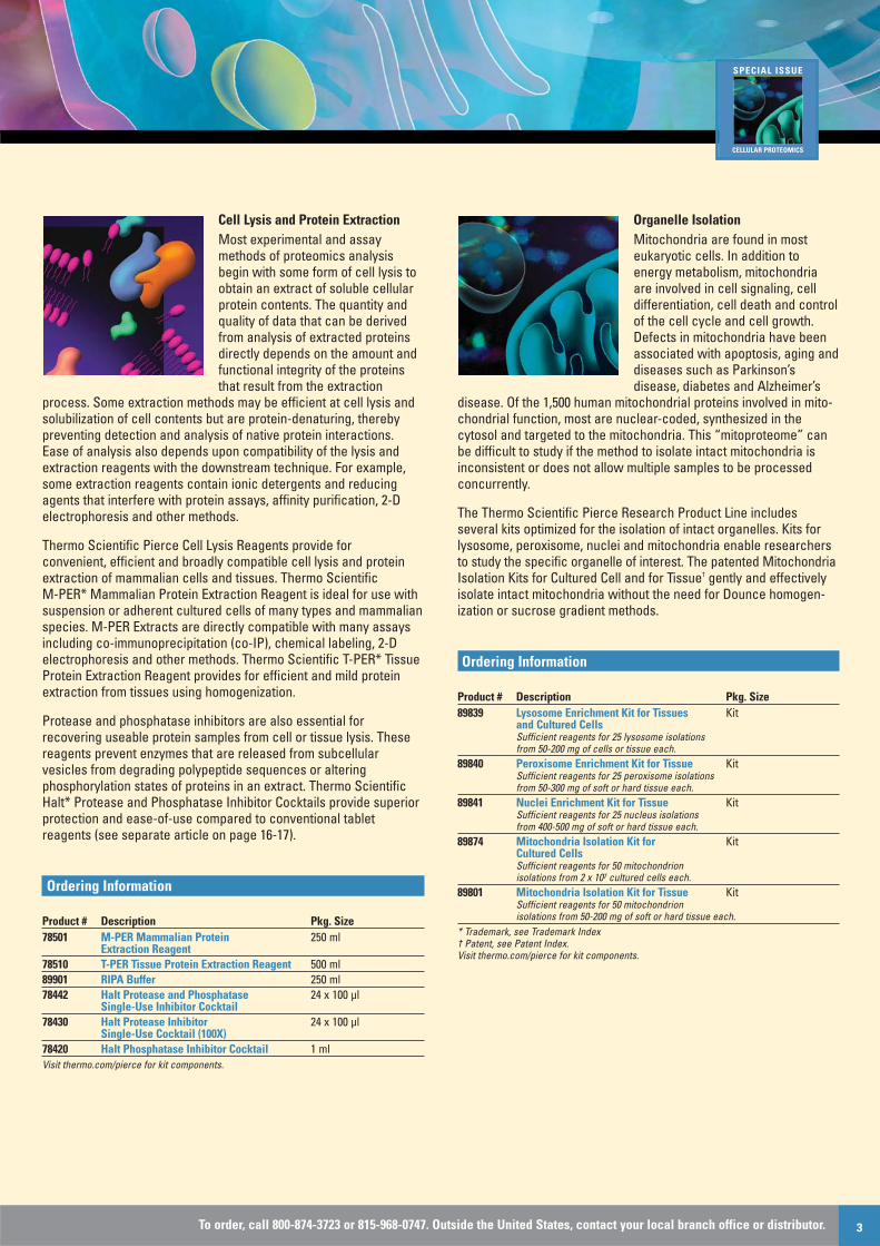
Organelle IsolationMitochondria are found in mosteukaryotic cells. In addition toenergy metabolism, mitochondriaare involved in cell signaling, celldifferentiation, cell death and controlof the cell cycle and cell growth.Defects in mitochondria have beenassociated with apoptosis, aging anddiseases such as Parkinson’sdisease, diabetes and Alzheimer’s
disease. Of the 1,500 human mitochondrial proteins involved in mito-chondrial function, most are nuclear-coded, synthesized in thecytosol and targeted to the mitochondria. This “mitoproteome” canbe difficult to study if the method to isolate intact mitochondria isinconsistent or does not allow multiple samples to be processedconcurrently.
The Thermo Scientific Pierce Research Product Line includesseveral kits optimized for the isolation of intact organelles. Kits forlysosome, peroxisome, nuclei and mitochondria enable researchersto study the specific organelle of interest. The patented MitochondriaIsolation Kits for Cultured Cell and for Tissue† gently and effectivelyisolate intact mitochondria without the need for Dounce homogen-ization or sucrose gradient methods.
Ordering Information
Product # Description Pkg. Size89839 Lysosome Enrichment Kit for Tissues Kit
and Cultured Cells Sufficient reagents for 25 lysosome isolations from 50-200 mg of cells or tissue each.
89840 Peroxisome Enrichment Kit for Tissue Kit Sufficient reagents for 25 peroxisome isolations from 50-300 mg of soft or hard tissue each.
89841 Nuclei Enrichment Kit for Tissue Kit Sufficient reagents for 25 nucleus isolations from 400-500 mg of soft or hard tissue each.
89874 Mitochondria Isolation Kit for Kit Cultured CellsSufficient reagents for 50 mitochondrion isolations from 2 x 10⁷ cultured cells each.
89801 Mitochondria Isolation Kit for Tissue Kit Sufficient reagents for 50 mitochondrion isolations from 50-200 mg of soft or hard tissue each.
* Trademark, see Trademark Index† Patent, see Patent Index.Visit thermo.com/pierce for kit components.
SPECIAL ISSUE
CELLULAR PROTEOMICS
www.thermo.com/pierce Vol. 12, Issue 14
Nuclear TargetsAlthough the “central dogma”describes the top-down flow of information from nuclear DNA totranscription of RNA to translationof proteins, the entire process isintimately controlled by bottom-upregulation from the cellularproteomic environment. For example,transcription factors are proteinsactivated by cell surface receptor-
ligand interactions and other intercellular signals and involved in the transcription of genes into RNA.
Measurement and analysis of transcription factors and otherprotein:DNA binding interactions of the nuclear proteome are facilitated by isolation or enrichment of the nuclear protein component of a sample. The Thermo Scientific NE-PER* Nuclear and Cytoplasmic Extraction Kit provides for efficient cell lysis andisolation of soluble cytoplasmic and nuclear proteins into two separate fractions. Specific transcription factors in the nuclearextract can be accurately measured in a microplate format using aChemiluminescent Transcription Factor Assay Kit for NFΚB p50,NFΚB p65 or c-Fos.
The Thermo Scientific LightShift* Chemiluminescent EMSA Kitenables accurate and highly sensitive, non-isotopic detection ofDNA binding proteins in nuclear protein extracts. Nearly any DNA-binding protein can be measured in this electrophoretic mobility shiftassay (EMSA) by supplying the appropriate biotin end-labeled DNAbinding sequence. Unlike traditional EMSAs, the LightShift Kit isformatted for chemiluminescent detection on a simple mini-blot.
Ordering Information
Product # Description Pkg. Size78833 NE-PER Nuclear and Cytoplasmic Kit
Extraction KitSufficient reagents for extracting 50 cell pelletfractions having packed cell volumes of 20 µl each(a total of ~ 2.0 g of cell paste).
89858 NFΚB p50 Chemiluminescent Transcription KitFactor Assay KitSufficient reagents for two 96-well plate assays.
89859 NFΚB p65 Chemiluminescent Transcription KitFactor Assay KitSufficient reagents for two 96-well plate assays.
89860 c-Fos Chemiluminescent Transcription KitFactor Assay KitSufficient reagents for two 96-well plate assays.
20148 LightShift Chemiluminescent EMSA Kit KitSufficient components for 100 binding reactions and detection reagents for ~800 cm2 of membrane.
89880 Chemiluminescent Nucleic Acid KitDetection Module Detection reagents from the LightShift Kit (Product # 20148).
Visit thermo.com/pierce for kit components.
Extracellular Targets Cells synthesize, present, releaseand secrete a variety of cytokines,soluble receptors, hormones andbyproducts to the extracellular environment. Synthesized inresponse to a variety of intracellularand extracellular signals, theseproducts act locally (autocrine andparacrine effects) or at a distance(endocrine effects) to communicate
and regulate cellular functions by interaction with cell surfacereceptors of specific cell signaling systems. Because their primaryfunctions are mediated by secretion in extracellular fluids, cytokinesand other such products are frequently assayed in serum, plasmaand cell culture supernatants.
Thermo Scientific Pierce Protein Research Products include ELISAkits, matched antibody pairs and recombinant proteins for analysis ofcytokines (interferons, interleukins, tumor necrosis factors, etc.),cytokine receptors (soluble and cell surface) and selected otherextracellular targets (cyclic AMP, cyclic GMP, nitric oxide,leukotriene B4, prostaglandin E4, matrix metalloproteases, etc.).These products allow scientists to characterize disease states anddrug responses by measuring production and extracellular expres-sion of soluble proteins relevant to inflammation and other criticalcellular functions.
Ordering Information
Product # Description Pkg. SizeEHIFNG Human IFN gamma ELISA Kit 1 Plate EHIFNG2 Human IFN gamma ELISA Kit 2 Plates EHIFNG5 Human IFN gamma ELISA Kit 5 Plates EH2IL6 Human IL-6 ELISA Kit 1 PlateEH2IL65 Human IL-6 ELISA Kit 5 Plates EH3TNFA Human TNF alpha ELISA Kit 1 Plate EH3TNFA2 Human TNF alpha ELISA Kit 2 PlatesEH3TNFA5 Human TNF alpha ELISA Kit 5 Plates Note: Selected mouse, rat, porcine and b ovine kits and antibodies are also available.Visit thermo.com/pierce for kit components.
Protein InteractionsInteractions among proteins areessential to most cellular functionsand critical to a full understanding ofcellular biology. For example, signalsoriginating at the surface of a cellare transmitted through the cellmembrane and to the appropriateintracellular target via protein inter-actions in the signal transductioncascade. Understanding these inter-
actions is key to unlocking the mysteries of normal biologicalprocesses as well as disease progression, which can provide thebasis for new therapeutic approaches.
Thermo Scientific Pierce Protein Research Products include avariety of tools to discover, confirm and characterize protein interactions. The photoreactive amino acid analogs L-Photo-Leucineand L-Photo-Methionine can be incorporated into proteins in a livingcell and then used to crosslink interacting proteins in their nativecellular context. Specific protein complexes can be isolated withThermo Scientific Pierce Co-immunoprecipitation Kits to search forinteracting proteins, confirm putative interactions or allow furtheranalysis of the individual proteins that make up a particular complex.The analysis of co-IP results is enhanced by using the ThermoScientific Clean-Blot IP Detection Kit, which easily binds to a primaryantibody used for Western blot detection but does not cross-reactwith the antibody used for precipitation (see separate article onpages 20-21). Thermo Scientific Pierce Crosslinkers such as BS3,DSS and formaldehyde are popular choices for fixing transientprotein interactions in place for isolation and analysis. Other kits forpull-down assays, far-Western blots and label transfer are alsoavailable to study protein interactions.
Ordering Information
Product # Description Pkg. Size22610 L-Photo-Leucine 100 mg
(L-2-amino-4, 4'-azi-pentanoic acid) 22615 L-Photo-Methionine 100 mg
(L-2-amino-5, 5'-azi-hexanoic acid) 30030 Dulbecco's Modified Eagle's Limiting 500 ml
Medium (DMEM-LM) Sterile filtered (-)L-leucine, (-)L-methionine with 4.5 g/L glucose, 4.0 mM L-glutamine, sodium pyruvate and phenol red
23600 Pierce Co-Immunoprecipitation Kit KitSufficient to immobilize up to 10 antibodiesand perform a minimum of 40 Co-IP reactions when 25 µl of antibody support is used.
23605 Pierce Mammalian KitCo-Immunoprecipitation Kit
Sufficient to immobilize up to 10 antibodies and perform a minimum of 40 Co-IP reactions when 25 µl of antibody support is used.
21580 BS3 (Bis[sulfosuccinimidyl] suberate) 50 mg21585 BS3, No-Weigh Format 8 x 2 mg 28908 16% Formaldehyde (w/v), Methanol-free 10 x 10
ml ampuleVisit thermo.com/pierce for kit components.
Post-Translational ModificationsCells rely on post-translational modification (PTM), a physiochem-ical event, to control vital processessuch as protein function, regulationand expression. Among the severalhundred known types of PTMs,protein phosphorylation, glycosyla-tion and ubiquitination are the mostintensely studied. Characterizationof these modifications, although
challenging, provides invaluable insight into the cellular functionsunderlying etiological processes.
The first challenge in studying post-translationally modified proteinsis to separate them from the sample source without damaging thestructural integrity or functionality of modified proteins. ThermoScientific Pierce Protein Research Products include a specializedline of kits and reagents that enable efficient extraction, purification,enrichment and detection of phosphorylated, ubiquitinyated andglycosylated proteins.
Ordering Information
Product # Description Pkg. Size90003 Pierce Phosphoprotein Enrichment Kit Kit
Sufficient for 10 purifications from up to 4 mgof cell lysate each.
89853 Pierce Phosphopeptide Isolation Kit KitSufficient for analyzing 30 protein digestions.
89804 Glycoprotein Isolation Kit, ConA KitSufficient reagents to isolate glycoproteins with strong affinity for ConA from 10 samples of up to 640 µl each (1-1.5 mg total protein).
89805 Glycoprotein Isolation Kit, WGA KitSufficient reagents to isolate glycoproteins with strong affinity for WGA from 10 samples of up to 640 µl each (1-1.5 mg total protein).
89899 Ubiquitin Enrichment Kit KitContains sufficient materials for enriching up to 15 lysate samples containing ~0.15 mg total protein per sample.
24565 O-GlcNAc Western Blot Detection Kit KitSufficient material to develop up to 10 mini-blots.
53074 Krypton* Glycoprotein Staining Kit KitSufficient reagents to stain 10 mini (8 cm x 10 cm) gels.
24562 GelCode* Glycoprotein Staining Kit KitSufficient reagents to stain 10 mini (8 cm x 10 cm) gels.
24550 GelCode Phosphoprotein Staining Kit KitSufficient reagents to stain 10 mini (8 cm x 10 cm) gels.
* Trademark, see Trademark IndexVisit thermo.com/pierce for kit components.
To order, call 800-874-3723 or 815-968-0747. Outside the United States, contact your local branch office or distributor. 5
SPECIAL ISSUE
CELLULAR PROTEOMICS

www.thermo.com/pierce Vol. 12, Issue 16
Cellular Imaging and AnalysisTools for the analysis of cellularprocesses consist of severallabeling technologies to facilitateanalysis by fluorescencemicroscopy, flow cytometry,Western blotting, high-contentscreening and other array platforms.In recent years a growing apprecia-tion for the complexity of cellularprocesses has increased demand
for higher resolution probing and imaging technologies that arecapable of measuring multiple parameters simultaneously and/or thedistribution and abundance of specific cellular targets over time. Wehave met the demand for such a technology by developing ThermoScientific DyLight* Fluors, a complete family of high-intensity, photo-stable fluorescent dyes for labeling antibodies and other molecularprobes. (See separate article on pages 9-12.)
DyLight Fluors provide superior performance to traditional fluorophoressuch as fluorescein and enable multiplexing analysis in several non-overlapping excitation/emission channels. DyLight Fluor-conjugatedsecondary antibodies are the basis for Thermo Scientific Cellomics*High Content Screening Reagent Kits (see text that follows thissection). Conjugated mouse and rabbit secondary antibodies are available, as well as both NHS-ester-activated and maleimide-activated forms, providing for efficient and specific labeling of primary amines and sulfhydryl groups, respectively.
Table 1. Thermo Scientific DyLight Fluorescent Dyes.
DyLight Fluor Ex/Em Emission ColorDyLight 405 400/420 BlueDyLight 488 493/518 GreenDyLight 549 550/568 YellowDyLight 633 638/658 RedDyLight 649 646/674 RedDyLight 680 682/715 Near IRDyLight 800 770/794 Infrared
Ordering Information
Product # Description Pkg. Size46407 DyLight 549 NHS Ester 1 mg46408 DyLight 549 NHS Ester 5 x 50 µg46607 DyLight 549 Maleimide 1 mg53035 DyLight 549 Microscale Antibody Kit
Labeling Kit53034 DyLight 549 Antibody Labeling Kit Kit 35507 DyLight 549 Goat Anti-Mouse 1 mg
IgG (H+L) Conjugate35557 DyLight 549 Goat Anti-Rabbit 1 mg
IgG (H+L) Conjugate21837 DyLight 549 Streptavidin Conjugate 1 mg22837 DyLight 549 NeutrAvidin Protein Conjugate 1 mgVisit thermo.com/pierce for kit components.Note: A similar range of reagents, kits and conjugates is available for all DyLight Fluors.Search for “DyLight” at www.thermo.com/pierce.
High-Content Screening in DrugDiscoveryHigh-content screening (HCS) isemerging as an indispensable toolfor analysis of cellular biologicalcomplexity in relation to drugdiscovery. HCS involves visualizationof target protein expression anddistribution in a cell population,single cell or subcellular structureand data analysis with image-anal-
ysis tools. The ability of HCS to measure multiple parameters relatedto spatial and temporal changes in populations of individual cellsallows for measurement of experimental effects from a systemperspective. HCS is widely used in all stages of target-based drugdiscovery that involve the study of cells, including target discovery,drug screening in cell-based assays, early safety evaluation, mode-of-action analysis and in vivo studies to monitor cell fate.
The Thermo Scientific Cellomics HCS Platform comprises a totalsolution for high-content screening. The platform includes fluores-cent reagents, imaging equipment and software for image analysis,data management, automation, and informatics. Cellomics HCSReagent Kits provide easy-to-use, validated probes (fluorescentstains, target-specific primary antibodies and fluorescent secondaryantibodies), reagents and protocols to prepare imaging-qualitysamples for HCS. (See separate article on pages 7-8.)
Available Thermo Scientific Cellomics HCS Reagent KitsCategory of Analysis Selected List of Kit TargetsCytotoxicity and Apoptosis Caspase 3, Caspase 9, Cleaved PARP,
Cytochrome C, LC3B (autophagy), Poly-ubiquitinand other parameters
Genotoxicity, DNA Damage Ku70/80, MDM2, Micronucleus, p21, p53, ATM,and Repair Chk2, H2AXInflammation and Cell Stress CHOP/GADD153, COX-2, FKBP52,
Heme Oxygenase 1, Hsp27, Hsp60, Hsp70, Hsp90,iNOS, MnSOD, NFAT-1, NFκB, oxidative stress,p38, 4E-BP1, c-Jun, S6, STAT1, 2 & 3
Cell Signaling and ATF-2, β-Catenin, ERK, FOXO1A & 3A, HIF-1α,Transcription Factors AKT, CREB, GSK-3, JNK, PKA, PKCα, Smad3Cell Cycle and Proliferation BrdU, Cyclin B1, Ki67, Histone H3, PLK1, Rb
and other parametersCell Morphology and F-actin, Tubulin, Whole Cell Stains and severalPhenotypic Changes other parametersAccessory Reagents Whole cell stains, fluorescent 2° antibodies, etc.Kits are specific for active (e.g., phosphorylated) forms of target proteins.
Thermo Scientific BioImage Redistribution* Technology is a cell-based assay platform that uses protein translocation as areadout for signaling pathway activity. Redistribution Assays arehigh-throughput, high-content assays ideal for the profiling of leadseries, primary screening of compound libraries and measuringgene silencing by siRNA.
Redistribution Technology is based on:• Labeling targets with green fluorescent protein (GFP)• Generation of stably transfected assay cell lines• Measurement of protein translocation• Image analysis and quantification of cellular responses
For more information about Cellomics and BioImage products and custom services,please visit www.thermo.com/HCS.
6 www.thermo.com/pierce Vol. 12, Issue 1

To order, call 800-874-3723 or 815-968-0747. Outside the United States, contact your local branch office or distributor. 7
Heat shock proteins (HSPs) are ubiquitous and essential forcellular homeostasis and survival in response to a variety ofstresses. HSPs have multiple cellular functions in signal trans-duction, cell survival and cell death, and protein dynamics,including synthesis, folding, degradation and translocation.There ar e five major HSP families: small HSPs, Hsp60 or chap-eronins, Hsp70, Hsp90, and Hsp110. The new ThermoScientific Cellomics* HCS Reagent Kits enable quantitative,cell-based, high-content screening (HCS) of different HSPs toanalyze stress response of cells. These kits and assays aredesigned for fluorescence labeling, detection and imaging ofdifferent HSPs in the cell, including Hsp27 and phospho-Hsp27, Hsp60 and Hsp90β, Hsp70 and Hsp90α, and FKBP52(Hsp56).
We offer drug discovery and systems biology researchers completesystems for high-content screening (HCS) and high-content analysis(HCA). The Cellomics Product Family offers a total solution platform,comprising automated imaging instrumentation, BioApplicationImage Analysis Software and high-content informatics, coupled withreagents, laboratory automation and services to provide the fastestpossible time-to-decision. Thermo Scientific Reagents includeCellomics HCS Reagent Kits and BioImage Redistribution*Technology. BioImage Redistribution Technology is a high-contentcell-based assay approach for monitoring cellular translocation ofGFP-tagged proteins in response to drug compounds, siRNA or otherstimuli. A wide range of target classes are amenable to redistributionassays, including kinases, GPCRs, transcription factors and others.
Thermo Scientific BioImage Products• Validated Redistribution Assays for screening or profiling
applications• CryoRedi Ready-to-use Cryopreserved Cells • High-content screening services and custom assay development
Cellomics HCS Reagent Kits are validated, ready-to-use HCAreagent assays. By eliminating assay development time and costs,productivity increases.
Highlights:• Flexible – available in multiplex or singleplex applications• Robust – Z´ factor > 0.3 for all assays• Optimized protocol and reagent preparations included• Validated on Thermo Scientific ArrayScan* Instruments and
compatible with other HCS platforms and with fluorescencemicroscopes
To demonstrate the performance of the Cellomics HCA Reagent Kits,we treated two representative cancer cell types, A549 and U2OS,with different known compounds and then imaged and analyzed thecellular responses with the HCS assays. The kits enabled us to
precisely determine and evaluate the cell stress caused by thecompounds. In summary, the use of automated, cell-based, high-content assays to monitor key HSPs is a suitable and effectivestrategy for cellular stress analysis.
Figure 1. Illustration of heat shock proteins’ response to cellular stress.
Figure 2. Staining of A549 cells with various Thermo Scientific Cellomics HCSKits for HSPs in response to CuSO4 treatment. The cellular levels of Hsp27,phospho-Hsp27, Hsp70, Hsp90 and FKBP52 are increased by CuSO4 (1 mM)treatment for 24 hours with HSPs accumulating in the nucleus. MitochondrialHsp60 is also increased. Treatment of specific Hsp90 inhibitor, 17-AAG,increased Hsp27 and Hsp70 levels by inhibiting Hsp90-mediated proteindegradation (data not shown).
Heat
Mitochondria
HSPs
Nuclear Translocation
Nucleus
OxidativeStressUV
Hsp90α
Hsp90β
Hsp90
HSF-1
Hsp27
Hsp27
Hsp60
Hsp70
PRAK FKBP52 (Hsp56)p38MAPK
pMAPKAPK-2
p
SPECIAL ISSUE
CELLULAR PROTEOMICS
Total Solution Platform for High-Content Screening
Detect cellular stress responses to drugsin cell-based, high-content assaysHeat shock proteins are powerful cell stress indicators
By Suk Jin Hong, Ph.D., Thermo Fisher Scientific Inc., Rockford, IL, USA
Hsp27 / phospho-Hsp27
Hsp70 / Hsp90α
Immunophilin FKBP52 (Hsp56)
Hsp60 / Hsp90β
To order, call 800-874-3723 or 815-968-0747. Outside the United States, contact your local branch office or distributor. 7
www.thermo.com/pierce Vol. 12, Issue 18
Total Solution Platform for High-Content Screening (Cont.)
Ordering Information
Product # Description Pkg. Size8406001 Hsp27 and Phospho-Hsp27 Detection Kit 1 x 968406002 5 x 968406101 Hsp27 Detection Kit 1 x 968406102 5 x 968406201 Phospho-Hsp27 Detection Kit 1 x 968406202 5 x 968406701 Hsp60 and Hsp90β Detection Kit 1 x 968406702 5 x 96
Ordering Information
Product # Description Pkg. Size8406801 Hsp60 Detection Kit 1 x 968406802 5 x 968406301 Hsp70 and Hsp90α Detection Kit 1 x 968406302 5 x 968406401 Hsp70 Detection Kit 1 x 968406402 5 x 968406501 Hsp90α Detection Kit 1 x 968406502 5 x 968406601 Immunophillin FKBP52 Detection Kit 1 x 968406602 5 x 96For a complete listing of Cellomics HCS Reagent Kits and for more information aboutBioImage Products, visit www.thermo.com/hcs.* Trademark, see Trademark Index
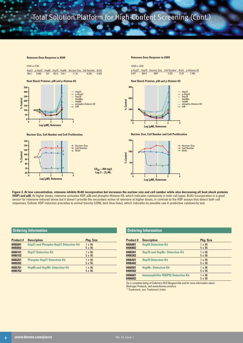
Figure 3. At low concentration, rotenone inhibits BrdU incorporation but increases the nuclear size and cell number while also decreasing all heat shock proteins(HSP) and p38. At higher doses, rotenone activates HSP, p38 and phospho-Histone H3, which indicates cytotoxicity in both cell types. BrdU incorporation is a goodsensor for rotenone-induced stress but it doesn't provide the secondary action of rotenone at higher doses, in contrast to the HSP assays that detect both cellresponses. Cellular HSP induction precedes to animal toxicity (LD50, dark blue lines), which indicates its possible use in predictive cytotoxicity test.
Rotenone Dose Response in A549 Rotenone Dose Response in U20S
Heat Shock Proteins, p38 and p-Histone H3
Nuclear Size, Cell Number and Cell Proliferation
LD50 = 394 mg/LLog 3 = [3μM]
EC50 or IC50 EC50 or IC50
Hsp27 p-Hsp27 Hsp60 Hsp70 Hsp90 Nuclear Size Cell Number BrdU 964.1 9.093 527 452.9 724.1 11.70 8.289 9.929
p-Hsp27 Hsp70 Nuclear Size Cell Number BrdU p-Histone H35.547 684.5 5087 5.291 13.35 7.390
Hsp27p-Hsp27Hsp70Hsp90αHsp60phospho-Histone H3p38
350
300
250
200
150
100
50
00 1 2 3 4
% C
ontr
ol
Log [µM], Rotenone
Heat Shock Proteins, p38 and p-Histone H3
Nuclear SizeCell NumberBrdU
Nuclear Size, Cell Number and Cell Proliferation
140
130
120
100
90
80
70
600 1 2 3 4
% C
ontr
ol
Log [µM], Rotenone
0
100
200
300
0 1 2 3 4%
Con
trol
Log [µM], Rotenone
Hsp27p-Hsp27Hsp70Hsp90α Hsp60phospho-Histone H3p38
0
50
100
150
0 1 2 3 4
% C
ontr
ol
Log [µM], Rotenone
Nuclear SizeCell NumberBrdU

Thermo Scientific DyLight* Dyes have absorption spectraranging from 400 nm to 770 nm (Table 1) and match the prin-cipal output wavelengths of common fluorescenceinstrumentation. The DyLight Dyes exhibit higher fluorescenceintensity and photostability than Alexa*, CyDye* and LI-CORDyes in many applications and remain highly fluorescent overa broad pH range (pH 4-9) (Figures 2 and 3). Additionally, thewater solubility of the DyLight Dyes allows a high dye-to-protein ratio without precipitation during conjugation.
Table 1. Spectral properties of Thermo Scientific DyLight Fluorescent Dyes.
Spectrally SimilarEmission DyLight Dye Ex/Em* ε† DyesBlue 405 400/420 30,000 Alexa 405Green 488 493/518 70,000 Alexa 488,
fluorescein and FITCYellow 549 550/568 150,000 Alexa 546, Alexa 555,
Cy3 and TRITCRed 633 638/658 170,000 Alexa 633Red 649 646/674 250,000 Alexa 647 and Cy5Near Infrared 680 682/715 140,000 Alexa 680 Infrared 800 770/794 270,000 IRDye 800* Excitation and emission maxima in nanometers† Molar extinction coefficient (M 1 cm 1)
Highlights:• Brightness – high-fluorescence intensity provides outstanding
sensitivity and requires less conjugate• Photostability – exceptional resistance to photobleaching enables
fluorescence imaging under the most demanding conditions (e.g., structured illumination, 4pi microscopy)
• Instrument compatibility – emission spectra match the principaloutput wavelengths of common fluorescence instrumentation
Figure 2. Thermo Scientific DyLight 488 and DyLight 633 Dyes exhibitoutstanding fluorescence in structured illumination. The uniform fluorescenceintensity throughout the images demonstrates the outstanding brightness andphotostability of DyLight 488 and 633 Dyes. Red: Alpha tubulin detected in HeLacells with anti-tubulin monoclonal antibody and DyLight 633 Dye-conjugatedsecondary antibody (highly cross-adsorbed). Green: Histone H4 detected withanti-histone monoclonal antibody and DyLight 488 Dye-conjugated secondaryantibody (highly cross-adsorbed). Blue: Nucleus counter-stained with fluores-cent mounting media containing DAPI. Images were acquired with the AxioImager.Z1 and ApoTome Slider (Zeiss MicroImaging, Inc). The ApoTome Moduleprovides confocal-like resolution allowing optical sectioning without using apinhole (e.g., confocal). No image enhancement was performed.
Bright new alternatives to conventionalfluorescent dyes
Fluorescent DyesEx
cita
tion
Emis
sion
DyLight 549
450 500 550 650600 700Wavelength (nm)
Abs max: 550 nmEm max: 568 nm
Exci
tatio
n
Emis
sion
DyLight 680
550 600 650 750700 800Wavelength (nm)
Abs max: 682 nmEm max: 715 nm
Exci
tatio
n
Emis
sion
DyLight 649
550 600 650 750700 800Wavelength (nm)
Abs max: 646 nmEm max: 674 nm
Exci
tatio
n
Emis
sion
DyLight 800
650 700 750 850800 900Wavelength (nm)
Abs max: 770 nmEm max: 794 nm
Abs max: 400 nmEm max: 420 nm
Exci
tatio
n
Emis
sion
DyLight 405
300 350 400 500450 550Wavelength (nm)
Abs max: 493 nmEm max: 518 nm
Exci
tatio
n
Emis
sion
DyLight 488
420370 470 520 620570 670Wavelength (nm)
Abs max: 638 nmEm max: 658 nm
Exci
tatio
n
Emis
sion
DyLight 633
550500 600 650 750700Wavelength (nm)
Figure 1. Excitation and emission spectra for Thermo Scientific DyLight Dyes.
To order, call 800-874-3723 or 815-968-0747. Outside the United States, contact your local branch office or distributor. 9
SPECIAL ISSUE
CELLULAR PROTEOMICS

www.thermo.com/pierce Vol. 12, Issue 110
Ordering Information
Product # Description Pkg. SizeAmine-Reactive Dyes46400 DyLight 405 NHS Ester 1 mg46401 DyLight 405 NHS Ester 5 x 50 µg46402 DyLight 488 NHS Ester 1 mg46403 DyLight 488 NHS Ester 5 x 50 µg46407 DyLight 549 NHS Ester 1 mg46408 DyLight 549 NHS Ester 5 x 50 µg46414 DyLight 633 NHS Ester 1 mg46417 DyLight 633 NHS Ester 5 x 50 µg46415 DyLight 649 NHS Ester 1 mg46416 DyLight 649 NHS Ester 5 x 50 µg
Product # Description Pkg. Size46418 DyLight 680 NHS Ester 1 mg46419 DyLight 680 NHS Ester 5 x 50 µg46421 DyLight 800 NHS Ester 1 mg46422 DyLight 800 NHS Ester 5 x 50 µgSulfhydryl-Reactive Dyes46600 DyLight 405 Maleimide 1 mg46602 DyLight 488 Maleimide 1 mg46607 DyLight 549 Maleimide 1 mg46613 DyLight 633 Maleimide 1 mg46615 DyLight 649 Maleimide 1 mg46618 DyLight 680 Maleimide 1 mg46621 DyLight 800 Maleimide 1 mg
Ordering InformationConjugates: Package size for these items is 1 mg at 1 mg/ml.
Product #DyLight DyLight DyLight DyLight DyLight
Description 488 Dye 549 Dye 649 Dye 680 Dye 800 DyeGoat Anti-Mouse IgG (H+L) 35502 35507 35515 35518 35521Goat Anti-Rabbit IgG (H+L) 35552 35557 35565 35568 35571Streptavidin 21832 21837 21845 21848 21851NeutrAvidin* Biotin-Binding Protein 22832 22837 22845 22848 22853* Trademark, see Trademark Index
Figure 2. 3D reconstruction of HeLa cells in doublefluorescence using Thermo Scientific DyLight 488 andDyLight 633 Dyes. The uniform fluorescence intensitythroughout the images demonstrates the outstandingbrightness and photostability of DyLight 488 and 633 Dyes.Structured illumination was used to acquire images of 56optical sections (~7 µm) and processed with AxioVisionSoftware to generate 3D reconstructions of HeLa cells.The image was not edited (e.g., contrast, brightness orgamma correction) before rendering. Green: Alpha tubulindetected with anti-tubulin monoclonal antibody andDyLight 488 Dye-conjugated secondary antibody (highlycross-adsorbed). Red: Hsp60 detected with anti-Hsp60polyclonal antibody and DyLight 633 Dye-conjugatedsecondary antibody. Blue: Nucleus counter-stained withfluorescent mounting media containing DAPI.

The Thermo Scientific DyLight Antibody Labeling Kits were specifically developed for fast, efficient labeling of anti-bodies. Two convenient kit formats are available toaccommodate varied labeling requirements. The AntibodyLabeling Kits contain all necessary components to performthree separate labeling reactions using 1 mg of IgG or similarquantities of other proteins. The DyLight MicroscaleAntibody Labeling Kits contain all the necessary componentsto perform five separate labeling reactions using 100 µg ofIgG. The labeling kits use high-performance spin desaltingcolumns to provide exceptional dye removal and antibodyrecovery (Figure 1).
Highlights: • Fast – fluorescently label and purify protein in approximately
one hour (Figure 2)• Amine-reactive dyes – label virtually any protein• Efficient non-reacted dye removal• Minimal sample dilution• Spin column format eliminates the need for column preparation,
fraction screening and waiting for protein to emerge from column
Figure 1. Thermo Scientific DyLight Antibody Labeling Kits provide outstandingrecovery.
Figure 2. Protocol summary for Thermo Scientific DyLight AntibodyLabeling Kits.
0
20
40
60
80
100
Thermo ScientificDyLight Antibody
Labeling Kit
84%(n=8)
83%(n=7)
Thermo ScientificDyLight Microscale
Antibody Labeling Kit
Ant
ibod
y Re
cove
ry
Step 1. Labeling reaction
Add antibody to vial containing pre-measured dye. Incubate 1 hour at
room temperature.
Apply labeling reaction to Spin
Desalting Column.
Recover labeled antibody.
Centrifuge
30 seconds
Step 2. Removal of excess fluorescent dye
SPECIAL ISSUE
CELLULAR PROTEOMICS
To order, call 800-874-3723 or 815-968-0747. Outside the United States, contact your local branch office or distributor. 11
Antibody labeling kitsLabel and purify antibodies in one hour
Microscale KitsContain sufficient reagents to label and purify 5 x 100 µg of IgG In addition to contents listed below, all Microscale Kits include: • Reaction Buffer, 1 ml• Spin Columns, 5 ea.• Microcentrifuge Collection Tubes, 10 ea.• Purification Resin, 5 ml
Ordering Information
Product # Description Pkg. Size53021 DyLight 405 Microscale Antibody Kit
Labeling KitDyLight 405 NHS Ester 5 vials
53025 DyLight 488 Microscale Antibody KitLabeling KitDyLight 488 NHS Ester 5 vials
53035 DyLight 549 Microscale Antibody KitLabeling KitDyLight 549 NHS Ester 5 vials
53047 DyLight 633 Microscale Antibody KitLabeling KitDyLight 633 NHS Ester 5 vials
53051 DyLight 649 Microscale Antibody t Kit Labeling KitDyLight 649 NHS Ester 5 vials
53057 DyLight 680 Microscale Antibody KitDyLight 680 NHS Ester 5 vials
53063 DyLight 800 Microscale Antibody Kit DyLight 800 NHS Ester 5 vials
Antibody Labeling KitsContain sufficient reagents to label and purify 3 x 1 mg of IgG orsimilar quantities of other proteinsIn addition to contents listed below, all Antibody Labeling Kitsinclude: • Reaction Buffer, 1 ml• Spin Columns, 6 ea.• Microcentrifuge Collection Tubes, 12 ea.• Purification Resin, 5 ml
Ordering Information
Product # Description Pkg. Size53020 DyLight 405 Antibody Labeling Kit Kit
DyLight 405 NHS Ester 3 vials53024 DyLight 488 Antibody Labeling Kit Kit
DyLight 488 NHS Ester 3 vials53034 DyLight 549 Antibody Labeling Kit Kit
DyLight 549 NHS Ester 3 vials53046 DyLight 633 Microscale Antibody Kit
Labeling Kit 3 vialsDyLight 633 NHS Ester
53050 DyLight 649 Antibody Labeling Kit Kit DyLight 649 NHS Ester 3 vials
53056 DyLight 680 Antibody Labeling Kit Kit DyLight 680 NHS Ester 3 vials
53062 DyLight 800 Antibody Labeling Kit Kit DyLight 800 NHS Ester 3 vials
Compatible Product22858 Dye Removal Columns Kit
Includes: Purification Resin 5 mlSpin Columns 10 ea. Microcentrifuge Collection Tubes 20 ea.

www.thermo.com/pierce Vol. 12, Issue 112
SPECIAL ISSUE
CELLULAR PROTEOMICS
Western Blotting
Multiplex fluorescent Western blotting kitsExcellent brightness make these conjugates a clear alternative
The fluorescence detection of two different targets on a singleWestern blot is now easier to perform with Thermo ScientificDyLight* Western Blotting Kits. The kits provide a highly optimized and convenient format to save you the time and frustration of having to evaluate reagents for compatibility withfluorescent Western blotting.
Two kit formats are available to provide compatibility with a widerange of fluorescence imaging systems (Table 1). The DyLight549/649 Western Blotting Kit utilizes DyLight 549 and DyLight 649labeled secondary antibodies for imaging in the visible region of thespectrum (Figure 1). The DyLight 680/800 Western Blotting Kit utilizesDyLight 680- and DyLight 800-labeled secondary antibodies forimaging in the near-infrared region (e.g., LI-COR Odyssey* ImagingSystem) of the spectrum (Figure 2).
Highlights:• Easily multiplexed – detect two different targets on a single
Western blot• Optimized format – protocol requires minimal to no optimization,
saving time and the frustration of having to evaluate and optimizereagents (e.g., secondary antibody dilutions, etc.)
• Plug and play – kits include DyLight Fluor-labeled secondary anti-bodies, blocking buffer, wash buffer, dual-labeled fluorescentmolecular weight markers and low-fluorescence membranes
• Instrument compatible – DyLight Fluor spectra match the principaloutput wavelengths of common fluorescence instrumentation
Figure 1. The Thermo Scientific DyLight 549/649 Western Blotting Kit provideslower background and higher signal in two-color Western blot detectioncompared to a competing fluorescent Western blotting kit. Proteins were separated in 4-20% Precise* Protein Gels and transferred to a low-fluorescencePVDF membrance. The membranes were blocked overnight in 1% BSA andtarget proteins were detected following manufacturer-recommended protocols.Membranes were imaged with a Typhoon* 9410.
Table 1. Thermo Scientific DyLight Western Blotting Kit spectral properties.
SpectrallyWestern Blotting Kit Ex/Em† ε‡ Similar DyesDyLight 549/649 Western Blotting Kit DyLight 549 Goat Anti-Mouse IgG (H+L) 550/568 150,000 Alexa 546,
Alexa 555, Cy3DyLight 649 Goat Anti-Mouse IgG (H+L) 646/674 250,000 Alexa 647,
Cy3DyLight 680/800 Western Blotting Kit DyLight 680 Goat Anti-Mouse IgG (H+L) 682/715 140,000 Alexa 680,
Cy 5.5DyLight 800 Goat Anti-Rabbit IgG (H+L) 770/794 270,000 IRDye* 800† Excitation and emission maxima in nanometers‡ Molar extinction coefficient (M 1 cm 1)
Figure 2. The Thermo Scientific DyLight 680/800 Western Blotting Kit provideslow background and high signal in two-color Western blot detection. Proteinswere separated in 4-20% Precise Protein Gels and transferred to Low-FluorescencePVDF Transfer Membrane (Product # 22860). The membrane was blockedovernight in SEA BLOCK Blocking Buffer (Product # 37527) and target proteins were detected following the recommended protocol. Membranes were imagedwith the LI-COR Odyssey Infrared Imaging System. Tubulin was detected from the indicated quantity of HeLa cell lysate. Purified TNFα was detected at the indicated quantity.
Ordering Information
Product # Description Pkg. Size 22855 DyLight 680/800 Western Blotting Kit Kit
Sufficient reagents for 10 Western blots.Includes: DyLight 680 Goat Anti-Mouse IgG (H+L) 120 µl
DyLight 800 Goat Anti-Rabbit IgG (H+L) 120 µl DyLight Infrared Protein Molecular 30 µl
Weight MarkersWash Buffer (30X) 200 mlSEA BLOCK Blocking Buffer 500 mlLow-Fluorescence PVDF 10 ea.
Transfer Membrane* Trademark, see Trademark Index
25 n
g
12 n
g
6 ng
3 ng
1 ng
0.5
ng
1 ng
3 ng
6 ng
12 n
g
25 n
g
25 n
g
12 n
g
6 ng
3 ng
1 ng
0.5
ng
1 ng
3 ng
6 ng
12 n
g
25 n
g
MW
Mar
ker
MW
Mar
ker
DyLight 549/649 Western Blotting Kit
ECL Plex Western Blotting Kit
TNFα
Tubulin
TNFα
Tubulin
Tubulin
DyLight 680/800 Western Blotting Kit
1 µg
0.5
µg
0.25
µg
0.12
5 µg
0.06
3 µg
6.25
µg
12.5
µg
25 µ
g
50 µ
g
TNFα

To order, call 800-874-3723 or 815-968-0747. Outside the United States, contact your local branch office or distributor. 13
Validated stand-alone antibodies and new siRNA Western blotting modules Rigorously tested antibodies for the most precise detection available
The newly expanded Thermo Scientific SuperSignal* siRNA-Western Blot Product Line now includes complete modules aswell as stand-alone antibodies for more than 100 targets.
The SuperSignal siRNA/Antibody Modules contain Thermo ScientificDharmacon ON-TARGETplus* SMARTpool* siRNA Reagents andvalidated antibodies to ensure accurate detection of the specifiedprotein. When used with the SuperSignal siRNA ChemiluminescentDetection Module (Product # 82200), each gene-specificsiRNA/Antibody Module provides for precise, efficient knockdownand accurate, sensitive Western blot detection in an optimizedprotocol using predetermined and validated reagent concentrations.Our paired validation of siRNA reagents and detection antibodies inthese modules means that you can focus on the science instead ofreagent optimization.
Highlights:• Validated antibodies for accurate protein detection• Thermo Scientific SuperSignal Chemiluminescent Substrate† for
sensitive protein detection• Thermo Scientific Dharmacon ON-TARGETplus SMARTpool siRNA
Reagents for potent gene knockdown and greatly reduced off-target effects
Gene Silencing ReagentsON-TARGETplus SMARTpool siRNA Reagents silence genes in amanner similar to the natural silencing pathway. The reagentsreduce false negatives by targeting four different mRNA regionssimultaneously and reducing the effective concentration of individual siRNA molecules. This SMARTpool Technology is a widelyaccepted strategy for reducing off-target effects. The use of proprietary ON-TARGETplus Chemical Modifications to the siRNAmolecule provide further reduction of off-target effects (Figure 1).The combination of validated antibody, potent gene knockdown andintracellular target coverage provides valuable tools for studyingcomplex intracellular pathways.
Figure 1. Reduction of off-target effects.
Stand-alone Antibodies for Western BlottingSpecific and reliable detection of target proteins is especially important in Western blot confirmation of siRNA knockdownbecause the purpose is to detect the absence or decrease of proteinrelative to controls. Therefore, our development process includedscreening more than 400 antibodies to 185 different targets to ensurethat we offered only those capable of accurate, sensitive and reliable detection. Many of the commercially available antibodiesscreened did not even detect the specified protein (Table 1 andFigure 2).
We now offer the validated antibodies both as integral validatedparts of each siRNA/Antibody Module and as stand-alone reagentsfor use in siRNA Western blotting or other Western blotting applications that you have developed independently. Don’t wasteyour time and money screening numerous antibodies just to find onethat detects your target protein! We’ve done the work for you,offering the best intracellular target antibodies available.
Table 1. Variable performance of commercially available antibodies.Results are listed as the percent of more than 400 antibodies tested.
Result Total (%)No band detected 24High background/unacceptable # nonspecific bands detected 26Incorrect band (as indicated by lack of siRNA knockdown) 7Band detected at appropriate MW 43
Figure 2. Validated Thermo Scientific SuperSignal siRNA AntibodyDetection Module Antibodies accurately recognize specific targets. Cells weretransfected with ON-TARGETplus SMARTpool siRNA Reagents (ILK2 or AKT2).Cell lysates were analyzed by Western blot, comparing detection by SupplierA and B antibodies and validated Thermo Scientific Antibodies (included inrespective ILK2 and AKT2 siRNA/Antibody Detection Modules or available asstand-alones). Lane 1. Mock transfection, Lane 2. control pool siRNA and Lane3. gene-specific siRNA. Arrows indicate ILK protein band (52 kDa) and AKT2 (60 kDa). Specific target proteins and associated knockdowns were detectedby the validated Thermo Scientific Antibodies, but not by the correspondingantibodies from Suppliers A and B.
1 2 3
Supplier A
ILK2 AKT2
Thermo Scientific Supplier B Thermo Scientific
1 2 3 1 2 31 2 3
IndividualsiRNA
ON-TARGETplus Individual siRNA
ON-TARGETplus SMARTpool
SMARTpool
Number Off-Target Genes
0 20 40 60 80 100 120
RNA Interference and Western Blotting

www.thermo.com/pierce Vol. 12, Issue 114
* Trademark, see Trademark Index† Patent, see Patent Index†† Antibody detects both MAPK1 and MAPK3 § Antibody detects SMAD2 and SMAD3
Target Mod Prod # Ab Prod #AKT1 82217 82310AKT2 82247 82311AR 82248 82312ARF6 82262 82313ATM 82211 82314ATR 82246 82315AURKB 82263 82316BAD 82286 82317BAX 82265 82318BCL2 82209 82319BCL2L1 (BclXL) 82223 82320BID 82283 82321BIRC4 82287 82322BIRC5 (Survivin) 82250 82323BRCA1 82238 82324BUB1B 82295 82325CASP3 82251 82326CASP8 82265 82327CASP9 82266 82328CAV1 82231 82329CCNB1 (Cyclin B) 82267 82330CCND1 (Cyclin D) 82215 82331CCNE1 (Cyclin E) 82268 82332CDC2 82229 82333CDC25C 82291 82334CDH1 (E-Cadherin) 82302 82335CDK2 82237 82336CDK4 82212 82337CDK5 82269 82338CDK6 82252 82339CDK9 82270 82340CDKN1A 82253 82341CDKN1B (p27) 82292 82342CDKN2A (p14ARF) 82309 82343CDKN2A (p16INK) 82271 82344CHEK1 82241 82345CHEK2 82220 82346CHUK 82227 82347CSNK2A1 (CKIIα) 82272 82348CTNNB1 (β Catenin) 82218 82349E2F1 82254 82354EGFR 82219 82355EP300 82205 82356ERBB2 82239 82357FOXO1A 82273 82358FRAP1 (mTOR) 82243 82359GRB2 82294 82360GSK3A 82298 82361GSK3B 82249 82362HDAC1 82226 82363HDAC2 82303 82364
Target Mod Prod # Ab Prod #HDAC3 82299 82365HIF-1A 82240 82366HRAS 82207 82367HSPA1A (HSP70) 82296 82368HSPB1 82256 82369IKBKB 82221 82370ILK 82274 82371IRS1 82306 82372ITGB1 (CD29) 82304 82373JUN 82206 82374KIF11 82257 82375LYN 82290 82376MAP2K1 (MEK1) 82236 82377MAP2K3 82308 82378MAP3K7 (TAK1) 82275 82379MAPK1 (ERK2) 82203 82380 ††
MAPK3 (ERK1) 82210 82380 ††
MAPK8 82293 82381MAPK14 (p38) 82208 82382MDM2 82258 82383MET 82228 82384MYC 82202 82385NCK1 82259 82386NFKB1 (p50/p105) 82297 82387PDPK1 82289 82388PLCG1 82300 82390PLK1 82216 82391PPP2CA 82260 82392PRKACA 82285 82393PRKCA 82276 82394PRKDC 82277 82395PRKR 82255 82389PTEN 82233 n/aPTK2 (FAK1) 82242 82396RAF1 82284 82397RB1 82278 82398RELA (NFΚB) 82222 82399RHOA 82235 82400RIPK1 82282 82401SMAD2 82232 82402SMAD3 82279 82403 §
SMAD4 82280 82404SOS1 82305 82405SP1 82213 82406SRC 82204 82407STAT1 82245 82408STAT3 82225 82409STAT6 82307 82410TP53 82001 82411TTK 82281 82412VIL2 (Ezrin) 82261 82413
Ordering Information
Product # Description Pkg. Size82200 SuperSignal siRNA Chemiluminescent Kit
Detection ModuleSee Below SuperSignal siRNA Module for Target KitSee table below for Product # and Module Targets, visit thermo.com/pierce for kit components.See Below Primary Antibody for Target KitSee table below for Product # and Antibody Targets, visit thermo.com/pierce for kit components.

To order, call 800-874-3723 or 815-968-0747. Outside the United States, contact your local branch office or distributor. 15
ELISA
ELISA assay development made easierNew screening sets quantify human and bovine cytokines
Thermo Scientific Screening Sets contain the basic components and protocol required to develop a colorimetricsandwich ELISA to quantify a specific protein in 100 µl of cell culture supernatant. Each set contains vigorously testedreagents for exceptional quality and lot-to-lot consistency.
Highlights:• Sensitive – detection in the picogram per milliliter range • Specific – antibodies are completely validated • Reliable – rigorous quality testing ensures lot-to-lot consistency • Adaptable – use reagents for 96-well plates and high-throughput
applications• Convenient – all key components are included • Flexible – plates may be coated and stored for easy experiment
planning
Figure 1. Thermo Scientific Screening Set protocol summary.
Components:Each kit contains sufficient reagents for five 96-well plates:Coating AntibodyDetection AntibodyRecombinant StandardStreptavidin-HRPTMB SubstrateStop Solution
Ordering Information
Product # Description Pkg. SizeESS0026B Bovine IFNγ Screening Set 5 plateESS0002 Human IFNγ Screening Set 5 plateESS0020 Mouse IFNγ Screening Set 5 plateESS0027 Bovine IL-1γ Screening Set 5 plateESS0008 Human IL-1γ Screening Set 5 plateESS0009 Primate IL-1γ Screening Set 5 plateESS0010 Human IL-2 Screening Set 5 plateESS0031 Bovine IL-4 Screening Set 5 plateESS0012 Human IL-4 Screening Set 5 plateESS0029 Bovine IL-6 Screening Set 5 plateESS0005 Human IL-6 Screening Set 5 plateESS0013 Human IL-8 Screening Set 5 plateESS0030 Human IL-17 Screening Set 5 plateESS0004 Mouse MCP-1 Screening Set 5 plateESS0011B Bovine TNFα Screening Set 5 plateESS0017 Equine TNFα Screening Set 5 plateESS0001 Human TNFα Screening Set 5 plateESS0003 Porcine TNFα Screening Set 5 plateESS0006 Primate TNFα Screening Set 5 plateRelated Products28374 BupH* Modified Dulbecco’s PBS 40 pack N502 ELISA Blocker Blocking Buffer 1 L28320 Surfact-Amps* 20 Detergent 6 x 10 ml 15031 Reacti-Bind* 96-well EIA Strip Plates 100 plates15036 Plate Sealers for 96-well EIA Plates 100 15075 Reagent Reservoirs 200* Trademark, see Trademark Index
We offer a full line of kits and reagents for cytokines and otherbiomarkers, including complete ELISA kits in one-, two- and five-plate pack sizes; ELISA Mini Kits for the development of 4096-well plates; and an extensive line of antibodies and recombinantproteins for use in many different applications.
Step 1Coat the microplate with coating antibody (analyte-specific). Block and wash.
Coating Antibody Analyte Streptavidin-HRPBiotinylated Detection Antibody
Step 2Add standards, samples and controls and incubate. Analyte is bound by the immobilized coating antibody. Aspirate and wash.
Step 4Add streptavidin-HRP and incubate. It binds the biotinylated detection antibody. Aspirate and wash.
Step 6Add stop solution and read. This stops the reaction and converts the blue solution to yellow.
Step 5Add TMB/peroxide substrate and incubate. The substrate is converted to a blue-coloredreaction product by HRP, in proportion to the amount of bound analyte.
Step 3Add biotinylation detection antibody (analyte-specific) and incubate. A second epitope on the analyte is bound, forming the coating antibody-analyte-detection antibody “sandwich” complex. Aspirate and wash.
New!
New!
New!

www.thermo.com/pierce Vol. 12, Issue 116
Complete protection from proteases and phosphatases in one reliable cocktailPerform your protein extractions and purifications with confidence
The new all-in-one Thermo Scientific Halt* Protease andPhosphatase Inhibitor Cocktail provides the convenience of asingle solution with full sample protection. This broad-spec-trum inhibitor cocktail is specifically optimized to protectproteins from degradation during extraction and purification.The cocktail contains inhibitors against the major classes ofproteases and phosphatases (Table 1), targeting aminopepti-dases, cysteine and serine proteases and serine/threonine andprotein tyrosine phosphatases. To promote the inhibition ofmetalloproteases, 0.5 M EDTA is provided in a separate tube.
Highlights:• Convenient all-in-one cocktail contains both protease and
phosphatase inhibitors• Four convenient formats are available: single-use, 1 ml, 5 x 1 ml
and 10 ml• Immediate inhibition – no need to thaw, open multiple tubes or
wait for tablets to dissolve• Compatible with Thermo Scientific Pierce Cell Lysis Reagents
(Figure 2)• Compatible with mass spectrometry (MS)
This combined cocktail is the first of its kind. The aqueous-basedformat allows for convenient cold-room storage without freezing,which is associated with DMSO-based cocktails, and is much easierto use than tablets that require tedious splitting with a razor blade totreat samples < 10 ml (the minimum sample volume for tablets is 10ml). More importantly, the cocktail formulation is more effective thantablet formats for inhibiting proteases in lysates (Figure 1) andpreserving protein phosphorylation during cell harvest (Figure 2). Theformulation is also MS-compatible because it does not containAEBSF, which can cause peaks to shift.
Table 1. Protease and phosphatase inhibitors included in the cocktail formula-tion and their targeted enzyme class.
Inhibitor TargetSodium Fluoride Ser/Thr and Acidic Phosphatases Sodium Orthovanadate Tyr and Alkaline Phosphatases β-glycerophosphate Ser/Thr Phosphatases Sodium Pyrophosphate Ser/Thr Phosphatases Aprotinin Ser Proteases Bestatin Amino-peptidases E64 Cysteine Proteases Leupeptin Ser/Cys Proteases EDTA (optional) Metalloproteases
Figure 1. Thermo Scientific Halt Protease and Phosphatase Inhibitor Cocktailsare more effective than tablet-format protease inhibitor cocktails. Using ageneral protease assay with a rat pancreas extract (1 mg/ml), the Halt Proteaseand Phosphatase Inhibitor Cocktails (± EDTA) were compared with the tablet-based protease inhibitor cocktails (± EDTA) of Competitor Tablet. The sameexperimental conditions were used for all samples tested. A 1X final concentra-tion of each inhibitor cocktail was added to the extract. The Halt Protease andPhosphatase Inhibitor Cocktails resulted in ≥ 97% inhibition compared to ≥ 62%inhibition for the tablets.
% Inhibition
Abs
orba
nce
Control; No Inhibitor
Competitor Tablet:Protease InhibitorCocktail EDTA-Free
Halt Protease &PhosphataseInhibitor CocktailEDTA-Free
Competitor Tablet:Protease InhibitorCocktail + EDTA
Halt Protease &PhosphataseInhibitor Cocktail +EDTA
0%
62%
97%
64%
98%
Protease and Phosphatase Inhibitor

To order, call 800-874-3723 or 815-968-0747. Outside the United States, contact your local branch office or distributor. 17
Figure 2. Thermo Scientific Halt Protease and Phosphatase Inhibitor Cocktailpreserves phosphorylation of phosphorylated PTEN and CDC2 in HeLa celllysate more effectively than tablet-format phosphatase inhibitor cocktail. HeLacells were serum-starved for 24 hours. Serum-containing media was added tothe cells for 15 minutes and cells were lysed with Thermo Scientific M-PER*Mammalian Protein Extraction Reagent (Product # 78501) in the presence orabsence of 1X inhibitor cocktail. Tablets were dissolved in 1 ml of buffer andthen diluted 1:100 into the lysis reagent. The Halt Protease and PhosphataseInhibitor Cocktail was added directly to the lysis reagent. Lysates were analyzedby Western blot for preservation of protein phosphorylation using the antibodiesindicated. Panel A. Phosphorylated PTEN, Panel B. Phosphorylated CDC2. The antibodies recognize phosphorylation on serine and tyrosine, respectively.
Ordering Information
Product # Description Pkg. Size78440 Halt Protease and Phosphatase 1 ml
Inhibitor Cocktail78441 Halt Protease and Phosphatase 1 ml
Inhibitor Cocktail, EDTA-free78442 Halt Protease and Phosphatase 24 x 100 µl
Single-Use Inhibitor CocktailEach 100 µl microtube contains sufficient cocktail to treat 10 ml of lysate.
78443 Halt Protease and Phosphatase 24 x 100 µl Inhibitor Single-Use Cocktail, EDTA-freeEach 100 µl microtube contains sufficient cocktail to treat 10 ml of lysate.
78444 Halt Protease and Phosphatase 5 x 1 mlInhibitor Cocktail
78445 Halt Protease and Phosphatase 5 x 1 mlInhibitor Cocktail, EDTA-free
78446 Halt Protease and Phosphatase 10 mlInhibitor Cocktail
78447 Halt Protease and Phosphatase 10 mlInhibitor Cocktail, EDTA-free
New Package Sizes for our Original Non-combined Halt Proteaseand Phosphatase Inhibitors!
Ordering Information
Product # Description Pkg. SizeProtease Inhibitor Cocktails78429 Halt Protease Inhibitor Cocktail 5 ml78437 Halt Protease Inhibitor Cocktail, EDTA-Free 5 ml78438 Halt Protease Inhibitor Cocktail 10 ml78430 Halt Protease Inhibitor Cocktail, EDTA-Free 10 mlPhosphatase Inhibitor Cocktails78426 Halt Phosphatase Inhibitor Cocktail 5 x 1 ml78427 Halt Phosphatase Inhibitor Cocktail 10 ml78428 Halt Phosphatase Inhibitor Single-Use 24 x 100 µl
CocktailEach 100 µl microtube contains sufficient cocktail to treat 10 ml of lysate.
Related Products78430 Halt Protease Inhibitor Single-Use 24 x 100 µl
Cocktail (100X)Each 100 µl microtube contains sufficientcocktail to treat 10 ml of lysate. Proteaseinhibitors prepared in DMSO solution.
78425 Halt Protease Inhibitor Single-Use 24 x 100 µlCocktail, EDTA-free (100X)Each 100 µl microtube contains sufficientcocktail to treat 10 ml of lysate. Proteaseinhibitors prepared in DMSO solution.
78420 Halt Phosphatase Inhibitor Cocktail 1 mlA mixture of four phosphatase inhibitors includingsodium fluoride, sodium orthovanadate, sodium pyrophosphate and beta-glycerophosphate.
78431 AEBSF 100 mg [4-(2-aminoethyl) benzenesulfonyl fluoride•HCl]
78432 Aprotinin 25 mg 78433 Bestatin 10 mg
N-[(2S,3R)-3-Amino-2-hydroxy-4-phenylbutyryl]-L-Leucine78434 E-64 10 mg
[N-trans-epoxysuccinyl-L-leucine-4-guanidinobuty-lamide]Also known as: N-[N-(L-3-trans-carboxirane-2-carbonyl)]-L-leucyl agmatin
78435 Leupeptin 50 mg[Acetyl-Leucyl-Leucyl-Argininal]•Hemisulfate
78436 Pepstatin A 25 mg* Trademark, see Trademark Index
ControlNo Inhibitor
CompetitorProtease
Inhibitor Tablet
Competitor Phosphatase
Inhibitor Tablet
Thermo Scientific Halt Protease &
PhosphataseInhibitor Cocktail
A.
B.
Phospho PTEN
Phospho CDC2
ComingSoon!

www.thermo.com/pierce Vol. 12, Issue 118
New Protein A/G Plus agarose beads withthree times more binding capacityBind a wider range of antibodies efficiently over a broad pH range
Protein A/G is an excellent tool for purifying immunoglobulins.This genetically engineered protein combines the IgG-bindingproperties of both Protein A and Protein G, but lacks the serumalbumin-binding sequence of Protein G. Protein A/G is recom-binantly expressed in E. coli with four binding domains fromProtein A and two from Protein G, resulting in a mass of 50,460Daltons. Protein A/G binds efficiently to immunoglobulins overa broad pH range (pH 5-8.2) (Table 1).
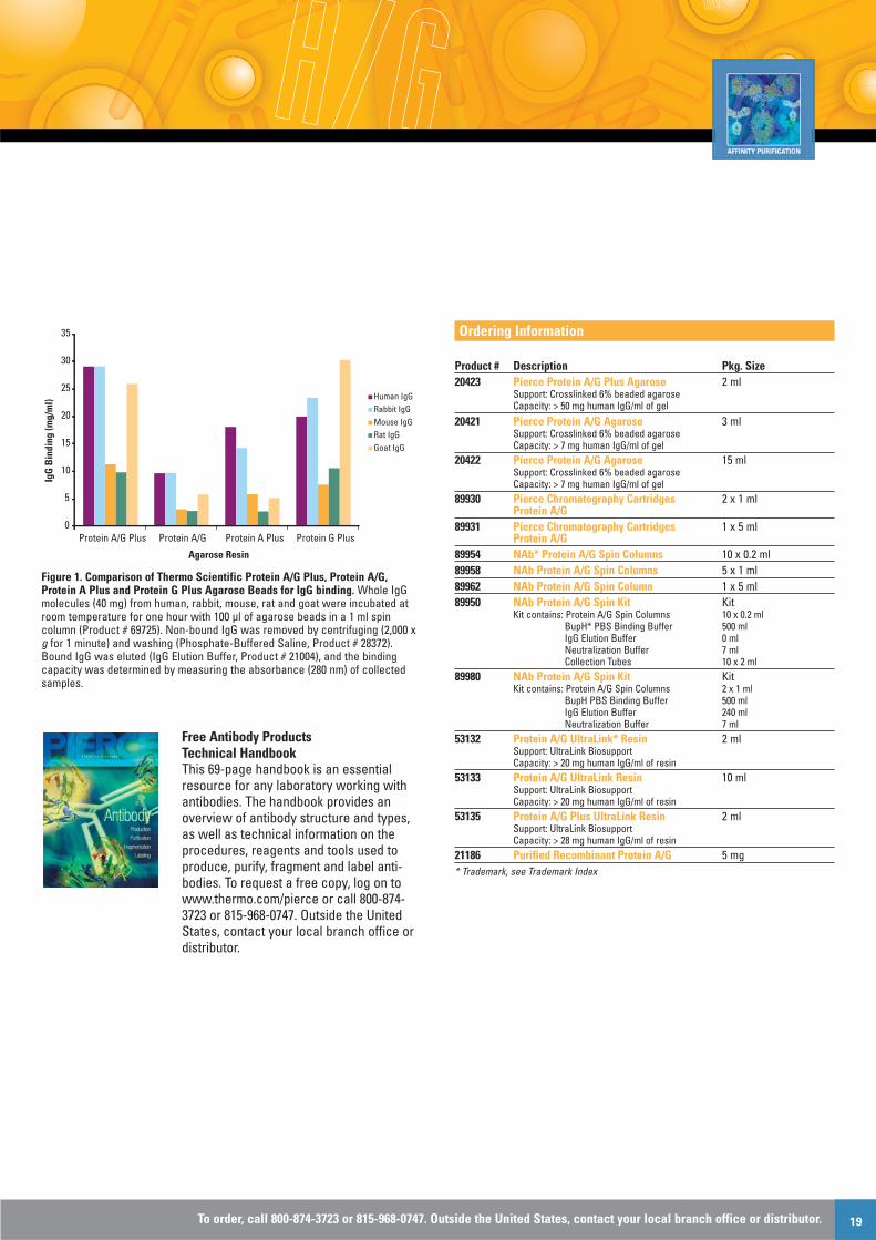
Thermo Scientific Pierce Protein A/G Plus Agarose (Product # 20423)has more than three times the binding capacity than our standardProtein A/G Agarose, purifying greater amounts of IgG for the sameresin volume and process time (Figure 1). As an added benefit, theproprietary manufacturing process for the Pierce Protein A/G PlusAgarose Beads reduces the amount of nonspecific binding.
Highlights:• Binds three times more IgG than the standard beads• The combined benefits of Protein A and Protein G in a single
purification system • Binds to a wider range of antibodies than Protein A or Protein G
beads alone (visit our website or contact us for more information)• Can be used in large- or small-scale gravity-flow or spin formats • Leach-resistant coupling chemistry for contamination-free results
Protein A/G binds to all human IgG subclasses, making it ideal forpurifying IgG from human samples whose subclasses have not beendetermined, and for binding IgA, IgE, IgM and, to a lesser extent,IgD. Protein A/G also binds well to all mouse IgG subclasses butdoes not bind mouse IgA, IgM or murine serum albumin,1 making itan excellent tool for purifying mouse monoclonal antibodies of allIgG subclasses. Individual subclasses of mouse monoclonal anti-bodies are likely to have a stronger affinity to the chimeric ProteinA/G than to either Protein A or Protein G.2
All of our Protein A/G-based products are coupled to beads usingleach-resistant chemistries that prevent Protein A/G from contam-inating the sample. These beads can be regenerated and reusedmultiple times for antibody purification.
Applications for our Protein A/G Plus Agarose beads• Purify any antibody that binds to either Protein A or Protein G • Purify monoclonal and polyclonal antibodies whose subclasses
have not been determined • Perform immunoprecipitation and co-immunoprecipitation
experiments• Remove IgG from sample
Properties of our Protein A/G Plus beads• Protein A/G source: recombinant protein produced in E. coli• Molecular weight: 50,460 Daltons• Support: 6% beaded agarose• Storage conditions: 4°C, do not freeze
References1. Sikkema, J.W.D. (1989). Amer. Biotech. Lab. 7, 42. 2. Eliasson, M., et al. (1988). J. Biol. Chem. 263, 4323-4327.
Table 1. Properties of antibody-binding proteins.
Recombinant Recombinant RecombinantProtein A Protein G Protein A/G
Expressed In Bacillus E. coli E. coliMolecular Weight 44,600 21,600 50,460Number of IgG 4 2 6Binding DomainsAlbumin-Binding Site No No NoOptimum Binding pH 8.2 5 5-8.2Binds to Fc Fc Fc
Protein Purification
To order, call 800-874-3723 or 815-968-0747. Outside the United States, contact your local branch office or distributor. 19
Figure 1. Comparison of Thermo Scientific Protein A/G Plus, Protein A/G,Protein A Plus and Protein G Plus Agarose Beads for IgG binding. Whole IgGmolecules (40 mg) from human, rabbit, mouse, rat and goat were incubated atroom temperature for one hour with 100 µl of agarose beads in a 1 ml spincolumn (Product # 69725). Non-bound IgG was removed by centrifuging (2,000 xg for 1 minute) and washing (Phosphate-Buffered Saline, Product # 28372).Bound IgG was eluted (IgG Elution Buffer, Product # 21004), and the bindingcapacity was determined by measuring the absorbance (280 nm) of collectedsamples.
Free Antibody ProductsTechnical HandbookThis 69-page handbook is an essentialresource for any laboratory working withantibodies. The handbook provides anoverview of antibody structure and types,as well as technical information on theprocedures, reagents and tools used toproduce, purify, fragment and label anti-bodies. To request a free copy, log on towww.thermo.com/pierce or call 800-874-3723 or 815-968-0747. Outside the UnitedStates, contact your local branch office ordistributor.
Ordering Information
Product # Description Pkg. Size 20423 Pierce Protein A/G Plus Agarose 2 ml
Support: Crosslinked 6% beaded agaroseCapacity: > 50 mg human IgG/ml of gel
20421 Pierce Protein A/G Agarose 3 ml Support: Crosslinked 6% beaded agaroseCapacity: > 7 mg human IgG/ml of gel
20422 Pierce Protein A/G Agarose 15 mlSupport: Crosslinked 6% beaded agaroseCapacity: > 7 mg human IgG/ml of gel
89930 Pierce Chromatography Cartridges 2 x 1 ml Protein A/G
89931 Pierce Chromatography Cartridges 1 x 5 ml Protein A/G
89954 NAb* Protein A/G Spin Columns 10 x 0.2 ml 89958 NAb Protein A/G Spin Columns 5 x 1 ml 89962 NAb Protein A/G Spin Column 1 x 5 ml 89950 NAb Protein A/G Spin Kit Kit
Kit contains: Protein A/G Spin Columns 10 x 0.2 mlBupH* PBS Binding Buffer 500 mlIgG Elution Buffer 0 mlNeutralization Buffer 7 mlCollection Tubes 10 x 2 ml
89980 NAb Protein A/G Spin Kit Kit Kit contains: Protein A/G Spin Columns 2 x 1 ml
BupH PBS Binding Buffer 500 mlIgG Elution Buffer 240 mlNeutralization Buffer 7 ml
53132 Protein A/G UltraLink* Resin 2 ml Support: UltraLink BiosupportCapacity: > 20 mg human IgG/ml of resin
53133 Protein A/G UltraLink Resin 10 ml Support: UltraLink BiosupportCapacity: > 20 mg human IgG/ml of resin
53135 Protein A/G Plus UltraLink Resin 2 ml Support: UltraLink BiosupportCapacity: > 28 mg human IgG/ml of resin
21186 Purified Recombinant Protein A/G 5 mg * Trademark, see Trademark Index
0
5
10
15
20
25
30
35
Protein A/G Plus Protein A/G Protein A Plus Protein G Plus
Agarose Resin
IgG
Bin
ding
(mg/
ml) Human IgG
Rabbit IgGMouse IgGRat IgGGoat IgG

www.thermo.com/pierce Vol. 12, Issue 120
Find just the protein bands you’re looking forClearly better Western blots
Western Blotting
Antibody bands often mask target proteins when performingWestern blots on immunoprecipitated samples. ThermoScientific Clean-Blot IP Detection Reagents are unique HRPand AP conjugates that reveal your target protein, allowingclear, specific Western blot detection from immunoprecipita-tion (IP) experiments and tissue extracts without anyinterference from denatured IgG (Figure 1). Whereas conven-tional secondary antibodies recognize both denatured andnative IgG, our new reagents bind to only native IgG (Figure 2).So unmask your results by simply substituting the secondaryantibody with Clean-Blot IP Detection Reagents for clearWestern blots (Figure 3).
Highlights:• Versatile – recognizes most native antibodies independent of the
host species (Table 1)• Compatible – clear results with IPs performed using Protein A,
Protein G or anti-IgG agarose beads and any blocking buffer (e.g.,milk, BSA or Thermo Scientific SuperBlock* or StartingBlock*Blocking Buffers)
• Cost effective – eliminates the need to immobilize IgG andpurchase separate kits specific for the primary antibody species;membranes can be stripped and reprobed when chemiluminescentsubstrate is used
• Flexible – use any HRP or AP substrate, including chemilumines-cent, fluorescent or colorimetric substrates
• Easy to use – simply replace the conventional secondary antibodywith the Clean-Blot IP Detection Reagents in your Western blottingprotocol
• Unobstructed detection – clear IP/Western blot results withoutinterference from denatured IgG bands
Our Clean-Blot IP Detection Reagents are the perfect substitute fortraditional secondary antibody conjugates. These unique conjugatesrecognize most primary antibodies, independent of the host species(Table 1), and can be used with IPs performed using Protein A or Gagarose resins. This versatility eliminates the need to buy separatedetection kits based on primary antibody species.
Our conjugates are conveniently stored at 2-8°C and are compatiblewith any HRP or AP substrate, including Thermo Scientific PierceECL, SuperSignal* Chemiluminescent† and Lumi-Phos* WBSubstrates (Table 2). For added convenience, the HRP conjugate isavailable in a kit that contains StartingBlock T20 Blocking Buffer andPierce ECL Chemiluminescent Substrate.
Table 1. Thermo Scientific Clean-Blot IP Detection Reagents recognize thevarious polyclonal antibodies and the specific monoclonal antibodies listed.To determine specific antibody compatibility, perform a dot-blot analysis.
Species Monoclonal Isotype(s)Bovine IgG2
Goat IgG2
Human IgG1, IgG2, IgG4
Mouse IgG2a, IgG2b, IgG3
Rat IgG2c
Sheep IgG2
Table 2. Recommended dilution ranges for the Thermo Scientific Clean-Blot IPDetection Reagents when using our chemiluminescent Substrates.
Chemiluminescent Recommended Product # Substrate Dilution Range34095 SuperSignal West Femto 1:200 to 1:4,000
Chemiluminescent Substrate34075 SuperSignal West Dura 1:200 to 1:2,000
Chemiluminescent Substrate34080 SuperSignal West Pico 1:40 to 1:1,000
Chemiluminescent Substrate32209 Pierce ECL Western 1:40 to 1:400
Blotting Substrate34150 Lumi-Phos WB Substrate 1:50 to 1:500
Figure 1. Immunoprecipitation (IP) and Western blot experiments demonstratespecificity of the Thermo Scientific Clean-Blot IP Detection Reagent (HRP).Lane 1. A431 total cell extract expressing p53 (positive control), Lanes 2 and 3.No-lysate negative control of IP wash (Lane 2) and elution (Lane 3) fractions,Lanes 4-6. Complete IP experiment of wash (Lane 4) and elution (Lanes 5 and 6)fractions. Lanes 1-5 were probed with Clean-Blot IP Detection Reagent (HRP)and Lane 6 was detected with GAM-HRP.
IP Conditions: Anti-p53 antibody (2 µg) was added to A431 total cellextract (500 µg) expressing p53 and incubated overnight with rockingat 4°C. Protein A Agarose Resin (Product # 20333) was washed threetimes with binding/wash buffer (50 mM Tris, 150 mM NaCl, 1% NP40,5% glycerol; pH 7.5). A 50% resin slurry (100 µl) prepared withbinding/wash buffer was used for each reaction. The tubes wererocked for 1 hour at 4°C. Non-bound protein was collected by
p53
Lane 1 Lane 2 Lane 3 Lane 4 Lane 5 Lane 6
Wash Elute
+ Control - Control Immunoprecipitation
Wash Elute Elute

To order, call 800-874-3723 or 815-968-0747. Outside the United States, contact your local branch office or distributor. 21
centrifugation at 1,000 x g for 1 minute. The resin-antibody-proteincomplexes were washed with 500 µl of binding/wash buffer threetimes. P53 protein-antibody complexes were eluted by suspendingthe Protein A Resin in 30 µl 5X Lane Marker Reducing Sample Buffer(Product # 39000) and boiled for 5 minutes.
Western Blot Conditions: Proteins separated on Tris-glycine gelswere transferred to PVDF membranes (Product # 88518). Themembranes were blocked with 5% milk in TBST and probed withmouse anti-p53 primary antibody (BD Biosciences) (1 µg/ml) andClean-Blot Detection Reagent (HRP) (0.1 µg/ml) or Goat Anti-MouseHRP (Product # 31430, 0.16 µg/ml) as secondary conjugates. P53protein was detected using SuperSignal West PicoChemiluminescent Substrate (Product # 34080).
Figure 2. Easily distinguish your target protein on a Western blot with ThermoScientific Clean-Blot Detection Reagent (HRP). Mouse liver extract (50 µg) totalprotein was separated on a Bio-Rad Criterion* Gel, transferred to PVDFmembrane and blocked with 5% milk in TBST. The membrane was probed withmouse monoclonal anti-Cdk1 (LabVision) (0.2 µg/ml) and goat anti-mouse HRP(0.16 µg/ml) or Clean-Blot Detection Reagent (HRP) (0.2 µg/ml). SuperSignalWest Pico Substrate (Product # 34080) was used for detection of Cdk1 protein.
Figure 3: Reveal your target protein with Thermo Scientific Clean-BlotDetection Reagent (HRP). To demonstrate unmasking of the target protein, weperformed IP and Western blot experiments. NFκB and Bax were immunopre-cipitated from A549 lysate using Protein A/G Agarose Resin and rabbitanti-NFκB (Panels A and B) and rabbit anti-Bax (Panels C and D). Panels A andC were detected with goat anti-rabbit HRP, which masked the target. Panels Band D were detected with the Clean-Blot Detection Reagent (HRP), revealingthe target protein.
IP Conditions: Four A549 cell lysate samples expressing NFκB andBax were incubated overnight on a rocking platform at 4°C with 4 µgof rabbit anti-NFκB (Upstate) or rabbit anti-Bax (Upstate). Protein A/GAgarose (Product # 20421) was washed three times withBinding/Wash Buffer (Product # 28376). A 50% Protein A/G slurry(100 µl), made with Binding/Wash Buffer, was added to each tubeand rocked for 2 hours at room temperature. Non-bound protein wascollected by centrifugation at 2,500 x g for 1 minute. The resin-anti-body-protein complexes were washed with 500 µl of Binding/Washbuffer three times. Bax protein- and NFκB protein-antibodycomplexes were eluted by suspending the Protein A/G Agarose in150 µl 5X Lane Marker Reducing Sample Buffer (Product # 39000)and boiled for 5 minutes.
Western Blot Conditions: Proteins were separated on Tris-HEPESPrecise* Protein Gels (Product # 25244) and transferred to PVDFmembranes (Product # 88518). The membranes were blocked withStartingBlock T20 Blocking Buffer (Product # 37543) and probed withrabbit anti-NFκB or rabbit anti-Bax primary antibodies (1 µg/ml,Upstate) and Clean-Blot Detection Reagent (HRP) at 0.4 µg/ml(Panels B and D) or Goat Anti-Rabbit HRP (Product # 31460) at 0.16µg/ml (Panels A and C). NFκB and Bax were detected using PierceECL Chemiluminescent Substrate (Product # 32106).
Ordering Information
Product # Description Pkg. Size21230 Clean-Blot IP Detection Reagent (HRP) 2.5 ml
Sufficient reagent for approximately 100 Western blots.
21232 Clean-Blot IP Detection Kit (HRP) KitSufficient reagents for approximately 2,000 cm2 of membrane. Clean-Blot Detection Reagent (HRP) 2.5 ml StartingBlock T20 (TBS) Blocking Buffer 1L Pierce ECL Detection Reagent 1, 125 ml
Peroxide SolutionPierce ECL Detection Reagent 2 125 ml
Luminol Enhancer Solution21233 Clean-Blot IP Detection Reagent (AP) 2.5 ml
Sufficient reagent for approximately 100 Western blots.
† Patent, see Patent Index.* Trademark, see Trademark IndexTo view data on our Clean-Blot Detection Reagents, visit www.thermo.com/pierce.
NFκB
Bax
A
GAR-HRP
C
GAR-HRP
BClean-BlotDetection Reagent
DClean-BlotDetection Reagent
WashElute
Lysate
WashElute
Lysate
WashElute
Lysate
WashElute
Lysate
Heavy Chain (55 kDa)
Cdk1 (34 kDa)
Light Chain (22 kDa)
Anti-
Mou
se H
RP
Mou
se A
nti-C
dk1
+ An
ti-M
ouse
HRP
Mou
se A
nti-C
dk1
+ Cl
ean-
Blot
Dete
ctio
n Re
agen
t (HR
P)Cl
ean-
Blot
Dete
ctio
n Re
agen
t (HR
P)
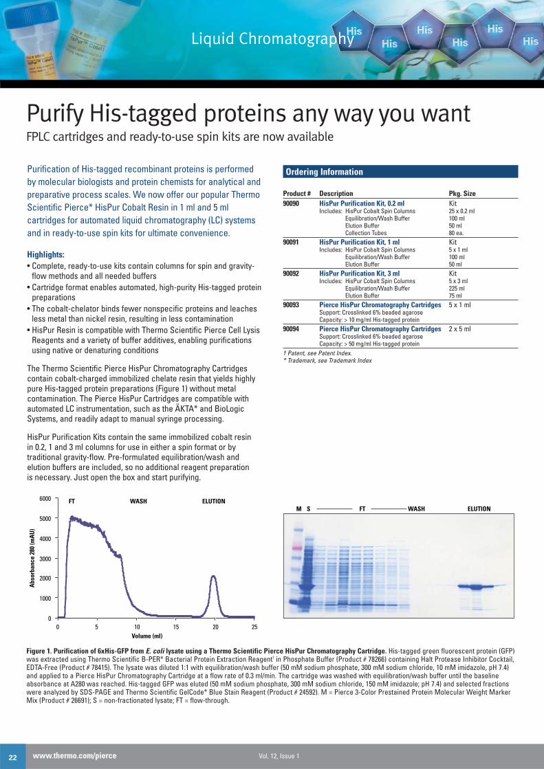
www.thermo.com/pierce Vol. 12, Issue 122
Purify His-tagged proteins any way you wantFPLC cartridges and ready-to-use spin kits are now available
Purification of His-tagged recombinant proteins is performedby molecular biologists and protein chemists for analytical andpreparative process scales. We now offer our popular ThermoScientific Pierce* HisPur Cobalt Resin in 1 ml and 5 mlcartridges for automated liquid chromatography (LC) systemsand in ready-to-use spin kits for ultimate convenience.
Highlights:• Complete, ready-to-use kits contain columns for spin and gravity-
flow methods and all needed buffers • Cartridge format enables automated, high-purity His-tagged protein
preparations• The cobalt-chelator binds fewer nonspecific proteins and leaches
less metal than nickel resin, resulting in less contamination• HisPur Resin is compatible with Thermo Scientific Pierce Cell Lysis
Reagents and a variety of buffer additives, enabling purificationsusing native or denaturing conditions
The Thermo Scientific Pierce HisPur Chromatography Cartridgescontain cobalt-charged immobilized chelate resin that yields highlypure His-tagged protein preparations (Figure 1) without metalcontamination. The Pierce HisPur Cartridges are compatible withautomated LC instrumentation, such as the ÄKTA* and BioLogicSystems, and readily adapt to manual syringe processing.
HisPur Purification Kits contain the same immobilized cobalt resinin 0.2, 1 and 3 ml columns for use in either a spin format or by traditional gravity-flow. Pre-formulated equilibration/wash andelution buffers are included, so no additional reagent preparationis necessary. Just open the box and start purifying.
Ordering Information
Product # Description Pkg. Size90090 HisPur Purification Kit, 0.2 ml Kit
Includes: HisPur Cobalt Spin Columns 25 x 0.2 mlEquilibration/Wash Buffer 100 mlElution Buffer 50 mlCollection Tubes 80 ea.
90091 HisPur Purification Kit, 1 ml KitIncludes: HisPur Cobalt Spin Columns 5 x 1 ml
Equilibration/Wash Buffer 100 mlElution Buffer 50 ml
90092 HisPur Purification Kit, 3 ml KitIncludes: HisPur Cobalt Spin Columns 5 x 3 ml
Equilibration/Wash Buffer 225 mlElution Buffer 75 ml
90093 Pierce HisPur Chromatography Cartridges 5 x 1 mlSupport: Crosslinked 6% beaded agaroseCapacity: > 10 mg/ml His-tagged protein
90094 Pierce HisPur Chromatography Cartridges 2 x 5 mlSupport: Crosslinked 6% beaded agaroseCapacity: > 50 mg/ml His-tagged protein
† Patent, see Patent Index.* Trademark, see Trademark Index
Liquid Chromatography
FTM S WASH ELUTION
0
1000
2000
3000
4000
5000
6000
0 5 10 15 20 25Volume (ml)
FT WASH ELUTION
Abs
orba
nce
280
(mA
U)
Figure 1. Purification of 6xHis-GFP from E. coli lysate using a Thermo Scientific Pierce HisPur Chromatography Cartridge. His-tagged green fluorescent protein (GFP)was extracted using Thermo Scientific B-PER* Bacterial Protein Extraction Reagent† in Phosphate Buffer (Product # 78266) containing Halt Protease Inhibitor Cocktail,EDTA-Free (Product # 78415). The lysate was diluted 1:1 with equilibration/wash buffer (50 mM sodium phosphate, 300 mM sodium chloride, 10 mM imidazole, pH 7.4)and applied to a Pierce HisPur Chromatography Cartridge at a flow rate of 0.3 ml/min. The cartridge was washed with equilibration/wash buffer until the baselineabsorbance at A280 was reached. His-tagged GFP was eluted (50 mM sodium phosphate, 300 mM sodium chloride, 150 mM imidazole; pH 7.4) and selected fractionswere analyzed by SDS-PAGE and Thermo Scientific GelCode* Blue Stain Reagent (Product # 24592). M = Pierce 3-Color Prestained Protein Molecular Weight MarkerMix (Product # 26691); S = non-fractionated lysate; FT = flow-through.

To order, call 800-874-3723 or 815-968-0747. Outside the United States, contact your local branch office or distributor. 23
*Trademark Index
ÄKTA is a trademark of GE Healthcare. Criterion™ is a trademark of Bio-Rad Laboratories, Inc. Alexa® is a trademark of Molecular Probes,Inc. CyDye™ and Typhoon™ are trademarks of GE Healthcare Bio-Sciences. Odyssey® and IRDye™ are trademarks of LI-CORBiosciences. Lumi-Phos™ is a trademark of and is sourced fromLumigen, Inc.
All other trademarks are property of Thermo Fisher Scientific Inc.and its subsidiaries.
† Patent Index
B-PER Technology is protected by US Patent # protected 6,174,704.Slide-A-Lyzer MINI Dialysis Unit Technology is protected by U.S. Patent#6,039,871. SuperSignal Technology is protected by U.S. patent # 6,432,662.US patent pending on Mitochondria Isolation Kit Technology.
Achieve recovery from small sample volumesIntroducing a 20K MWCO dialysis unit for 10-100 μl samples
The efficient Thermo Scientific Slide-A-Lyzer* MINI Dialysis Unit† isdesigned for high recovery from small sample volumes (10-100 µl).The device’s patented design, a disposable cup composed ofpolypropylene and a regenerated cellulose membrane, allows easysample application and removal using a standard laboratory pipette.A specialized foam float holds the unit upright with the membrane incontact with the dialysis buffer for maximum performance.
Highlights:• Easy to use – simply use a standard pipette to add and
remove samples• Robust performance – rigorous quality testing for
maximum consistency• Efficient – recover > 87% of protein from small sample
volumes (10-100 µl)
Dialysis is a method for removing low molecular-weight solutes frommacromolecules in solution by selective diffusion across a porousmembrane. The membrane’s pore size determines the molecular-weight cutoff (MWCO) characterized by the molecular weight atwhich 90% of the solute is retained. The permeability of a solutedepends on the shape of the molecule, the degree of hydration andits charge. Each of these factors may be influenced by the solvent’snature, pH and ionic strength.
Protein Recovery Protein solutions (100 µl, 0.5 mg/ml, pH 7.2) in PBS were separatelyadded to dialysis units and dialyzed overnight at 4°C. Proteinconcentrations of the recovered samples (~100 µl) were determinedby the Pierce* BCA Protein Assay (Product # 23225). Proteinrecovery for all samples was > 87% (Table 1).
Table 1. High protein recovery from Thermo Scientific Slide-A-Lyzer MINIDialysis Units (20K MWCO).
Protein Recovery (%)Myoglobin (17 kDa) 93.8Soybean Trypsin Inhibitor (21 kDa) 87.4Carbonic Anhydrase (29 kDa) 90.36Ovalbumin (44 kDa) 89.3BSA (67 kDa) 89.5
Desalting RateSamples (100 µl) of 1 M NaCl were dialyzed against 1 L of water at4°C and the rate of NaCl removal was determined by measuring theretentate’s conductivity at 10 minute intervals. Within 20 and 100minutes, > 50% and > 99% of the NaCl was removed, respectively(Figure 1).
Figure 1. Desalting rate using a Thermo Scientific Slide-A-Lyzer MINI DialysisUnit (20K MWCO).
Ordering Information
Product # Description Pkg. Size69590 20K MWCO Slide-A-Lyzer 50/pkg.
MINI Dialysis Units69555 20K MWCO Slide-A-Lyzer 250/pkg.
MINI Dialysis UnitsSlide-A-Lyzer MINI Dialysis Units are also available with 3.5K, 7K and 10K MWCOmembranes. Visit www.thermo.com/dialysis for more information.
† Patent, see Patent Index* Trademark, see Trademark Index
0
20
40
60
80
100
120
0 20 40 60 80 100 120 Time (Minutes)
% R
esid
ual N
aCl
High-Performance Dialysis

www.thermo.com/pierce
Pierce ®
now sold as
Thermo Scientific
Double the Precise, not the price:Get two gels for the price of one.
©20
08 T
herm
o Fi
sher
Sci
enti
fic In
c. A
ll ri
ghts
rese
rved
. All
trad
emar
ks a
re th
e pr
oper
ty o
f Th
erm
o Fi
sher
Sci
enti
fic In
c. a
nd it
s su
bsid
iari
es.
Thermo Scientific Precise* Protein Gels are cast in a durable plastic
cassette using a neutral pH buffer that prevents polyacrylamide
breakdown. The gels are individually packaged and are ready to run
with no comb or tape to remove. They are available in both gradient
and fixed concentrations and in 10-, 12- and 15-well formats.
Highlights:
• Easy-to-load, large-volume wells with durable plastic dividers
• Long shelf life – 12-month guarantee ensures consistent performance
• Easy to open – no need for special tools
Buy 1 package of Precise Gels,get 1 identical package FREE!Offer valid 4/1-5/30/2008.Visit www.thermo.com/pierce for details
Tel: 815-968-0747 or 800-874-3723 • Fax: 815-968-7316 • Customer Assistance E-mail: [email protected]© 2008 Thermo Fisher Scientific Inc. Unless otherwise noted on page 23, all t ademarks belong to Thermo Fisher Scientific Inc. and its subsidiaries.Prices listed herein were accurate at he time of printing. Thermo Scientific Pierce Products are sold for research and manufacturing purposes only.
PRSTD STDU.S. POSTAGE
PAIDPERMIT NO. 4346Milwaukee, WI
Pierce Protein Research Products
PO Box 117
Rockford, IL
61105-0117
Attention mailroom/recipient: Help keep our mail list current. Please fax (800-842-5007 or 815-968-8148) back this panel with your corrections or deletions made to the address.
1601
615
03/
08