Physiological Assessment and Intra-Coronary Imaging … · Physiological Assessment and...
78
Physiological Assessment and Intra-Coronary Imaging in the Cath Lab Ronen Jaffe, MD Department of Cardiology Lady Davis Carmel Medical Center Rappaport School of Medicine, IIT Haifa
Transcript of Physiological Assessment and Intra-Coronary Imaging … · Physiological Assessment and...

Physiological Assessment and
Intra-Coronary Imaging in the Cath
Lab
Ronen Jaffe, MD
Department of Cardiology
Lady Davis Carmel Medical Center
Rappaport School of Medicine, IIT
Haifa

?


Kern JACC 2010;55;173-185

Angiography Has Major Limitations
in Assessing Complicated Lesions
Angiography Has Major Limitations
in Assessing Complicated Lesions
Nissen SE et al in Textbook of Cardiovascular Medicine, 1998; Topol EJ et al, Circulation, 1995.

Limitations of Coronary Angiography
• Coronary angiography does not reliably detect the hemodynamic significance of coronary stenosis
• Coronary angiography does not reliably delineate the anatomy of coronary lesions

IC Imaging: ANATOMY
Functional assessment: PHYSIOLOGY
?

Improved Assessment of Coronary Lesions
• Fractional Flow Reserve (FFR):
–Accurate assessment of hemodynamic significance
• Intravascular imaging (IVUS/OCT):
–Precise visualization of intra-
coronary anatomy

Anatomy Vs. Physiology
Two clinical settings:
1. Is PCI indicated?
2. Technical aspects of PCI:
– Characterization of lesion
– Is the PCI result optimal?
– ….

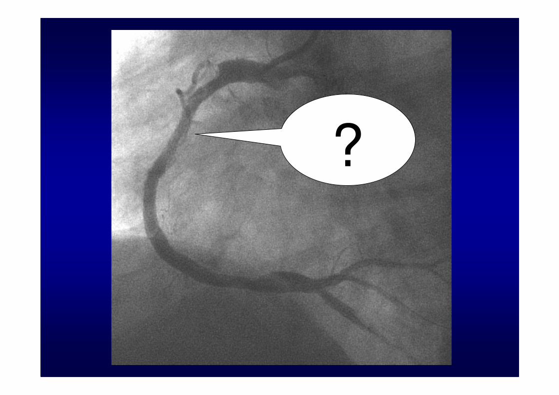
P distalP proximal
FFR=P dist / P prox

Fractional Flow Reserve (FFR)
NormalFFR > 0.8
?FFR: 0.75-0.8
IschemiaFFR < 0.75

Microvascular hyperemia: Adenosine
• Adenosine: 6mg/500cc NS (12mcg/cc)
• IC boluses:
–60 mcg (5 cc)
–96 mcg (8 cc)
–120 mcg (10 cc)
• IV drip 140 mcg/Kg/Min

A: FFR=1.00B: FFR=0.65
B
A

2.7
1.9
18.3
13.2
78
81
FAME study
1005 patients with stenosis >50% randomized: PCI or FFR (PCI if FFR<0.8)
FAME NEJM 2009;360:213-24
PCI FFR PCI PCIFFR FFR
# stents 1-yr D/MI/TVR 1-yr angina-free
% % %
P<0.001 P<0.02 P=NS
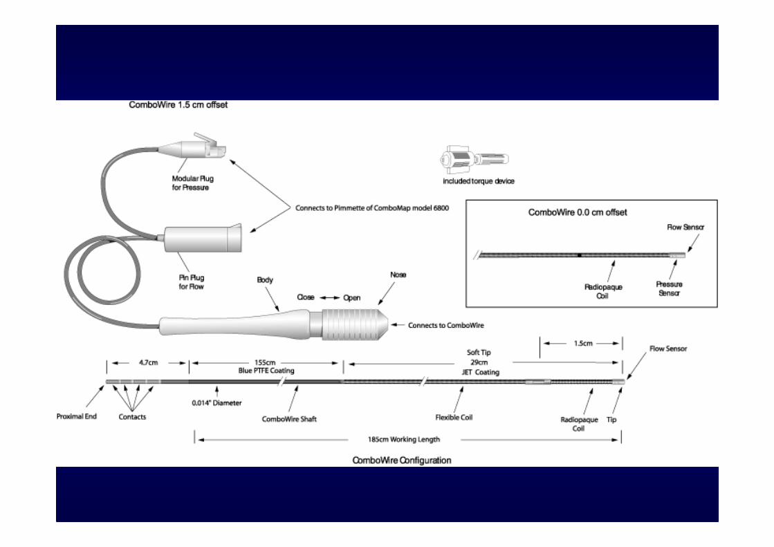
LDCMC Analysis:First 20 FFR Cases in Intermediate
Lesions
• Mean FFR = 0.85±0.08
(range: 0.71-1.00)
• FFR<0.80 was measured in only 5 stenoses (25%)
• Comparison of FFR to opinion of 2 experienced cardiologists

25
0
20
40
60
80
100
FFR<0.8
%

25
60
0
20
40
60
80
100
FFR<0.8
MD-1
%

25
60
95
0
20
40
60
80
100
FFR<0.8
MD-1
MD-2
%

FFR=0.88
FFR=0.88

FFR=0.84


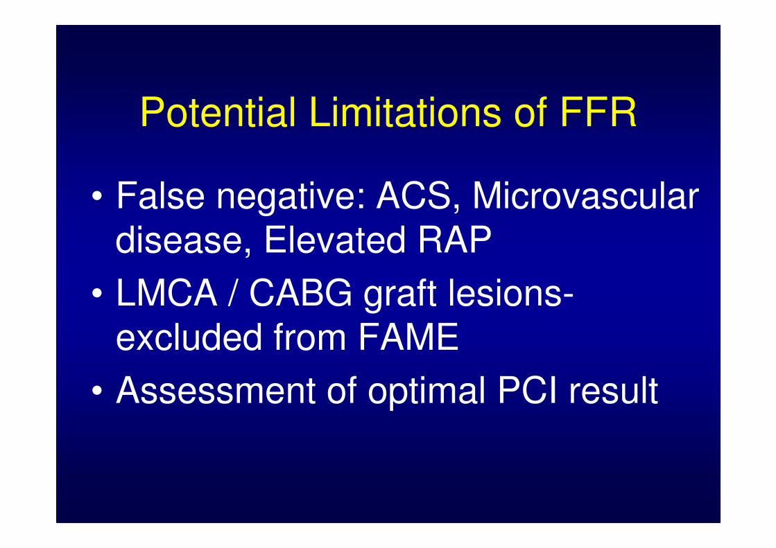
Potential Limitations of FFR
• False negative: ACS, Microvascular disease, Elevated RAP
• LMCA / CABG graft lesions-excluded from FAME
• Assessment of optimal PCI result

IVUS / OCT
• Accurate visualization of coronary anatomy
• Analysis of plaque composition & distribution, vessel and lumen geometry
• Identify dissections, stent apposition etc…
• Virtual histology

IVUS Transducers
A: Mechanical rotating transducer
B: Electronic phased array
A
B


3.1 mm3.1 mm
3.1 mm3.1 mm
Angiography Cannot Account forCoronary Remodeling

LAO RAO
Angiography Masks Complicated LesionsAngiography Masks Complicated Lesions

IVUS GUIDED STENT DEPLOYMENT
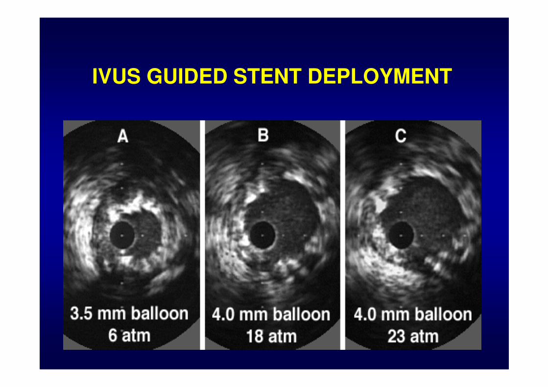
IVUS GUIDED STENT DEPLOYMENT


How many previous
stents have been
placed in this lesion?
1) 1
2) 2
3) 3
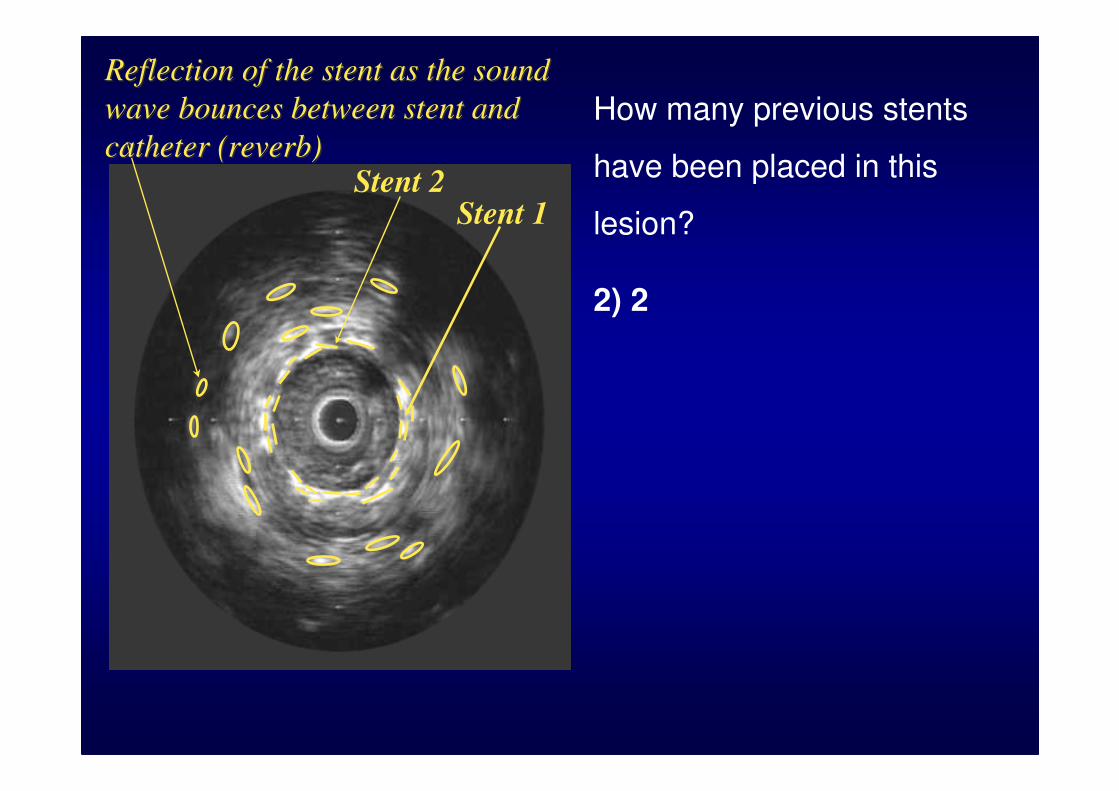
How many previous stents
have been placed in this
lesion?
2) 2
Stent 1
Stent 2
Reflection of the stent as the sound
wave bounces between stent and
catheter (reverb)
Reflection of the stent as the sound
wave bounces between stent and
catheter (reverb)

The most likely cause of this
image is:
1) Inadequate flushing
2) Bend in catheter
3) Calcified Plaque
4) Wrong Frequency

The most likely cause of this image is:
2) Bend in catheter
A bend in a mechanical IVUS
catheter may cause unnecessary
friction and generate Non-Uniform
Rotational Distortion (NURD), which
results in a smeared image. This
affect can be minimized by removing
bends in the catheter and checking
the tension on the Y-adaptor. NURD
can also occur when imaging in
torturous anatomy.
NURD

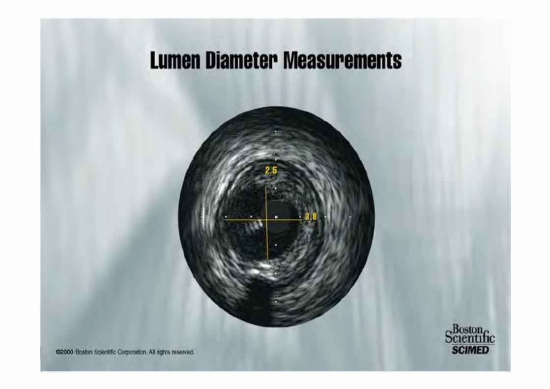
What is the present
diameter of the stent?
1) 2.0mm
2) 2.5mm
3) 3.0mm
4) 4.5mm
5) 4.0mm1.0mm/div.

What is the present
diameter of the
stent?
2) 2.5 mm
Since each division is
1 mm, the diameter is
approximately 2.5
mm.
1.0mm/div.
1mm1mm
1mm1mm
0.5mm0.5mm

What size balloon
should be used to
adequately deploy
this stent?
1) 3.0 mm
2) 4.0 mm
3) 5.0 mm
1.0 mm/div.

What size balloon should be
used to adequately deploy this
stent?
2) 4.0mm
The external elastic lamina
(EEL) appears to be 4.0 mm
and the lesion is primarily
fibrofatty plaque. Therefore, a
4.0 mm balloon should
produce the desired result.
1.0 mm/div.

IVUS
• Threshold for PCI (vs FFR/CFR)
–LMCA: CSA=6 mm2
–Prox vessels: CSA=4 mm2
• Missing: Robust outcome data
• IVUS less specific than FFR for ischemia
• “If you want PCI then IVUS, if not-then FFR”
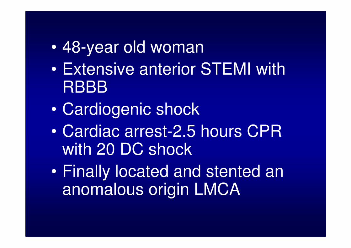
• 48-year old woman
• Extensive anterior STEMI with RBBB
• Cardiogenic shock
• Cardiac arrest-2.5 hours CPR with 20 DC shock
• Finally located and stented an anomalous origin LMCA


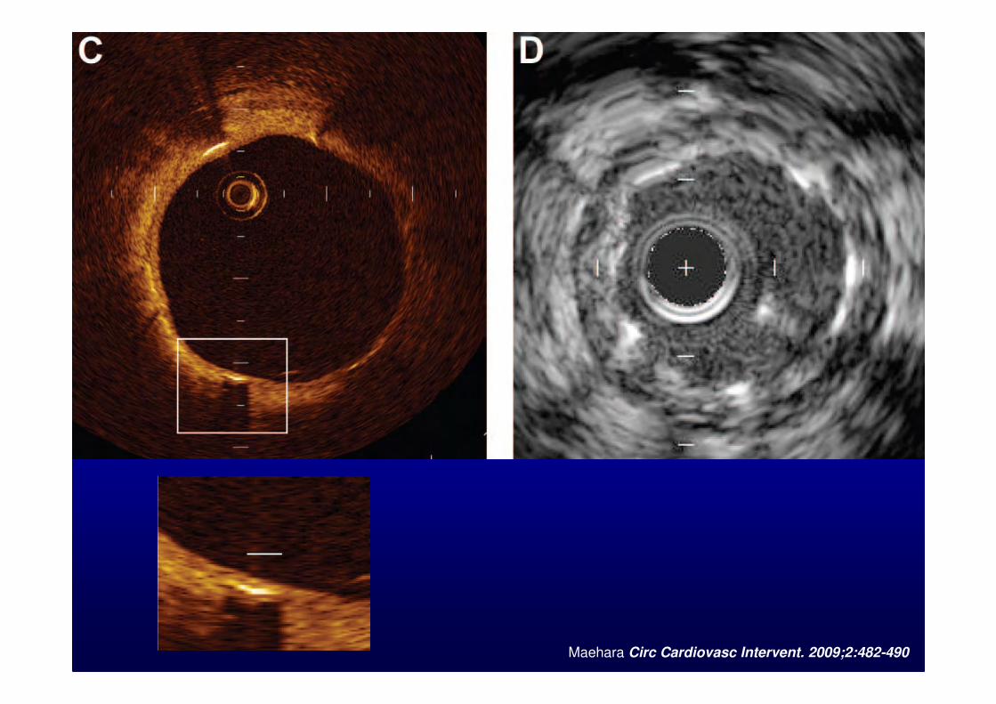
CSA=9.3 mm2CSA=6.3 mm2
Mid LMCAOstial LMCA

Thin Cap With Lipid CoreThin Cap With Lipid CoreThin Cap With Lipid Core Thick Stable Fibrotic CapThick Stable Fibrotic CapThick Stable Fibrotic Cap
Same Lumen Size: Different AtheromasSame Lumen Size: Different Atheromas

Kern SCAI 2006

intimal thickening thick-cap fibroatheroma
Maehara Circ Cardiovasc Intervent. 2009;2:482-490


IVUS vs. OCT
Kern SCAI 2006

OCT IVUS
Resolution 15 µm 100 µm
Penetration* 2 mm 10 mm
Penetration
requires blood
clearance
Yes No

Maehara Circ Cardiovasc Intervent. 2009;2:482-490

The Future?

•51 year-old diabetic woman
•Atypical angina•? Positive exercise test
Angiography: ≈ 50% stenosis in ostial LAD


CSA=4.1 MM2

FFR=0.93

CSA=4.1 mm2
Plaque volume 59 mm3

•Decision not to perform PCI
•Medical therapy:
Lipitor and Aspirin
•Follow-up CTA after 1 year

CSA=6.3 mm2
Plaque volume 32 mm3

Conclusions

Conclusions
• IVUS/OCT and FFR are complementary techniques
• “Is PCI indicated?”–Shift from anatomic revasc to
physiological revasc-FFR more applicable
–IVUS has a role in LMCA and prox LAD
• “Is PCI result optimal?”–IVUS preferred


Thank-you

FAME study
1005 patients with stenosis > 50% randomized: PCI or FFR (PCI if FFR<0.8)
FAME NEJM 2009;360:213-24
2.7 1.9
18.313.2
78 81
0
10
20
30
40
50
60
70
80
90
# stents D/MI/TVR No angina
P<0.001
P<0.02
P=NS
%

Raffel Heart 2008;94:1200-1210

Raffel Heart 2008;94:1200-1210

For a 30-40 MHz IVUS transducer: Axial resolution=80-100 µ
Lateral resolution=200-250 µ

Kawaguchi JACC 2007 50:1641

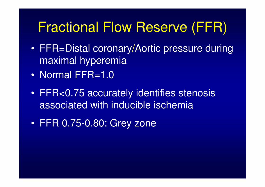
Fractional Flow Reserve (FFR)
• FFR=Distal coronary/Aortic pressure during maximal hyperemia
• Normal FFR=1.0
• FFR<0.75 accurately identifies stenosis associated with inducible ischemia
• FFR 0.75-0.80: Grey zone