Physical Therapy Management of Chronic …c.ymcdn.com/sites/ Therapy Management of Chronic Diseases:...
114
Physical Therapy Management of Chronic Diseases: COPD & CHF Suzanne Greenwalt, PT, DPT, CCS, GCS TPTA Spring State Meeting, April 9, 2016
-
Upload
nguyendiep -
Category
Documents
-
view
217 -
download
0
Transcript of Physical Therapy Management of Chronic …c.ymcdn.com/sites/ Therapy Management of Chronic Diseases:...

Physical Therapy Management of
Chronic Diseases: COPD & CHF
Suzanne Greenwalt, PT, DPT, CCS, GCS
TPTA Spring State Meeting, April 9, 2016

Statistics
Chronic diseases were the leading causes of death among U.S. adults aged 65 or older in 2007-2009 (CDC, 2013)
Among those chronic diseases, heart disease was the #1 cause of death and respiratory disease was the #3 leading cause of death in those 65 and older (CDC, 2013)
As the aging population increases in the US, the prevalence of chronic heart and respiratory disease is also increasing

Statistics (CDC, 2013)
More than 2/3 of all health care costs are
for treating chronic illnesses
95% of the cost of health care in older
adults in the US is in managing chronic
diseases, the majority of which are
cardiopulmonary in nature
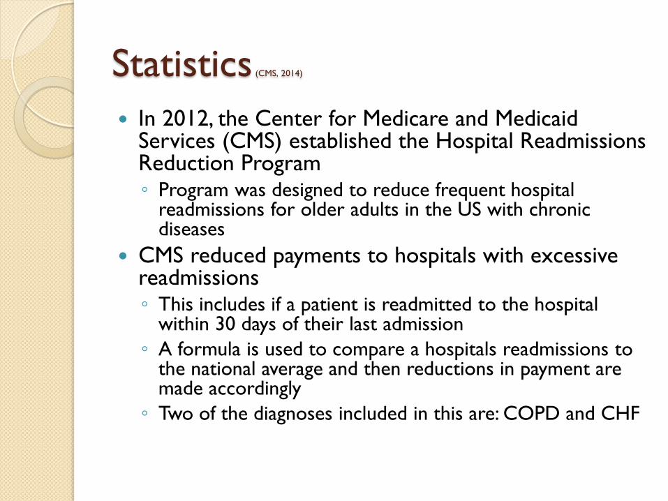
Statistics (CMS, 2014)
In 2012, the Center for Medicare and Medicaid Services (CMS) established the Hospital Readmissions Reduction Program ◦ Program was designed to reduce frequent hospital
readmissions for older adults in the US with chronic diseases
CMS reduced payments to hospitals with excessive readmissions ◦ This includes if a patient is readmitted to the hospital
within 30 days of their last admission
◦ A formula is used to compare a hospitals readmissions to the national average and then reductions in payment are made accordingly
◦ Two of the diagnoses included in this are: COPD and CHF

Statistics
What does this mean for rehab professionals?
All rehab professionals must take an active role in chronic disease management at all levels of care: acute, skilled nursing facilities, home health, out-patient.
These diseases and comorbidities can not be ignored!
Rehab professionals have the ability to aid in reducing hospital readmissions, improving quality of life and improving health care costs for those with chronic cardiac and pulmonary diseases.
WHAT IS COPD?

COPD Chronic Obstructive Pulmonary Disease
Obstructive Pulmonary Disease
◦ Any diseases of the airway that produce
restriction to expiratory airflow, which relates
to:
Retained secretions
Inflammation of the mucosal lining of airway walls
Bronchial constriction related to increased tone or
spasm of bronchial smooth mm
Weakening of the support structure of airway walls

Obstructive Pulmonary Disease
Chronic Bronchitis
Emphysema
Asthma
◦ Airway resistance caused by constriction of the bronchial smooth muscle cells and mucus production within the airway
Cystic Fibrosis
◦ Affects every organ system that has epithelial surfaces – most predominant systems are the lungs and the pancreas
◦ Pulmonary system is affected with chronic obstruction and inflammation, thick mucus, and recurrent bacterial infections.
◦ Pancreas develops exocrine pancreatic insufficiency which effect GI function (fat maldigestion) and growth and development
Bronchiectasis
◦ Irreversible dilation of one or more bronchi with chronic inflammation and infection
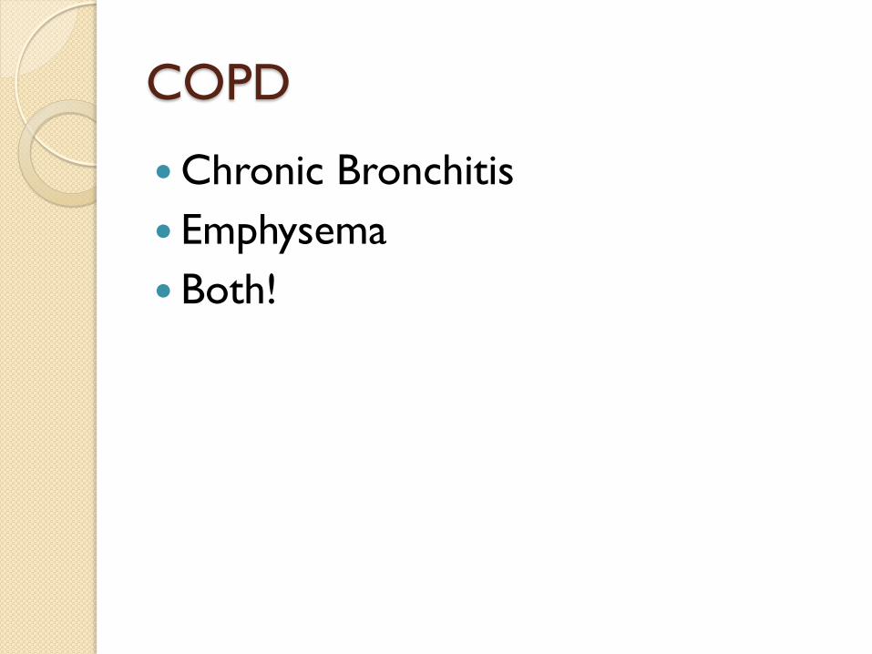
COPD
Chronic Bronchitis
Emphysema
Both!

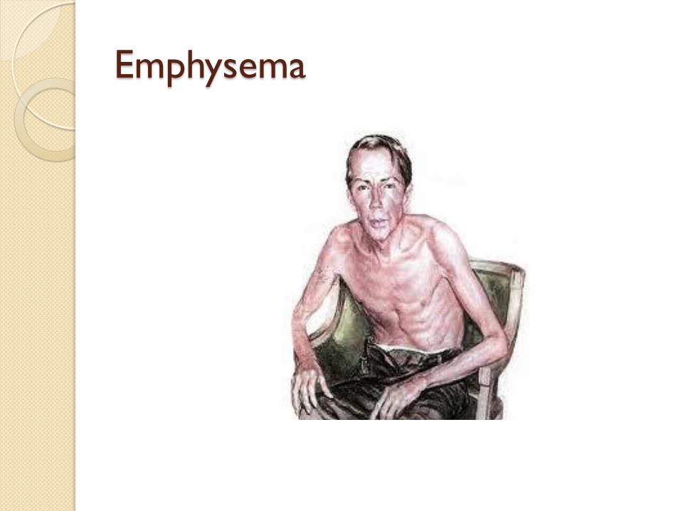
Emphysema
Emphysema: permanent enlargement of the bronchioles and destructive changes in the alveoli ◦ Loss of elastic recoil
of lungs
◦ Excessive collapsing of airways upon exhalation
◦ Chronic airflow obstruction

Emphysema
Signs and Symptoms of Emphysema:
◦ Dyspnea (most common symptom)
◦ Breathe with accessory muscles of respiration
◦ Wheezing
◦ Breathe through pursed lips on expiration
◦ Rapid and shallow respirations
◦ Tripod positioning
Other Characteristics:
◦ Little to no sputum production
◦ Thin with elevated shoulders
◦ Increased anterioposterior chest diameter
Emphysema
Chronic Bronchitis
Disease process that is caused by long-term irritation of the tracheobronchial tree
◦ Cigarette smoke
◦ Environmental pollutants
◦ Occupational irritants
Chronic bronchitis: diagnosis is based on symptoms
◦ Chronic or productive cough on most days for minimum of 3 months/year in >2 consecutive years

Chronic Bronchitis
Signs & Symptoms: ◦ Chronic productive cough
◦ Increased sputum production
◦ DOE
◦ Frequent respiratory infections
◦ Cyanosis
◦ Decreased capillary refill
◦ Clubbing fingers
◦ Decreased vital capacity due to increased residual volume
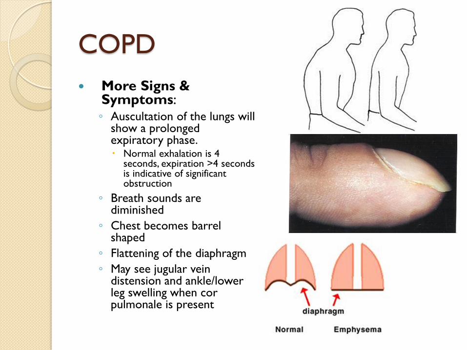
COPD
More Signs & Symptoms: ◦ Auscultation of the lungs will
show a prolonged expiratory phase. Normal exhalation is 4
seconds, expiration >4 seconds is indicative of significant obstruction
◦ Breath sounds are diminished
◦ Chest becomes barrel shaped
◦ Flattening of the diaphragm
◦ May see jugular vein distension and ankle/lower leg swelling when cor pulmonale is present

Hillegass, E. (2011)

COPD Chodbury, G., Rabinovich, R., & MacNee, W. (2014)
In addition to the lung abnormalities COPD causes, it also causes significant systemic effects ◦ Cardiovascular disease – most significant
nonrespiratory contributor to death in COPD Includes HTN, CAD, heart failure
◦ Skeletal muscle dysfunction: reduced peripheral muscle mass, skeletal muscle wasting and overall dysfunction Leads to reduced exercise capacity
◦ Osteoporosis
◦ Lung Cancer

COPD
Patients with COPD often retain CO2 –
monitor them carefully
Those with CO2 retention have a decreased
ventilatory drive unless oxygen levels are low
Oxygen through a nasal cannula can not get too
high or they will become apneic
Maintaining levels around 1-2 L/minute may be
required with some of these patients
May even see an increase in oxygen saturation
levels with a decrease in supplemental oxygen

COPD PATIENT ASSESSMENT

Components of Patient Assessment
Vital Signs
COPD Assessment Tool
Physical Appearance
Breathing Patterns
Functional Assessments
Strength and Flexibility

Vital Signs
Heart Rate
Blood pressure
Respiratory rate
Oxygen saturation level
◦ If oxygen saturation levels are less than 88%
then a patient with COPD is a candidate for
supplemental oxygen
◦ The average “normal” range for a patient with
COPD is 88-92%

Patient Questionnaire
COPD Assessment Tool (CAT) ◦ Standardized questionnaire that evaluates the
impact that COPD has on a patient’s daily life
◦ This test should be administered during a patient’s rehab admission Upon admission
Prior to discharge
◦ There is a maximum of 40 points on the test
◦ COPD management guidelines do exist based on the score obtained
◦ Research is ongoing to determine the Minimally Clinically Important Difference (MCID) Appears to be 2 but more research is needed
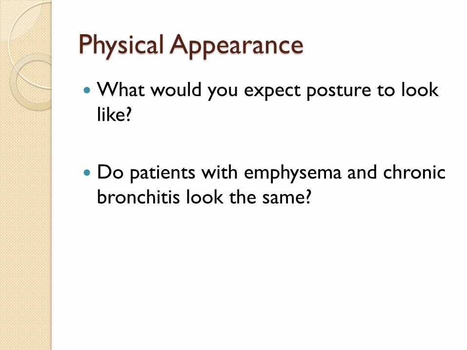
Physical Appearance
What would you expect posture to look
like?
Do patients with emphysema and chronic
bronchitis look the same?

Breathing Patterns
◦ Pursed lip breathing – clinical sign of COPD, is performed to alleviate the trapping of air in the lungs and to improve gas exchange
◦ Patients breathe out against lips that are mostly closed and shaped in a circular fashion
◦ Used to decrease symptoms of dyspnea
◦ Slows down respiratory rate, decreases pressure and reduced airway collapse during expiration

Breathing Patterns
o Paradoxical breathing:
◦ Abnormal chest movement where the chest moves inward during inhalation rather than outward.
◦ This abnormal movement impairs the ability to effectively inhale, limiting the amount of oxygen you can take in
◦ Patients will then have to actively contract the abdominal musculature during expiration to decrease the air trapped in the lungs – this results in the chest moving outward
◦ This abnormal breathing pattern is an indicator of
advanced COPD

Breathing Patterns
Accessory Muscle Use

Functional Assessments
BORG
Ventilatory Response Index
6 Minute Walk Test
Gait Velocity
2 Minute Step Test

BORG
Used to assess a patient’s level of dyspnea
Can be used in writing goals, tracking
progress of treatment

BORG vs. Modified BORG
BORG (6-20) Modified BORG (1-10)

Ventilatory Response Index (VRI)
Uses a scale from 0-4
Assesses how many breaths a patient
needs to take in order to count aloud to
15
There are not specific norms
Each number should be said in .5 seconds
(total time = 7.5 seconds)

Ventilatory Response Index
VRI Level Definition
0 Needs no additional breaths in order to complete counting
aloud to 15
1 Needs to take 1 breath in order to complete counting aloud
to 15
2 Needs to take 2 breaths in order to complete counting aloud
to 15
3 Needs to take 3 breaths in order to complete counting aloud
to 15
4 Needs to take 4 or more breaths in order to complete
counting aloud to 15

6 Minute Walk Test
6 Minute Walk Test ◦ Tests for exercise capacity and endurance
◦ “Valid and reliable test when assessing exercise capacity in older patients with chronic heart failure, those in phase II/III cardiac rehabilitation programs and patients with COPD.” Lewis & Shaw, 2005
◦ Walk as far as possible in 6 minutes
◦ Have a 100 ft (30 meter) course designated so that patient turns around each 100 feet
◦ Patient begins test from a seated position – there is no warm up phase to this test
◦ Instruct patient not to talk when walking
◦ Use standardized phrases when the patient is walking: “Keep up the good work” or “You are doing great”
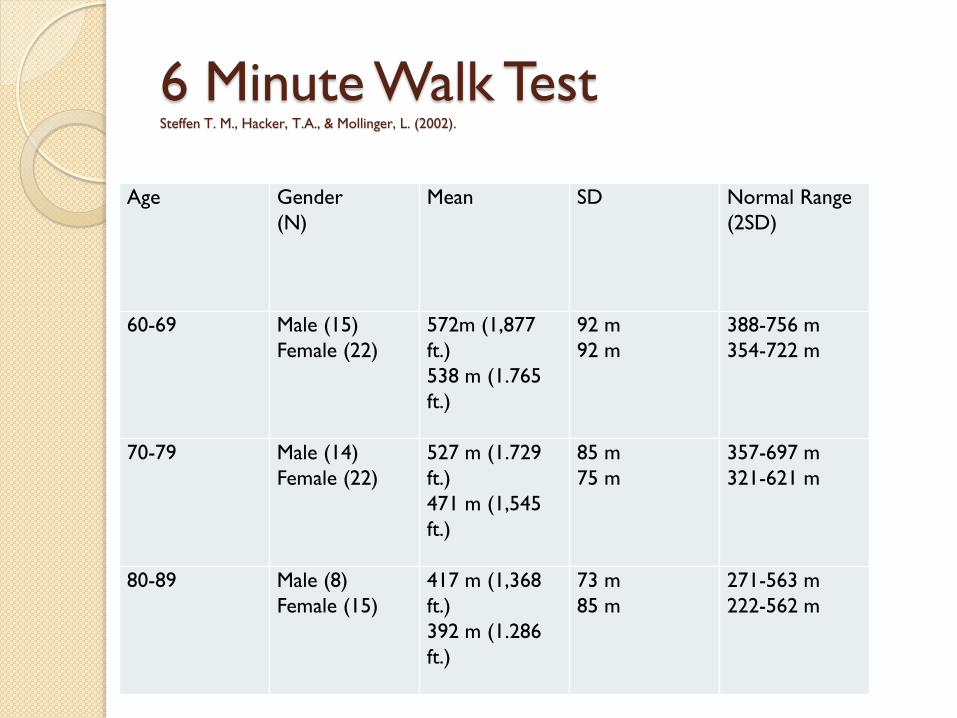
6 Minute Walk Test Steffen T. M., Hacker, T.A., & Mollinger, L. (2002).
Age Gender
(N)
Mean SD Normal Range
(2SD)
60-69 Male (15)
Female (22)
572m (1,877
ft.)
538 m (1.765
ft.)
92 m
92 m
388-756 m
354-722 m
70-79 Male (14)
Female (22)
527 m (1.729
ft.)
471 m (1,545
ft.)
85 m
75 m
357-697 m
321-621 m
80-89 Male (8)
Female (15)
417 m (1,368
ft.)
392 m (1.286
ft.)
73 m
85 m
271-563 m
222-562 m

Gait Velocity
Measures gait speed at preferred and maximum speeds
Is an indicator for falls risk and home safety
6th vital sign
>1.0 m/sec is doing well, means low fall risk
<0.6 m/sec is at high risk for falls
.4 m/sec or less are typically homebound
Assistive devices can be used when measuring gait velocity
3.3 feet = 1 meter
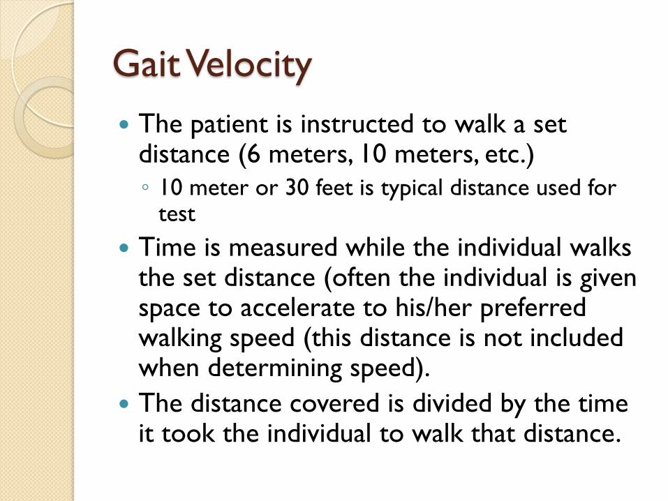
Gait Velocity
The patient is instructed to walk a set distance (6 meters, 10 meters, etc.)
◦ 10 meter or 30 feet is typical distance used for test
Time is measured while the individual walks the set distance (often the individual is given space to accelerate to his/her preferred walking speed (this distance is not included when determining speed).
The distance covered is divided by the time it took the individual to walk that distance.

2 Minute Step Test
Test for physical endurance
Stand facing wall with line marked halfway between patella and iliac crest
Count number of full steps completed in 2 minutes (number of times right knee reaches marked height)
Less than 65 steps (men or women) is indicative of problem (in general)
Measure HR, BP and RR prior to beginning test and after the test
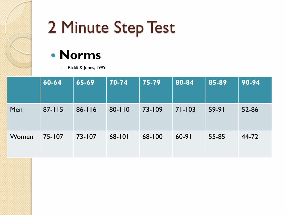
2 Minute Step Test
Norms ◦ Rickli & Jones, 1999
60-64 65-69 70-74 75-79 80-84 85-89 90-94
Men 87-115 86-116 80-110 73-109 71-103 59-91 52-86
Women 75-107 73-107 68-101 68-100 60-91 55-85 44-72

Strength & Flexibility
Upper extremity ROM and strength
Trunk ROM and strength
Lower extremity ROM and strength

TREATMENT OF COPD

Treatment of COPD
Airway Clearance Techniques
Breathing Techniques
Positioning
Incentive Spirometers
Flexibility Exercises
Strengthening
Energy Conservation
Patient/Family Education

Airway Clearance Techniques
Manual or mechanical procedures that facilitate mobilization of secretions from the airways
Include: ◦ Percussion
◦ Vibration
◦ Postural Drainage
◦ Active cycle of breathing
◦ Deep Breathing
◦ Coughing
◦ Positive Expiratory Pressure
Airway Clearance Techniques
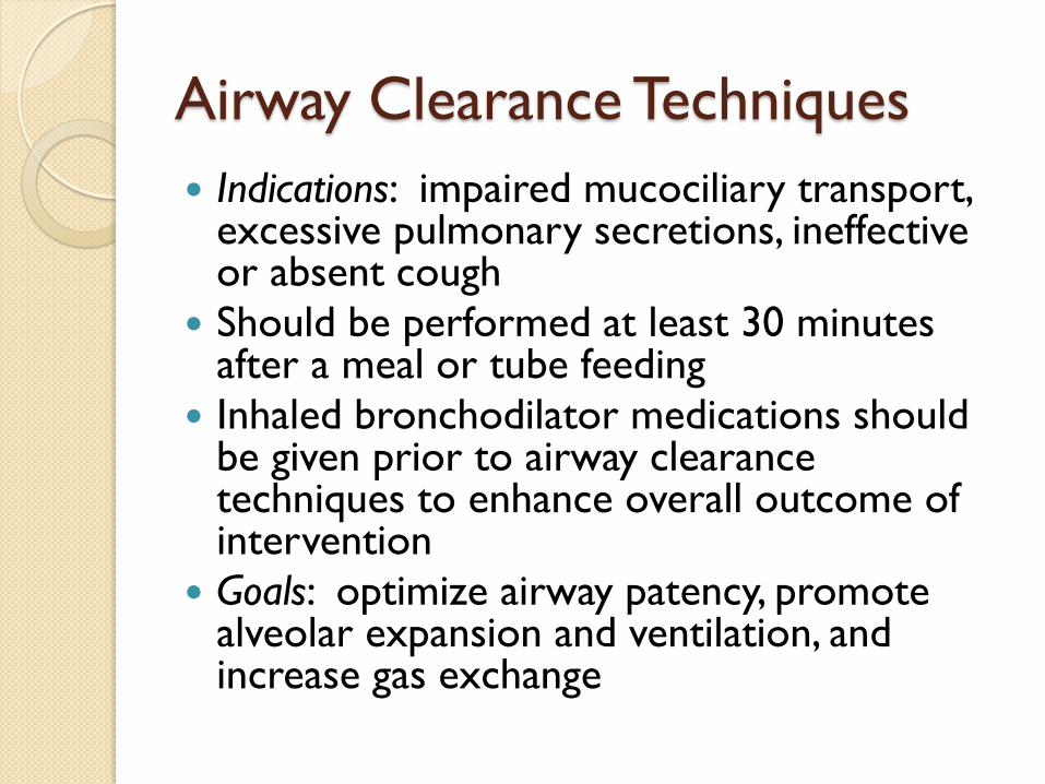
Indications: impaired mucociliary transport, excessive pulmonary secretions, ineffective or absent cough
Should be performed at least 30 minutes after a meal or tube feeding
Inhaled bronchodilator medications should be given prior to airway clearance techniques to enhance overall outcome of intervention
Goals: optimize airway patency, promote alveolar expansion and ventilation, and increase gas exchange

Airway Clearance Techniques
Historically physical therapists addressed this with percussion, vibration and postural drainage
Other devices and techniques have been shown to be as effective
When trying to decide what method of secretion clearance to use, consider the following: ◦ Is it adaptable
◦ Can it be performed independently
◦ Is it cost effective
◦ Is it time efficient

Active Cycle of Breathing
Effective form of airway clearance
◦ Very effective way to clear excess bronchial
secretions
Active participation from the patient
Cyclic repetition of 3 phases:
1. breathing control
2. thoracic expansion
3. forced expiratory technique

Active Cycle of Breathing
Breathing Control
◦ Diaphragmatic breathing at a normal tidal volume
◦ Rest one hand on the stomach and allow shoulders to relax
◦ Breathe quietly and gently.
◦ With inspiration the stomach should rise slightly, it should fall with expiration
Thoracic Expansion
◦ Deep inhalation with relaxed exhalation
◦ Used to get air behind the sputum stuck in small airways
◦ Relax upper chest
◦ Breathe in slowly and deeply – will see rib cage expand laterally
◦ Breathe out gently until your lungs are empty – don’t force the air out
Active Cycle of Breathing
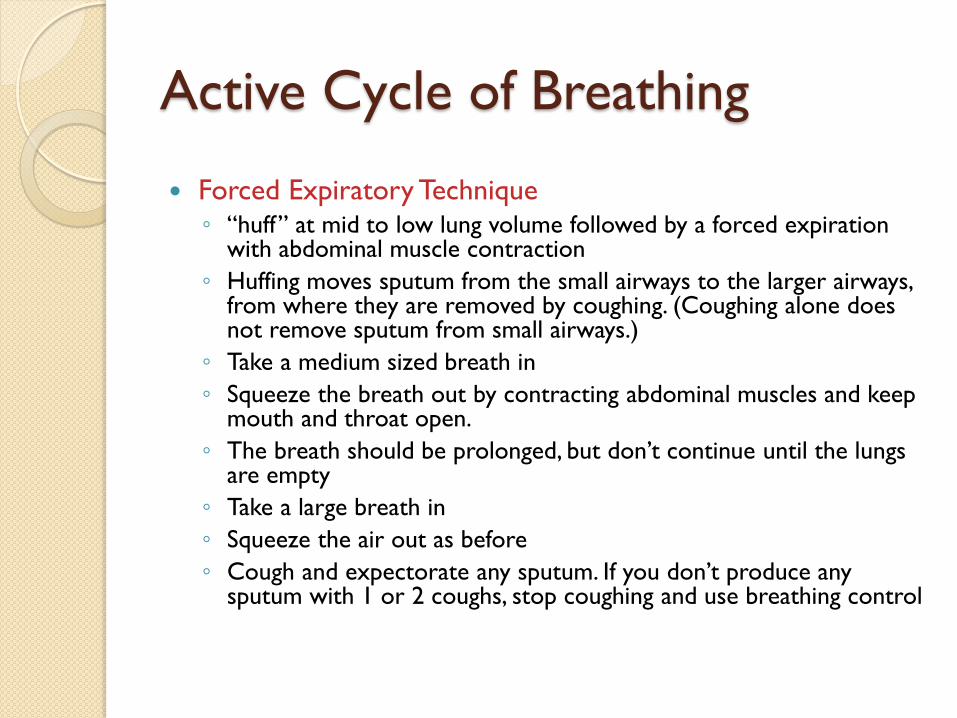
Forced Expiratory Technique ◦ “huff” at mid to low lung volume followed by a forced expiration
with abdominal muscle contraction
◦ Huffing moves sputum from the small airways to the larger airways, from where they are removed by coughing. (Coughing alone does not remove sputum from small airways.)
◦ Take a medium sized breath in
◦ Squeeze the breath out by contracting abdominal muscles and keep mouth and throat open.
◦ The breath should be prolonged, but don’t continue until the lungs are empty
◦ Take a large breath in
◦ Squeeze the air out as before
◦ Cough and expectorate any sputum. If you don’t produce any sputum with 1 or 2 coughs, stop coughing and use breathing control
Active Cycle of Breathing
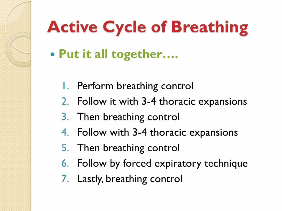
Put it all together….
1. Perform breathing control
2. Follow it with 3-4 thoracic expansions
3. Then breathing control
4. Follow with 3-4 thoracic expansions
5. Then breathing control
6. Follow by forced expiratory technique
7. Lastly, breathing control
Active Cycle of Breathing
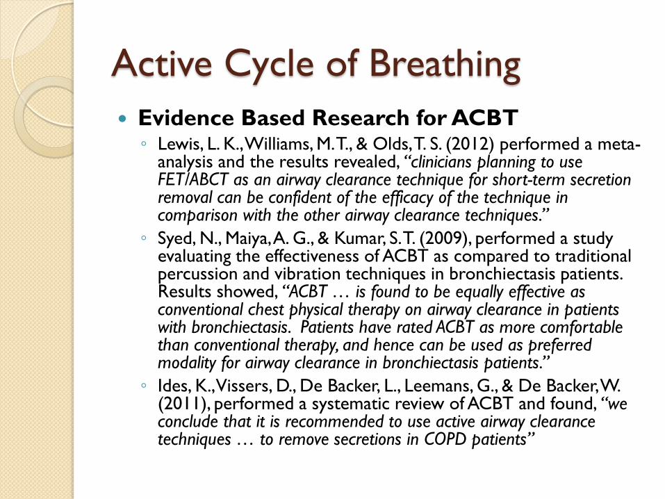
Evidence Based Research for ACBT ◦ Lewis, L. K., Williams, M. T., & Olds, T. S. (2012) performed a meta-
analysis and the results revealed, “clinicians planning to use FET/ABCT as an airway clearance technique for short-term secretion removal can be confident of the efficacy of the technique in comparison with the other airway clearance techniques.”
◦ Syed, N., Maiya, A. G., & Kumar, S. T. (2009), performed a study evaluating the effectiveness of ACBT as compared to traditional percussion and vibration techniques in bronchiectasis patients. Results showed, “ACBT … is found to be equally effective as conventional chest physical therapy on airway clearance in patients with bronchiectasis. Patients have rated ACBT as more comfortable than conventional therapy, and hence can be used as preferred modality for airway clearance in bronchiectasis patients.”
◦ Ides, K., Vissers, D., De Backer, L., Leemans, G., & De Backer, W. (2011), performed a systematic review of ACBT and found, “we conclude that it is recommended to use active airway clearance techniques … to remove secretions in COPD patients”
Airway Clearance Techniques
Deep breathing exercises
◦ Aid in secretion clearance
◦ Perform as described on the active cycle of
breathing slides
Airway Clearance Techniques
Cough
◦ An effective cough is one of the best ways to clear the airway!
◦ An initial intervention should be to make sure that patients are taught how to perform an effective cough and that they are proficient with it Position the patient to allow for trunk extension and flexion
Inspiration – combine with trunk extension and UE elevation
Expiration – combine with trunk flexion and lowering UEs
Improve the inspiratory hold by giving VCs and by positioning
Maximize intrathoracic and intraabdominal pressures with mm contractions or trunk movement
Airway Clearance Techniques
Coughing continued….
◦ If a patient is unable to cough, then teach them to “huff”
◦ Deep inspiration followed by forced expiration without glottis closure (as in coughing)
Often used in post-op patients when coughing would be too painful
◦ Coughing exercises are beneficial after bronchodilator treatments or before or after each exercise session
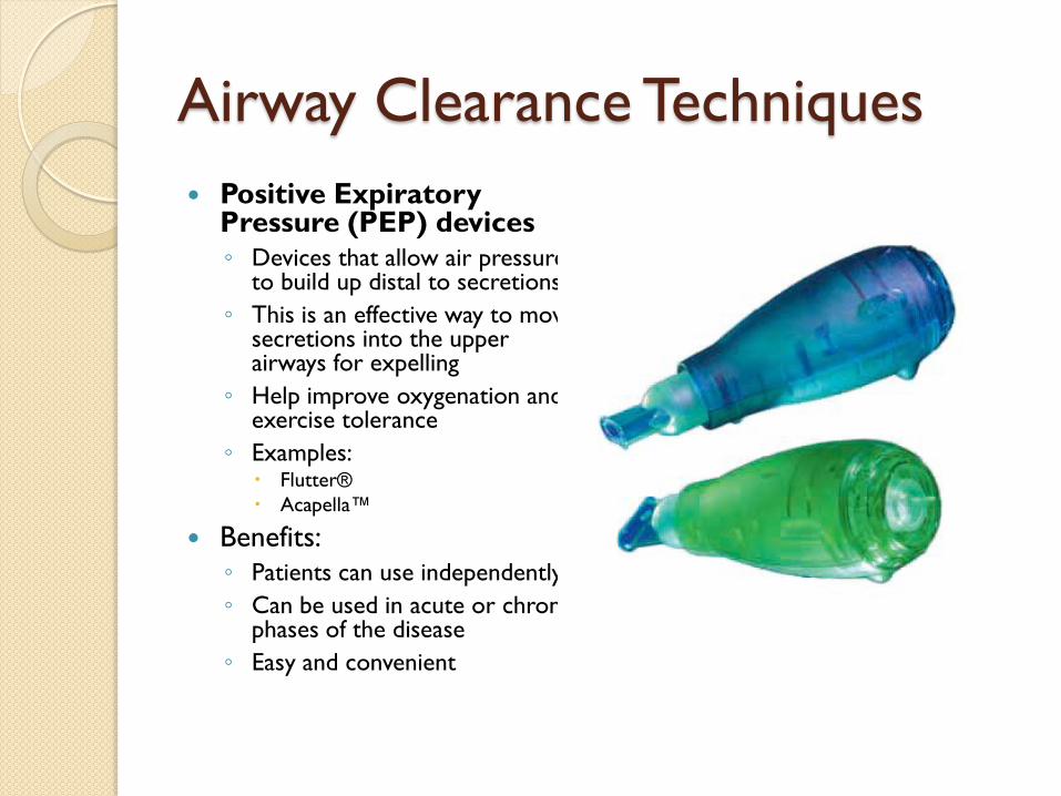
Airway Clearance Techniques
Positive Expiratory Pressure (PEP) devices ◦ Devices that allow air pressure
to build up distal to secretions
◦ This is an effective way to move secretions into the upper airways for expelling
◦ Help improve oxygenation and exercise tolerance
◦ Examples: Flutter®
Acapella™
Benefits: ◦ Patients can use independently
◦ Can be used in acute or chronic phases of the disease
◦ Easy and convenient
Airway Clearance Techniques
Sustained aerobic exercise, if tolerated by
the patient, can have very beneficial
airway clearance effects

Airway Clearance Techniques
Summary:
◦ Vibration
◦ Percussion
◦ Postural Drainage
◦ Active Cycle of Breathing
◦ Deep breathing
◦ Coughing
◦ PEP devices
◦ Aerobic exercise
Techniques to Control Dyspnea
Pursed Lip Breathing ◦ Used to decrease symptoms of dyspnea
◦ Encourage patients to use this with exercise and activity
Paced Breathing ◦ Volitional control of breathing during activity
◦ During rhythmic activities, coordinate the breathing with the rhythm of the activity
◦ During non-rhythmic activities have the patient breathe in at the beginning of the activity and push out during the activity
◦ This can help the patient with their feelings of dyspnea and to control their breathing
◦ The basic concepts of paced breathing are inhale with rest and exhale with work; slow down, set priorities, get organized and take rest breaks

Techniques to Control Dyspnea
Inspiratory Hold Technique ◦ Prolonged holding of breath at maximum inspiration
◦ Can be used in conjunction with vibration to aid in
airway clearance
◦ Can improve flow of air to poorly ventilated regions
of the lungs
◦ Patient is instructed to hold his or her breath at the
peak of inspiration for 2-3 seconds and then do a
relaxed exhale
Techniques to Control Dyspnea
Diaphragmatic Controlled
Breathing ◦ Can be used to manage dyspnea, reduce atelectasis
and increase oxygenation
◦ Facilitating outward motion of the abdominal wall
while reducing upper rib cage motion of the
abdominal wall during inspiration
◦ Instruct patients in this technique in all positions
◦ Overall goal is to decrease use of accessory mm and
increase the recruitment of the diaphragm
Techniques to Control Dyspnea
Diaphragmatic Controlled Breathing Position the patient with a posterior pelvic tilt to
facilitate use of the diaphragm
Relax accessory mm through VCs
If desired breathing pattern is not noticed yet, instruct patient to “sniff” to help engage the diaphragm
Patients hands should be positioned on the abdomen for proprioceptive FB – patient is asked to sniff 3 times and exhale (feel for abdominal rise)
Will progress to 2 sniffs and then just one sniff and then progress to breathing in a relaxed manner
Semi-fowler and side-lying positions work well for this because they are gravity eliminated

Breathing Techniques for Dyspnea
How do I know when to encourage a patient in a specific technique? Which one is best?
◦ Be aware of what each is beneficial for
Diaphragmatic Breathing – manages dyspnea and improves oxygenation
Expiratory Hold Technique – gets air to poorly ventilated areas of the lungs and aids in airway clearance
Paced Breathing – controls dyspnea, especially during activity
Pursed Lip Breathing – aids in dyspnea, especially with activity and with emphysema

Positions to Relieve Dyspnea Hillegass, E. (2011)

Positions to Relieve Dyspnea
Semi-fowlers position (HOB elevated 45°)
◦ Many cardiopulmonary patients may prefer
Tripod Position
◦ Leaning forward on supported hands
◦ Intraabdominal pressures rise and the diaphragm is pushed
up in a lengthened position
◦ This position increase the strength/tension relationship of
the diaphragm
◦ The diaphragm has increased strength of contraction so
the patient will feel relief from dyspnea

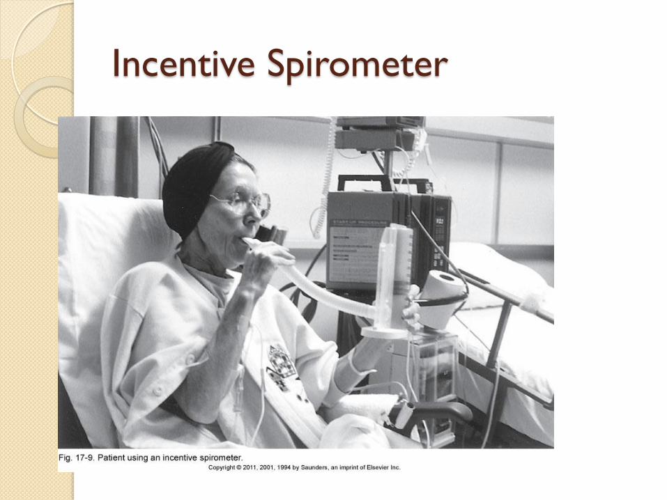
Incentive Spirometers
Incentive spirometers are an effective way to practice diaphragmatic breathing, prevent or reduce atelectasis and stimulate a cough
◦ Perform 10 reps every hour
◦ Recommended for all patients post surgery because surfactant is reduced postoperatively which makes patients at increased risk for pneumonia and respiratory failure
◦ Recommended for patients with a weak cough and weak abdominal muscles
PTs need to encourage the use of these!!
Incentive Spirometer
Flexibility & Strengthening
Flexibility exercises should be included for posture, to increase joint ROM, and decrease stiffness
Trunk and UE flexibility are essential for breathing and coughing techniques
Full shoulder flexion and back extension performed with inspiration
Exercises with forward reaching and trunk flexion with unilateral or bilateral hip and lower trunk flexion may be combined with expiration
Flexibility & Strengthening
Thoracic Mobilization Techniques
◦ If the mobility of the thorax is restricted it may be difficult for the patient to improve his or her breathing pattern through controlled breathing alone
◦ Techniques to Mobilize the Thorax: Place a towel roll vertically down the thoracic spine while the
patient is in supine will improve anterior chest wall mobility
Place the patient in side-lying over a towel roll will increase lateral chest wall mobility
UEs can be actively or passively flexed to further stretch the trunk
PNF techniques:
D2 flexion – UE flex, abd, ER
D2extension – UE ext, add, and IR
Flexibility & Strengthening
Respiratory Muscle Training
◦ Aerobic exercise training of the UE and LEs
that is moderate to high intensity is an
adequate stimulus to improve respiratory
muscle endurance and strength
◦ Instruction in breathing re-training exercise,
such as diaphragmatic breathing, can improve
strength, awareness, coordination of the
diaphragm muscle
Flexibility & Strengthening
Upper Body work and UE exercise….
◦ Associated with high metabolic demand and
ventilatory demand.
◦ Activities involving the UEs can lead to
irregular or dyssynchronous breathing (such
as ADLS), therefore, it is important to
incorporate UE exercise into the program
◦ Patients are more likely to hold their breath
and develop dyspnea with upper body work
◦ Upper body resistance training is essential!
Flexibility & Strengthening
Weight training should start with low
resistance and progress repetitions first;
increase to 20 reps before increasing
resistance
E-stim can be used to increase muscle
strength even in the absence of traditional
cardiovascular exercise

Energy Conservation
Prioritize daily tasks
In an inpatient setting nursing and therapy
must coordinate daily activities
◦ Patients can not be expected to take a
shower, go to physical therapy and go to the
dining room for lunch all back to back

PATIENT/FAMILY EDUCATION

Education
Use of supplemental oxygen
◦ Do the patients know when it is safe for them to turn up their oxygen and when it is not?
Exercise program
◦ Flexibility, strength, and aerobic exercise are essential in the management of COPD.
◦ Patients need to be setup with daily home programs that are direct, efficient and reasonable
Monitoring signs and symptoms of dyspnea
◦ Use the BORG everyday with patients – when ambulating, when taking a shower, when getting dressed
◦ Do they know what activities make their dyspnea worse?
◦ Do they recognize the signs?
◦ Have laminated BORG scales in every facility so that they can be used in therapy sessions
Education
Breathing techniques ◦ Do your patients know when to use PLB?
◦ When should they use active cycle of breathing?
◦ What works best for them and when?
◦ Practice these daily so that they become independent
Coughing techniques ◦ Is their cough weak or strong?
◦ If it is weak do they know to use trunk flexion and extension to enhance the force of the cough?
Positions for relief of dyspnea ◦ Practice dyspnea relief positions with COPD whenever
they are having exacerbations
◦ Before discharge from PT patients should know what positions provide them with the most relief
Break!!

WHAT IS CHF?
Understanding CHF
It is characterized by the inability of the
heart to maintain adequate cardiac output
Essentially, the heart muscle can not pump
enough blood through the heart to meet
the body’s needs for blood and oxygen

Understanding CHF
With heart failure the pumping action of
the heart becomes less and less powerful.
When this happens, blood does not move
efficiently through the circulatory system
and starts to back up.
Symptoms depend on which area of the
body is most involved in the reduced
pumping action
Understanding CHF
Types of Congestive Heart Failure:
◦ Left Side
◦ Right Side
◦ Diastolic
◦ Systolic

Understanding CHF
Left Side Heart Failure:
◦ Left ventricle is unable to maintain normal cardiac output
◦ When the left side of the heart (left ventricle) starts to fail, fluid collects in the lungs – this is pulmonary edema or congestion.

Understanding CHF
◦ This extra fluid in the lungs makes it more difficult for the airways to expand as a person inhales.
◦ Breathing becomes more difficult and the person may feel short of breath, particularly with activity or when lying down.
◦ Fluid also diffuses into the pleural spaces and can cause pleural effusion
Understanding CHF
Right Side Heart Failure
◦ When the right side of the heart (right ventricle) starts to fail, fluid begins to collect in the feet and lower legs.
◦ This usually occurs secondary to left side heart failure or if a right ventricular impairment is present (such as PE)
When right side of the heart fails due to left heart failure this is known as biventricular heart failure
◦ Edema is a sign of right heart failure, especially if the edema is pitting edema.
Understanding CHF
Right Side Heart Failure
◦ As the right heart failure worsens, the upper
legs swell and eventually the abdomen collects
fluid - ascites.
◦ Weight gain accompanies the fluid retention
and is a reliable measure of how much fluid is
being retained.
◦ Jugular vein distension is common
◦ Right Side Heart Failure is referred to as Cor
Pulmonale
Understanding CHF
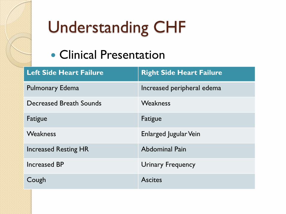
Clinical Presentation
Left Side Heart Failure Right Side Heart Failure
Pulmonary Edema Increased peripheral edema
Decreased Breath Sounds Weakness
Fatigue Fatigue
Weakness Enlarged Jugular Vein
Increased Resting HR Abdominal Pain
Increased BP Urinary Frequency
Cough Ascites

Understanding CHF
Risk Factors:
◦ Smoking
◦ Diabetes
◦ Anemia
◦ Psychological Stress
◦ Alcohol and drug abuse
Understanding CHF
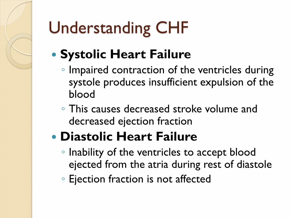
Systolic Heart Failure
◦ Impaired contraction of the ventricles during systole produces insufficient expulsion of the blood
◦ This causes decreased stroke volume and decreased ejection fraction
Diastolic Heart Failure
◦ Inability of the ventricles to accept blood ejected from the atria during rest of diastole
◦ Ejection fraction is not affected

Understanding CHF
Ejection Fraction
◦ Best indicator of cardiac function
◦ Ratio or percentage of the blood volume ejected out of the ventricles relative to the blood volume received by the ventricles before contraction
◦ Normal: 60-70%
◦ A certain volume of blood must always remain in the ventricles in order for the myocardium to maintain a certain level of stretch

Understanding CHF
Other Signs and Symptoms….
◦ Crackles in the bases of the lungs
◦ S3 Heart Sound
◦ Weight gain Greater than 3lbs per day is a concern
◦ Sinus tachycardia
Body attempts to increase the delivery of fluid and oxygen to the peripheral tissues where it is needed; compounds the problem more and makes the heart work harder
◦ Decreased exercise tolerance Decreased blood flow to the muscles

CHF PATIENT ASSESSMENT
Patient Assessment
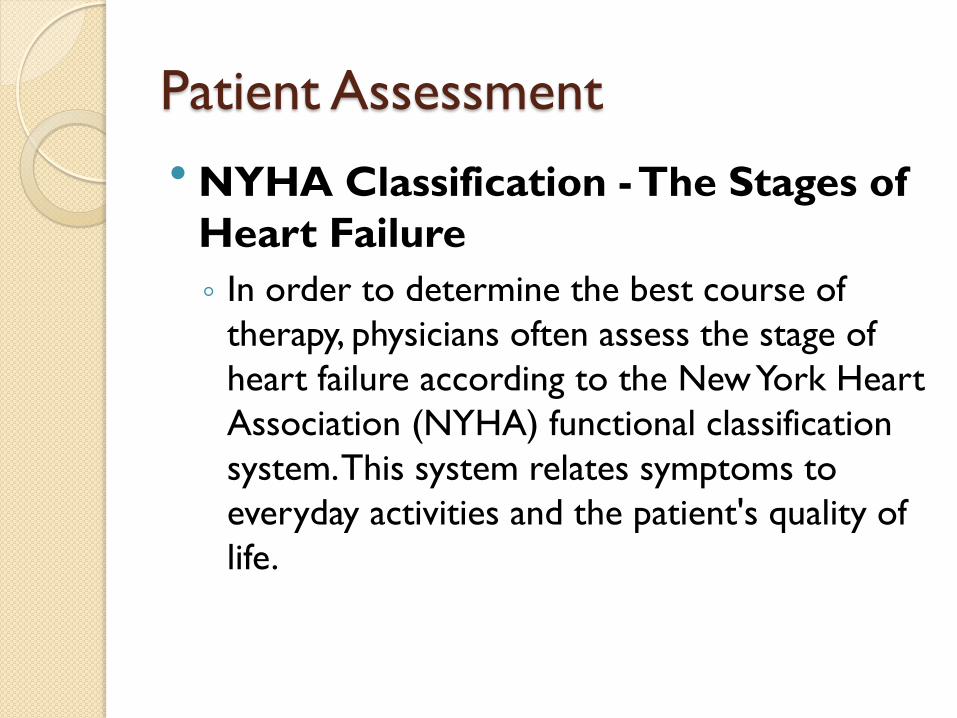
NYHA Classification - The Stages of
Heart Failure
◦ In order to determine the best course of
therapy, physicians often assess the stage of
heart failure according to the New York Heart
Association (NYHA) functional classification
system. This system relates symptoms to
everyday activities and the patient's quality of
life.

Patient Assessment
Class Patient Symptoms
Class I (Mild) No limitation of physical activity. Ordinary physical activity does
not cause undue fatigue, palpitation, or dyspnea (shortness of
breath).
Class II (Mild) Slight limitation of physical activity. Comfortable at rest, but
ordinary physical activity results in fatigue, palpitation, or dyspnea.
Class III
(Moderate)
Marked limitation of physical activity. Comfortable at rest, but less
than ordinary activity causes fatigue, palpitation, or dyspnea.
Class IV
(Severe)
Unable to carry out any physical activity without discomfort.
Symptoms of cardiac insufficiency at rest. If any physical activity is
undertaken, discomfort is increased.
Patient Assessment
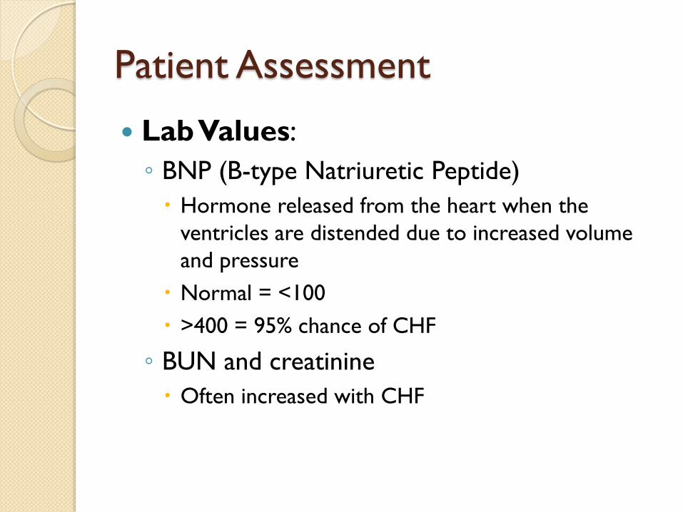
Lab Values:
◦ BNP (B-type Natriuretic Peptide)
Hormone released from the heart when the
ventricles are distended due to increased volume
and pressure
Normal = <100
>400 = 95% chance of CHF
◦ BUN and creatinine
Often increased with CHF

Patient Assessment
Evaluation
◦ Physical Appearance:
Peripheral edema
Jugular Vein distension
Posture
◦ Mobility Assessment
◦ ROM, strength
◦ Peripheral pulses and overall circulation
◦ Vital Signs
◦ Edema assessment
◦ Cardiopulmonary Activity Tolerance

Patient Assessment
Vital Signs
◦ Heart Rate
Pay attention to rate and rhythm
◦ Blood pressure
◦ Respiratory rate
◦ Oxygen saturation level
>90%
Is it necessary to monitor the pulmonary system or can you just do HR and BP?
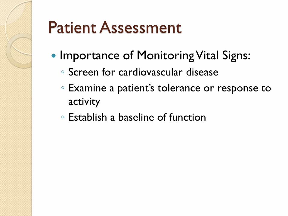
Patient Assessment
Importance of Monitoring Vital Signs:
◦ Screen for cardiovascular disease
◦ Examine a patient’s tolerance or response to
activity
◦ Establish a baseline of function
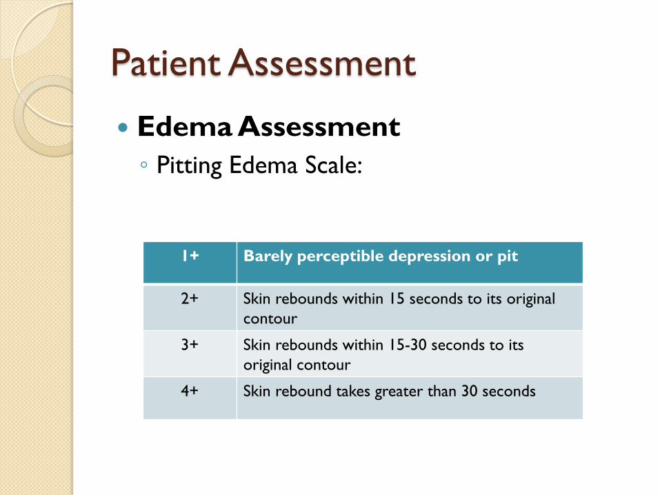
Patient Assessment
Edema Assessment
◦ Pitting Edema Scale:
1+ Barely perceptible depression or pit
2+ Skin rebounds within 15 seconds to its original
contour
3+ Skin rebounds within 15-30 seconds to its
original contour
4+ Skin rebound takes greater than 30 seconds

Patient Assessment
Edema
◦ Girth measurements
Ankles: figure 8
Circumferential lower legs
Circumferential abdomen

Patient Assessment
Cardiopulmonary Activity Tolerance
◦ BORG Scale of Perceived Exertion
◦ Gait Velocity
◦ 6 Minute Walk Test
◦ 2 Minute Step Test
◦ Ventilatory Response Index (VRI)
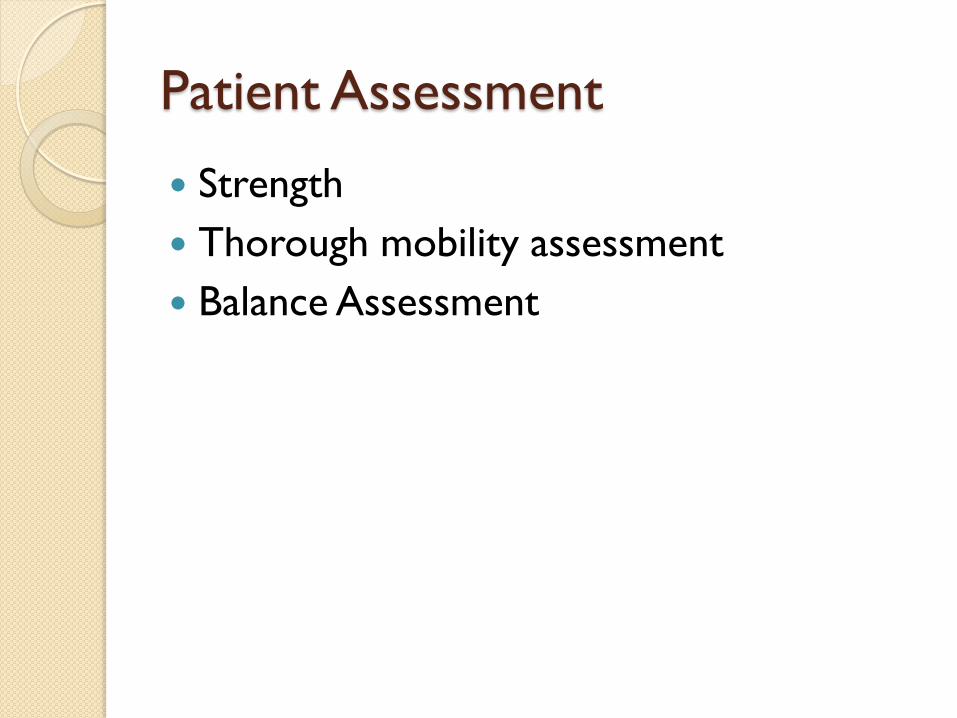
Patient Assessment
Strength
Thorough mobility assessment
Balance Assessment

TREATMENT OF THE PATIENT WITH CHF

Treatment of CHF
Exercise
◦ Resistance training, aerobic exercise and
flexibility
Functional Mobility
◦ Think energy conservation and safety
◦ Issue assistive devices as appropriate
Fluctuations in LE edema can impact balance and
safety with mobility
Patient Education

Treatment of CHF
Exercise
◦ Monitor vital signs before, during and after
exercise
◦ Be aware of other clinical signs – increased
peripheral edema, dyspnea
◦ BORG scale of perceived exertion

Treatment of CHF
Contraindications to Exercise ◦ If HR is >100bpm before therapy they are
(tachycardic)
◦ Resting HR <50bpm
◦ Resting systolic BP >200mmHg or <90mmHg
◦ Resting diastolic BP >110mmHg
◦ Oxygen saturations <90%
◦ Cyanosis
◦ Edema in patient with CHF and has gained 3lbs of weight in one day
◦ Worsening dyspnea over 1-3 days
◦ Class IV New York Heart Association CHF
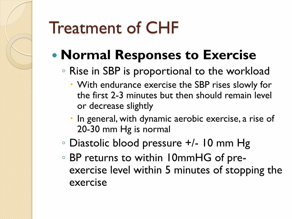
Treatment of CHF
Normal Responses to Exercise ◦ Rise in SBP is proportional to the workload
With endurance exercise the SBP rises slowly for the first 2-3 minutes but then should remain level or decrease slightly
In general, with dynamic aerobic exercise, a rise of 20-30 mm Hg is normal
◦ Diastolic blood pressure +/- 10 mm Hg
◦ BP returns to within 10mmHG of pre-exercise level within 5 minutes of stopping the exercise
Treatment of CHF
Normal Responses to Exercise
◦ HR gradually increases proportional to the workload With low to moderate intensity exercise the HR rise should
be fairly small: <20-40 beats/minute
HR response with exercise will be blunted in those taking Beta blockers
◦ During static or isometric exercises the HR increase is less pronounced than with dynamic exercise due to reduced cardiac output requirements
◦ HR should return to within 10bpm of pre-exercise within 5 min of stopping exercise
◦ If elderly, HR response may be less brisk overall

Treatment of CHF
Normal Responses to Exercise
◦ Rate of Perceived Exertion
RPE 13/20 (indicating moderate exercise)
Increases gradually in proportion to the progressive workload
Correlated highly with HR response
More rapid increase in RPE with gradually increasing workloads indicate marked impairment in exercise tolerance, which is often due to cardiovascular and pulmonary dysfunction
◦ Respiratory Rate
Gradually increases with exercise
Treatment of CHF
Exercise Recommendations
◦ Aerobic activity = moderate intensity for
minimum of 30 minutes, 5days/wk or vigorous
activity for a minimum of 20 minutes 3days/wk
◦ Muscle strengthening = minimum of 2 days/wk, 8-
10 exercises using the major mm groups (weight
training, calisthenics) on non-consecutive days
◦ Flexibility exercises = at least 2 days/wk for at
least 10 min each day
Recommendations from the American College of Sports Medicine and the American Heart Association

Treatment of CHF
Exercise Prescription
◦ When working with patients with CHF, therapists
and other health care providers have to be able to
prescribe the correct intensity of exercise.
◦ Use the BORG Rate of Perceived Exertion Scale to
determine how a patient is tolerating the exercises
prescribed
Recommendation is that a patient with CHF should rate
themselves from 11-14 (light to somewhat hard)
Treatment of CHF
Edema Management
◦ Compression stockings
Can make symptoms worse!
Must have clearance from cardiologist
◦ Daily weight monitoring
Can the patient independently get on and off of the scale?
Can the patient see the scale?
Can the patient track their weight so that they understand if they have had a 3lb. weight gain that warrants calling their cardiologist?

Treatment of CHF
Patient Education
◦ Understanding of CHF
◦ Medications
What they are taking, when they should take them,
and why
◦ Self monitoring during functional mobility
activities and ADLS
BORG
◦ Self monitoring of weight
Treatment of CHF
Patient Education Regarding
Lifestyle Changes
◦ Stress management and relaxation techniques
◦ Smoking cessation
◦ Diet: low fat, low cholesterol, low sodium,
fluid restriction
◦ Home exercise programs and lifelong exercise
programs – ambulation is a very effective mode
of exercise for CHF patients!

Treatment of CHF
Many patients with CHF will also develop
COPD!
Even in CHF patients who do not have
COPD, breathing exercises are important!
◦ Facilitation of diaphragmatic breathing and
inhibition of accessory muscle use may
decrease the work of breathing in CHF
◦ Pursed lip breathing is effective in assisting
with dyspnea management in those with CHF

References
Center for Disease Control and Prevention (2013). The State of Aging
Health in America. Retrieved from:
http://www.cdc.gov/features/agingandhealth/state_of_aging_and_heal
th_in_am erica_2013.pdf
Centers for Medicare and Medicaid Services (2014). Readmissions
Reduction Program. Retrieved from:
http://www.cms.gov/Medicare/Medicare-Fee-for-Service-
Payment/AcuteInpatientPPS/Readmissions-Reduction-Program.html
Chodbury, G., Rabinovich, R., & MacNee, W. (2014). Comorbidities and
Systemic Effects on Chronic Obstructive Pulmonary Disease. Clinics
in Chest Medicine, 35(1), 101-130. Doi: 10.1016/j.ccm.2013.10.007.
COPD Assessment Test (2013). Retrieved from
http://www.catestonline.org/
Frownfelter, Donna & Dean, Elizabeth. Cardiovascular and Pulmonary Physical
Therapy. St. Louis: Mosby, 2006. 4th Edition.

References
Hillegass, E. (2011). Essentials of Cardiopulmonary Physical Therapy. St. Louis: Elsevier Saunders. 3rd Edition
• Ides, K., Vissers, D., De Backer, L., Leemans, G., & De Backer, W. (2011). Airway clearance for COPD: Need for a breath of fresh air? A systemic review. Journal of Chronic Obstructive Pulmonary Disease, 8(3), 196-205. Doi: 10.3109/15412555.2011.560582.
Levine, P. (2010). Using gait speed as a marker for progress. Advance, 21(6), 38.
Lewis, L. K., Williams, M. T., & Olds, T. S. (2012). The active cycle of breathing technique: A systematic review and meta- analysis. Respiratory Medicine, 106(2), p. 155-172. Doi: 10.1016/j.rmed.2011.10.014
Moffat, M. & Frownfelter, D. (2007). Cardiovascular/Pulmonary Essentials: Applying the Preferred Physical Therapist Practice Patterns . New Jersey: SLACK Incorporated.

References
Rikli R. E. & Jones C. J. (1999). Functional fitness normative scores for
community residing older adults ages 60-94. Journal of Aging and
Physical Activity, 7,160-179.
Steffen T. M., Hacker, T.A., & Mollinger, L. (2002). Age and gender related test
performance in community-dwelling elderly people: 6mw test, BBS,
TUG, and gait speed. Physical Therapy, 82(2), 128-137.
Syed, N., Maiya, A. G., & Kumar, S. (2009). Active cycles of breathing technique
(ACBT) versus conventional chest physical therapy on airway clearance
in bronchiectasis – A crossover trial. Advances in Physiotherapy 11(4),
193-198. Doi: 10.3109/14038190802294856.
Watchie, J. (2003). Monitoring Clinical Responses to Exercise.
Cardiovascular & Pulmonary Section, American Physical Therapy
Association.
Wills, K. & Mai, J. (2012). Congestive heart failure: A review for physical
therapist practitioners. GeriNotes, 19(3), 12-15