Physical Examination Of The Dialysis Vascular Access · 2017-10-20 · Physical Examination of the...
20
A Practitioner’s Resource Guide To Physical Examination of Dialysis Vascular Access The ESRD Network of Texas, Inc., is proud to offer this excellent educational resource written by Gerald Beathard, MD for distribution to the nephrology & surgical community in support of the Fistula First Project. The End Stage Renal Disease Network of Texas, Inc (#14) www.esrdnetwork.org 972-503-3215 14114 Dallas Parkway #660, Dallas, Texas, 75254
Transcript of Physical Examination Of The Dialysis Vascular Access · 2017-10-20 · Physical Examination of the...

A Practitioner’s Resource Guide To
Physical Examination of
Dialysis Vascular Access The ESRD Network of Texas, Inc., is proud to offer this excellent educational resource written by Gerald Beathard, MD for distribution to the nephrology & surgical community in support of the Fistula First Project.
The End Stage Renal Disease Network of Texas, Inc (#14) www.esrdnetwork.org
972-503-3215 14114 Dallas Parkway #660, Dallas, Texas, 75254

This article is used with the permission of the author, Gerald A. Beathard, MD, PhD, FACP, FCAP. Dr. Beathard is Medical Director of RMS Lifeline and Clinical Associate Professor, Internal Medicine, at the University of Texas Medical Branch at Galveston. He is a member of the American Society of Diagnostic and Interventional Nephrology and is board certified in Internal Medicine, Nephrology, Pathology (Anatomical) and Allergy and Clinical Immunology.
TABLE OF CONTENTS
Physical Examination Related to Arteriovenous Fistulae (AVF)……………………………..1
Evaluation Prior to Access Placement…………………………………………………………….1 Arterial Evaluation………………………………………………………………………...1 Venous Evaluation………………………………………………………………………...2 Evaluation of Early AVF Failures………………………………………………………………...3 Juxta-Anastomotic Venous Stenosis……………………………………………………....4 Accessory Veins…………………………………………………………………………...5 Evaluation of Late Fistula Problems……………………………………………………………....6 Assessing Strength of Arterial Inflow……………………………………………………..6 Venous Stenosis…………………………………………………………………………...7 Aneurysm Formation……………………………………………………………………...7 Ischemia…………………………………………………………………………………...8 Infection of AVF…………………………………………………………………………..9 Physical Examination Related to Arteriovenous Grafts……………………………………....9 Detection of Direction of Flow……………………………………………………………………9 Detecting Recirculation…………………………………………………………………………...9 Diagnosis of Venous Stenosis…………………………………………………………………....10 Graft Infection……………………………………………………………………………………12 Ischemia Associated with a Graft………………………………………………………………..13 Ischemic Monomelic Neuropathy………………………………………………………..13 Vascular Steal Syndrome………………………………………………………………...14 Pseudoaneurysm Associated with a Graft………………………………………………………..15 Conclusion……………………………………………………………………………………... 16 References…………………………………………………………………………………….…17
Distributed by: End Stage Renal Disease Network of Texas, Inc. (#14)
14114 Dallas Parkway #660, Dallas, Texas 75254 Phone: 972-503-3215 http://www.esrdnetwork.org/
Distributed November 2003
The End Stage Renal Disease Network of Texas, Inc. (#14) is under contract with the Centers for Medicare & Medicaid Services (CMS). The contents presented do not necessarily reflect CMS policy.
ii

Physical Examination of the Dialysis Vascular Access “The examining physician often hesitates to make the necessary examination because it involves soiling the finger” William J. Mayo, MD, 1915. Physical examination, i.e., the use of the hands, eyes and ears as diagnostic instruments is a skill that has been basic to medicine from its earliest origins. Unfortunately, in many disciplines of medicine it is one that has been largely abandoned in favor of more elaborate and costly technical approaches to diagnosis. In the evaluation of the dialysis vascular access, one could argue that it never existed. Nevertheless, physical examination is easily performed, inexpensive to apply and provides a high level of accuracy (1-4) to the physician who understands its principles and is inclined toward the art of medicine as well as its science. Physical Examination Related to Arteriovenous Fistulae (AVF) Evaluation Prior to Access Placement Evaluation of the new end stage renal disease (ESRD) patient in preparation for the placement of a peripheral venous access is extremely important. Proper patient selection will materially enhance the opportunity to place an arteriovenous fistula (AVF). In order to determine the type of access most suitable for an ESRD patient a general physical examination as well as a detailed focused medical history is important (5). Emphasis should be placed upon aspects that might affect the placement of the vascular access. Any physical evidence (scars) that the patient has had previous central venous catheters should be documented. In most instances a patient will give a positive history for such an occurrence, but this is not always the case (6). The patient’s chest, breast and upper arms should be evaluated for the presence of swelling or collateral veins. In patients with normal venous pressure, central venous occlusion may not be associated with swelling; however, the presence of collateral veins should alert the examiner to the problem. The presence of scars indicating previous neck or thoracic surgery or trauma should also raise the possibility of a venous anomaly that might affect access creation. Specially directed evaluations of both the arterial and venous anatomy should be completed. Arterial Evaluation In relation to the arterial system, two issues are important. Firstly, the vessel must be capable of delivering blood flow at a rate adequate to support dialysis and secondly, the utilization of the vessel for the creation of an access must not jeopardize the viability of the digits and hand. Arterial narrowing and calcification are relatively common in ESRD patients, especially those that are diabetic and hypertensive. This problem can usually be diagnosed before the patient is sent for surgery. A significant amount of data can be obtained by physical examination relative to the adequacy of arterial inflow for the creation of an arteriovenous fistula. There are three evaluations (Table 1) that lend themselves to this purpose - pulse examination, segmental blood pressure measurement and the Allen Test. Pulse examination - The axillary, brachial, radial and ulnar pulses should be examined in both upper extremities. The quality of these pulses should be scored as either normal, diminished or absent (7).
1

Segmental blood pressures - Differences in blood pressure between the two arms should be should be reported and graded (7). A difference of less than 10 mm Hg should be considered as normal. A difference of 10 to 20 mm Hg is considered marginal and over 20 as problematic.
Table 1 - Arterial Evaluation for AVF
• • •
Pulse examination Segmental blood pressures - Pressure differential < 20 mmHg between armsPatent palmar arch (Negative Allen Test)
The Allen Test - The Allen Test (Table 2) is used to determine competence of the palmar arch. Use of vascular Doppler can increase the effectiveness of the Allen test in predicting collateral arterial perfusion of the hand. Given adequate collateral flow, the Doppler should detect augmented pulsation in the palmar arch during occlusion of either the radial or ulnar artery (8). Failure of palmar arch pressures to increase during this maneuver suggests inadequate collateral circulation in the hand and predicts a higher risk for vascular steal if the dominant artery is used for access creation.
Table 2 - The Allen Test
1.
2. 3.
4.
5.
.
Position the patient so that they are facing you with their arm extended with the palm turned upward. Compress both the radial and ulnar arteries at the wrist. With the arteries compressed firmly, instruct the patient to create a fist repetitively in order to cause the palm to blanch. When the patient’s hand is blanched, release the compression of the ulnar artery and watch the palm to determine if it becomes pink. Then release all compression. Repeat steps 2-4 for the radial artery.
Interpretation – When color returns to the blanched palm upon release of the arterial compression it indicatesarterial patency and reflects upon adequacy of flow. Blanching that persists for 5 seconds or more after releaseof the ulnar artery is a positive test for radial artery insufficiency. Likewise, blanching that persists for 5 secondsor more following release of the radial artery is positive for ulnar insufficiency
If clinical examination results are clearly satisfactory, no further testing is mandatory. However, if problems are apparent, then noninvasive or even invasive studies should be used for evaluation (7). Duplex ultrasound is a particularly valuable tool for vascular evaluation relative to access placement. It has been found that a preoperative radial artery diameter of less than 1.6 mm is associated with a high risk of failure in radial-cephalic fistulae (9, 10). Others have suggested that a diameter of 2 mm or greater is optimal (11). Duplex ultrasound can also be used to evaluate blood flow and determine resistive index in specific vessels. Venous Evaluation Venous anatomy is extremely important for access creation; either an AVF or an arteriovenous graft (graft). Most problems incurred with access creation are actually venous problems. The primary goal when evaluating any patient for a dialysis vascular access should be the identification of a venous anatomy conducive to the creation of an AVF. This is best done by vein mapping.
2

The cephalic vein is ideal for an AVF because it is located on ventral surface of the forearm and the lateral surface of the upper arm (Figure 1). These features make for easy access in the dialysis facility with the patient in a sitting position. Ideally, the cephalic should have a straight segment long enough to allow for rotation of cannulation sites and lie within 1 cm of the surface.
Figure 1 - Appearance of veins of lower arm To evaluate a patient for these optimum characteristics, venous mapping should be performed. In some patients this can be done adequately by physical examination (Figure 1). It is essential that the patient be evaluated with outflow obstruction so as to dilate the veins of the arm adequately for evaluation. This is best done using a blood pressure cuff inflated to a pressure about 5mm Hg above diastolic pressure. This should be left in place for periods of no more than 5 minutes at a time. While this provides excellent information in many patients, most surgeons will want a more detailed venogram performed using color flow Doppler ultrasound prior to surgery. Optimum features on venogram (Table 3) for the creation of an AVF are a luminal diameter at the point of anastomosis of 2.5 mm or greater, a straight segment of vein, absence of obstruction and continuity with the proximal central veins (11).
Table 3 – Venous Requirements for AVF
• • • • •
Luminal diameter 2.5 mm or greater at anastomosis point Absence of obstruction Straight segment for cannulation Within 1 cm of surface Continuity with proximal central veins
Evaluation of Early AVF Failures Not all attempts at AVF creation are successful; this is especially true if the surgeon is appropriately aggressive in attempting to create an AVF. Fistulae that never develop adequately for use or those that fail within the first 3 months of use are classified as early failures. While there are multiple causes of early failure, the two most frequent causes, if the patients were
3

adequately evaluated prior to placement, are juxta-anastomotic venous stenosis and the presence of cephalic vein side branches referred to as accessory veins (12). Both of these anomalies can be easily diagnosed by physical examination.
Figure 2 – Juxta-anastomotic stenosis. A – radial artery, B – stenotic lesion, C – cephalic vein.
Juxta-Anastomotic Venous Stenosis The most common site for venous stenosis to occur in relation to an AVF is in the segment of vein that is immediately adjacent to the anastomosis (12, 13, 14). The etiology of this juxta-anastomotic lesion (Figure 2) is not clear. However, this is the segment of vein that is mobilized and manipulated by the surgeon in creating the fistula. It may be related to stretching, torsion or other types of trauma. The effect of the lesion is to obstruct fistula inflow. Since it occurs early, it results in early access failure. This lesion can be easily diagnosed by palpation of the anastomosis and distal vein (15, 16). Normally, a very prominent thrill is present at the anastomosis. In the absence of abnormalities, the pulse is soft and easily compressible. With juxta-anastomotic stenosis, a water-hammer pulse is felt at the anastomosis. The thrill, which is normally continuous, is present only in systole. As one moves up the vein from the anastomosis with the palpating finger (Figure 3), the pulse goes away rather abruptly as the site of stenosis is encountered. Above this level, the pulse is very weak and the vein is poorly developed. The stenosis itself can frequently be felt as an abrupt diminution in the size of the vein, almost like a shelf. Once these typical physical findings are detected, the cause for poor fistula development becomes obvious.
4

Figure 3 - Physical examination of juxta-anastomotpresent at the anastomosis (B). It disappears as one
Figure 4 – Accessory vein. A – Accessory bran Accessory Veins As previously stated, the optimum venous avein stretching from the wrist to the antecubitcase. The cephalic vein may have one or accessory veins diverts blood flow from theflow and pressure on the vein wall that is e(maturation) to occur. In many cases this ipermitting the development of multiple venouwhere flow is less than optimum, the accessor
C
ic stenosis moves up
ch coming
natomy fal space. several
main chssential fs not a ps sites foy vein(s)
5
B
A
. A – radial artery. A strong pulse and thrill are the fistula to the level of the lesion (C).
off of the cephalic vein, B – cephalic vein.
or AVF development is a single cephalic In many instances, however, this is not the side branches (Figure 3). Each of these annel. This has the effect of reducing the or expansion, dilation, and arterialization roblem; in fact it can be an advantage,
r access cannulation. However, in the case can result in early fistula failure.

Figure 5 - Physical examination of accessory vein. When the fistula is occluded at point A, the thrill will disappear at the anastomosis. As the point of occlusion is moved upward past the accessory vein to point B, the thrill will continue when the fistula is occluded. Accessory veins can be easily identified through physical examination (15, 16). Frequently they are visible. If not, they can be detected by palpating the fistula. Normally, the thrill that is palpable over the arterial anastomosis disappears when the down stream (antegrade) fistula is manually occluded (this causes flow to stop). If it does not disappear, an outflow channel (accessory vein) is present below the point of occlusion. Palpation of the fistula below the occlusion point will generally reveal the location of the accessory vein by the presence of a thrill over its trunk. As long as the main channel can be identified for occlusion, the entire length of the vein can be evaluated by moving the point of fistula occlusion progressively upward (Figure 5). Ligation of these accessory veins will redirect flow to the main channel and promote the development of a usable AVF (12, 13). If the AVF is created using a side-to-side anastomosis (the typical Cimino-Brescia fistula), an abnormal flow pattern in the venous system beyond the fistula over the back of the hand can occur. This can prevent the fistula from developing and can result in venous hypertension in the hand (Figure 6). This can cause pain, edema, and limitation of motion. Evaluation of Late Fistula Problems Once an AVF is functional, it is associated with much fewer problems than is seen with arteriovenous grafts. Nevertheless, problems can occur. In general, physical examination plays a major role in the evaluation of these problems. The most common complications associated with the established AVF are venous stenosis, thrombosis, ischemia, aneurysm formation and infection. Assessing Strength of Arterial Inflow Normally, a fistula is not pulsatile; it is very soft and compressible. If it is occluded; however, it becomes very pulsatile. The strength of the pulse is directly proportional to the arterial inflow pressure. This is a useful test in assessing the strength of the arterial inflow. It is certainly subjective, but the experienced examiner can glean a great deal from the evaluation. The term “augmentation” is used to describe this assessment. The fistula is said to augment well, meaning it has a very strong pulse and by inference, a good arterial inflow. Conversely, it may be found to augment poorly, meaning a weak pulse with obstruction and by inference, a poor arterial inflow. This test is also helpful in assessing the severity of a venous stenosis. The fistula will be pulsatile due to obstruction caused by the stenotic lesion. If this is compared to the augmented pulse obtained by total manual occlusion of the fistula, the severity of the lesion can be estimated by the comparison.
6

Figure 6 - Venous hypertension in hand secondary to side to side anastomosis. a – appearance of hand, note multiple dilated vessels, b – angiogram, arrow indicates anastomosis, note multiple collateral vessels (dim opacification due to rapid flow) Venous Stenosis The details of physical diagnosis of venous stenosis are discussed in detail below in conjunction with dialysis access grafts and in Table 4. They are basically the same for the AVF with only a few unique differences. Normally, the mature AVF has a soft pulse and the entire structure is easily compressed. When the extremity is elevated the entire fistula will generally collapse, at least partially. With down stream (antegrade) stenosis, the AVF becomes more forcibly pulsatile and firm. It also enlarges rapidly, often taking on aneurysmal or near-aneurysmal proportions. When the extremity is elevated, that portion of the fistula distal to point of stenosis remains distended, while the proximal portion collapses in the normal fashion (Figure 7). This phenomenon allows one to localize the site of obstruction. In addition, the pulse diminishes abruptly as does the caliber of the vessel. Changes in the location and character of thrills and bruits also occur as described later in association with grafts. Aneurysm Formation An aneurysm in an AVF is recognized as a localized ballooning of the vein (Figure 8). This is very much analogous to the pseudoaneurysm seen with the graft. The difference being that with a graft there is no vessel wall involved, thus the term pseudoaneurysm. With time, flow in an otherwise normal AVF continues to increase and the vein may continue to enlarge. Eventually, the AVF may become quite large and somewhat tortuous. It can reach aneurysmal proportions. Aneurysms are more likely to develop up stream (retrograde) from a venous stenosis, especially at sites of repetitive needle insertion. The examiner can recognize these easily. Their progress should be followed and any associated skin changes should be noted.
7
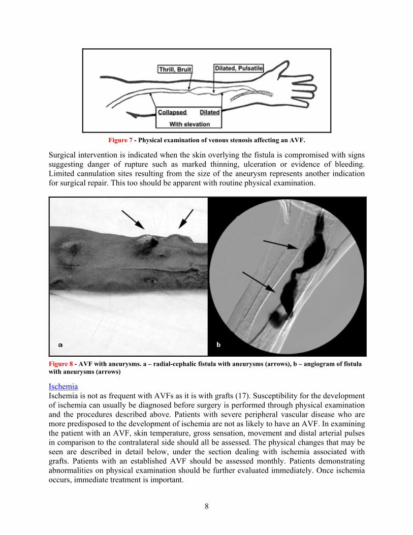
Figure 7 - Physical examination of venous stenosis affecting an AVF.
Surgical intervention is indicated when the skin overlying the fistula is compromised with signs suggesting danger of rupture such as marked thinning, ulceration or evidence of bleeding. Limited cannulation sites resulting from the size of the aneurysm represents another indication for surgical repair. This too should be apparent with routine physical examination.
Figure 8 - AVF with aneurysms. a – radial-cephalic fistula with aneurysms (arrows), b – angiogram of fistula with aneurysms (arrows)
Ischemia Ischemia is not as frequent with AVFs as it is with grafts (17). Susceptibility for the development of ischemia can usually be diagnosed before surgery is performed through physical examination and the procedures described above. Patients with severe peripheral vascular disease who are more predisposed to the development of ischemia are not as likely to have an AVF. In examining the patient with an AVF, skin temperature, gross sensation, movement and distal arterial pulses in comparison to the contralateral side should all be assessed. The physical changes that may be seen are described in detail below, under the section dealing with ischemia associated with grafts. Patients with an established AVF should be assessed monthly. Patients demonstrating abnormalities on physical examination should be further evaluated immediately. Once ischemia occurs, immediate treatment is important.
8

Infection of AVF Infection associated with an AVF occurs at a rate of about one-tenth that seen in grafts (18). Most infections associated with an AVF are actually perivascular cellulitis recognized by localized erythema, swelling and tenderness on physical examination. These are usually easily treated. Much more serious is the occasional infection associated anatomic abnormalities such as aneurysms, perigraft hematomas, or associated abscesses from infected needle puncture sites. These lesions are frequently associated with drainage and may be fluctuant to palpation. Such perivascular abscesses require surgical drainage or excision with access revision (18). Physical Examination Related To Arteriovenous Grafts Detection of Direction of Flow Most grafts are created with a standard configuration; however, occasionally it is necessary to deviate from the usual pattern of placement in order to accomplish the task. When this occurs, the orientation of the dialysis needles must correspond to the direction of blood flow or gross recirculation will result. In order to avoid this occurrence, the direction of blood flow should be determined and documented for each patient in the dialysis facility. This can be accomplished easily by occluding the graft with the tip of the finger and palpating on each side of the occlusion point for a pulse (Figure 9). The side without a pulse is the downstream side of the graft. The upstream pulse will increase in intensity during the occlusion. This is called augmentation. While it is easier to accomplish this when the patient is not on dialysis, the maneuver can generally be adequately performed with needles in place if they are not placed too closely together.
Figure 9 - Detection of direction of flow in a graft. When the graft is occluded, the upstream portion (A- arterial) will continue to be pulsatile while the downstream portion (V-venous) will be non pulsatile. Detecting Recirculation Recirculation occurs when the blood flow of the graft falls below the rate demanded by the blood pump. This results in varying degrees of reversal of flow between the needles depending upon the severity of the problem. If the degree of recirculation is more than minimal, it can frequently be detected by physical examination. To perform this maneuver, simply occlude the graft between the two needles during dialysis and observe the venous and arterial pressure gauges (Figure 10). A hard object such as a closed hemostat seems to work more efficiently than a finger in affecting occlusion. With a normal graft, very little or no change is seen in either the venous or arterial pressure readings. If recirculation is secondary to outflow obstruction (venous stenosis), the pressure will rise in the venous return because the lower resistance, recirculation route has been occluded. As pressure limits are exceeded, the alarm will sound and the blood pump will
9

stop. The arterial pressure may become slightly more negative as the pressure head generated by the venous side is no longer transmitted through the occluded site in the graft. If recirculation is due to poor inflow (arterial stenosis or insufficiency), the major pressure change observed will be a drop (become more negative) as the blood pump demands more blood than is available with the recirculation route cut-off. In this instance the venous pressure may change very little. If the needles are too close together, this examination will not be possible. This maneuver can also be used to detect reversed placement of needles since malposition of needle sites results in gross recirculation.
Figure 10 - This demonstrates the technique of graft occlusion to detect recirculation. When the graft that is recirculating is occluded as shown, the venous pressure will quickly rise causing the alarm to sound and the blood pump to stop. The arterial pressure may go down slightly in this instance. Diagnosis of Venous Stenosis Significant venous stenosis causes hemodynamic changes in the graft. These changes result in abnormalities that can be detected by physical examination. Unfortunately, venous stenosis is such a common occurrence that many nephrologists do not recognize these changes as being abnormal. A strong pulse or a vigorous thrill is often misinterpreted as evidence of a good access with excellent flow rather than as a sign of a pathological lesion. A properly functioning graft has a soft easily compressible pulse with a continuous thrill palpable (without compression) only at the arterial anastomosis. The normal graft has a low-pitched bruit, which is continuous with both systolic and diastolic components (Table 4). With the development of significant venous stenosis, down stream (antegrade) resistance is increased. This causes an increase in the force of the pulse within the graft below the stenosis. To determine if the pulse is increased, compare it with the intensity of the pulse in the graft when it is totally occluded. As resistance increases, the pulse will eventually equal the augmented pulse obtained by total manual occlusion of the graft. At this point, it is said to have a “water-hammer” character. Narrowing within the blood flow channel causes turbulence. Turbulent blood flow results in a palpable thrill. The greater the turbulence, the stronger the thrill. Both the turbulence and the thrill are localized to the site of the stenotic lesion. By palpating over the venous anastomosis and the course of the veins that drain the graft, a stenotic site can be identified by detecting the thrill. Even the thrill generated by a central venous stenosis may be palpable, especially in thin
10

chested individuals. These abnormal thrills may not be continuous. As the stenosis becomes more severe and the resistance increases, it will eventually exceed the diastolic pressure. Then the thrill will only be systolic.
Parameter
Thrill O
Pulse S
Bruit L C D
*Abnormalities listed are fstenosis, the changes will be
The examiner should auditory frequency (pivelocity of flow increaduration of the diastolmade from the presenstenosis the bruit is higthe veins draining thedifficult to hear a bruitIf a bruit is heard, it isthere is turbulence in athe bruit suggests narrohead of the stethoscopinto the upper arm or stenosis at that point as By examining the grafand intensity of thrills,risk for thrombosis secdependent upon the sev Intra-graft stenosis caninstances, it is possiblelesion. However, in mthese instances the gramanually occluded, thgraft stenosis this augmcharacteristic of a steno Many cases of centralstenosis. In cases of se
Table 4 – Physical Findings of Venous Stenosis
Normal Stenosis*
nly at the arterial anastomosis At site of stenotic lesion oft, easily compressible Water hammer ow pitch High pitched ontinuous Discontinuous iastolic and systolic Systolic only
or the two extremes: completely normal and severe stenosis. With lesser degrees of intermediate. Significant stenosis tends toward the characteristics of a severe lesion.
also listen to the graft with a stethoscope paying attention to both the tch) and the duration of the bruit. As the degree of stenosis increases the ses and the pitch of the bruit rises. As the resistance to flow increases, the ic component decreases. An estimate of the severity of the lesion can be ce and duration of the diastolic component of the bruit. With severe h pitched and only a systolic component is audible. The entire length of graft should also be examined with the stethoscope. Normally, it is in the upper arm unless there is some degree of compression of the vein. of low pitch and decreases in pitch as one moves up the arm. Because n area of stenosis, a localized bruit or a localized increase in the pitch of wing. To aid in localizing stenoses, Depner (19) suggested removing the
e and listening with the open end of the tubing. Continuing to listen high even into the axilla or subclavian area may sometimes reveal a venous a localized bruit or increase in the pitch of a bruit.
t and draining veins to determine the character of the pulse, the location and the duration and pitch of the bruit it is possible to detect the graft at ondary to venous stenosis. The degree to which these changes occur are erity of the stenotic lesion (Table 4).
cause confusion. Abnormal thrills are generally not present. In some to detect a change in pulsation within the graft as one crosses the stenotic any cases the intra-graft lesion is so diffuse that this is not possible. In ft may be relatively pulseless. Normally, if the outflow of the PBG is
ere is considerable augmentation of the pulse. In cases of diffuse intra-entation does not occur. The bruit does reflect the hemodynamic changes tic lesion – it is high pitched and of short duration.
vein stenosis present a feature generally not seen with more peripheral vere stenosis, gross swelling of the access arm is frequently seen. This
11

physical finding is virtually pathognomonic for central vein stenosis. When it is associated with a catheter scar over the subclavian or a cardiac pacemaker, both the disease and its etiology become obvious (Figure 11). Multiple subcutaneous collaterals will frequently be evident over the neck, upper chest and shoulder. It is important for the examiner to realize that not all central venous lesions cause arm swelling and not all cases are associated with a previous history of a central venous catheter. Figure 11 - Ce(arrow). Graft InfectInfection of dialysis accinflammatiofollowing grbright red fTypically, itflare does nitself. ThereThis is proba Infection assdo not involpustule or apustular lesi
ntral vein stenosis. Note the swelling of the arm. Also note the tell-tale scars over subclavian site
ion the PBG is a serious complication. It has been reported to account for 20% of all
ess complications (20) and to be the second leading cause of graft loss. Not all n associated with the graft is indicative of infection. Occasionally, immediately aft placement, a cutaneous inflammatory reaction occurs. This is characterized by a lare that is restricted to the skin immediately overlying the graft (Figure 12). is generalized to the entire course of the graft. With a loop graft, this erythematous ot spread to the skin lying within the loop; only the immediate course of the graft is generally little to no swelling, it is not fluctuant and there is frequently no pain. bly a dermal reaction related to the graft being placed more superficially than usual.
ociated with a graft may be classified as superficial or deep. Superficial infections ve the graft itself. These are generally related to a cannulation site and represent by a localized area of cellulitis. On physical examination, they are recognized as small ons with minimal or no inflammation, swelling or pain. They are not fluctuant.
12

Figure 12 – Cutaneous flare, redness over the course of a relatively new graft Deep infections involve the graft and are serious problems. Surgical treatment is always required. These infections are recognized on physical exam by the classic combination of erythema, frequently localized, but not just to the skin overlying the graft and swelling which on occasion is also fluctuant. Pain may be present, but is variable. The area generally feels warm, but this is not a very reliable sign because the skin overlying a flowing graft is always warmer than normal. Ischemia Associated with a Graft Two distinct clinical variants of hand ischemia are recognized following the placement of an graft: vascular steal syndrome, in which ischemic changes affect all tissues of the hand to a varying degree of severity; and ischemic monomelic neuropathy, where change is confined to the nerves of the hand (21). Ischemic Monomelic Neuropathy Ischemic monomelic neuropathy is an under-recognized ischemic complication of vascular access creation related to nerve ischemia or infarction (22). It is most commonly seen in diabetics with severe peripheral artery disease, especially when the brachial artery is used for the creation of the vascular access. When this occurs, the patient complains of profound weakness of the hand immediately postoperatively, and frequently associated severe pain and paraesthesia. On physical examination it is characterized by weakness in the distal muscle groups and a sensory defect characterized by reduced or absent responses to pinprick and vibration. These
13

findings may be located in the distribution of the median, ulnar or radial nerves and may affect any or all three of these. Typically, there is no associated appearance of ischemia of the other tissues of the hand. The hand appears warm and well perfused and the pulses are normal or at least comparable to the opposite side. When this is recognized immediate treatment is indicated.
Figure 13 – Ischemic fingertip due to steal syndrome
Vascular Steal Syndrome Vascular steal syndrome occurs when blood derived destined for the hand and the finger is shunted through the graft, depriving the hand of perfusion. This is due to the marked difference in resistance to flow presented by the arteriovenous shunt and the microcirculation of the hand. Diabetics, elderly individuals, patients with severe peripheral vascular disease and those who have had multiple access attempts are at high risk for this complication. Ischemia is most often seen early after access construction but it can occur at any time. It may first appear after the treatment of a venous stenotic lesion associated with a graft. Such treatment reduces the resistance within the vascular access circuit and promotes shunting from the higher resistance circuit of the hand. Physical findings in patients with vascular steal syndrome are somewhat variable depending upon the severity of the problem and the pre-existing status of the peripheral circulation. In most instances it is very helpful to compare the affected side to the opposite normal or relatively normal side. In the mildest cases the affected hand is pale or cyanotic in appearance, it feels cold and has a diminished or absent radial pulse. It is very helpful to know if a radial pulse was present prior to surgery. Occlusion of the graft may be found to either increase the strength of a previously weak radial pulse or result in the appearance of a previously absent pulse. Using a
14

Doppler to listen to the bruit over the vessel frequently aids in this examination. The sound is significantly augmented when the graft is occluded. In more severe cases, evidence of ischemic changes in the skin, especially at the fingertips may be present (Figure 13). At this time the patient generally has significant pain and neuropathic changes. Signs and symptoms of ischemia are generally more prominent during dialysis.
a b
Figure 14 – Pseudoaneurysm. a –physical appearance, note appearance of overlying skin, b – radiographic appearance with radiocontrast injection. Pseudoaneurysm Associated with a Graft Dialysis needles are unique; they are extremely sharp and have very thin walls. Because of this, they act like a cookie cutter. When the graft is cannulated, the needle either cuts a core or creates a flap. In either instance, the graft is left with a defect. If cannulation sites are too close together, these defects become confluent. On physical examination these confluent cannulation sites can be felt as a defect in the graft, the roof of the graft is missing. Significant scarring of the skin overlying the defect is generally apparent as well. It is important to recognize the sites prior to performing endovascular procedures on a graft. High-pressure dilatation of an angioplasty balloon within such an area can cause it to extend. In patients with an associated venous stenosis, the increased pressure within the graft results in the formation of an area of dilatation at the site of the graft defect, the formation of a pseudoaneurysm (Figure 14). It takes both of these factors, a defect and increased venous pressure to generate a pseudoaneurysm. This differs from the aneurysm seen in association with an AVF in that here there is no vessel present in the wall of the dilatation, nor is there any graft material. There is only skin and a thin layer of fibrous connective tissue. Physical examination of the pseudoaneurysm is important. Firstly, its presence should raise the concern for the associated venous stenosis that is present in most cases. Secondly, the overlying skin should be closely evaluated. With progressive enlargement, a pseudoaneurysm can eventually compromise circulation to the skin covering the graft and may ultimately lead to
15

rupture and severe hemorrhage. Any evidence of thinning of the skin, scarring, ulceration or spontaneous bleeding should be noted (Figure 15).
Figure 15 - P Thirdly, its sipseudoaneurysmthereby limitingthe adjacent nopseudoaneurysmpseudoaneurysmFrequently, thisusing endovaspseudoaneurysmrevision combin Conclusion Hemodialysis vaof the managemgained through is where access this time of constenosis, we oftpatient’s access “More mistakesRussel John How
seudoaneurysm associated with an graft. Note the thinning of the skin (arrow).
ze relative to the diameter of the graft should be determined. Large s can prevent access to the adjacent areas of the PBG for needle placement, potential puncture sites. If the pseudoaneurysm exceeds twice the diameter of rmal graft, it should be referred to surgery for revision (23). Fourthly, the should be palpated for firmness, especially if the graft is thrombosed. A hard in a thrombosed graft is indicative of the presence of retained thrombus.
is represented by chronic, layered blood clot that is very resistant to removal cular techniques. This appearance associated with the fact that the is present, is justification for referring the patient to surgery for a declot-ation therapy.
scular access and its associated problems represent an extremely important part ent of the chronic dialysis patient. A great deal of useful information can be
a simple, but thorough physical examination of the dialysis vascular access. This evaluation should start and it should be done on a regular basis. Unfortunately, in cern over medical costs and debate over the best method to screen for venous en ignore an effective technique that is literally at the tip of our fingers. The has a lot to say if we will but listen.
are made from want of a proper examination than for any other reason.” ard (1875-1942)
16

References 1. Beathard GA. Physical Examination of AV Grafts. Semin Dial 1992; 5: 74. 2. Trerotola SO, Scheel PJ, Powe NR, Prescott C, Feeley N, He J, Watson A. Screening for
access graft malfunction: comparison of physical examination with ultrasound. J Vasc Interv Radiol 1996; 7:16-20.
3. Safa AA, Valji K, Roberts AC, Ziegler TW, Hye RJ, Ogletree SB. Detection and treatment of dysfunctional hemodialysis access grafts: effect of a surveillance program on graft patency and incidence of thrombosis. Radiology 1996; 199:653-657.
4. Migliacci R, Selli ML, Falcinelli F, Vandelli L, Lusvarghi E, Santucci A, Nenci GG, Gresele P: Assessment of occlusion of the vascular access in patients on chronic hemodialysis: comparison of physical examination with continuous-wave Doppler ultrasound. Nephron 1999;82:7-11.
5. NKF-DOQI Clinical Practice Guidelines For Vascular Access. Guideline 1: patient evaluation prior to access placement. Am J Kidney Dis 2001; 37 (Suppl 1):S141.
6. Beathard GA: Percutaneous transvenous angioplasty in the treatment of vascular access stenosis. Kidney Int 1992; 42:1390-1397.
7. Sidawy AN, Gray R, Besarab A, Henry M, Ascher E, Silva M Jr, Miller A, Scher L, Trerotola S, Gregory RT, Rutherford RB, Kent KC. Recommended standards for reports dealing with arteriovenous hemodialysis accesses. J Vasc Surg. 2002 :603-610.
8. Kamienski RW, Barnes RW. Critique of the Allen test for continuity of the palmar arch assessed by Doppler ultrasound. Surg Gynecol Obstet 142:861-864, 1976.
9. Wong V, Ward R, Taylor J, Selvakumar S, How TV, Bakran A. Factors associated with early failure of arteriovenous fistulae for haemodialysis access. Eur J Vasc Endovasc Surg. 1996 12:207-213.
10. Malovrh M. Native arteriovenous fistula: Preoperative evaluation. Am J Kidney Dis. 2002 39:1218-25.
11. Silva MB Jr, Hobson RW 2nd, Pappas PJ, Jamil Z, Araki CT, Goldberg MC, Gwertzman G, Padberg FT Jr: A strategy for increasing use of autogenous hemodialysis access. procedures: impact of preoperative noninvasive evaluation. J Vasc Surg 1998; 27:302-307.
12. Beathard GA, Settle SM, Shields MW: Salvage of the nonfunctioning arteriovenous fistula. Am J Kidney Dis 1999; 33:910-916.
13. Beathard GA, Arnold P, Jackson J, Litchfield T, Physician Operators Forum of RMS Lifeline, Inc: Aggressive Treatment of Early Fistula Failure. Kidney Int (pending publication).
14. Romero A, Polo JR, Morato EG, Garcia Sabrido JL, Quintans A, Ferreiroa JP. Salvage of angioaccess after late thrombosis of radiocephalic fistulas for hemodialysis. Int Surg 1986; 71:122-124.
15. Beathard GA: Physical Examination Of The Dialysis Vascular Access. Semin Dial 1998; 11:231-236.
16. Beathard GA: Physical examination: The forgotten tool, in A Multidisciplinary Approach for Hemodialysis Access, edited by Gray R and Sands J, New York New York, Lippincott Williams & Wilkins, 2002, pp 111 – 118.
17

17. Morsy AH, Kulbaski M, Chen C, Isiklar H, Lumsden AB. Incidence and characteristics of patients with hand ischemia after a hemodialysis access procedure. J Surg Res 1998; 74:8-10.
18. Albers F. Causes of hemodialysis access failure. Adv Ren Replace Ther 1994; 1:107-118. 19. Depner TA. Techniques for Prospective Detection of Venous Stenosis. Adv Renal
Replace Therapy 1994; 1: 119-130. 20. Butterly DW. A quality improvement program for hemodialysis vascular access. Adv
Renal Replace Therapy 1994; 1:163-166.
21. Miles AM. Upper limb ischemia after vascular access surgery: differential diagnosis and management. Semin Dial 2000; 13:312-316.
22. Hye RJ, Wolf YG. Ischemic monomelic neuropathy: an under-recognized complication of hemodialysis access. Ann Vasc Surg 1994; 8:578-582.
23. NKF-DOQI Clinical Practice Guidelines For Vascular Access. Guideline 27: treatment of pseudoaneurysm of dialysis AV grafts. Am J Kidney Dis 2001; 37 (Suppl 1):S167-168.
18