Phylum Porifera - Mr.Nolan's Science...
53
Phylum Porifera You will need: four colours of pencil crayon or pen (preferably red, blue, green, orange)
Transcript of Phylum Porifera - Mr.Nolan's Science...

Phylum Porifera
You will need:
four colours of pencil crayon or
pen (preferably red, blue, green,
orange)

Phylum Porifera
no true tissues
no germ layers
no body cavity
no body symmetry
simplest form of
animal, mostly marine


Sponge anatomy
sponges have four different cells:
1. choanocytes = flagellated cells with a comb-
like border that traps food
2. porocytes = tubular cells that allow water to
pass into the body of the sponge
3. pinacocytes = flattened cells that form the
outer layer of the sponge
4. amoebocytes = amoeboid cells which create
the skeleton and form the gametes

pinacocyte
porocyte
Anatomy of a
typical sponge
choanocyte
amoebocyte
spicule

Sponge feeding
1. water enters through
porocytes
2. choanocytes create a
water current with
flagella, trapping food
as it passes
3. water leaves through a
large opening called the
osculum
1
2
3

osculum

water flow
food is trapped
and consumed
waste is
released
food is
digested
choanocyte
feeding

Asbestopluma hypogea –
the ‘carnivorous sponge’
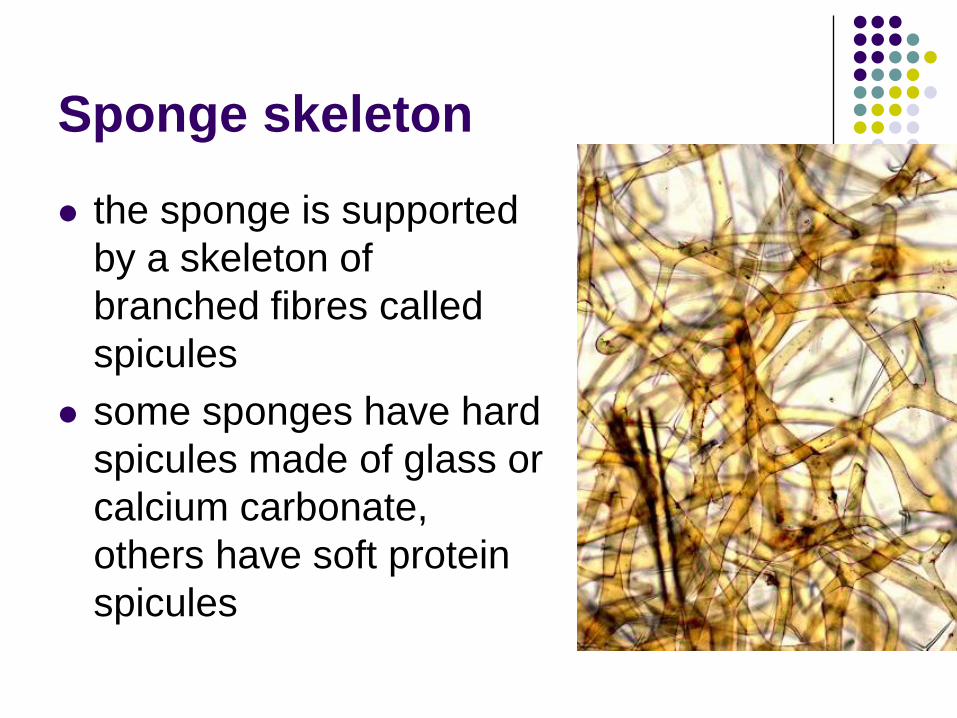
Sponge skeleton
the sponge is supported
by a skeleton of
branched fibres called
spicules
some sponges have hard
spicules made of glass or
calcium carbonate,
others have soft protein
spicules

glass sponge

red volcano sponge –
protein spicules
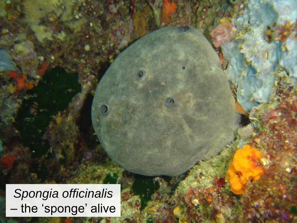
Spongia officinalis
– the ‘sponge’ alive
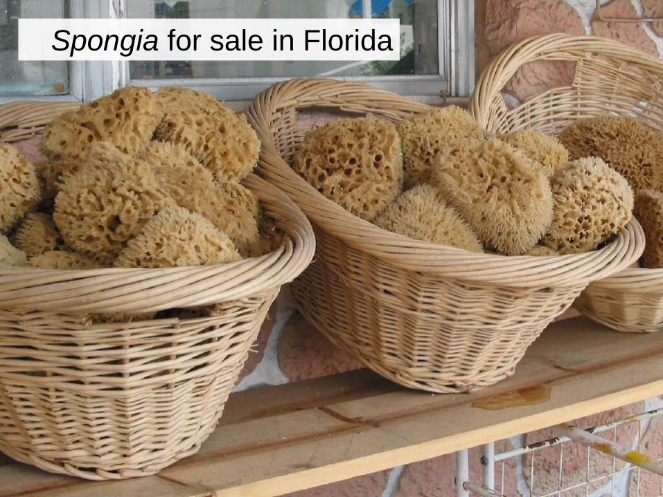
Spongia for sale in Florida

Is this a
sponge?

Organ systems
as small, simple organisms, sponges do not
require complicated organ systems
individual choanocytes digest nutrients and
excrete their own waste
circulation is accomplished by the water
current flowing through the sponge
reproduction occurs asexually through
budding, or sexually by the release of
gametes from the osculum


The birth of animals
choanocytes of sponges are nearly identical
to certain protists
because they lack true tissues, sponges are
not truly multicellular
they probably represent a transitional stage
between colonial protists and multicellular
animals

Phylum Cnidaria
You will need:
four colours of pencil crayon or
pen (preferably blue, green, red,
orange)

Phylum Cnidaria
radial symmetry
first animals with true tissues
only 2 embryonic germ layers: ectoderm and
endoderm
no body cavity
all aquatic, mostly marine

A local cnidarian, the
Lion’s Mane jellyfish

Cnidarian anatomy
Cnidarians come in two different forms:
1. polyp – a sessile form with the mouth and
tentacles facing upward
2. medusa – a free-swimming form with the
mouth and tentacles facing downward
polyp medusa

The sea anemone,
a polyp
mouth

The sea nettle,
a medusa
mouth
Organ systems
an outer layer (from ectoderm) forms the skin
and weak muscle fibres
an inner layer (from endoderm) lines the gut
cavity and digests food
between the two is a jelly-like layer called
mesoglea (NOT mesoderm)
simple net-like nervous system
circulation and excretion occur by diffusion

mesoglea ectoderm
endoderm

Feeding in cnidarians
ring of tentacles surrounds a central mouth
stinging cells on the tentacles, cnidocytes,
paralyse and capture prey
first animals with a digestive system: gut has
one opening serving as both mouth and anus
gastrovascular cavity = simple digestive system
with only one opening

tentacles
mouth
gastrovascular
cavity
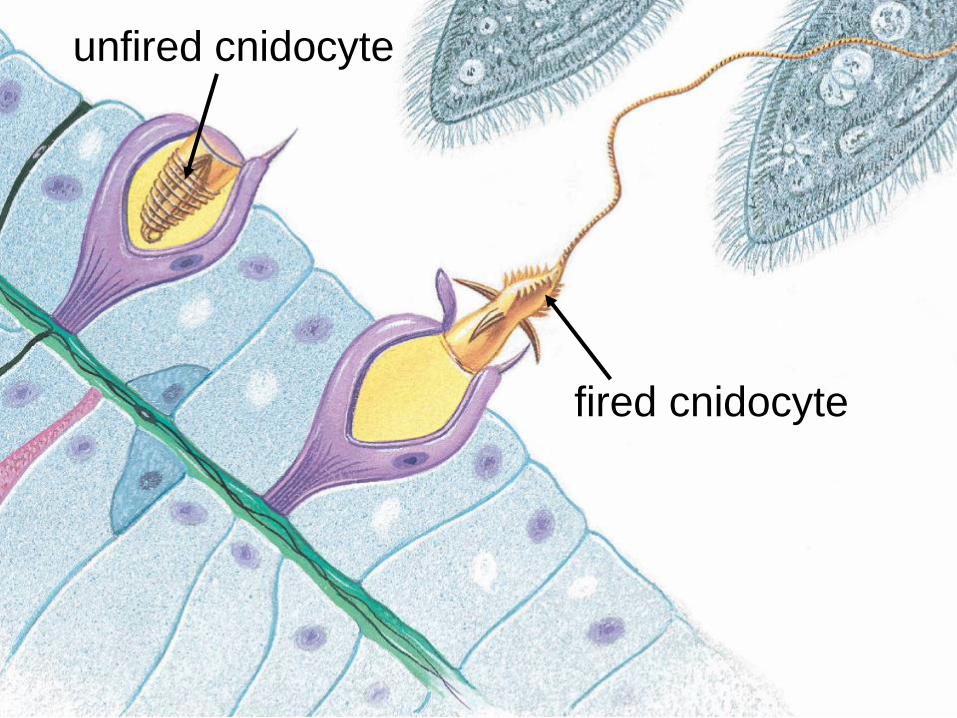
unfired cnidocyte
fired cnidocyte

Anatomy of Obelia
Obelia polyps form small branched colonies
that can reproduce asexually by budding
there are two kinds of polyps:
1. feeding polyps – have tentacles for feeding
2. medusa polyps – produce medusae by budding
mature medusae have gonads for sexual
reproduction

medusa
polyp colony
mouth
medusa
polyp
feeding
polyps
gonad
tentacles

Phylum Cnidaria observation
1. Hydra whole mount
mouth, tentacles, bud (if present)
2. Hydra cross section
endoderm, ectoderm, gastrovascular cavity
3. Obelia polyp colony
feeding polyp, medusa polyp, medusa
4. Obelia medusa
tentacles, gonad

Drawing 1: Hydra
whole mount
mouth
tentacle
bud

Drawing 2: Hydra
cross section
endoderm
ectoderm
gastrovascular
cavity detail

Drawing 3: Obelia
polyp colony
medusae (inside
medusa polyp)
feeding polyp
medusa polyp
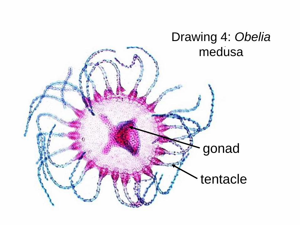
Drawing 4: Obelia
medusa
tentacle
gonad

Cnidarian diversity
Much ado about jellyfish…

Life cycle of Obelia
polyps reproduce asexually by budding to
form small colonies
medusae bud asexually inside the medusa
polyps
medusa are released and mature
medusae release haploid gametes which
fertilise each other making a diploid zygote
the zygote matures into a larva which plants
itself and turns into a new polyp

Life cycle of
Obelia
polyp
colony
medusa
sperm
zygote egg
polyp
larva
medusa
polyp

Cnidarian diversity
Class Hydrozoa
alternate between polyps and medusae but
spend most of their adult existence as a
colonial polyp. Example is the Obelia
the Portuguese Man O’ War is actually a
colony containing both polyps and medusae
Class Cubozoa
only medusa forms
includes the box jellies, the most venomous
living organisms

NO!
Hydra?

Hydra – a
hydrozoan

Portuguese Man O’ War

A sign warning against
Man O’ Wars

The box jellyfish

Net protecting against box
jellyfish - Queensland

Cnidarian diversity
Class Anthozoa
exclusively polyp forms
includes the sea anemones and corals
corals form calcium carbonate reefs, and
are symbiotic with algae
Class Scyphozoa
mostly medusa forms
includes the true jellyfish

The sea nettle – a
scyphozoan

Coral – an
anthozoan
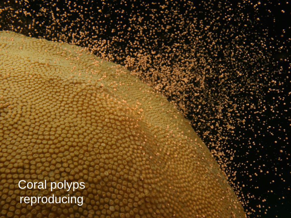
Coral polyps
reproducing