Phyllanthus urinaria and - Hindawi Publishing...
10
Hindawi Publishing Corporation Evidence-Based Complementary and Alternative Medicine Volume 2013, Article ID 603634, 9 pages http://dx.doi.org/10.1155/2013/603634 Research Article Inhibitory Effects of Standardized Extracts of Phyllanthus amarus and Phyllanthus urinaria and Their Marker Compounds on Phagocytic Activity of Human Neutrophils Yuandani, Menaga Ilangkovan, Ibrahim Jantan, Hazni Falina Mohamad, Khairana Husain, and Amirul Faiz Abdul Razak Drug and Herbal Research Center, Faculty of Pharmacy, Universiti Kebangsaan Malaysia, Jalan Raja Muda Abdul Aziz, Kuala Lumpur 50300, Malaysia Correspondence should be addressed to Ibrahim Jantan; [email protected] Received 11 February 2013; Revised 6 April 2013; Accepted 9 April 2013 Academic Editor: Jairo Kenupp Bastos Copyright © 2013 Yuandani et al. is is an open access article distributed under the Creative Commons Attribution License, which permits unrestricted use, distribution, and reproduction in any medium, provided the original work is properly cited. e standardized methanol extracts of Phyllanthus amarus and P. urinaria, collected from Malaysia and Indonesia, and their isolated chemical markers, phyllanthin and hypophyllanthin, were evaluated for their effects on the chemotaxis, phagocytosis and chemiluminescence of human phagocytes. All the plant extracts strongly inhibited the migration of polymorphonuclear leukocytes (PMNs) with the Malaysian P. amarus showing the strongest inhibitory activity (IC 50 value, 1.1 g/mL). ere was moderate inhibition by the extracts of the bacteria engulfment by the phagocytes with the Malaysian P. amarus exhibiting the highest inhibition (50.8% of phagocytizing cells). e Malaysian P. amarus and P. urinaria showed strong reactive oxygen species (ROS) inhibitory activity, with both extracts exhibiting IC 50 value of 0.7 g/mL. Phyllanthin and hypophyllanthin exhibited relatively strong activity against PMNs chemotaxis, with IC 50 values slightly lower than that of ibuprofen (1.4 g/mL). Phyllanthin exhibited strong inhibitory activity on the oxidative burst with an IC 50 value comparable to that of aspirin (1.9 g/mL). Phyllanthin exhibited strong engulfment inhibitory activity with percentage of phagocytizing cells of 14.2 and 27.1% for neutrophils and monocytes, respectively. e strong inhibitory activity of the extracts was due to the presence of high amounts of phyllanthin and hypophyllanthin although other constituents may also contribute. 1. Introduction Phagocytosis is an important response of innate immu- nity which is mediated by professional phagocytes such as polymorphonuclear neutrophils (PMNs), peripheral blood mononuclear, and macrophage cells [1, 2]. Phagocytes play important roles in our innate immune defence against infectious microbes and in activating the adaptive immune response [3]. Phagocytic activity consists of several steps, that is, migration of phagocyte cells to the site of infection, adherence to vascular endothelial cells, recognition of the tar- get structures, and subsequent engulfment of the pathogen, followed by intracellular destruction [4]. At the earliest stage of immune response, active recruit- ment of neutrophils to sites of infection is fundamentally important. is process involves mobilizations of PMNs from circulation in response to host- and pathogen-derived chemotactic factors. Phagocytes pass through the capillary wall, surveying tissue, membranes, and lymphatic organs for signs of tissue distress and the presence of chemoat- tractant [4]. Endogenous substances such as interleukin 8 (IL-8), leukotriene B4 (LTB4), platelet-activating factor (PAF), and exogenous substances such as formyl methionyl- leucyl-phenylalanine (fMLP) derived from bacterial cell products are the important neutrophil chemoattractants [5]. e phagocytes adhere stably to the endothelium cells because they possess cell surface expression of all three CD11/CD18 leucocyte integrins [6]. e phagocytes directly recognize surface bound or freely secreted molecules pro- duced by pathogen and interact directly with a number of
Transcript of Phyllanthus urinaria and - Hindawi Publishing...

Hindawi Publishing CorporationEvidence-Based Complementary and Alternative MedicineVolume 2013, Article ID 603634, 9 pageshttp://dx.doi.org/10.1155/2013/603634
Research ArticleInhibitory Effects of Standardized Extracts ofPhyllanthus amarus and Phyllanthus urinaria andTheir Marker Compounds on Phagocytic Activity ofHuman Neutrophils
Yuandani, Menaga Ilangkovan, Ibrahim Jantan, Hazni Falina Mohamad,Khairana Husain, and Amirul Faiz Abdul Razak
Drug and Herbal Research Center, Faculty of Pharmacy, Universiti Kebangsaan Malaysia, Jalan Raja Muda Abdul Aziz,Kuala Lumpur 50300, Malaysia
Correspondence should be addressed to Ibrahim Jantan; [email protected]
Received 11 February 2013; Revised 6 April 2013; Accepted 9 April 2013
Academic Editor: Jairo Kenupp Bastos
Copyright © 2013 Yuandani et al.This is an open access article distributed under theCreative CommonsAttribution License, whichpermits unrestricted use, distribution, and reproduction in any medium, provided the original work is properly cited.
The standardized methanol extracts of Phyllanthus amarus and P. urinaria, collected from Malaysia and Indonesia, and theirisolated chemical markers, phyllanthin and hypophyllanthin, were evaluated for their effects on the chemotaxis, phagocytosisand chemiluminescence of human phagocytes. All the plant extracts strongly inhibited the migration of polymorphonuclearleukocytes (PMNs) with the Malaysian P. amarus showing the strongest inhibitory activity (IC
50value, 1.1 𝜇g/mL). There was
moderate inhibition by the extracts of the bacteria engulfment by the phagocytes with the Malaysian P. amarus exhibiting thehighest inhibition (50.8% of phagocytizing cells). The Malaysian P. amarus and P. urinaria showed strong reactive oxygen species(ROS) inhibitory activity, with both extracts exhibiting IC
50value of 0.7 𝜇g/mL. Phyllanthin and hypophyllanthin exhibited
relatively strong activity against PMNs chemotaxis, with IC50values slightly lower than that of ibuprofen (1.4𝜇g/mL). Phyllanthin
exhibited strong inhibitory activity on the oxidative burst with an IC50value comparable to that of aspirin (1.9 𝜇g/mL). Phyllanthin
exhibited strong engulfment inhibitory activity with percentage of phagocytizing cells of 14.2 and 27.1% for neutrophils andmonocytes, respectively. The strong inhibitory activity of the extracts was due to the presence of high amounts of phyllanthinand hypophyllanthin although other constituents may also contribute.
1. Introduction
Phagocytosis is an important response of innate immu-nity which is mediated by professional phagocytes such aspolymorphonuclear neutrophils (PMNs), peripheral bloodmononuclear, and macrophage cells [1, 2]. Phagocytes playimportant roles in our innate immune defence againstinfectious microbes and in activating the adaptive immuneresponse [3]. Phagocytic activity consists of several steps,that is, migration of phagocyte cells to the site of infection,adherence to vascular endothelial cells, recognition of the tar-get structures, and subsequent engulfment of the pathogen,followed by intracellular destruction [4].
At the earliest stage of immune response, active recruit-ment of neutrophils to sites of infection is fundamentally
important. This process involves mobilizations of PMNsfrom circulation in response to host- and pathogen-derivedchemotactic factors. Phagocytes pass through the capillarywall, surveying tissue, membranes, and lymphatic organsfor signs of tissue distress and the presence of chemoat-tractant [4]. Endogenous substances such as interleukin8 (IL-8), leukotriene B4 (LTB4), platelet-activating factor(PAF), and exogenous substances such as formyl methionyl-leucyl-phenylalanine (fMLP) derived from bacterial cellproducts are the important neutrophil chemoattractants[5]. The phagocytes adhere stably to the endothelium cellsbecause they possess cell surface expression of all threeCD11/CD18 leucocyte integrins [6]. The phagocytes directlyrecognize surface bound or freely secreted molecules pro-duced by pathogen and interact directly with a number of

2 Evidence-Based Complementary and Alternative Medicine
pattern-recognition receptor expressed on the cell surface,such as TLRs, CD14, and Fc𝛾R [5, 7]. After adherence ofthe pathogen to the surface of phagocytes through recogni-tion receptor, engulfment phase is initiated. The pathogensare then destroyed by microbicidal mechanism that is oftenreferred to as oxidative burst [5].
Theoxidative burst involves the generation of superoxidesby NADPH-oxidase complex through a series of molecularreaction that consume oxygen. Myeloperoxidase (MPO) inthe phagosome catalyzes the transformation of superoxideinto a variety of toxic molecules for microorganisms, such ashypochlorous acid, chlorines, chloramines, hydroxyl radicals,and single oxygen [3]. Besides the defensive roles during theinfections, the phagocyte-microbe interactions when exces-sively or inappropriately deployed can damage host tissuesand contribute to the pathogeny of various immune andnonimmune chronic inflammatory diseases, including somerheumatoid disorders. Therefore, the inhibitors of phagocytereactive oxygen species (ROS) production can be used in thetreatment of a variety of disorders including inflammation[8].
Many therapeutic effects of plant extracts have beensuggested to be due to their influence on the immunesystem of the human body [9]. Many herbal preparationssuch as Tinospora cordifolia, Centella asiatica, Phyllanthusdebilis, Trigonella foenum graecum, Pouteria cambodiana,Panax ginseng, and Picrorhiza scrophulariiflora have beenshown to alter the immune function and possess a wide arrayof immunomodulatory effects [10–13]. Our previous studyon the screening of 20 medicinal plants for their phago-cytic properties has indicated that the methanol extractsof some plants including Phyllanthus amarus exhibitedstrong immunomodulatory effects on polymorphonuclearneutrophils and macrophage cells [14]. P. amarus Schum.& Thonn. and P. urinaria Linn (family Euphorbiaceae) arewidely used in traditional medicine to treat various diseasessuch as viral hepatitis, diarrhoea, jaundice kidney disorders,influenza, diabetes, bronchial infections, sores, swelling, itch-iness, cardiovascular problems, and inflammatory disorders[15, 16].
P. amarus was found to be rich in lignans, polyphe-nols, flavonoids, hydrolysable tannins, triterpenes, sterols,and alkaloids [15–18]. The extract and compounds isolatedfrom P. amarus have shown a wide spectrum of phar-macological activities including antiviral, hepatoprotectiveanti-inflammatory, antioxidant, antiplasmodial, antimalarial,antidiabetic, hypolipidemic, antihyperuremic, nephroprotec-tive, and diuretic properties [18–24].The extract and purifiedlignans such as phyltetralin, nirtetralin, and niranthin fromP. amarus exhibited important in vivo and in vitro anti-inflammatory actions [20]. Several studies have indicated thatP. amarus was able to suppress the growth and replication ofhepatitis B virus [23].The hepatoprotective effect of P. amarusand its ability to protect hepatocytes against carbon tetrachlo-ride, paracetamol, ethanol, aflatoxin B1, and galactosamine-induced liver toxicity in various animal models have beenwell documented [24]. Phyllanthin and hypophyllanthinpresent in P. amarus have been shown to inactivate hepatitisB, both in vitro and in vivo, and these two lignans were
also reported to be the active principles accountable for thehepatoprotective property of many Phyllanthus species [24].Phytochemical studies on P. urinaria have resulted in theisolation of various compounds, mainly lignans, flavonoids,tannins, and other benzenoid constituents [25–27]. Vari-ous biological activities of P. urinaria have been reported,including hepatoprotective effect, antihepatitis B virus, anti-Epstein-Barr virus, antiretroviral reverse transcriptase, andantiherpes simplex virus type I and type 2 [28, 29]. Antiox-idant and inflammatory mediator’s growth inhibitory effectsof compounds isolated from P. urinaria have been reported[30].
In the present study the standardized methanol extractsof P. amarus and P. urinaria, collected from Malaysia andIndonesia, were investigated for their inhibitory effects onphagocytic activity of human neutrophils. The biochemicalmarkers of the extracts were isolated, and their immunomod-ulatory effects were determined in an effort to correlate theinhibitory activity of the extracts with their active compo-nents. The results of this study may provide some insightson the ability of these plants and their marker compoundsto modulate the innate immune response of phagocytes atdifferent steps.
2. Materials and Methods
2.1. Chemicals and Reagents. The chemicals used in thisstudy were of analytical grade. Serum opsonized zymosan A(Saccharomyces cerevisiae suspensions and serum), luminol(3-aminophthalhydrazide), phosphate buffer saline tablet(PBS), Hanks Balance Salt Solutions (HBSS), Ficoll, HanksBalance Salt Solution (HBSS), N-formyl-methionylleucyl-phenylalanine (fMLP), acetyl salicylic acid (purity 99%),ibuprofen (purity 99%), and dimethylsulfoxide (DMSO)werepurchased from Sigma (St Louis, MO, USA). Fetal calf serumwas obtained from PAA Laboratories (USA). Chemilumi-nescence measurements were carried out on a LuminoskanAscent luminometer (Thermo Scientific, UK). fMLP wasstored as a stock solution of 10−8M in DMSO at −80∘Cand diluted in Hanks solution, prior to assay. Haematoxylinand xylene for staining were obtained from BDH, UK. ABoyden 48-well chamber with a 2𝜇m polycarbonate mem-brane filter separating the upper and lower compartmentswas purchased from Neuro Probe (Cabin John, MD, USA).Phyllanthin and hypophyllanthin standards (purity >98%)were purchased from ChromaDex (CA, USA). MethanolHPLC grade, acetonitrile HPLC grade, and, trifluoroaceticacid AR grade were obtained from E-Merck. Phagotest kitwas obtained fromGlycotopeTechnology,Germany.Theflowcytometer BDFACS Canto II equipped with 488 nm argon-ion laser was used. A CO
2incubator (Shell Lab, USA), light
microscope, and high-performance liquid chromatograph(Waters 2998) (LeitzWatzler, Germany)were also used in thisassay. Molecular weights of the compounds were recordedby ESIMS using ESI-TOFF MS (Bruker MicroToF-Q 86,Switzerland). The 1H and 13C NMR spectra were carried outon a JOEL NMR 500MHz (JOEL Ltd, Japan) with TMS asinternal standard.

Evidence-Based Complementary and Alternative Medicine 3
2.2. Plant Collection. Thewhole plants of Phyllanthus amarusand P. urinaria were collected from Marang, Kuala Tereng-ganu, Malaysia and Tanjung Anom, Northern Sumatera,Indonesia, between February and June 2012. The voucherspecimens (P. amarus UKMB 30078 and P. urinaria UKMB30077)were identified byDrAbdul LatifMohamad of Facultyof Science and Technology, Universiti Kebangsaan Malaysia(UKM), and deposited at the Herbarium of UKM, Bangi,Malaysia.
2.3. Extraction and Isolation of Phyllanthin and Hypophyllan-thin. The plant materials were allowed to dry under shade.500 g of dried material of each plant sample were groundand macerated in methanol at the ratio of 1 : 10 (w/v). Theextraction was repeated thrice on the residue. The filtrateswere combined and the solvent was removed under reducedpressure to obtain extracts of P. amarus (Malaysia, 55.2 g,11.04% w/w; Indonesia, 49.7 g, 10.18% w/w) and P. urinaria(Malaysia, 52.7 g, 10.54% w/w; Indonesia, 47.2 g, 9.44% w/w).Ten g of P. amarus extract was fractionated by vacuumliquid chromatography (VLC) on silica gel type H (10–40 𝜇m, 7 × 30 cm) and eluted with a gradient system ofhexane : CHCl
3(10 : 0–1 : 9, v/v) and CHCl
3: MeOH (10 : 0–
0 : 10, v/v); repeated silica gel column (40–63𝜇m, 3 × 60 cm)was eluted with a gradient system of n-hexane : ethyl acetate(10 : 0–1 : 9, v/v), followed by recrystallization from EtOAc:hexane to yield different amounts of phyllanthin (Malaysiansample: 228.5mg, 2.29%; Indonesian sample: 115.0mg, 1.15%)and hypophyllanthin (Malaysian sample: 321.2mg, 3.21%;Indonesian sample: 235.5mg, 2.35%). Purity of the com-pounds was >98%, based on their physicochemical proper-ties, NMR and ESI-MS data.
2.4. Standardization of the Methanol Extracts of PhyllanthusSpecies by HPLC. Twenty mg of the methanol extracts of theplant in 10mL of methanol and 1mg each of the referencestandards of phyllanthin and hypophyllanthin in 1mL ofmethanol were filtered through 0.45 𝜇m Millipore MillexPTFE membrane (Maidstone, Kent, UK) before injection.The diluted solutions of the extracts and the reference stan-dards were analyzed separately by HPLC using the followingconditions: column: Reverse Phase, C-18 column (250mm ×4.6mm i.d., 5𝜇m, Xbridge, Waters, Ireland), detector: PDA(Waters 2998), wavelength: 205 nm, flow rate: 0.4mL/min,Mobile phase: A. acetonitrile : B. water (acidified with 0.1%orthophosphoric acid) isocratically eluted with 5% B, andthen increased to 95% over 20min and had been hold at 95%for 15min.
2.5. Validation Procedures for HPLC Analysis. The reversed-phase HPLC method was validated by determination oflinearity, precision, limits of quantification (LOQ), and detec-tion (LOD). Linearity was evaluated by linear calibrationanalysis and correlation coefficient (R2), calculated fromthe calibration curves. Calibration standards were preparedat concentrations of 250, 125, 62.5, and 31.25 𝜇g/mL ofphyllanthin and hypophyllanthin. A graph was plotted forthe area versus concentration of corresponding compound.
Table 1: Amounts of phyllanthin and hypophyllanthin (𝜇g/mL) inPhyllanthus amarus and P. urinaria obtained from HPLC measure-ments.
Species Phyllanthin HypophyllanthinP. amarus (Mal) 172.4 214.1P. amarus (Ind) 93.3 124.6P. urinaria (Mal) 96.8 111.2P. urinaria (Ind) 9.0 14.8
Precision of the method was determined by intra-assayand interassay validation. Separately, one concentration ofextract (2mg/mL) and reference compounds (1mg/mL) wereinjected three times in one day and on three different days.LOD and LOQwere calculated from the RSD and slope (𝑆) ofthe calibration curves using equations: LOD= 3.3 × (RSD/𝑆)and LOQ = 10 × (RSD/𝑆).
2.6. Isolation of Neutrophils. Fresh blood was collectedin heparin-containing tubes from healthy human volun-teers who fulfilled the following criteria: nonsmoker, fastedovernight and had not taking any medicine or supplements.PMNs were isolated from the blood by Ficol-gradient sepa-ration as described previously [14]. Using a haemocytometerand light microscope, cell suspensions were counted. Cellsuspensions were diluted with HBSS to obtain a final cell sus-pension of 1 × 10⁶/mL.The use of human blood was approvedby the Human Ethical Committee of Universiti KebangsaanMalaysia (approval number FF/2012/Ibrahim/23-May/432-May 2012–August 2013).
2.7. Cell Viability. Cell viability was determined by the stan-dard trypan blue exclusion method. The neutrophils (1 ×10⁶/mL) were incubated with 6.25 or 100𝜇g/mL of plantsextracts and 3.125 to 100 𝜇g/mL of pure compounds in trip-licate at 37∘C for 2 h. The blue dye uptake was an indicationof cell death.The percentage viability was calculated from thetotal cell counts.
2.8. Chemiluminescence Assay. Luminol-amplified chemilu-minescence assays were carried out as described by Koko etal. [31]. Briefly, 25 𝜇L diluted whole blood or 25 𝜇L PMN (1× 10⁶/mL) suspended in HBSS++ were incubated with 25 𝜇Lof five different concentrations (6.25–100 𝜇g/mL). The cellswere induced with 25𝜇L of opsonized zymosan followed by25 𝜇L of luminol as a probe, and then HBSS++ was added toadjust the final volume to 200𝜇L. The final concentrationsof the samples in the mixture were 12.5, 6.25, 3.13, 1.56, and0.78𝜇g/mL. Tests were performed in 96-well microplateswhich were incubated at 37∘C for 50min in the thermostatedchamber of the luminometer. Luminol, 0.6%DMSO, HBSS++and cells were added as a control, and the acetylsalicylicacid was used as a positive control. The final concentrationof DMSO in the mixture was 0.6% to eliminate the effectof the solvent on the chemiluminescence. The luminometerresults were monitored as chemiluminescence RLU (readingper luminometer unit) with peak and total integral values set

4 Evidence-Based Complementary and Alternative Medicine
Table 2: Percentage of phagocytic activity (%) of neutrophils and monocytes at various concentrations of Phyllanthus extracts and theirchemical markers (Mean ± SEM, 𝑛 = 3).
Sample (𝜇g/mL) Neutrophils Monocytes100 50 6.25 3.125 100 50 6.25 3.125
P. amarus (Ind) 89.2 ± 3.2 89.8 ± 6.2 70.8 ± 8.2 71.0 ± 4.1
P. urinaria (Ind) 85.1 ± 2.4 86.5 ± 7.5 52.3 ± 9.8 86.4 ± 4.5
P. amarus (Mal) 69.2 ± 3.6 81.7 ± 2.9 50.8 ± 2.3 55.5 ± 1.6
P. urinaria (Mal) 73.7 ± 6.2 74 ± 4.0 52.3 ± 6.6 60.1 ± 5.0
Hypophyllanthin 49.1 ± 4 80.5 ± 3.8 64.6 ± 10.8 67.0 ± 3.0
Phyllanthin 14.2 ± 2 34.9 ± 4.2 27.1 ± 5.7 52.1 ± 2.6
(AU
)
0
0.5
1
1.5
Minutes0 2 4 6 8 10 12 14 16 18 20 22 24 26 28 30 32 34
1 2
(a)
Minutes0 2 4 6 8 10 12 14 16 18 20 22 24 26 28 30 32 34
0
1
2
(AU
)
1 2
(b)
Minutes0 2 4 6 8 10 12 14 16 18 20 22 24 26 28 30 32 34
00.5
11.5
22.5
(AU
)
1
(c)
Minutes0 2 4 6 8 10 12 14 16 18 20 22 24 26 28 30 32 34
00.5
11.5
22.5
(AU
)
2
(d)
Figure 1: Representative HPLC chromatograms of (a) Phyllanthus amarus (Mal), (b) P. urinaria (Ind) standardized to phyllanthin (1) atRT = 27.251min and hypophyllanthin (2) at RT = 28.079min, (c) reference standard (phyllanthin) at 27.265min, and (d) reference standard(hypophyllanthin) at 27.883min.
with repeated scans at 30 s intervals and 1 s points measuringtime.
2.9. Chemotaxis Assay. The assay was performed using amodified 48-well Boyden chamber with formyl-methionyl-leucyl-phenylalanine (fMLP) a bacterial peptide as achemoattractant, as previously described by Sacerdoteet al. [32]. Briefly, aliquots of 25𝜇L of fMLP (10−8M)
were added to the lower chamber. Serial dilutions 5 𝜇L ofeach extract (6.25–100𝜇g/mL) were added to the upperchamber containing PMNs (1 × 10⁶ cells per mL) suspendedin HBSS++. The final concentrations of the samples in themixture were 10, 5, 2.5, 1.25, and 0.625 𝜇g/mL.Then cells wereincubated for 1 h at 37∘C in a CO
2incubator. Migrated cells
which had adhered to the distal part of the filters were fixedand stained by haematoxylin and xylene. The cell migrationdistance was measured by using a light microscope. Control
Evidence-Based Complementary and Alternative Medicine 5
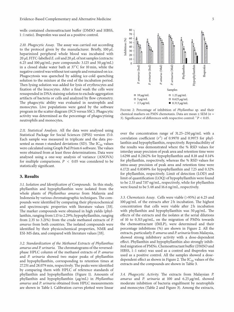
wells contained chemoattractant buffer (DMSO and HBSS,1 : 1 ratio). Ibuprofen was used as a positive control.
2.10. Phagocytic Assay. The assay was carried out accordingto the protocol given by the manufacturer. Briefly, 100 𝜇Lheparinized peripheral whole blood was incubated with20𝜇LFITC-labelledE. coli and 20 𝜇Lof test samples (extracts:6.25 and 100 𝜇g/mL; pure compounds: 3.125 and 50𝜇g/mL)in a closed shake water bath at 37∘C for 10min, while thenegative control waswithout test sample and remained on ice.Phagocytosis was quenched by adding ice-cold quenchingsolution to the mixture at the end of the incubation period.Then lysing solution was added for lysis of erythrocytes andfixation of the leucocytes. After a final wash the cells wereresuspended inDNA staining solution to exclude aggregationartifacts of bacteria or cells and analyzed by flow cytometry.The phagocytic ability was evaluated in neutrophils andmonocytes. Live populations were gated by the softwareprogram in the scatter diagram (FCS versus SSC). Phagocyticactivity was determined as the percentage of phagocytizingneutrophils and monocytes.
2.11. Statistical Analysis. All the data were analysed usingStatistical Package for Social Sciences (SPSS) version 15.0.Each sample was measured in triplicate and the data pre-sented as mean± standard deviations (SD). The IC
50values
were calculated usingGraph Pad Prism 6 software.The valueswere obtained from at least three determinations. Data wereanalysed using a one-way analysis of variance (ANOVA)for multiple comparisons. 𝑃 < 0.05 was considered to bestatistically significant.
3. Results
3.1. Isolation and Identification of Compounds. In this study,phyllanthin and hypophyllanthin were isolated from thewhole plants of Phyllanthus amarus from Malaysia andIndonesia by various chromatographic techniques. The com-pounds were identified by comparing their physicochemicaland spectroscopic properties with literature values [33].The marker compounds were obtained in high yields (phyl-lanthin, ranging from 1.15 to 2.29%; hypophyllanthin, rangingfrom 2.35 to 3.21%) from the crude methanol extracts of P.amarus from both countries. The isolated compounds wereidentified by their physicochemical properties, NMR andESI-MS data, and compared with literature values [33].
3.2. Standardization of the Methanol Extracts of Phyllanthusamarus and P. urinaria. The chromatograms of the reversed-phase HPLC column of the methanol extracts of P. amarusand P. urinaria showed two major peaks of phyllanthinand hypophyllanthin, corresponding to retention times at27.251 and 28.079min, respectively.The peaks were identifiedby comparing them with HPLC of reference standards ofphyllanthin and hypophyllanthin (Figure 1). Amounts ofphyllanthin and hypophyllanthin (𝜇g/mL) in Phyllanthusamarus and P. urinaria obtained from HPLC measurementsare shown in Table 1. Calibration curves plotted were linear
8070605040302010
0
Inhi
bitio
n (%
)
Phyl
lant
hin
Hyp
ophy
llant
hin
Ibup
rofe
n
Samples
∗∗
∗
∗∗∗
∗∗∗
∗
∗
∗
∗
∗
∗
∗
∗
∗
∗ ∗
∗
∗∗
∗
∗∗
∗
∗∗
10𝜇g/mL5𝜇g/mL2.5 𝜇g/mL
1.25 𝜇g/mL0.625𝜇g/mL0.313𝜇g/mL
(Ind)
P. am
arus
P. ur
inar
ia
P. am
arus
P. ur
inar
ia
(Ind)
(Mal
)
(Mal
)
Figure 2: Percentage of inhibition of Phyllanthus sp. and theirchemical markers on PMN chemotaxis. Data are mean ± SEM (𝑛 =3). Significance of differences with respective control: ∗𝑃 < 0.05.
over the concentration range of 31.25–250 𝜇g/mL with acorrelation coefficient (𝑟2) of 0.9970 and 0.9973 for phyl-lanthin and hypophyllanthin, respectively. Reproducibility ofthe results was demonstrated where the % RSD values forinterday assay precision of peak area and retention time were1.6298 and 0.2162% for hypophyllanthin and 8.18 and 0.14%for phyllanthin, respectively, whereas the % RSD values forintra-assay precision of peak area and retention time were6.1721 and 0.8589% for hypophyllanthin and 7.25 and 0.32%for phyllanthin, respectively. Limit of detection (LOD) andlimit of quantification (LOQ) of hypophyllanthin were foundto be 2.33 and 7.07 ng/mL, respectively, while for phyllanthinwere found to be 5.48 and 16.6 ng/mL, respectively.
3.3. Chemotaxis Assay. Cells were viable (>95%) at 6.25 and100 𝜇g/mL of the extracts after 2 h incubation. The highestconcentration that cells were viable after 2 h incubationwith phyllanthin and hypophyllanthin was 50𝜇g/mL. Theeffects of the extracts and the isolates at the serial dilutionsof 10 to 0.313 𝜇g/mL, on the migration of PMNs towardsthe chemoattractant (fMLP), were determined and theirpercentage inhibitions (%) are shown in Figure 2. All theextracts, particularlyP. amarus andP. urinaria fromMalaysia,showed strong inhibitory activity with a dose-dependenteffect. Phyllanthin and hypophyllanthin also strongly inhib-itedmigration of PMNs. Chemoattractant buffer (DMSO andHBSS, 1 : 1 ratio) was used as a control and ibuprofen wasused as a positive control. All the samples showed a dose-dependent effect as shown in Figure 2. The IC
50values of the
extracts and the compounds are shown in Table 3.
3.4. Phagocytic Activity. The extracts from Malaysian P.amarus and P. urinaria at 100 and 6.25 𝜇g/mL showedmoderate inhibition of bacteria engulfment by neutrophilsand monocytes (Table 2 and Figure 3). Among the extracts,

6 Evidence-Based Complementary and Alternative Medicine
400350300250200150100
500
Cou
nt
102
103
104
105
FITC-A
(A) (B)
(C)
(D)
E. coli+
E. coli
400350300250200150100
500
Cou
nt
102
103
104
105
FITC-A
(A)(B)
(E)
(F)
E. coli E. coli
400350300250200150100
500
Cou
nt
102
103
104
105
FITC-A
(A) (B)
(G)
(H)
E. coli+
(a)
E. co
li+
10
7.5
5
2.5
0
Cou
nt
102
103
104
105
FITC-A FITC-A FITC-A
(A)
(B)
(C)
(D)
10
7.5
5
2.5
0
Cou
nt
102
103
104
105
(A)(B)
(G)
(F)
10
7.5
5
2.5
0
Cou
nt
102
103
104
105
(A) (B)
(G)
(H)
(A))A))))))))))A)AAAAA (B)))))))))))
(G)GG
(H)H
(b)
Figure 3: Representation of E. coli engulfment by neutrophils (3a) and monocytes (3b). (A) negative control, (B) positive control, (C)methanol extract of P. amarus (Mal) (D) methanol extract of P. amarus (Ind)(E) methanol extract of P. urinaria (Mal), (F) methanol extractof P. urinaria, (Ind), (G) hypophyllanthin (H) phyllanthin.
P. amarus (Malaysia) at 100 𝜇g/mL exhibited the highestengulfment inhibitory activity with percentage of phagocy-tizing cells of 69.2 and 50.8% for neutrophils and monocytes,respectively. The engulfment inhibitory activity at normalcondition at 37∘C was used as a positive control and normalcondition at 0∘C as negative control (Table 2). Phyllanthin at50𝜇g/mL exhibited strong inhibitory activitywith percentageof phagocytizing cells of 14.2 and 27.1% for neutrophils andmonocytes, respectively.
3.5. Inhibition of Reactive Oxygen Species Generation. Prelim-inary screening of the extracts of P. amarus and P. urinariaon the whole blood showed that P. amarus and P. urinariafromMalaysia exhibited high inhibitory activity for luminol-enhanced chemiluminescence with IC
50values of 1.1 and
1.6 𝜇g/mL, respectively (Table 3 and Figure 4). Phyllanthinand hypophyllanthin exhibited IC
50values of 8.8 and 8.4 𝜇M,
respectively, indicating that they were more potent than thepositive control, aspirin (12.2𝜇M) (Table 3).The extracts werefurther investigated for their effects on the oxidative burst ofPMNs. Of all the extracts, P. amarus and P. urinaria fromMalaysia were the more potent samples against PMN withboth extracts exhibited IC
50value of 0.7𝜇g/mL which was
much lower than that of aspirin (1.9 𝜇g/mL). All the extractsand isolates showed a dose-dependent effect as shown in
Figure 4. Phyllanthin also showed lower IC50value (7.6𝜇M)
than hypophyllanthin (14.2 𝜇M) and aspirin (10.5 𝜇M) on theoxidative burst of PMNs, indicating that it was a more potentinhibitor of ROS generation.
4. Discussion
The extracts of Phyllanthus species were standardized forphyllanthin and hypophyllanthin contents by HPLC analysis.Quantitative determination of the marker compounds byHPLC indicated that P. amarus from Malaysia contained thehighest amounts of phyllanthin (172.4 𝜇g/mL) and hypophyl-lanthin (214.1𝜇g/mL) while P. urinaria from Indonesia con-tained the lowest concentrations of these marker compounds(Table 1). The variations in the quantitative amounts of themarker compounds in the plants of similar species collectedfrom the two different locations are a response of theindividual plant to environmental factors related to altitudeor a genetic adaptation of the populations growing at differentaltitudes to specific environment [34].
The cell viability test was performed using trypan blueto determine the nontoxic concentrations of P. amarus andP. urinaria extracts, and the isolated marker compounds(phyllanthin and hypophyllanthin). The high cell viabilityindicated that the extracts and the compounds were nontoxicto immune cells and could potentially modulate the cellular
Evidence-Based Complementary and Alternative Medicine 7
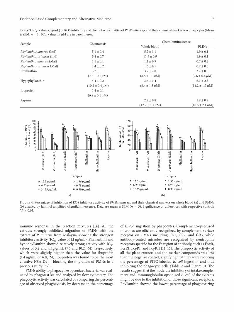
Table 3: IC50 values (𝜇g/mL) of ROS inhibitory and chemotaxis activities of Phyllanthus sp. and their chemical markers on phagocytes (Mean± SEM, 𝑛 = 3). IC50 values in 𝜇M are in parentheses.
Sample Chemotaxis ChemiluminescenceWhole blood PMNs
Phyllanthus amarus (Ind) 5.1 ± 0.4 5.2 ± 1.1 1.9 ± 0.1
Phyllanthus urinaria (Ind) 5.4 ± 0.7 11.9 ± 0.9 1.9 ± 0.1
Phyllanthus amarus (Mal) 1.1 ± 0.1 1.1 ± 0.9 0.7 ± 0.2
Phyllanthus urinaria (Mal) 1.4 ± 0.2 1.6 ± 0.5 0.7 ± 0.3
Phyllanthin 3.2 ± 0.1 3.7 ± 2.8 3.2 ± 0.8
(7.6 ± 0.1 𝜇M) (8.8 ± 1.0 𝜇M) (7.6 ± 0.4 𝜇M)Hypophyllanthin 4.4 ± 0.2 3.6 ± 1.4 6.1 ± 2.3
(10.2 ± 0.4 𝜇M) (8.4 ± 1.3 𝜇M) (14.2 ± 1.7 𝜇M)Ibuprofen 1.4 ± 0.1
(6.8 ± 0.1 𝜇M)Aspirin 2.2 ± 0.8 1.9 ± 0.2
(12.2 ± 1.1 𝜇M) (10.5 ± 2.1 𝜇M)
8090
100
70605040302010
0
Inhi
bitio
n of
RO
S
Phyl
lant
hin
Hyp
ophy
llant
hin
Asp
irin
Samples
∗
∗
∗
∗
∗∗
∗
∗∗
∗ ∗∗ ∗
∗∗
12.5 𝜇g/mL6.25 𝜇g/mL3.125 𝜇g/mL
1.56𝜇g/mL0.78𝜇g/mL0.39𝜇g/mL
on w
hole
blo
od (%
)
(Ind)
P. am
arus
P. ur
inar
ia
P. am
arus
P. ur
inar
ia
(Ind)
(Mal
)
(Mal
)
(a)
80100120
604020
0
Inhi
bitio
n of
RO
S
Phyl
lant
hin
Hyp
ophy
llant
hin
Asp
irin
Samples
∗∗∗
∗∗∗
∗
∗∗
∗∗
∗ ∗∗∗
12.5 𝜇g/mL6.25 𝜇g/mL3.125 𝜇g/mL
1.56𝜇g/mL0.78𝜇g/mL0.39𝜇g/mL
(Ind)
P. am
arus
P. ur
inar
ia
P. am
arus
P. ur
inar
ia
(Ind)
(Mal
)
(Mal
)
on P
MN
s (%
)
(b)
Figure 4: Percentage of inhibition of ROS inhibitory activity of Phyllanthus sp. and their chemical markers on whole blood (a) and PMNs(b) assayed by luminol amplified chemiluminescence. Data are mean ± SEM (𝑛 = 3). Significance of differences with respective control:∗𝑃 < 0.05.
immune response in the reaction mixtures [14]. All theextracts strongly inhibited migration of PMNs with theextract of P. amarus from Malaysia showing the strongestinhibitory activity (IC
50value of 1.1 𝜇g/mL). Phyllanthin and
hypophyllanthin showed relatively strong activity with IC50
values of 3.2 and 4.4 𝜇g/mL (7.6 and 10.2 𝜇M), respectively,which were slightly higher than the value for ibuprofen(1.4 𝜇g/mL or 6.8 𝜇M). Ibuprofen was found to be the mosteffective NSAIDs in blocking the migration of PMNs in aprevious study [35].
PMNs ability to phagocytize opsonized bacteria was eval-uated by phagotest kit and analyzed by flow cytometry. Thephagocytic activity was calculated by comparing the percent-age of observed phagocytosis, by decrease in the percentage
of E. coli ingestion by phagocytes. Complement-opsonizedmicrobes are efficiently recognized by complement surfacereceptor on PMNs including CR1, CR2, and CR3, whileantibody-coated microbes are recognized by neutrophilsreceptors specific for the Fc region of antibody, such as Fc𝛼R,Fc𝜀RI, Fc𝛾RI, and Fc𝛾RII [14, 16]. The phagocytic activity ofall the plant extracts and the marker compounds was lessthan the negative control, signifying that they were reducingthe percentage of FITC-labelled E. coli ingestion and thusinhibiting the phagocytic cells (Table 2 and Figure 3). Theresults suggest that themoderate inhibitory of intake comple-ment and immunoglobulin opsonized E. coli of the extractsmight be due to the inhibition of those significant receptors.Phyllanthin showed the lowest percentage of phagocytizing

8 Evidence-Based Complementary and Alternative Medicine
cells, indicating the strongest inhibitory activity. The resultssuggest that phyllanthin might be the major contributor ofthe phagocytic activity of the plant extracts.
PMNs upon activation by serum-opsonized zymosan(SOZ) would accumulate at the site of inflammation and bindand destroy invading microorganisms by phagocytosis pro-cess. This mechanism triggers the generation of superoxideradicals and other secondarily derivedROS, such as hydrogenperoxide, hypochlorous acid, and cholarimines. The ROSwere then quantified by the luminol-enhanced chemilumi-nescence assay.The luminol was used as probes to assess ROSproduction from plant extracts in PMN cells. The relativelysmall molecular weight of luminol enables it to enter the cellsand subsequently to be oxidized by hydrogen peroxide toform a luminol radical, which eventually yields an unstableendoperoxide due to the interaction of luminol radical withother radicals [36]. This endoperoxide decomposes to anexcited aminophthalate, which contributes to the emission oflight [37]. In this study, all the extracts and isolates exhibitedthe inhibitory activity on ROS production by phagocytesin a concentration-dependent manner. Among the extracts,P. amarus and P. urinaria from Malaysia exhibited activitystronger than the positive control, aspirin. This inhibitoryactivity could be due to the ability of the extracts to block theinteraction of SOZwith complement receptors, consequentlyinhibiting NADPH oxidase [36]. Phyllanthin and hypophyl-lanthin exhibited strong inhibitory activity on the oxidativeburst in whole blood with IC
50values (8.8 and 8.4 𝜇M, resp.)
lower than that of aspirin (12.2 𝜇M). In agreement with theinhibition on phagocytic activity, phyllanthin also showed alower IC
50value (7.6 𝜇M) than hypophyllanthin and aspirin
on the oxidative burst of PMNs, indicating that it was a morepotent inhibitor of ROS generation. Aspirin was used as apositive control based on a previous report that the druginhibited luminol-amplified chemiluminescence of humanneutrophils [38].
5. Conclusion
The standardized methanol extracts of P. amarus and P.urinaria and their biomarkers phyllanthin and hypophyl-lanthin were able to modulate the innate immune responseof phagocytes especially on the chemotactic migrationof phagocytes, phagocytic ability, and on the release ofROS. Among the extracts, the Malaysian P. amarus con-sistently showed strong inhibition at different steps of thephagocytosis, emphasizing its potential to be developed intoa standardized immunomodulating product. Phyllanthinexhibited higher inhibitory effects on the phagocytic activityof neutrophils particularly in inhibiting ROS production andbacteria engulfment as compared to hypophyllanthin. Thehigh inhibitory activity of the extracts could be due to thehigh amounts of phyllanthin and hypophyllanthin presentalthough the synergistic effect of the other constituentsof the plant extracts should not be excluded. P. amarusand P. urinaria and their biomarkers have potential to besources of leads for development of new immunomodulatoryagents. However, further studies are required to elucidate
their activities on other mechanisms of immunomodulatoryresponses.
Conflict of Interests
The authors declare that they have no conflict of interests.
Acknowledgment
The work was supported by the Ministry of AgricultureMalaysia, under the NKEA Research Grant Scheme (NRGS)(Grant no. NH0811D003).
References
[1] J. Arnhold, “Properties, functions, and secretion of humanmyeloperoxidase,” Biochemistry, vol. 69, no. 1, pp. 4–9, 2004.
[2] A. Filias, G. L. Theodorou, S. Mouzopoulou, A. A. Varvarigou,S. Mantagos, and M. Karakantza, “Phagocytic ability of neu-trophils and monocytes in neonates,” BMC Pediatrics, vol. 11,article 29, 2011.
[3] A. Savina and S. Amigorena, “Phagocytosis and antigen presen-tation in dendritic cells,” Immunological Reviews, vol. 219, no. 1,pp. 143–156, 2007.
[4] B. Beutler, “Innate immunity: an overview,” MolecularImmunology, vol. 40, no. 12, pp. 845–859, 2004.
[5] S. D. Kobayashi, J. M. Voyich, C. Burlak, and F. R. DeLeo, “Neu-trophils in the innate immune response,” Archivum Immunolo-giae etTherapiae Experimentalis, vol. 53, no. 6, pp. 505–517, 2005.
[6] H. Nagahata, H. Higuchi, N. Yamashiki, and M. Yamaguchi,“Analysis of the functional characteristics of L-selectin and itsexpression on normal and CD18-deficient bovine neutrophils,”Immunology and Cell Biology, vol. 78, no. 3, pp. 264–271, 2000.
[7] W. L. Lee, R. E. Harrison, and S. Grinstein, “Phagocytosis byneutrophils,” Microbes and Infection, vol. 5, no. 14, pp. 1299–1306, 2003.
[8] V. Afonso, R. Champy, D. Mitrovic, P. Collin, and A. Lomri,“Reactive oxygen species and superoxide dismutases: role injoint diseases,” Joint Bone Spine, vol. 74, no. 4, pp. 324–329, 2007.
[9] J. M. van der Nat, J. P. Klerx, H. van Dijk, K. T. de Silva, and R. P.Labadie, “Immunomodulatory activity of an aqueous extract ofAzadirachta indica stem bark,” Journal of Ethnopharmacology,vol. 19, no. 2, pp. 125–131, 1987.
[10] S. Diwanay, D. Chitre, and B. Patwardhan, “Immunoprotec-tion by botanical drugs in cancer chemotherapy,” Journal ofEthnopharmacology, vol. 90, no. 1, pp. 49–55, 2004.
[11] M. Gautam, S. Diwanay, S. Gairola, Y. Shinde, P. Patki, andB. Patwardhan, “Immunoadjuvant potential of Asparagus race-mosus aqueous extract in experimental system,” Journal ofEthnopharmacology, vol. 91, no. 2-3, pp. 251–255, 2004.
[12] M. G. Jayathirtha and S. H.Mishra, “Preliminary immunomod-ulatory activities of methanol extracts of Eclipta alba andCentella asiatica,” Phytomedicine, vol. 11, no. 4, pp. 361–365,2004.
[13] L. Yu, M. Zhao, B. Yang, and W. Bai, “Immunomodulatory andanticancer activities of phenolics from Garcinia mangostanafruit pericarp,” Food Chemistry, vol. 116, no. 4, pp. 969–973,2009.
[14] I. Jantan,N.HikmahHarun,A.Wira Septama, S.Murad, andM.A. Mesaik, “Inhibition of chemiluminescence and chemotactic

Evidence-Based Complementary and Alternative Medicine 9
activity of phagocytes in vitro by the extracts of selectedmedicinal plants,” Journal of Natural Medicines, vol. 65, no. 2,pp. 400–405, 2011.
[15] L. Y. Foo, “Amarulone, a novel cyclic hydrolysable tannin fromPhyllanthus amarus,” Natural Product Letters, vol. 3, no. 1, pp.45–52, 1993.
[16] O. S. Shokunbi and A. A. Odotela, “Gastroprotective andatioxidant activities of Phyllanthus amarus extracts on absoluteethanol-induced ulcer in albino rats,” Journal of MedicinalPlants Research, vol. 2, pp. 261–267, 2008.
[17] B. Joseph and S. J. Raj, “An overview: phannacognostic prop-erties of Phyllanthus atnarus Linn,” International Journal ofPharmacology, vol. 7, no. 1, pp. 40–45, 2011.
[18] J. R. Patel, P. Tripathi, V. Sharma, N. S. Chauhan, andV. K. Dixit,“Phyllanthus amarus: ethnomedical uses, phytochemistry andpharmacology: a review,” Journal of Ethnopharmacology, vol.138, pp. 286–313, 2011.
[19] C. A. L. Kassuya, A. A. Silvestre, V. L. G. Rehder, and J. B.Calixto, “Anti-allodynic and anti-oedematogenic properties ofthe extract and lignans from Phyllanthus amarus in modelsof persistent inflammatory and neuropathic pain,” EuropeanJournal of Pharmacology, vol. 478, no. 2-3, pp. 145–153, 2003.
[20] C. A. L. Kassuya, D. F. P. Leite, L. V. de Melo, V. L. C. Rehder,and J. B. Calixto, “Anti-inflammatory properties of extracts,fractions and lignans isolated from Phyllanthus amarus,” PlantaMedica, vol. 71, no. 8, pp. 721–726, 2005.
[21] A. K. Kiemer, T. Hartung, C. Huber, and A. M. Vollmar, “Phyl-lanthus amarushas anti-inflammatory potential by inhibition ofiNOS, COX-2, and cytokines via the NF-𝜅B pathway,” Journal ofHepatology, vol. 38, no. 3, pp. 289–297, 2003.
[22] A.A.Adeneye,O.O.Amole, andA.K.Adeneye, “Hypoglycemicand hypocholesterolemic activities of the aqueous leaf and seedextract of Phyllanthus amarus in mice,” Fitoterapia, vol. 77, no.7-8, pp. 511–514, 2006.
[23] M.Wang,H. Cheng, Y. Li, L.Meng, G. Zhao, andK.Mai, “Herbsof the genus Phyllanthus in the treatment of chronic hepatitis B:observations with three preparations from different geographicsites,”The Journal of Laboratory and Clinical Medicine, vol. 126,no. 4, pp. 350–352, 1995.
[24] R. Krithika, R. Mohankumar, R. J. Verma et al., “Isolation,characterization and antioxidative effect of phyllanthin againstCCl4-induced toxicity in HepG2 cell line,” Chemico-BiologicalInteractions, vol. 181, no. 3, pp. 351–358, 2009.
[25] C. C. Chang, Y. C. Lien, K. C. S. C. Liu, and S. S. Lee, “Lignansfrom Phyllanthus urinaria,” Phytochemistry, vol. 63, no. 7, pp.825–833, 2003.
[26] K. C. S. C. Liu, M. T. Lin, S. S. Lee, J. F. Chiou, S. Ren, and E.J. Lien, “Antiviral tannins from two Phyllanthus species,” PlantaMedica, vol. 65, no. 1, pp. 43–46, 1999.
[27] L. Z. Zhang, Y. J. Guo, G. Z. Tu, W. B. Guo, and F. Miao, “Isola-tion and identification of a novel ellagitannin from Phyllanthusurinaria L.,” Acta Pharmaceutica Sinica, vol. 39, no. 2, pp. 119–122, 2004.
[28] C. M. Yang, H. Y. Cheng, T. C. Lin, L. C. Chiang, and C.C. Lin, “The in vitro activity of geraniin and 1,3,4,6-tetra-O-galloyl-𝛽-d-glucose isolated from Phyllanthus urinaria againstherpes simplex virus type 1 and type 2 infection,” Journal ofEthnopharmacology, vol. 110, no. 3, pp. 555–558, 2007.
[29] C. M. Yang, H. Y. Cheng, T. C. Lin, L. C. Chiang, and C. C. Lin,“Acetone, ethanol andmethanol extracts ofPhyllanthus urinariainhibit HSV-2 infection in vitro,” Antiviral Research, vol. 67, no.1, pp. 24–30, 2005.
[30] S. H. Fang, Y. K. Rao, and Y. M. Tzeng, “Anti-oxidantand inflammatory mediator’s growth inhibitory effects ofcompounds isolated from Phyllanthus urinaria,” Journal ofEthnopharmacology, vol. 116, no. 2, pp. 333–340, 2008.
[31] W. S. Koko, M. A. Mesaik, S. Yousaf, M. Galal, and M. I.Choudhary, “In vitro immunomodulating properties of selectedSudanese medicinal plants,” Journal of Ethnopharmacology, vol.118, no. 1, pp. 26–34, 2008.
[32] P. Sacerdote, P. Massi, A. E. Panerai, and D. Parolaro, “Invivo and in vitro treatment with the synthetic cannabinoidCP55,940 decreases the in vitro migration of macrophages inthe rat: involvement of both CB1 and CB2 receptors,” Journal ofNeuroimmunology, vol. 109, no. 2, pp. 155–163, 2000.
[33] A. Somanabandhu, S. Nitayangkura, C.Mahidol et al., “1H- and13C-NMRassignments of phyllanthin and hypophyllanthin: lig-nans that enhance cytotoxic responseswith culturedmultidrug-resistant cells,” Journal of Natural Products, vol. 56, no. 2, pp.233–239, 1993.
[34] S. Khan, R. K. Singla, and M. Z. Abdin, “Assessment ofphytochemical diversity in Phyllanthus amarus using HPTLCfingerprints,” Indo-Global Journal of Pharmaceutical Sciences,vol. 1, pp. 1–12, 2011.
[35] S. Spisani, V. Gabriella, and T. Serena, “Inhibition of humanleukocytes locomotion by anti-inflammatory drugs,” Specialia,vol. 1, pp. 803–804, 1978.
[36] C. Rathakrishnan and M. L. Tiku, “Lucigenin-dependentchemiluminescence in articular chondrocytes,” Free RadicalBiology and Medicine, vol. 15, no. 2, pp. 143–149, 1993.
[37] S. Backa, K. Jansbo, and T. Reitberger, “Detection of hydroxylradicals by a chemiluminescence method—a critical review,”Holzforschung, vol. 51, no. 6, pp. 557–564, 1997.
[38] N. Parij, A. M. Nagy, P. Fondu, and J. Neve, “Effects of non-steroidal anti-inflammatory drugs on the luminol and lucigeninamplified chemiluminescence of human neutrophils,” EuropeanJournal of Pharmacology, vol. 352, no. 2-3, pp. 299–305, 1998.

Submit your manuscripts athttp://www.hindawi.com
Stem CellsInternational
Hindawi Publishing Corporationhttp://www.hindawi.com Volume 2014
Hindawi Publishing Corporationhttp://www.hindawi.com Volume 2014
MEDIATORSINFLAMMATION
of
Hindawi Publishing Corporationhttp://www.hindawi.com Volume 2014
Behavioural Neurology
EndocrinologyInternational Journal of
Hindawi Publishing Corporationhttp://www.hindawi.com Volume 2014
Hindawi Publishing Corporationhttp://www.hindawi.com Volume 2014
Disease Markers
Hindawi Publishing Corporationhttp://www.hindawi.com Volume 2014
BioMed Research International
OncologyJournal of
Hindawi Publishing Corporationhttp://www.hindawi.com Volume 2014
Hindawi Publishing Corporationhttp://www.hindawi.com Volume 2014
Oxidative Medicine and Cellular Longevity
Hindawi Publishing Corporationhttp://www.hindawi.com Volume 2014
PPAR Research
The Scientific World JournalHindawi Publishing Corporation http://www.hindawi.com Volume 2014
Immunology ResearchHindawi Publishing Corporationhttp://www.hindawi.com Volume 2014
Journal of
ObesityJournal of
Hindawi Publishing Corporationhttp://www.hindawi.com Volume 2014
Hindawi Publishing Corporationhttp://www.hindawi.com Volume 2014
Computational and Mathematical Methods in Medicine
OphthalmologyJournal of
Hindawi Publishing Corporationhttp://www.hindawi.com Volume 2014
Diabetes ResearchJournal of
Hindawi Publishing Corporationhttp://www.hindawi.com Volume 2014
Hindawi Publishing Corporationhttp://www.hindawi.com Volume 2014
Research and TreatmentAIDS
Hindawi Publishing Corporationhttp://www.hindawi.com Volume 2014
Gastroenterology Research and Practice
Hindawi Publishing Corporationhttp://www.hindawi.com Volume 2014
Parkinson’s Disease
Evidence-Based Complementary and Alternative Medicine
Volume 2014Hindawi Publishing Corporationhttp://www.hindawi.com

![PENGARUH PERASAN DAUN CEREMAI (Phyllanthus acidus [L.] …repository.unissula.ac.id/7692/3/1. Cover.pdf · 2017. 7. 25. · judul “PENGARUH PERASAN DAUN CEREMAI (Phyllanthus acidus](https://static.fdocuments.net/doc/165x107/60dafa2c4cb74308d5003545/pengaruh-perasan-daun-ceremai-phyllanthus-acidus-l-coverpdf-2017-7-25.jpg)


![Evidence-Based Complementary and AlternativeMedicine · 2019. 7. 31. · Evidence-Based Complementary and AlternativeMedicine vomiting, diarrhoea, ulceration, and bleeding [ ]. Gas-trointestinal](https://static.fdocuments.net/doc/165x107/613baed6f8f21c0c826922b1/evidence-based-complementary-and-alternativemedicine-2019-7-31-evidence-based.jpg)