PHT 416 Lab 7. Steps Microscopic Morphology Growth Biochemical Tests Nutrient agar Blood agar...
40
PHT 416 Lab 7
-
Upload
abigayle-kennedy -
Category
Documents
-
view
230 -
download
2
Transcript of PHT 416 Lab 7. Steps Microscopic Morphology Growth Biochemical Tests Nutrient agar Blood agar...

PHT 416 Lab 7
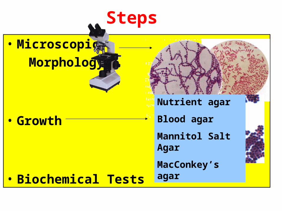
Steps
• Microscopic
Morphology
• Growth
• Biochemical Tests
Nutrient agar
Blood agar
Mannitol Salt Agar
MacConkey’s agar
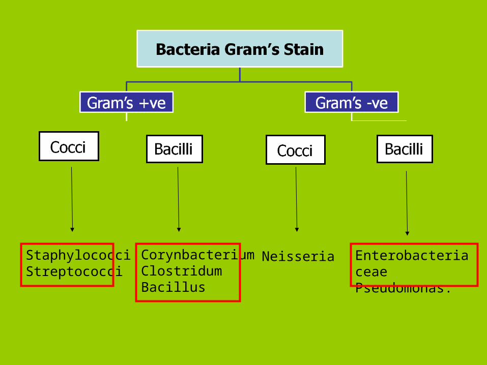
StaphylococciStreptococci
NeisseriaCorynbacteriumClostridumBacillus
Enterobacteriaceae Pseudomonas.
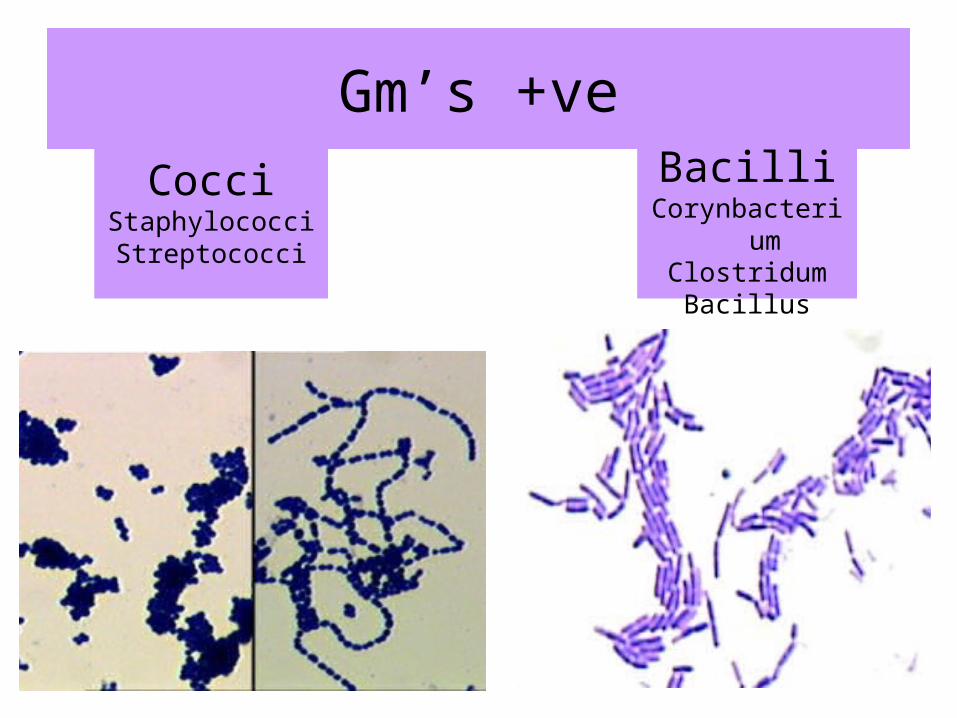
Gm’s +ve
CocciStaphylococciStreptococci
BacilliCorynbacterium
ClostridumBacillus
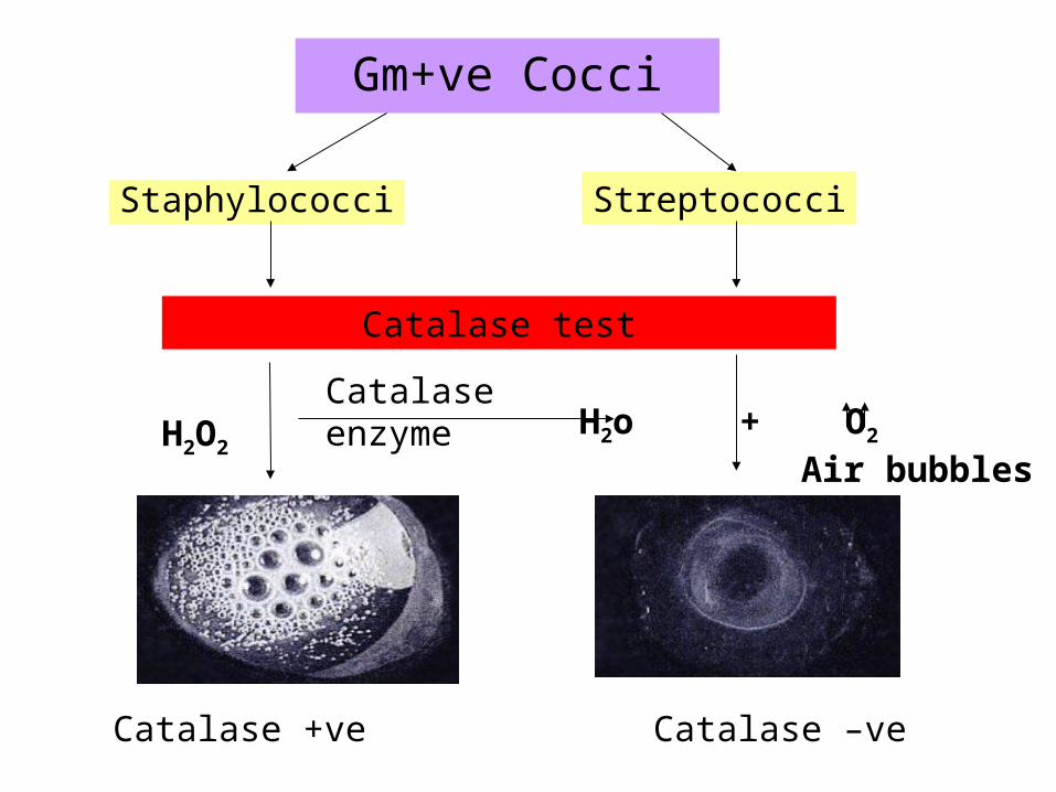
Gm+ve Cocci
Staphylococci Streptococci
H2O2
Catalase enzymeH2o + O2
Air bubbles
Catalase test
Catalase –veCatalase +ve

• Gram positive cocci, arranged in grape like clusters, non-motile, non-spore forming.
Staphylococci

Staphylococci
Staph. saprophyticus Staph. aureus Staph. epidermidis
Coagulase Test
Staph. saprophyticus Staph. aureus Staph. epidermidisFibrinogen
Plasma
Coagulase enzymeFibrin
Visible Clot
(+ve) Formation of visible clot -ve
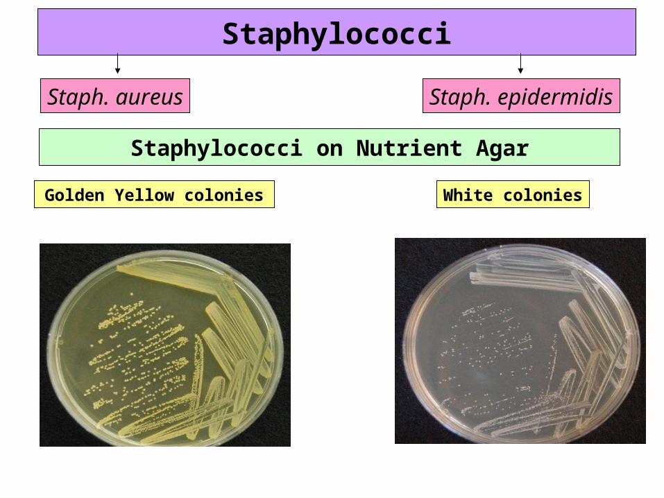
Staphylococci
Staph. aureus Staph. epidermidis
Staphylococci on Nutrient Agar
Golden Yellow colonies White colonies
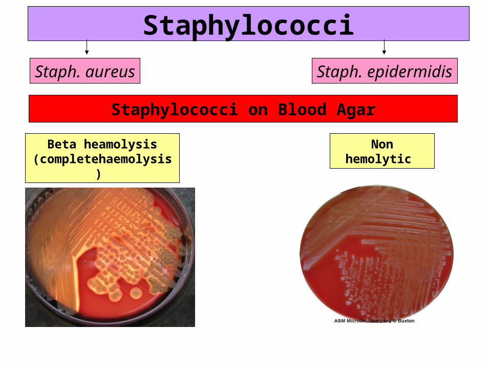
Staphylococci
Staph. aureus Staph. epidermidis
Staphylococci on Blood Agar
Beta heamolysis(completehaemolysis)
Non hemolytic
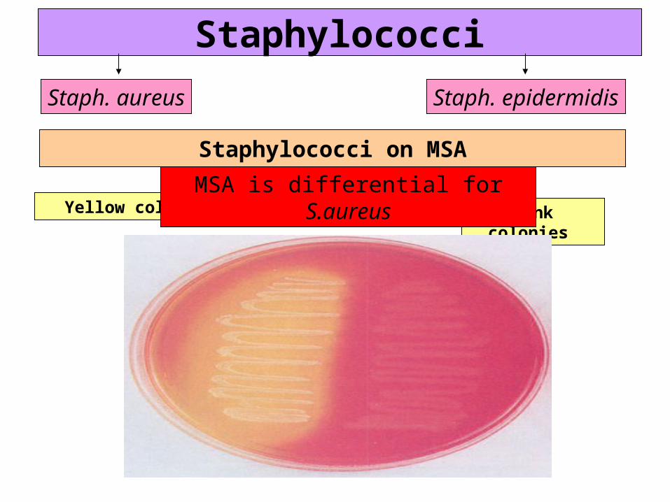
Staphylococci
Staph. aureus Staph. epidermidis
Staphylococci on MSA
Yellow colonies Pink colonies
MSA is differential for S.aureus

Deoxyribonuclease (DNase) Test
Staphylococci
Staph. aureus Staph. epidermidis
DNADNase enzyme
NucleotidesInsoluble in acid soluble in acid
DNase activity is indicated by a clear zone around the growth after addition of Hcl
Clear zone around the growth while the rest of the plate appears cloudy
Cloudiness in all the plate
S.epidermidis
S.aureus

Gm+ve Cocci
Staphylococci Streptococci
H2O2
Catalase enzymeH2o + O2
Air bubbles
Catalase test
Catalase –veCatalase +ve

Gram positive cocci,arranged in chains or pairs, non-motile,
non-spore forming.
Streptococci

(Fastidious organism)
Complex nutritional requirements
Blood agar
Streptococci
β-hemolyticS. pyogenes
Non-hemolyticα-hemolyticS.pneumonia ,
viridans streptococci

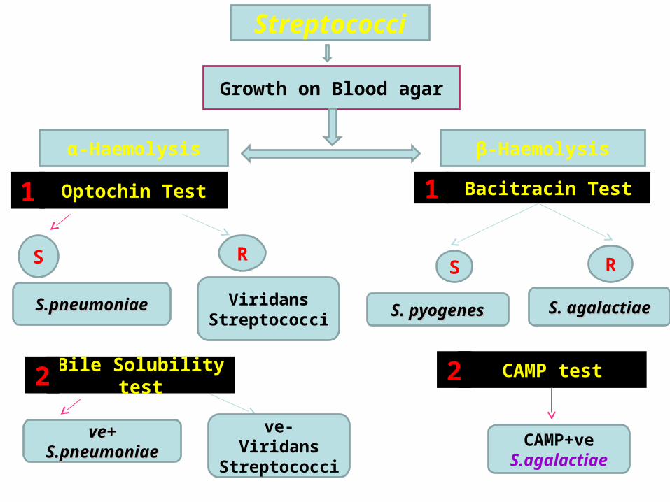
Streptococci
Growth on Blood agar
α-Haemolysis β-Haemolysis
Optochin Test Bacitracin Test
S RS R
S.pneumoniaeS.pneumoniae Viridans Streptococci
CAMP+veS.agalactiae
S. agalactiaeS. agalactiae
Bile Solubility test CAMP test
1 1
2 2
S. pyogenesS. pyogenes
++veveS.pneumoniaeS.pneumoniae
-veViridans
Streptococci

Optochin Test
α-Haemolysis
Principle:S.pneumoniae is inhibited by less than 5µg/ml Optochin reagent giving a zone of inhibition more than 15 mm in diameter.
Positive test: any zone of inhibition around the disc.
SR
S.pneumoniaS.pneumoniaee Viridans
Streptococci

Streptococci
Growth on Blood agar
α-Haemolysis β-Haemolysis
Bacitracin Test
S RS R
S.pneumoniaeS.pneumoniae Viridans Streptococci
CAMP+veS.agalactiae
S. agalactiaeS. agalactiae
Bile Solubility test CAMP test
1 1
2 2
S. pyogenesS. pyogenes
++veveS.pneumoniaeS.pneumoniae
-veViridans
Streptococci
Bile Solubility testOptochin Test

α-Haemolysis
Bile Solubility testS.pneumoniae produces a self-lysing enzyme to depress the growth of old colonies. The presence of bile salt accelerate this process.
Principle:
Visible clearance
S.Pneumoniae
Remain turbid
Viridans Streptococci
Results

Streptococci
Growth on Blood agar
α-Haemolysis β-Haemolysis
Optochin Test Bacitracin Test
S RS R
S.pneumoniaeS.pneumoniae Viridans Streptococci
CAMP+veS.agalactiae
S. agalactiaeS. agalactiae
Bile Solubility test CAMP test
1 1
2 2
S. pyogenesS. pyogenes
++veveS.pneumoniaeS.pneumoniae
-veViridans
Streptococci

β-Haemolysis
Bacitracin TestBacitracin Test
Principle:A low conc. of Bacitracin (0.04 units) will selectively inhibit the growth of S.pyogenes giving a zone of inhibition around the disc
B
Positive test: any zone of inhibition around the disc.
BS RS. S.
pyogenespyogenesS. S.
agalactiaeagalactiae
Streptococci
Growth on Blood agar
α-Haemolysis β-Haemolysis
Optochin Test Bacitracin Test
S RS R
S.pneumoniaeS.pneumoniae Viridans Streptococci
CAMP+veS.agalactiae
S. agalactiaeS. agalactiae
Bile Solubility test CAMP test
1 1
2 2
S. pyogenesS. pyogenes
++veveS.pneumoniaeS.pneumoniae
-veViridans
Streptococci
β-Haemolysis
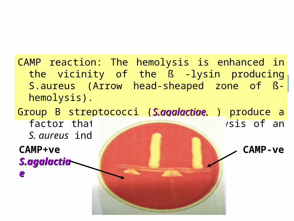
CAMP test
CAMP reaction: The hemolysis is enhanced in the vicinity of the ß -lysin producing S.aureus (Arrow head-sheaped zone of ß-hemolysis).
Group B streptococci (S.agalactiae.S.agalactiae. ) produce a factor that increases beta hemolysis of an S. aureus indicator strain.
CAMP+veS.agalactS.agalactiaeiae
CAMP-ve

Gm’s +ve
CocciStaphylococciStreptococci
BacilliCorynbacterium
ClostridumBacillus

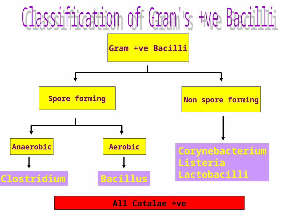
Gram +ve Bacilli
Spore forming Non spore forming
AerobicAnaerobic
BacillusClostridium
CorynebacteriumListeriaLactobacilli
All Catalae +ve
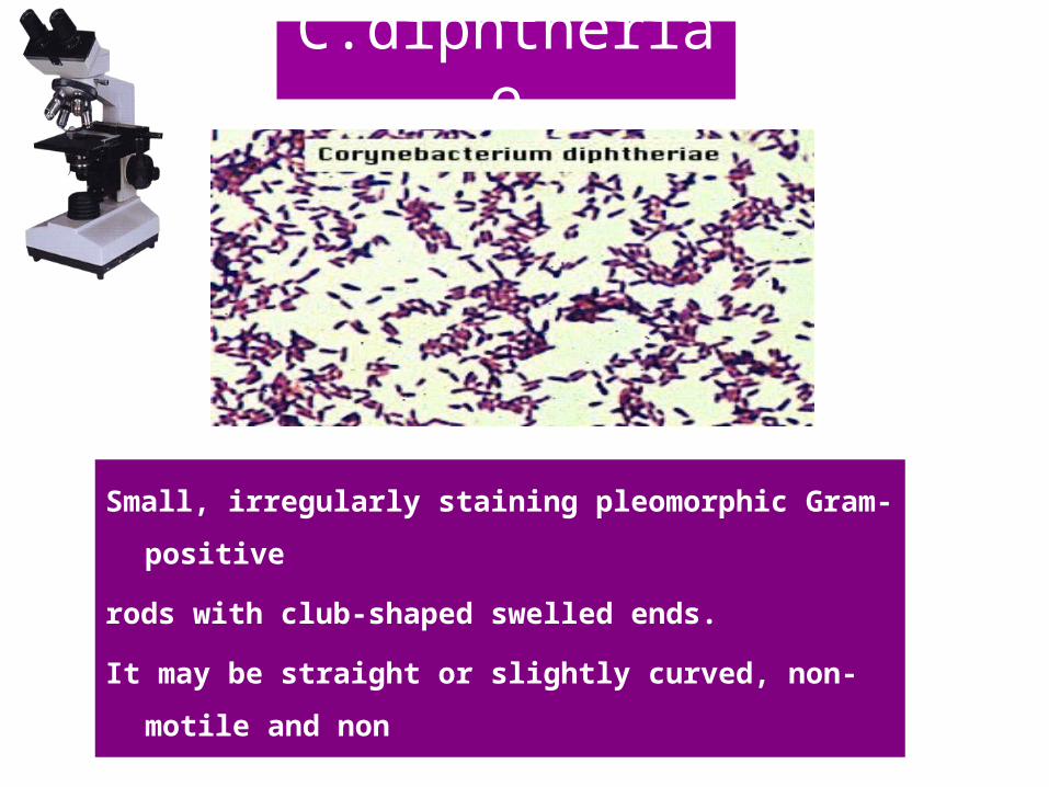
Small, irregularly staining pleomorphic Gram-positive
rods with club-shaped swelled ends.
It may be straight or slightly curved, non-motile and non
spore-forming.
"Chinese letters"
C.diphtheriae

Corynebacterium
.Growth occurs on media containing blood or serum
-On blood tellurite medium (selective & differential medium) colonies appear grey to black.
-On Loeffler’s serum

Corynebacterium
C.diphtheriaeOther
corynebacterium species
Carbohydrate Fermentation Test
Principle:
Each species of corynebacteria has its specific carbohydrate fermentation
pattaern.
C.diphtheriae can be differentiated from other corynebacterium species by
fermentation of glucose and maltose but not sucrose, with production of acid
without gas.

Glucose Maltose Sucrose
Results:
Sugar fermentation can be indicated by change of color of the medium from red to yellow.
Glucose Maltose Sucrose
C. xerosis
Glucose Maltose Sucrose
C. diphtheriae

Toxigenicity testing of C.diphtheriae strains
Elek’s Toxigenicity TestElek’s Toxigenicity Test
Results:Positive test: The antitoxin diffusing from the filter paper strip will form precipitation lines with the toxin diffusing from the toxigenic strain.
Absence of precipitation lines indicates that the strain is non-toxigenic.
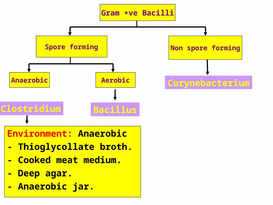
Gram +ve Bacilli
Spore forming Non spore forming
AerobicAnaerobic
BacillusClostridium
CorynebacteriumListeriaLactobacilli
All Catalae +ve

Gram-positive non-motile rectangular large
bacilli, that occur singly, in pairs, or in
chains and spore forming
Bacillus

Spore Stain
It has oval central spores.Using the Spore stainingTechnique(Malachite green &safranin), the sporesappear GREEN while thevegetative cells appearRED.
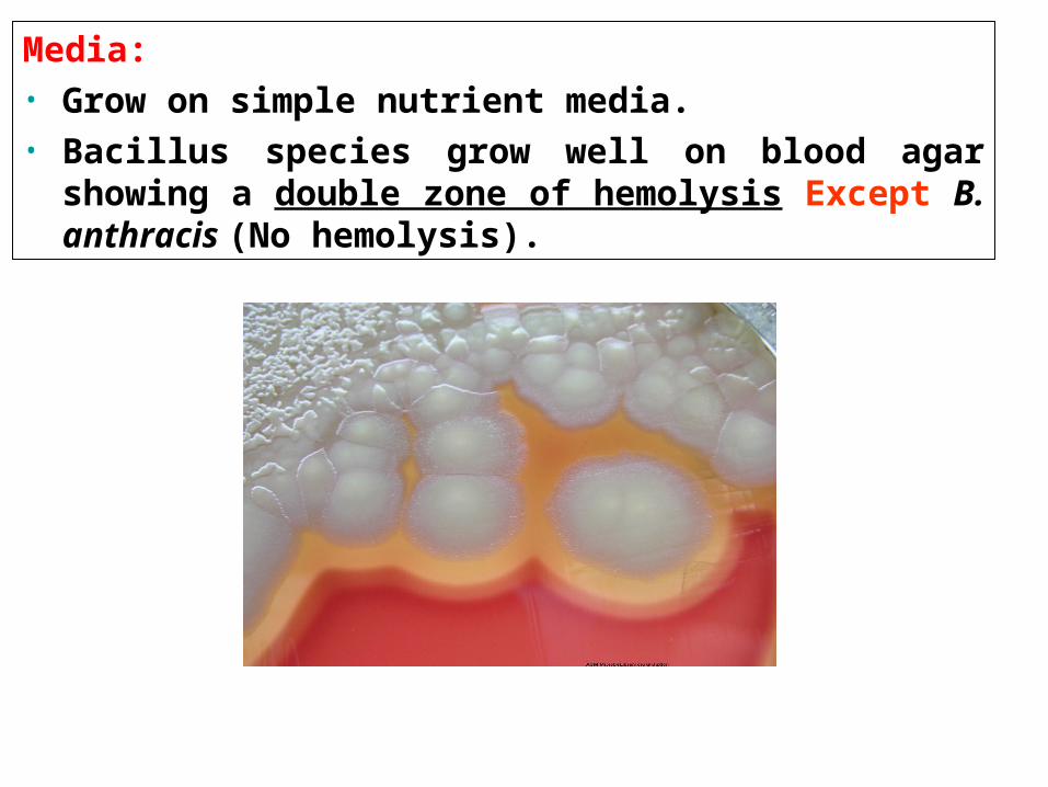
Media: • Grow on simple nutrient media.• Bacillus species grow well on blood agar showing a
double zone of hemolysis Except B. anthracis (No hemolysis).

Starch Hydrolysis Test:
Principle:
Starchamylase enzyme
glucose
Blue colour No colour
I2 I2
Biochemical reactions:
Result:Amylase activity is indicated by a clear zone around the growth while the rest of the plate gives blue color after addition of iodine solution.

Gram +ve Bacilli
Spore forming Non spore forming
AerobicAnaerobic
BacillusClostridium
CorynebacteriumListeriaLactobacilli
All Catalae +ve
Gram +ve Bacilli
Spore forming Non spore forming
AerobicAnaerobic
BacillusClostridium
Corynebacterium
Environment: Anaerobic
- Thioglycollate broth.
- Cooked meat medium.
- Deep agar.
- Anaerobic jar.

Anaerobic Medium
Anaerobic jar
principle:
Removal of O2 & replacing it with an inert gas→ Blood agar plates in Anaerobic Jar.
Cooked meat mediumI
anaerobic medium due to presence of:
Meat particles (prepared from heart muscles) which contain hematin and glutathione that act as reducing agents.

Biochemical reaction
It Contains: • Skimmed milk (without fat) i.e: contains only sugar (Lactose) and protein (casine)• Litmus indicator (acid base and redox indicator).
Litmus milk medium:
Reactions:Acidic reaction:
Lactose (milk sugar)
Fermentation acid Litmus indicator pink colour
Clostridium
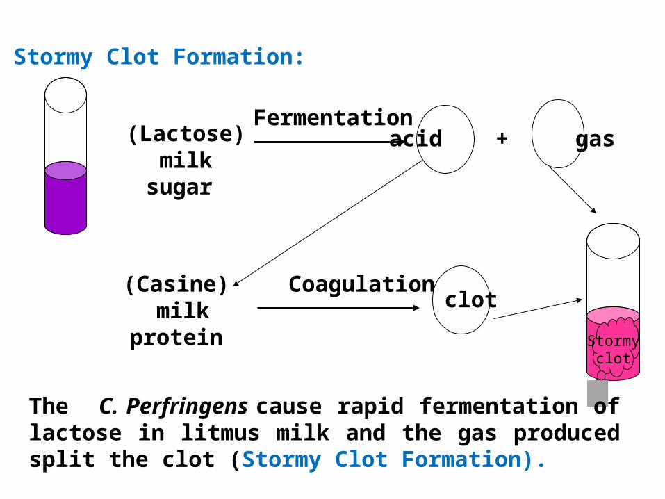
Stormy Clot Formation:
Fermentation(Lactose)
milk sugar
acid + gas
(Casine) milk
protein
Coagulation clot
Stormy clot
The C. Perfringens cause rapid fermentation of lactose in litmus milk and the gas produced split the clot (Stormy Clot Formation).