Photoplethysmograph - Frank's Hospital · PDF filePhotoplethysmograph 1 ... Photoelectric...
5
Photoplethysmograph 1 Photoplethysmograph Representative PPG taken from an ear pulse oximeter. Variation in amplitude are from Respiratory Induced Variation. A photoplethysmograph (PPG) is an optically obtained plethysmograph, a volumetric measurement of an organ. A PPG is often obtained by using a pulse oximeter which illuminates the skin and measures changes in light absorption (Shelley and Shelley, 2001). A conventional pulse oximeter monitors the perfusion of blood to the dermis and subcutaneous tissue of the skin. Diagram of the layers of human skin With each cardiac cycle the heart pumps blood to the periphery. Even though this pressure pulse is somewhat damped by the time it reaches the skin, it is enough to distend the arteries and arterioles in the subcutaneous tissue. If the pulse oximeter is attached without compressing the skin, a pressure pulse can also be seen from the venous plexus, as a small secondary peak. The change in volume caused by the pressure pulse is detected by illuminating the skin with the light from a Light Emitting Diode (LED) and then measuring the amount of light either transmitted or reflected to a photodiode. Each cardiac cycle appears as a peak, as seen in the figure. Because blood flow to the skin can be modulated by multiple other physiological systems, the PPG can also be used to monitor breathing, hypovolemia, and other circulatory conditions (Reisner, et al., 2008) [1] . Additionally, the shape of the PPG waveform differs from subject to subject, and varies with the location and manner in which the pulse oximeter is attached. Sites for measuring PPG While pulse oximeters are a commonly used medical device the PPG derived from them is rarely displayed, and is nominally only processed to determine heart rate. PPGs can be obtained from transmissive absorption (as at the finger tip) or reflective (as on the forehead). In outpatient setting pulse oximeters are commonly worn on the finger and ear. However, in cases of shock, hypothermia, etc. blood flow to the periphery can be reduced, resulting in a PPG without a discernible cardiac pulse. In this case, a PPG can be obtained from a pulse oximeter on the head, with the most common sites being the ear, nasal septum, and forehead. PPGs can also be obtained from the vagina and esophagus.
Transcript of Photoplethysmograph - Frank's Hospital · PDF filePhotoplethysmograph 1 ... Photoelectric...

Photoplethysmograph 1
Photoplethysmograph
Representative PPG taken from an ear pulse oximeter. Variation in amplitude arefrom Respiratory Induced Variation.
A photoplethysmograph (PPG) is anoptically obtained plethysmograph, avolumetric measurement of an organ. APPG is often obtained by using a pulseoximeter which illuminates the skin andmeasures changes in light absorption(Shelley and Shelley, 2001). A conventionalpulse oximeter monitors the perfusion ofblood to the dermis and subcutaneous tissueof the skin.
Diagram of the layers of human skin
With each cardiac cycle the heart pumps blood to theperiphery. Even though this pressure pulse is somewhatdamped by the time it reaches the skin, it is enough todistend the arteries and arterioles in the subcutaneoustissue. If the pulse oximeter is attached withoutcompressing the skin, a pressure pulse can also be seenfrom the venous plexus, as a small secondary peak.
The change in volume caused by the pressure pulse isdetected by illuminating the skin with the light from aLight Emitting Diode (LED) and then measuring theamount of light either transmitted or reflected to aphotodiode. Each cardiac cycle appears as a peak, asseen in the figure. Because blood flow to the skin canbe modulated by multiple other physiological systems,the PPG can also be used to monitor breathing,hypovolemia, and other circulatory conditions (Reisner,et al., 2008) [1]. Additionally, the shape of the PPGwaveform differs from subject to subject, and varieswith the location and manner in which the pulse oximeter is attached.
Sites for measuring PPGWhile pulse oximeters are a commonly used medical device the PPG derived from them is rarely displayed, and isnominally only processed to determine heart rate. PPGs can be obtained from transmissive absorption (as at thefinger tip) or reflective (as on the forehead).In outpatient setting pulse oximeters are commonly worn on the finger and ear. However, in cases of shock,hypothermia, etc. blood flow to the periphery can be reduced, resulting in a PPG without a discernible cardiac pulse.In this case, a PPG can be obtained from a pulse oximeter on the head, with the most common sites being the ear,nasal septum, and forehead.PPGs can also be obtained from the vagina and esophagus.
Photoplethysmograph 2
Uses
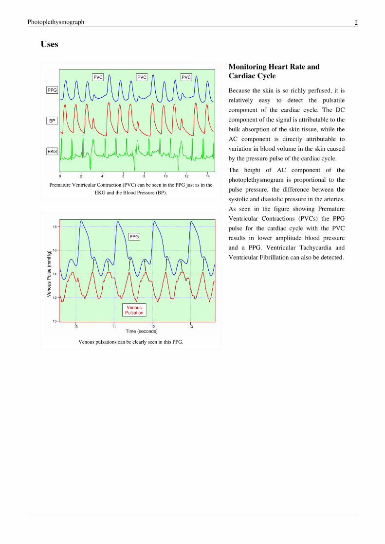
Premature Ventricular Contraction (PVC) can be seen in the PPG just as in theEKG and the Blood Pressure (BP).
Venous pulsations can be clearly seen in this PPG.
Monitoring Heart Rate andCardiac Cycle
Because the skin is so richly perfused, it isrelatively easy to detect the pulsatilecomponent of the cardiac cycle. The DCcomponent of the signal is attributable to thebulk absorption of the skin tissue, while theAC component is directly attributable tovariation in blood volume in the skin causedby the pressure pulse of the cardiac cycle.The height of AC component of thephotoplethysmogram is proportional to thepulse pressure, the difference between thesystolic and diastolic pressure in the arteries.As seen in the figure showing PrematureVentricular Contractions (PVCs) the PPGpulse for the cardiac cycle with the PVCresults in lower amplitude blood pressureand a PPG. Ventricular Tachycardia andVentricular Fibrillation can also be detected.

Photoplethysmograph 3
Monitoring Respiration
The effects of Sodium Nitroprusside(Nipride) a peripheral vasodilator on the fingerPPG of a sedated subject. As expected the PPG amplitude increases after infusion
and additionally the Respiratory Induced Variation (RIV) becomes enhanced.
Respiration affects the cardiac cycle byvarying the intrapleural pressure, thepressure between the thoracic wall and thelungs. Since the heart resides in the thoraciccavity between the lungs, the partialpressure of inhaling and exhaling greatlyinfluence the pressure on the vena cava andthe filling of the right atrium. This effect isoften referred to as normal sinus arrhythmia.During inspiration, intrapleural pressuredecreases by up to 4 mm Hg, which distendsthe right atrium, allowing for faster fillingfrom the vena cava, increasing ventricularpreload, and increasing the stroke volume.Conversely during expiration, the heart iscompressed, decreasing cardiac efficiencyand reducing stroke volume. However, theoverall net effect of respiration is to act as pump for the cardiovascular system. When the frequency and depth ofrespiration increases, the venous return increases, leading to increased cardiac output. (Shelley, et al., 2006)
Monitoring Depth of Anesthesia
Effects of an incision on a subject under general anesthesia on thephotoplethysmograph (PPG) and blood pressure (BP).
Anesthesiologist must often judgesubjectively whether a patient is sufficientlyanesthetized for surgery. As seen in thefigure if a patient is not sufficientlyanesthetized the sympathetic nervous systemresponse to an incision can generate animmediate response in the amplitude of thePPG.
Monitoring Hypo- andHyper-volemia
Shamir, Eidelman, et al. studied theinteraction between inspiration and removalof 10% of a patient’s blood volume forblood banking before surgery (Shamir,Eidelman et al. 1999). They found thatblood loss could be detected both from the photoplethysmogram from a pulse oximeter and an arterial catheter.Patients showed a decrease in the cardiac pulse amplitude caused by reduced cardiac preload during exhalation whenthe heart is being compressed.

Photoplethysmograph 4
References• M. Shamir, L. A. Eidelman, Y. Floman, L. Kaplan, and R. Pi-zov, Pulse Oximetry Plethysmographic Waveform
During Changes in Blood Volume, Br. J. Anaesth., vol. 82, pp. 178-181, 1999.• K. Shelley and S. Shelley, Pulse Oximeter Waveform: Photoelectric Plethysmography,in Clinical Monitoring,
Carol Lake, R. Hines, and C. Blitt, Eds.: W.B. Saunders Company, 2001, pp. 420-428.• K. H. Shelley, D. H. Jablonka, A. A. Awad, R. G. Stout, H. Rezkanna, and D. G. Silverman, What Is the Best Site
for Measuring the Effect of Ventilation on the Pulse Oximeter Waveform? Anesth Analg, vol. 103, pp. 372-377,2006.
• A. T. Reisner, P. A. Shaltis, D. McCombie, and H. H. Asada, Utility of the Photoplethysmogram in CirculatoryMonitoring, Anesthesiology, vol. 108, pp. 950-958, 2008. [1]
External linksA student project: building a device for collecting PPGs [2]
See alsoHemodynamics
References[1] http:/ / journals. lww. com/ anesthesiology/ Fulltext/ 2008/ 05000/ Utility_of_the_Photoplethysmogram_in_Circulatory. 24. aspx[2] http:/ / www. swarthmore. edu/ NatSci/ echeeve1/ Ref/ E72Projects/ E72Cardio/ E72Cardio. html

Article Sources and Contributors 5
Article Sources and ContributorsPhotoplethysmograph Source: http://en.wikipedia.org/w/index.php?oldid=340008684 Contributors: A2Kafir, Anthony5429, Arcadian, Bgranat, Epeper, Geni, Hu12, Johnpseudo, Karada, Spl4,Srvora, WriterHound, 16 anonymous edits
Image Sources, Licenses and ContributorsImage:PPG.PNG Source: http://en.wikipedia.org/w/index.php?title=File:PPG.PNG License: Public Domain Contributors: Spl4Image:skin.jpg Source: http://en.wikipedia.org/w/index.php?title=File:Skin.jpg License: Public Domain Contributors: Denniss, John Biancato, Lennert B, Lipothymia, NikNaks93, 1anonymous editsImage:PVC_detectionUsing_PGG.png Source: http://en.wikipedia.org/w/index.php?title=File:PVC_detectionUsing_PGG.png License: GNU Free Documentation License Contributors: Spl4Image:VenousPulsationAnd_PGG.png Source: http://en.wikipedia.org/w/index.php?title=File:VenousPulsationAnd_PGG.png License: GNU Free Documentation License Contributors: Spl4Image:NiprideEffectOnPPG-large.png Source: http://en.wikipedia.org/w/index.php?title=File:NiprideEffectOnPPG-large.png License: GNU Free Documentation License Contributors: Spl4Image:EffectsOfIncisionOnPPG-Large.png Source: http://en.wikipedia.org/w/index.php?title=File:EffectsOfIncisionOnPPG-Large.png License: GNU Free Documentation License Contributors: Spl4
LicenseCreative Commons Attribution-Share Alike 3.0 Unportedhttp:/ / creativecommons. org/ licenses/ by-sa/ 3. 0/