PHOTOCHEMISTRY AND PHOTOPHYSICS OF …shodhganga.inflibnet.ac.in/.../07_chapter-1-12size.pdf · 6...
56
CHAPTER 1 PHOTOCHEMISTRY AND PHOTOPHYSICS OF MULTICHROMOPHORIC COMPOUNDS: FUNDAMENTAL CONCEPTS AND APPLICATIONS 1.1. Abstract The present chapter gives an overview of multichromophoric compounds, which includes photophysical properties and mechanism of energy transfer in these systems. A survey including several selected examples of multichromophoric arrays is presented. Furthermore, a brief description of bischromophoric systems and the objectives of the present investigation are also discussed. 1.2. Introduction Large, well-defined multichromophoric arrays are receiving much attention due to the control they offer over the energy and electron transfer processes in restricted space. Formation of these arrays can be accomplished by either covalent coupling of chromophore building blocks into a single molecule or by the self assembly of interacting monochromophoric species where they are held together by relatively weak non-covalent interactions like hydrogen bonds, van der Waals interactions, electrostatic interactions, metal ion co-ordination and donor-acceptor interactions or π-stacking. 1,2 Different interactions of monochromophoric systems are shown in Figure 1.1.
Transcript of PHOTOCHEMISTRY AND PHOTOPHYSICS OF …shodhganga.inflibnet.ac.in/.../07_chapter-1-12size.pdf · 6...
CHAPTER 1
PHOTOCHEMISTRY AND PHOTOPHYSICS OF
MULTICHROMOPHORIC COMPOUNDS: FUNDAMENTAL
CONCEPTS AND APPLICATIONS
1.1. Abstract
The present chapter gives an overview of multichromophoric
compounds, which includes photophysical properties and mechanism of
energy transfer in these systems. A survey including several selected examples
of multichromophoric arrays is presented. Furthermore, a brief description of
bischromophoric systems and the objectives of the present investigation are
also discussed.
1.2. Introduction
Large, well-defined multichromophoric arrays are receiving much
attention due to the control they offer over the energy and electron transfer
processes in restricted space. Formation of these arrays can be accomplished
by either covalent coupling of chromophore building blocks into a single
molecule or by the self assembly of interacting monochromophoric species
where they are held together by relatively weak non-covalent interactions like
hydrogen bonds, van der Waals interactions, electrostatic interactions, metal
ion co-ordination and donor-acceptor interactions or π-stacking.1,2 Different
interactions of monochromophoric systems are shown in Figure 1.1.

2 Chapter 1
Figure 1.1: Schematic representation of the morphology of a
donor-acceptor system.
The combined strength of these relatively weak intermolecular
interactions, possessing different degrees of strength, directionality and
dependence on distance and angles, determines the final architecture of the
assembly. The close proximity of, or the interactions between multicomponent
arrays as well as between individual segments that make up such arrays; can
significantly affect their photophysical properties. The individual
chromophores are weakly coupled electronically, such that the key optical,
redox, and vibrational properties of a dimer (and larger arrays) are essentially
the sum of those of the individual molecular components.
Despite the progress that has been made in the synthesis of complex
molecules,3 formation of multichromophoric arrays in single molecule of high
symmetry is a challenge on its own. From mechanistic studies of the
Chapter 1 3
intramolecular energy transfer process, it has been clearly demonstrated that
factors at the molecular level, such as the nature of the chromophores,
interchromophoric distance and orientation, and so on, play important roles in
affecting the energy transfer process. In addition to the relative orientation of
chromophores and size of the chromophoric system, nature of the linker also
dictates the properties. Covalent multichromophoric arrays are connected
through linkers in such a manner that they can be rigid or non-rigid controlling
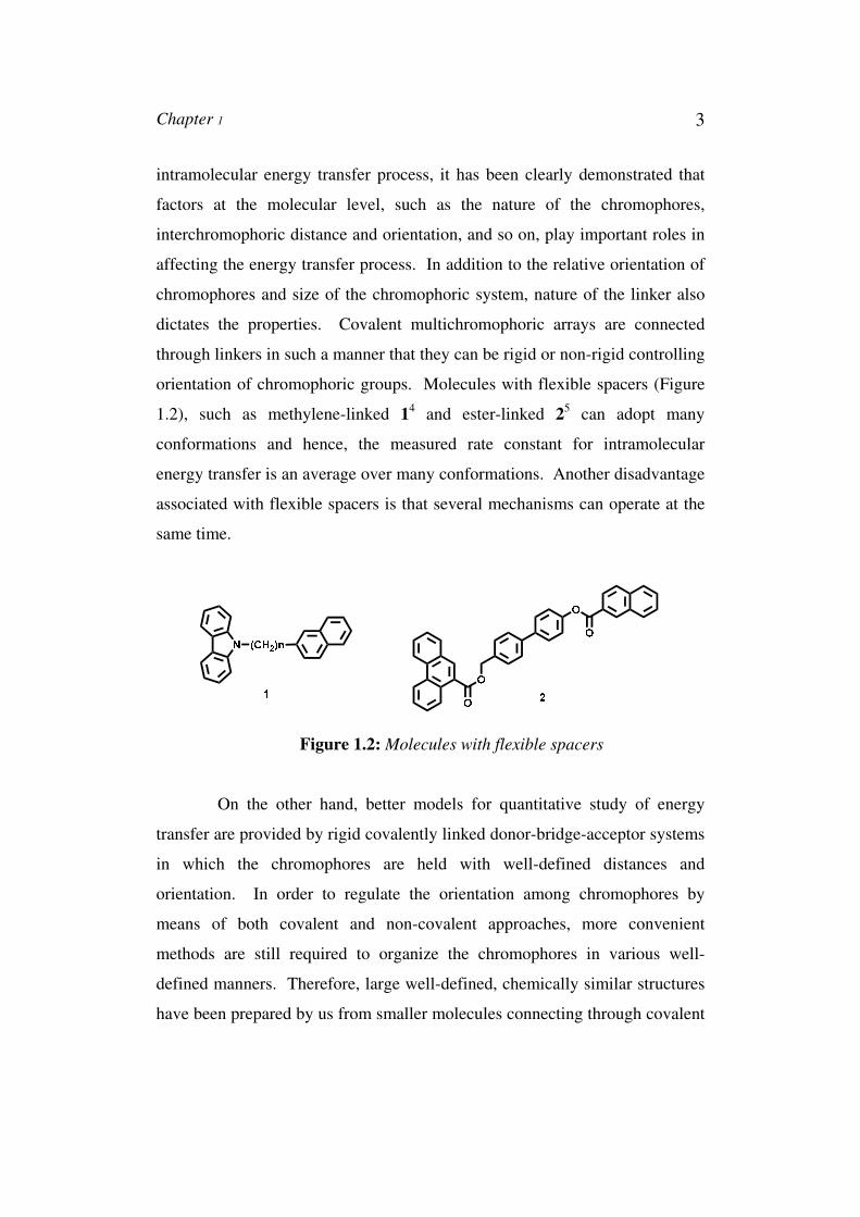
orientation of chromophoric groups. Molecules with flexible spacers (Figure
1.2), such as methylene-linked 14 and ester-linked 2
5 can adopt many
conformations and hence, the measured rate constant for intramolecular
energy transfer is an average over many conformations. Another disadvantage
associated with flexible spacers is that several mechanisms can operate at the
same time.
Figure 1.2: Molecules with flexible spacers
On the other hand, better models for quantitative study of energy
transfer are provided by rigid covalently linked donor-bridge-acceptor systems
in which the chromophores are held with well-defined distances and
orientation. In order to regulate the orientation among chromophores by
means of both covalent and non-covalent approaches, more convenient
methods are still required to organize the chromophores in various well-
defined manners. Therefore, large well-defined, chemically similar structures
have been prepared by us from smaller molecules connecting through covalent
4 Chapter 1
linkers. Slight variations in the nature of spacers have brought drastic changes
in the orientation of the structures. This thesis addresses the photochemical
and photophysical properties of such bischromophoric systems, and also is an
attempt to understand the effect of molecular geometry and nature of
chromophore in determining those properties. We investigated the effects of
geometry in a more systematic way. Though orientation effects are well
studied in covalent donor-acceptor arrays, the determination of geometry of
aggregates is currently a tremendously active field; the influence of
organizational effects on the photophysics within a superstructure is rarely the
topic of interest.
Investigations of such well defined bischromophoric arrays might
give information on the relation between the properties of these arrays and
those of monochromophoric building blocks. Extrapolation of this knowledge
by applying these relations to even larger architectures might facilitate relating
molecular and material properties conceivable. A variety of techniques to
study photophysical properties are available (e.g. UV/Vis and fluorescence
spectroscopy, CD/ORD spectroscopy, non-linear optical techniques like
electric field induced second harmonic generation (EFISHG)6,7,8 and hyper
Rayleigh scattering (HRS),9,10,11 sub-picosecond transient spectroscopy, time
resolved microwave conductivity (TRMC)12,13 etc.
1.3. Photophysical Processes
Two different chromophores in close proximity with complementary
electronic properties often exhibit photophysical processes that are not found
in either of the pure materials. Two of the most fundamental and potentially
useful photophysical reactions are energy transfer and electron (or charge)
transfer. These transfer reactions form the heart of natural photosynthesis and
are fundamental to the operation of artificial organic systems for solar light
harvesting antennae complexes and photovoltaics. In this section the basic
Chapter 1 5
principles of photoinduced energy and electron transfer reaction will be briefly
discussed.
1.3.1. Electron Transfer Process
Electron transfer14 occurs upon illumination of a chromophore pair.
For electron transfer, either the donor or the acceptor may be photoexcited. In
a system consisting of an electron donating moiety (D) and an electron
accepting group (A), upon photoexcitation, an electron transfer can occur from
the donor to the acceptor moiety, resulting in the creation of a charge-
separated (CS) state (consisting of the corresponding radical cation and anion),
from which charge recombination can take place to bring the system back to
the ground state. Electron transfer can occur when the change in free energy
for charge separation (∆G0) is negative. As a first approximation, this is
achieved when the excited-state energy exceeds the energy difference between
the oxidation potential of the donor [Eox(D)] and the reduction potential of the
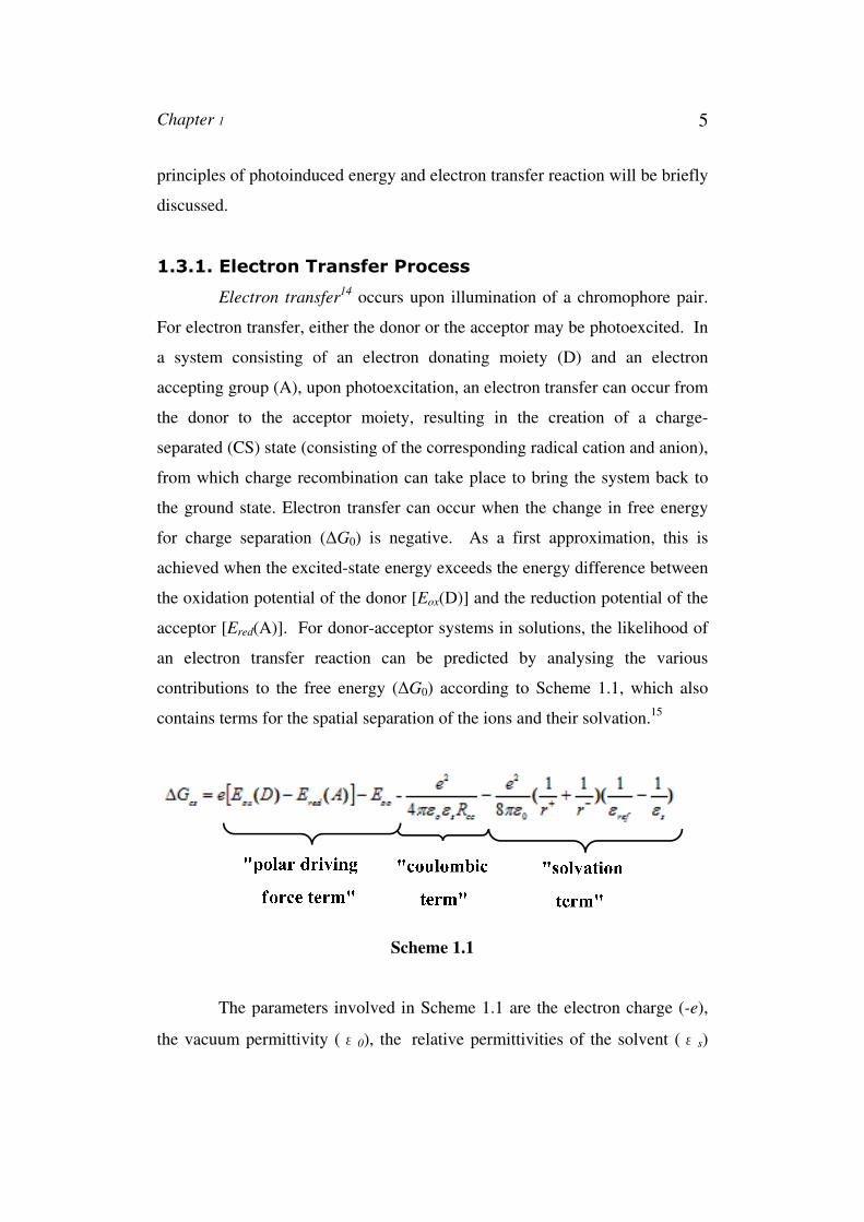
acceptor [Ered(A)]. For donor-acceptor systems in solutions, the likelihood of
an electron transfer reaction can be predicted by analysing the various
contributions to the free energy (∆G0) according to Scheme 1.1, which also
contains terms for the spatial separation of the ions and their solvation.15
Scheme 1.1
The parameters involved in Scheme 1.1 are the electron charge (-e),
the vacuum permittivity (ε0), the relative permittivities of the solvent (εs)
6 Chapter 1
and the reference solvent (εref) used to determine the redox potentials, the
centre-to-centre distance of positive and negative charges in the chromophores
(Rcc), and the radii of the positive and negative ions (r+ and r–). The driving
force (− ΔG0) can be directly linked to the kinetics of the reaction by the
theory proposed by Marcus.16
1.3.2. Energy-Transfer Mechanism in Multichromophoric
Systems
Multichromophoric systems can exhibit interesting optical and
electronic properties due to the interactions between chromophore units that
are near to each other. This may result in electron energy (exciton) transfer
over the system, a process that constitutes the physical working mechanism in
various kinds of relevant complex molecular aggregates, such as
photosynthetic and autofluorescent proteins,17,18 light-harvesting assemblies19
and conjugated polymers.20
In absence of orbital overlap, electron energy transfer between nearby
chromophores is generally caused by the transition dipole moment coupling,
an interaction whose strength (U) decreases with the third power of the
interchromophoric distance. The rate of the transfer process, however, is
constrained by static (energy and geometry fluctuations) and dynamic
(coupling to phonons) disorder in the system (∆), which counter-balances the
effect of dipole coupling on the optical properties of the assembly. Two
limiting situations can be distinguished according to the U/∆ ratio. For large
U/∆ values (i.e., large coupling strength due to short interchromophoric
distances, low disorder), coherent energy transfer between adjacent
chromophores holds, whose individual electronic functions superimpose to
yield new exciton states delocalised over the entire system. The underlying
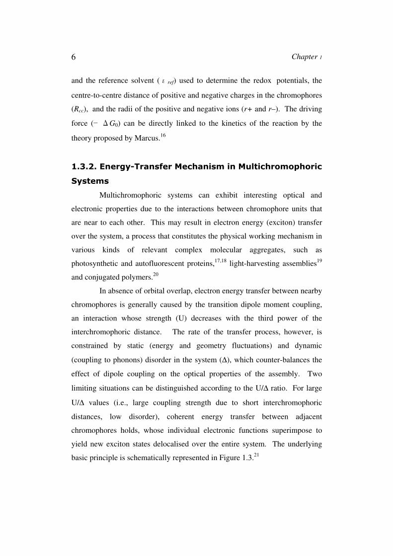
basic principle is schematically represented in Figure 1.3.21
Chapter 1 7
Figure 1.3: Two limiting energy-transfer processes occurring
on close-by molecules; a) In the strong coupling regime, the
assembly behaves as a new single quantum system, and the
energy is delocalised over the entire system. b) In the weak
coupling regime, the molecules behave as separate entities
preserving their spectral properties. Incoherent energy transfer
occurs from a donor to an acceptor molecule.
Accordingly, the assembly behaves as a new single-quantum system,
whose optical properties significantly differs from those of the separate
chromophores and critically depends on the orientation of the interacting
dipoles. Whereas a head-to-tail alignment (J configuration) leads to an
enhancement of the excited-state radiative rate (superradiance) together with
red-shifted absorption and emission, a parallel transition dipole arrangement
(H configuration) results in blue-shifted absorption followed by suppression of
fluorescence emission.21 On the other extreme, very low U/∆21 values (i.e.,
small coupling strength due to large interchromophoric distances, high
disorder) lead to Forster-type incoherent energy transfer between nearby
chromophores (Figure 1.3), vibrational relaxation in the excited unit (donor)
occurring before transfer to the adjacent chromophore (acceptor).22 To attain
optimal excitonic behaviour in multichromophoric assemblies, a well-defined
close packing of the chromophores is thus required.
8 Chapter 1
1.4. Applications of Multichromophoric Systems
It has been estimated that the average yearly incidence of solar
radiation at the earth’s surface amounts to several orders of magnitude more
energy than is consumed by its population.23 Clearly, harnessing this energy is
an important endeavour that will reduce our dependence on fossil fuels.
Indeed, our own existence depends on light-harvesting by the plethora of
photosynthetic organisms in the biosphere. These organisms have evolved
intricate and extremely efficient mechanisms for the transduction of light into
chemical energy in the form of ATP.24 The most studied of all photosynthetic
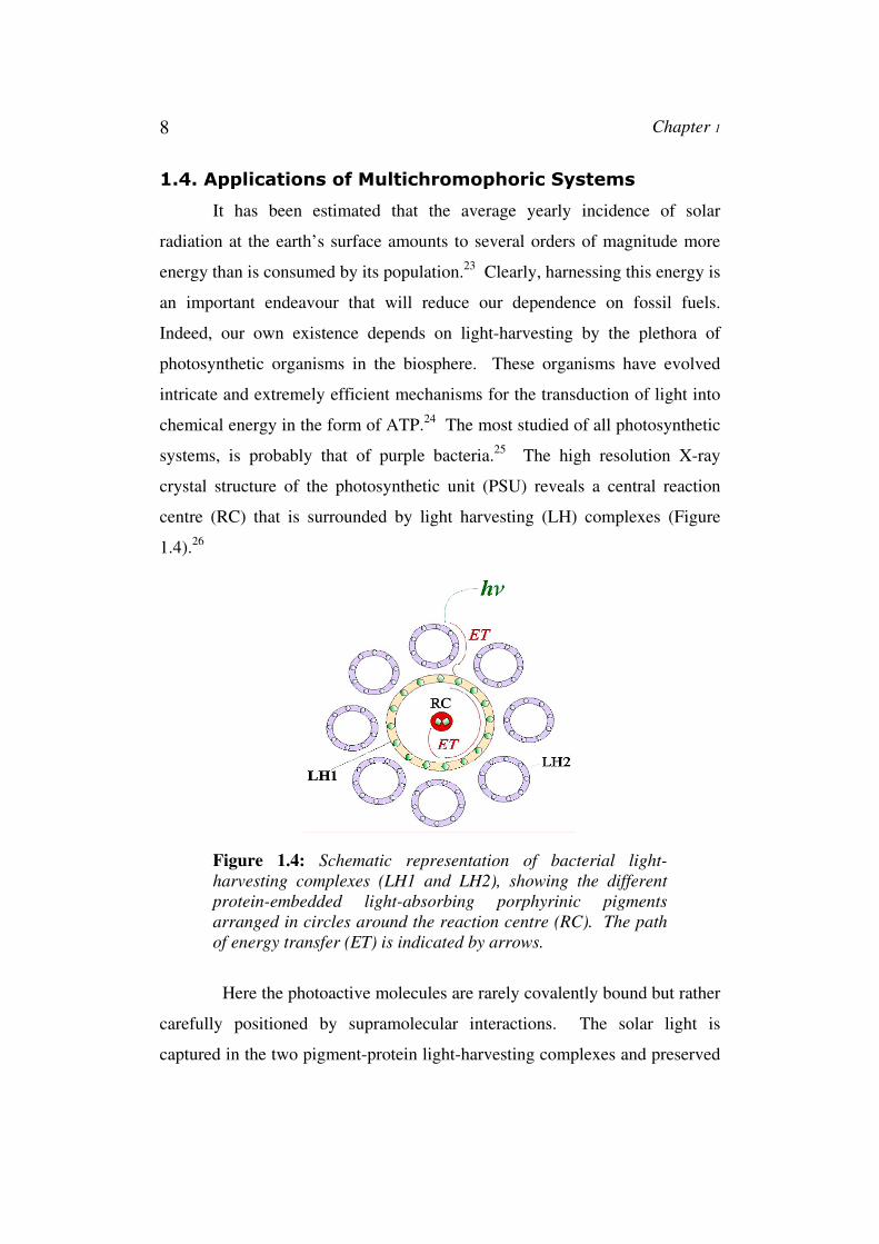
systems, is probably that of purple bacteria.25 The high resolution X-ray
crystal structure of the photosynthetic unit (PSU) reveals a central reaction
centre (RC) that is surrounded by light harvesting (LH) complexes (Figure
1.4).26
Figure 1.4: Schematic representation of bacterial light-
harvesting complexes (LH1 and LH2), showing the different
protein-embedded light-absorbing porphyrinic pigments
arranged in circles around the reaction centre (RC). The path
of energy transfer (ET) is indicated by arrows.
Here the photoactive molecules are rarely covalently bound but rather
carefully positioned by supramolecular interactions. The solar light is
captured in the two pigment-protein light-harvesting complexes and preserved
Chapter 1 9
by circulating the energy within the cyclic arrays of bacteriochlorophyll
molecules.
The LH1 complex is composed of a ring-shaped assembly of
chlorophyll and carotenoid moieties embedded in a protein matrix that
immediately surrounds the reaction centre. Similar ring-shaped assemblies,
somewhat further removed from the reaction centre, make up the LH1 and
LH2 complexes.25 The role of these chlorophyll-containing assemblies is that
of an antenna, absorbing photons that strike the relatively large surface area
that they cover. Remarkably, the energy of any photon that strikes any of the
several hundred chlorophylls within the extensive light harvesting system is
transferred to the reaction centre with unit efficiency.25 Following the initial
charge separation reaction in the heart of the reaction centre, the
photogenerated positive and negative charges are moved apart to suppress the
recombination reaction. The energy of the resulting long-lived charges is
subsequently used to drive biochemical processes within the bacteria. The
conversion of solar light into valuable energy by natural or artificial
photosynthesis is an appealing and intriguing process, making it an interesting
topic for researchers in biology, physics and chemistry.27 The most important,
well-studied and illustrative system citing the application of
multichromophoric system in nature is the photosynthetic reaction system in
purple bacteria.25
In natural photosynthetic systems, by employing elaborate light
harvesting antenna complexes, low intensity sunlight (280-900 nm) is
efficiently absorbed and energy is funnelled to the reaction centres. The
important mechanistic information extracted from studies on natural
processes28 led to ideas for the design of artificial reaction systems to mimic
and possibly improve the solar energy conversion process based on photo
induced electron transfer between electron donor and acceptor systems.29
Therefore in analogy to natural multichromophoric system, a large number of
multichromophoric systems have been synthesized. The preparation of
10 Chapter 1
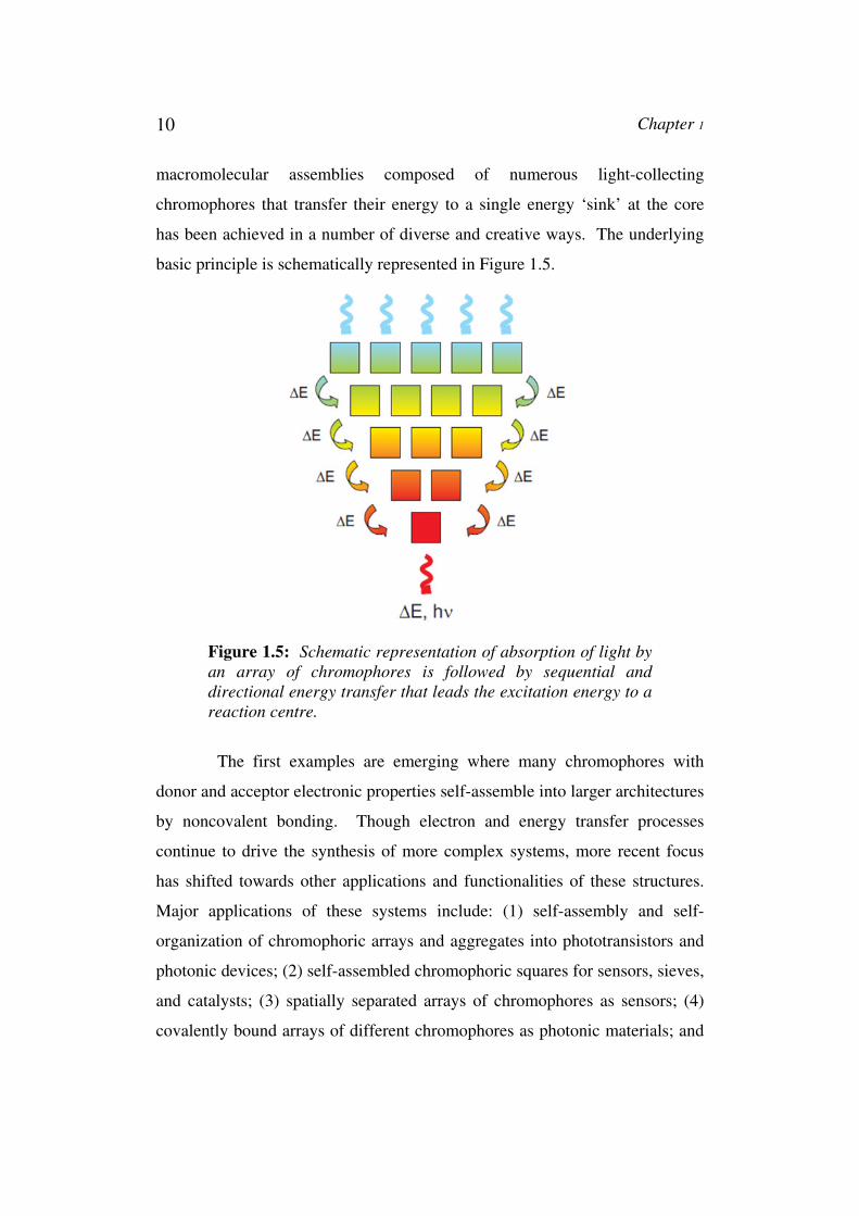
macromolecular assemblies composed of numerous light-collecting
chromophores that transfer their energy to a single energy ‘sink’ at the core
has been achieved in a number of diverse and creative ways. The underlying
basic principle is schematically represented in Figure 1.5.
Figure 1.5: Schematic representation of absorption of light by
an array of chromophores is followed by sequential and
directional energy transfer that leads the excitation energy to a
reaction centre.
The first examples are emerging where many chromophores with
donor and acceptor electronic properties self-assemble into larger architectures
by noncovalent bonding. Though electron and energy transfer processes
continue to drive the synthesis of more complex systems, more recent focus
has shifted towards other applications and functionalities of these structures.
Major applications of these systems include: (1) self-assembly and self-
organization of chromophoric arrays and aggregates into phototransistors and
photonic devices; (2) self-assembled chromophoric squares for sensors, sieves,
and catalysts; (3) spatially separated arrays of chromophores as sensors; (4)
covalently bound arrays of different chromophores as photonic materials; and
Chapter 1 11
(5) the development of molecular photonic devices such as solar cells,
photonated molecular wires, light-emitting diodes, organic electronic
components. All these takes place by absorbing light of specific wavelengths
and undergoing excited-state energy and or charge transfer reactions. The
versatile, optical, redox and photochemical properties of chromophores makes
them ideally suited as components of the above molecular devices.
To introduce photoexcitation at a specific site, the array requires the
use of accessory pigments. The ideal accessory pigment would exhibit the
following features: (1) absorb strongly in the trough between the B and Q
bands, (2) exhibit a singlet excited-state lifetime (preferably monophasic) that
is sufficiently long to support highly efficient energy transfer, (3) undergo
energy transfer without deleterious excited state quenching reactions, (4)
exhibit a high level of stability, (5) provide compatibility with the synthetic
building block approach, and (6) exhibit sufficient solubility for chemical
processing.
In those arrays, photochemical processes, especially photo-induced
energy and electron transfer are significantly affected by both electronic
properties (excitation energy and redox potentials) and structural elements (the
interchromophoric distance, orientation, and nature of the bridge) are critical
parameters that control the kinetics of charge transfer reactions in molecular
systems.
In the following paragraphs some illustrative examples of carefully
designed multichromophoric systems with covalent/non-covalent linkers are
explained. A covalent linker between the chromophores facilitated study of
interactions between different photoactive and electroactive chromophores in
close proximity.
Several elegant multi-porphyrin covalent arrays30 have been prepared
and used to study energy transport. Synthetic porphyrin-based arrays with
well-defined architectures provide essential models for probing the
mechanisms of energy and electron transfer analogous to those occurring in
12 Chapter 1
biological systems and can serve as molecular photonic devices. They are
important because of their striking photophysical and electrochemical
properties, their remarkable stability, and their predictable and rigid structure.
The multiporphyrin arrays are soluble in various organic solvents, have
controlled interporphyrin distances, incorporate porphyrins in predetermined
metallation states, and have visible absorption spectra that are nearly the sum
of spectra of the component parts. Their applications include nonlinear optics,
catalysts, sensors, actuators, molecular sieves and therapeutics. Arrays with
large numbers of porphyrins provide an ideal vehicle for studying energy
migration processes underlying photosynthetic light harvesting phenomena.
Multichromophoric arrays, usually comprising photoactive porphyrin
building blocks coupled to a quenching core, have been specifically
engineered for the study of novel photophysical processes achievable with
high powered laser sources. In this area, numerous biomimetic analogues of
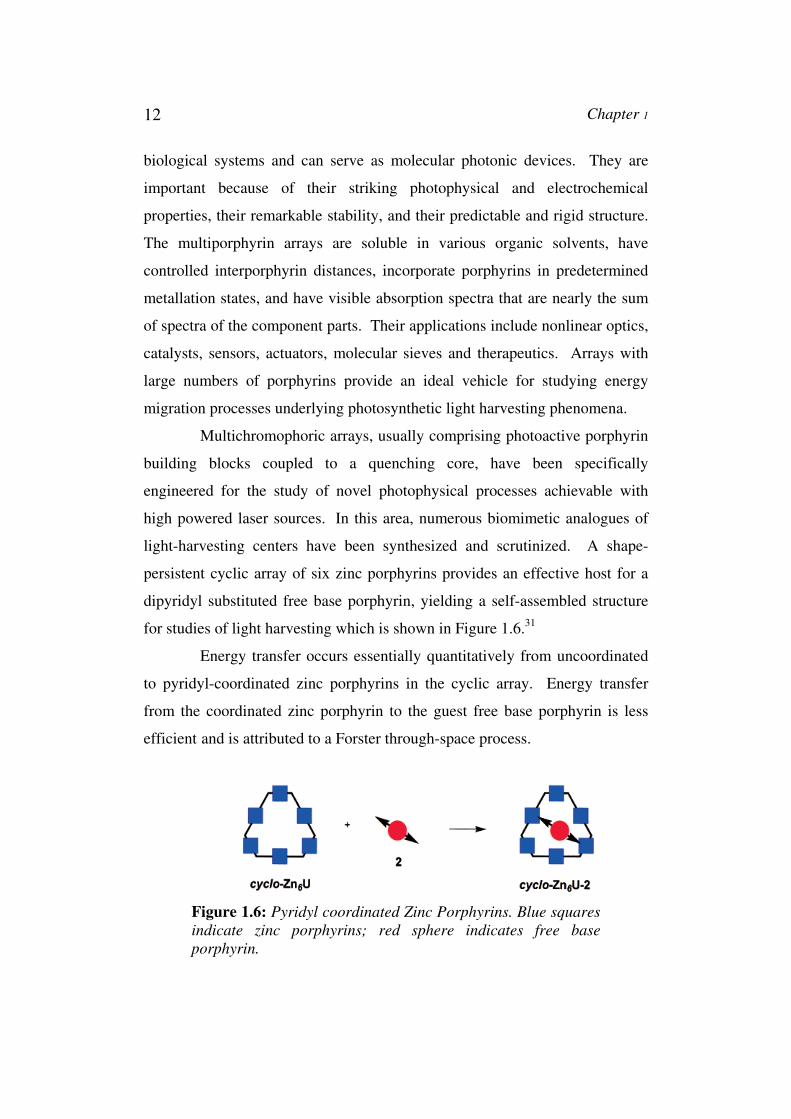
light-harvesting centers have been synthesized and scrutinized. A shape-
persistent cyclic array of six zinc porphyrins provides an effective host for a
dipyridyl substituted free base porphyrin, yielding a self-assembled structure
for studies of light harvesting which is shown in Figure 1.6.31
Energy transfer occurs essentially quantitatively from uncoordinated
to pyridyl-coordinated zinc porphyrins in the cyclic array. Energy transfer
from the coordinated zinc porphyrin to the guest free base porphyrin is less
efficient and is attributed to a Forster through-space process.
Figure 1.6: Pyridyl coordinated Zinc Porphyrins. Blue squares
indicate zinc porphyrins; red sphere indicates free base
porphyrin.

Chapter 1 13
As for another example, covalent arrays of five or more porphyrins32
in various metallation states with diphenylethyne spacer (Figure 1.7)33 provide
rigid centre to centre distances, is constructed from readily available
tetraarylporphyrin building blocks, and appears to be of appropriate length for
efficient light-harvesting. This synthetic approach enables incorporation of
peripherally-functionalized, facially-encumbered, free base porphyrins or
metalloporphyrins in rigid but soluble molecular architectures. The
diphenylethyne spacer providing rigid centre to centre distances, is constructed
from readily available tetraarylporphyrin building blocks, and appears to be of
appropriate length for efficient light-harvesting. A key feature of this
synthetic strategy is that the tetraarylporphyrins are intrinsic building blocks
and comprise an integral part of the array scaffolding. This approach should
provide a robust pathway to larger arrays resembling those found in the natural
photosynthetic systems.
Figure 1.7: Pentameric arrays of porphyrins through
dibutadiyne-linkage where M=Zn and M’=H, H.

14 Chapter 1
Another example of the use of self assembled donor-acceptor
multichromophoric porphyrin based systems is shown by Wasielewski et al.
They have shown that zinc 5,10,15,20-tetrakis (perylenediimide) porphyrin
(ZnTPP-PDI4)34 molecules (Figure 1.8), self-assemble into ordered
nanoparticles both in solution and in the solid state driven by van der Waals
stacking of the PDI molecules.
Photoexcitation of the nanoparticles results in quantitative charge
separation in 3.2 ps to form ZnTPP+ PDI- radical ion pairs in which the radical
anion rapidly migrates to stacked PDI molecules that are on average 5 layers
removed from the site of their generation as evidenced by magnetic field
effects on the yield of the PDI triplet state that results from radical ion pair
recombination. These field effects also show that the electron hopping rate
within the PDI stacks is about 2 x 10¹⁰ s-1. These nanoparticles can exhibit
charge transport properties that combine important features from both
photosynthetic and semiconductor photoconversion systems. So these
molecular systems can be used as components of photonic devices.
Figure 1.8: Structure and cartoon of the self-assembled
nanoparticles, ZnTPP-PDI4.

Chapter 1 15
Porphyrin-Fullerene Linked Dyads35 and Triads constitute highly
efficient photosynthetic energy transfer systems. The type of linkage is still of
major influence, in particular control over the rigidity of the linker deepened
the knowledge as the orientation of the donor and acceptor could be fixed.36
Synthetically more demanding is the extension of the lifetime of the created
charges by the incorporation of additional chromophores in the system, much
like in the natural photosynthetic reaction centre.37 This has led to the creation
of appealing, complex structures and demonstrates the large degree of control
that has been achieved in fine tuning energy levels towards tailoring the
outcome of the photophysical processes.
Of the many examples that can be found in literature, only two are
briefly mentioned here as an illustration of the state-of-the art in this field.
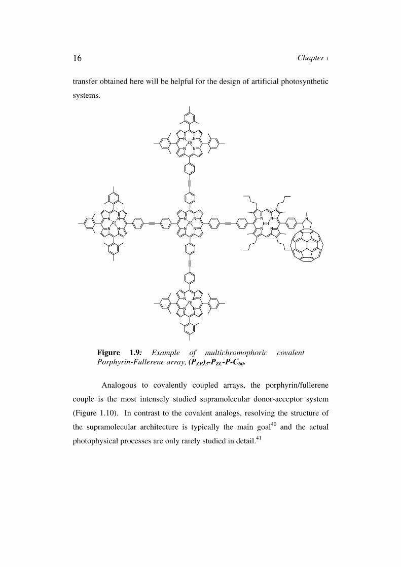
The hexadic porphyrin-C60 compound described by Kuciauskas et al.
shows how an artificial system can benefit from knowledge obtained from the
natural photosynthetic reaction centre (Figure 1.9).38 In a fullerene-containing
triad, efficient stepwise charge separation39 along a well-designed redox
gradient can be realized regardless of the solvent environment, whereas this
takes place only occasionally in conventional systems. The four zinc
porphyrins in (PZP)3-PZC-P-C60 are used as a light-harvesting antenna.
Subsequently, an energy transfer reaction occurs from the excited state of the
zinc porphyrin to the free base porphyrin. Finally, a charge transfer reaction
results in the formation of the (PZP)3-PZC-P+-C60– radical ion pair. Such control
over reaction pathways results in an exceptional enhancement of the lifetime
of the photoinduced charges.
It should be mentioned that these effects are the small reorganization
energy in electron-transfer and its consequences resulting from the fullerene’s
unique structure and symmetry, which are ultimately responsible for its high
degree of delocalization and structural rigidity. Energy transfer rate constants
depend on both the distance and driving force in a series of homologous
porphyrin-fullerene linked systems. The fundamental information on energy
16 Chapter 1
transfer obtained here will be helpful for the design of artificial photosynthetic
systems.
Figure 1.9: Example of multichromophoric covalent
Porphyrin-Fullerene array, (PZP)3-PZC-P-C60.
Analogous to covalently coupled arrays, the porphyrin/fullerene
couple is the most intensely studied supramolecular donor-acceptor system
(Figure 1.10). In contrast to the covalent analogs, resolving the structure of
the supramolecular architecture is typically the main goal40 and the actual
photophysical processes are only rarely studied in detail.41
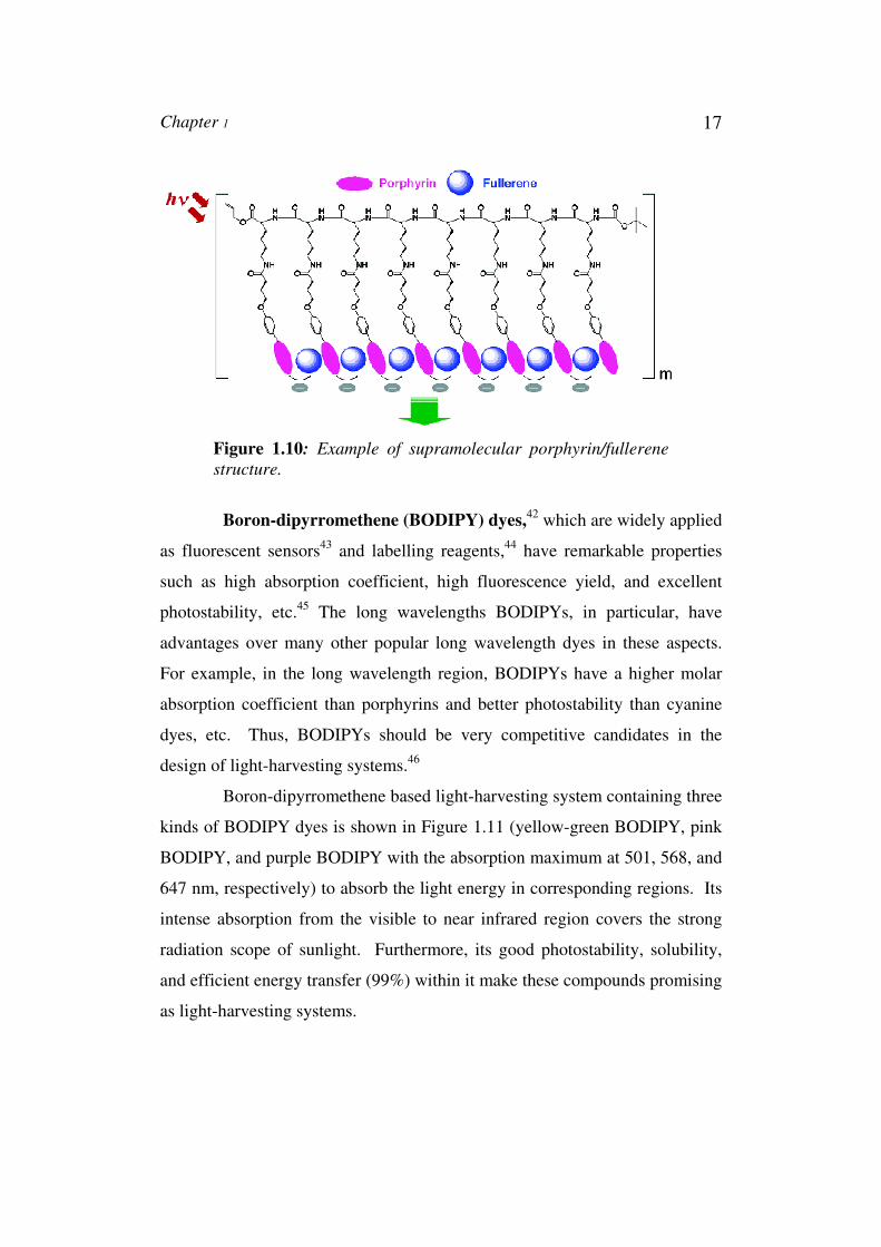
Chapter 1 17
Figure 1.10: Example of supramolecular porphyrin/fullerene
structure.
Boron-dipyrromethene (BODIPY) dyes,42 which are widely applied
as fluorescent sensors43 and labelling reagents,44 have remarkable properties
such as high absorption coefficient, high fluorescence yield, and excellent
photostability, etc.45 The long wavelengths BODIPYs, in particular, have
advantages over many other popular long wavelength dyes in these aspects.
For example, in the long wavelength region, BODIPYs have a higher molar
absorption coefficient than porphyrins and better photostability than cyanine
dyes, etc. Thus, BODIPYs should be very competitive candidates in the
design of light-harvesting systems.46
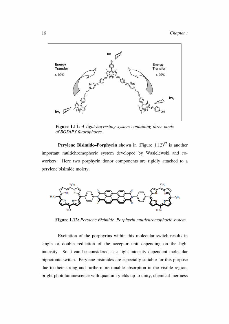
Boron-dipyrromethene based light-harvesting system containing three
kinds of BODIPY dyes is shown in Figure 1.11 (yellow-green BODIPY, pink
BODIPY, and purple BODIPY with the absorption maximum at 501, 568, and
647 nm, respectively) to absorb the light energy in corresponding regions. Its
intense absorption from the visible to near infrared region covers the strong
radiation scope of sunlight. Furthermore, its good photostability, solubility,
and efficient energy transfer (99%) within it make these compounds promising
as light-harvesting systems.
18 Chapter 1
Figure 1.11: A light-harvesting system containing three kinds
of BODIPY fluorophores.
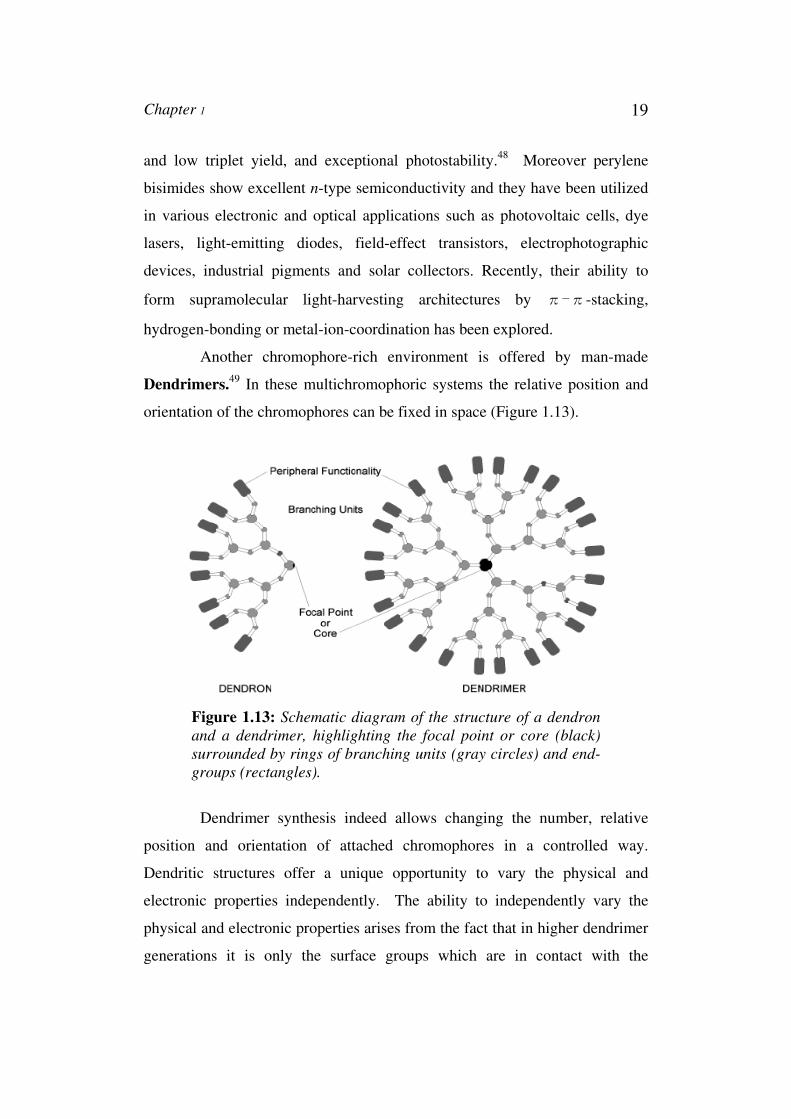
Perylene Bisimide–Porphyrin shown in (Figure 1.12)47 is another
important multichromophoric system developed by Wasielewski and co-
workers. Here two porphyrin donor components are rigidly attached to a
perylene bisimide moiety.
Figure 1.12: Perylene Bisimide–Porphyrin multichromophoric system.
Excitation of the porphyrins within this molecular switch results in
single or double reduction of the acceptor unit depending on the light
intensity. So it can be considered as a light-intensity dependent molecular
biphotonic switch. Perylene bisimides are especially suitable for this purpose
due to their strong and furthermore tunable absorption in the visible region,
bright photoluminescence with quantum yields up to unity, chemical inertness
Chapter 1 19
and low triplet yield, and exceptional photostability.48 Moreover perylene
bisimides show excellent n-type semiconductivity and they have been utilized
in various electronic and optical applications such as photovoltaic cells, dye
lasers, light-emitting diodes, field-effect transistors, electrophotographic
devices, industrial pigments and solar collectors. Recently, their ability to
form supramolecular light-harvesting architectures by π-π-stacking,
hydrogen-bonding or metal-ion-coordination has been explored.
Another chromophore-rich environment is offered by man-made
Dendrimers.49 In these multichromophoric systems the relative position and
orientation of the chromophores can be fixed in space (Figure 1.13).
Figure 1.13: Schematic diagram of the structure of a dendron
and a dendrimer, highlighting the focal point or core (black)
surrounded by rings of branching units (gray circles) and end-
groups (rectangles).
Dendrimer synthesis indeed allows changing the number, relative
position and orientation of attached chromophores in a controlled way.
Dendritic structures offer a unique opportunity to vary the physical and
electronic properties independently. The ability to independently vary the
physical and electronic properties arises from the fact that in higher dendrimer
generations it is only the surface groups which are in contact with the
20 Chapter 1
environment. Many different forms of light-sensitive building blocks have
been used in dendrimer construction, yielding macromolecules with
interesting and diverse photophysical properties. In fact, traits of natural light-
harvesting antenna systems themselves have been identified and studied.
A number of fascinating dendritic derivatives have been synthesized
and studied by the groups of Mullen and De Schryver. Dendrimeric systems
having many aromatic molecules, such as polyphenylbenzenes and perylene-
3,4-dicarboximides, have been used to elucidate the details of energy transport
within complex covalent structures as well as the photophysics of single
molecules having multiple chromophores.
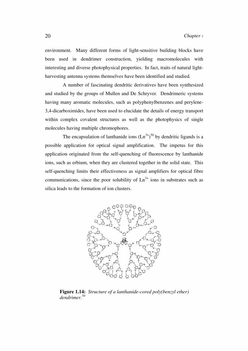
The encapsulation of lanthanide ions (Ln3+)50 by dendritic ligands is a
possible application for optical signal amplification. The impetus for this
application originated from the self-quenching of fluorescence by lanthanide
ions, such as erbium, when they are clustered together in the solid state. This
self-quenching limits their effectiveness as signal amplifiers for optical fibre
communications, since the poor solubility of Ln3+ ions in substrates such as
silica leads to the formation of ion clusters.
Figure 1.14: Structure of a lanthanide-cored poly(benzyl ether)
dendrimer.50
Chapter 1 21
Encapsulation of individual Er3+ and Tb3+ ions within a dendritic shell
(Figure 1.14) was expected to lead to their site isolation, thereby increasing
interchromophoric distance and decreasing the self-quenching effect. Indeed,
this site isolation was realized by self-assembly of suitably functionalized
carboxylate-cored dendrons around the lanthanide ion, and the resulting
assemblies possessed all the characteristics desired for use in signal
amplification.
During the course of photophysical studies on these ionically bound
supramolecular assemblies, it was found that irradiation at wavelengths where
the dendrimer backbone absorbed (280–290 nm) resulted in strong
luminescence from the lanthanide core. Apparently, energy absorbed by the
peripheral dendrimer shell was efficiently transferred to the luminescent Ln3+
at the focal point by a mechanism postulated to be of the Förster type. At
these wavelengths, energy transfer to Tb3+ was found to be more efficient than
in the case of Er3+, likely due to the better overlap of dendrimer emission with
Tb3+ absorption. This channeling of excitation energy from a dendrimer shell
to a single core unit was termed the ‘antenna effect’. Interestingly, it was also
found that this energy transfer phenomenon was critically dependent on the
substitution pattern within the dendritic shell. The findings made with the
dendrimer antennas can be extended to the design of single-layer
multichromophoric light-emitting diodes. The dendritic framework provides
for both the energy transfer interaction and the site-isolation of different
chromophores, enabling them to fluoresce simultaneously.51
Thus far, studies of energy transport in arrays consisting of many
chromophores have been focused largely on polymers, self-assembled
structures made from single chromophores and dendrimeric metal complexes.
Relevant examples include the trinuclear ruthenium complex prepared by
Scandola and Bignozzi which showed a remarkable photovoltaic performance
in dye-sensitized solar cells (DSSCs) due to its high absorptivity. As example,
a ruthenium polypyridine complex connected to a high absorber, either a
22 Chapter 1
difluorobora-zaindacene or a zinc phthalocyanine proved to be efficient
sensitizers in DSSCs. The underlying basic principle behind the operation of
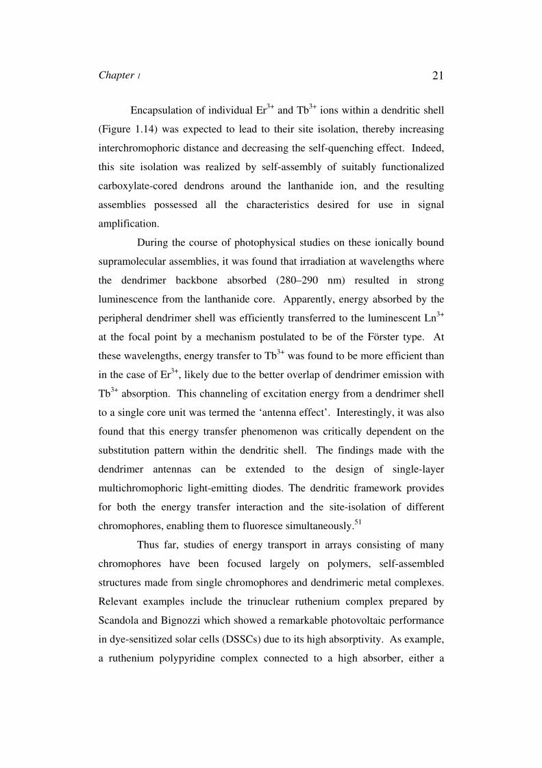
this multichromophoric system is schematically represented in Figure 1.15.52
A B C
Figure 1.15: Operation of Dyads.
In the above case, light excitation of the ruthenium complex with a
photon of energy hν1 results in electron injection in the conduction band of
TiO2. This step can be followed by a hole shift to the nearby antenna (leading
to state C). Light excitation of the antenna with a photon of energy hν2 can
undergo energy or electron transfer to the ruthenium complex. In the case of
electron transfer, the reduced ruthenium complex can also inject an electron
into TiO2. In the case of energy transfer, the sensitized ruthenium complex
excited state can subsequently inject an electron into the conduction band of
TiO2.
Multichromophoric cyclodextrins53 are good models for the study
of excitation energy transport (within a limited number of chromophores) and
antenna effect (i.e. energy transfer from the appended antenna chromophores
to an encased acceptor). They are of major interest for mimicking the
photosynthetic light-harvesting antennae because the circular arrangement of

Chapter 1 23
the chromophores is reminiscent of the light harvesting complexes of
photosynthetic bacteria (LH1 and LH2). The underlying basic principle
behind the operation of this multichromophoric system is schematically
represented in Figure 1.16.
Figure 1.16: Schematic representation of complex between β-
CD-PT7 and a dye molecule, Bottom picture shows transfer of
energy between chromophores and from chromophores to dye
molecule.54
In search of better mimics, Jullien and co-workers studied a number
of naphthyl substituted β-cyclodextrins (β-CDs) containing up to 14
chromophore units.55 The structural rigidity of these systems held the
naphthyl chromophores in spatially well-defined positions and in close
proximity with each other. This led to efficient energy hopping between the
naphthyl units in these molecules. Multichromophoric cyclodextrins can also
be used as nanoreactors for inducing selective photoreactions within the
cavity by energy transfer from the antenna chromophores, and as fluorescent
nanosensors of cationic surfactants or other cationic species.
A β-cyclodextrin bearing seven push-pull chromophores exhibits
interesting linear and nonlinear optical properties. For example,
Dye molecule

24 Chapter 1
heptachromophoric β-cyclodextrin56 (β-CD-PT7) with appended pyridin-2’-yl-
1,2,3-triazole groups (Figure 1.17) exhibit great diversity in cation-induced
photophysical effects.
Figure 1.17: β-CD-PT7
Calixarenes, the cyclic oligomeric products formed from phenol and
formaldehyde condensation, occupy a unique position in the annals of
‘supramolecular chemistry’- the chemistry of non-covalent bonds.
Multichromophoric system can be developed from this system by arranging
the chromophores in a circular fashion. Calixarene-based multichromophoric
systems that undergo changes in fluorescence emission upon ion binding are
of great interest, especially as molecular sensors.
Calix[4]arenes57 bearing more than two chromophores, have mainly
been designed for the selective detection of metal ions. The luminescence
properties of metal complexed calixarene derivatives, as investigated by
Pochin et al., show different phosphorescent behaviour for different metal
ions. Furthermore, due to the alignment of four aromatic units in
calix[4]arenes, Reinhoudt et al. were able to demonstrate that donor/acceptor
Chapter 1 25
substituted derivatives exhibit nonlinear optical behaviour in a poled polymer
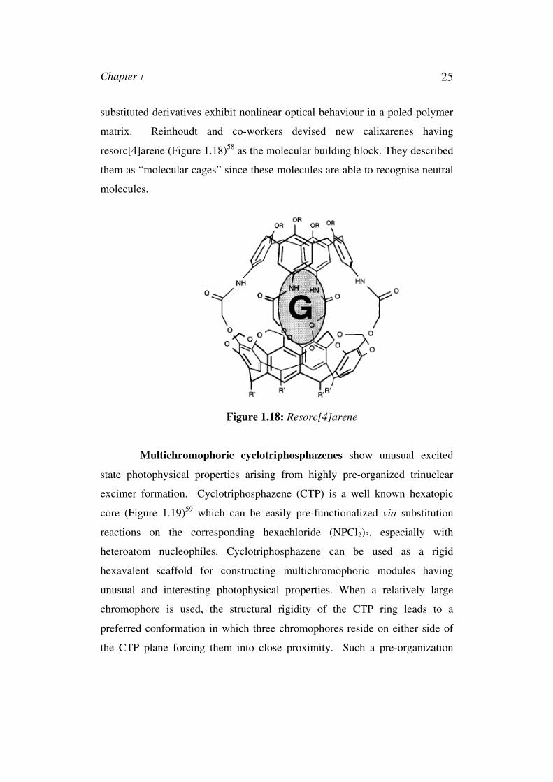
matrix. Reinhoudt and co-workers devised new calixarenes having
resorc[4]arene (Figure 1.18)58 as the molecular building block. They described
them as “molecular cages” since these molecules are able to recognise neutral
molecules.
Figure 1.18: Resorc[4]arene
Multichromophoric cyclotriphosphazenes show unusual excited
state photophysical properties arising from highly pre-organized trinuclear
excimer formation. Cyclotriphosphazene (CTP) is a well known hexatopic
core (Figure 1.19)59 which can be easily pre-functionalized via substitution
reactions on the corresponding hexachloride (NPCl2)3, especially with
heteroatom nucleophiles. Cyclotriphosphazene can be used as a rigid
hexavalent scaffold for constructing multichromophoric modules having
unusual and interesting photophysical properties. When a relatively large
chromophore is used, the structural rigidity of the CTP ring leads to a
preferred conformation in which three chromophores reside on either side of
the CTP plane forcing them into close proximity. Such a pre-organization
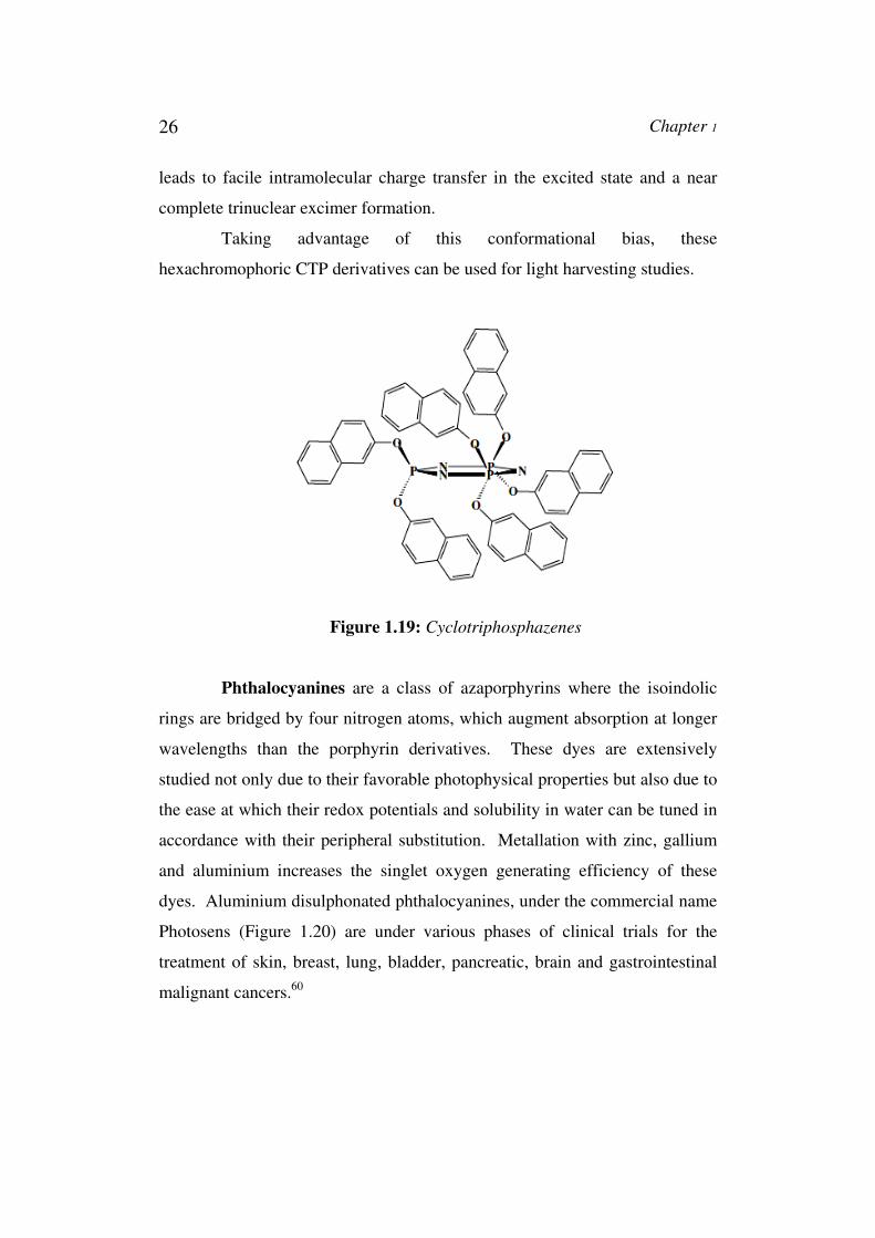
26 Chapter 1
leads to facile intramolecular charge transfer in the excited state and a near
complete trinuclear excimer formation.
Taking advantage of this conformational bias, these
hexachromophoric CTP derivatives can be used for light harvesting studies.
Figure 1.19: Cyclotriphosphazenes
Phthalocyanines are a class of azaporphyrins where the isoindolic
rings are bridged by four nitrogen atoms, which augment absorption at longer
wavelengths than the porphyrin derivatives. These dyes are extensively
studied not only due to their favorable photophysical properties but also due to
the ease at which their redox potentials and solubility in water can be tuned in
accordance with their peripheral substitution. Metallation with zinc, gallium
and aluminium increases the singlet oxygen generating efficiency of these
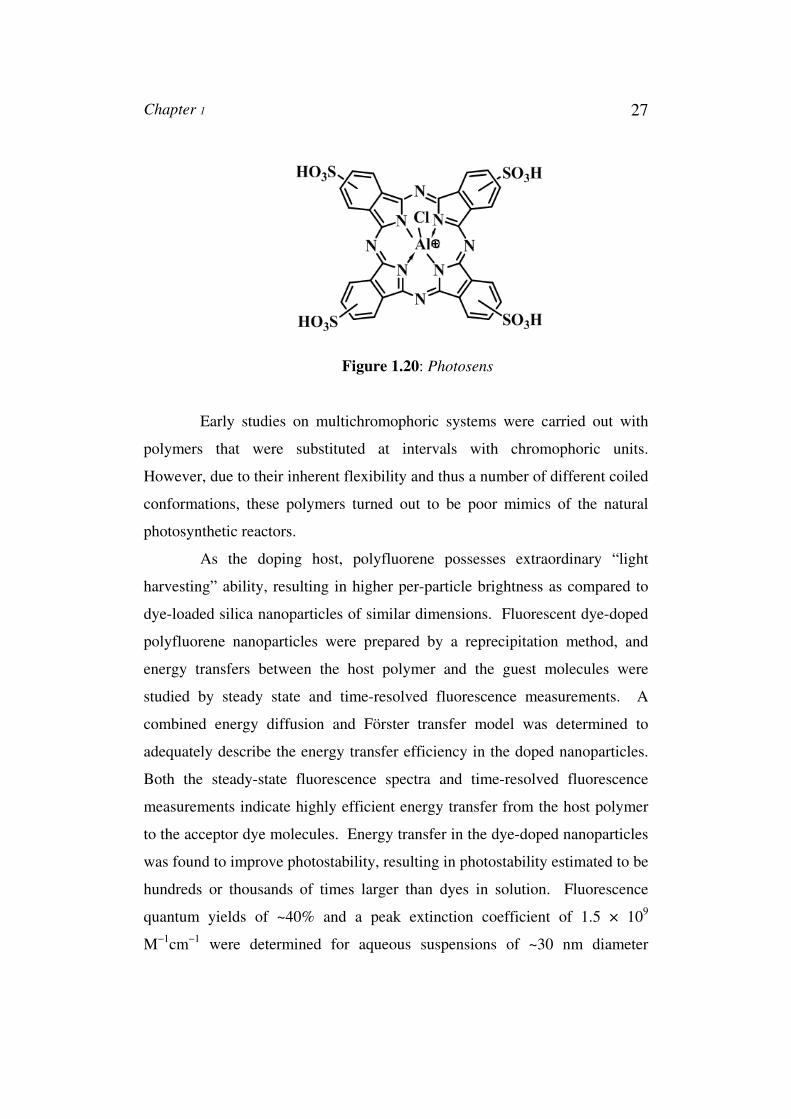
dyes. Aluminium disulphonated phthalocyanines, under the commercial name
Photosens (Figure 1.20) are under various phases of clinical trials for the
treatment of skin, breast, lung, bladder, pancreatic, brain and gastrointestinal
malignant cancers.60
Chapter 1 27
Figure 1.20: Photosens
Early studies on multichromophoric systems were carried out with
polymers that were substituted at intervals with chromophoric units.
However, due to their inherent flexibility and thus a number of different coiled
conformations, these polymers turned out to be poor mimics of the natural
photosynthetic reactors.
As the doping host, polyfluorene possesses extraordinary “light
harvesting” ability, resulting in higher per-particle brightness as compared to
dye-loaded silica nanoparticles of similar dimensions. Fluorescent dye-doped
polyfluorene nanoparticles were prepared by a reprecipitation method, and
energy transfers between the host polymer and the guest molecules were
studied by steady state and time-resolved fluorescence measurements. A
combined energy diffusion and Förster transfer model was determined to
adequately describe the energy transfer efficiency in the doped nanoparticles.
Both the steady-state fluorescence spectra and time-resolved fluorescence
measurements indicate highly efficient energy transfer from the host polymer
to the acceptor dye molecules. Energy transfer in the dye-doped nanoparticles
was found to improve photostability, resulting in photostability estimated to be
hundreds or thousands of times larger than dyes in solution. Fluorescence
quantum yields of ~40% and a peak extinction coefficient of 1.5 × 10⁹
M−1cm−1 were determined for aqueous suspensions of ~30 nm diameter
28 Chapter 1
polymer nanoparticles doped with perylene or coumarin-6 (2 wt %). The
combination of high brightness, highly red-shifted emission spectrum, and
excellent photostability are promising for fluorescence imaging and
multiplexing applications.
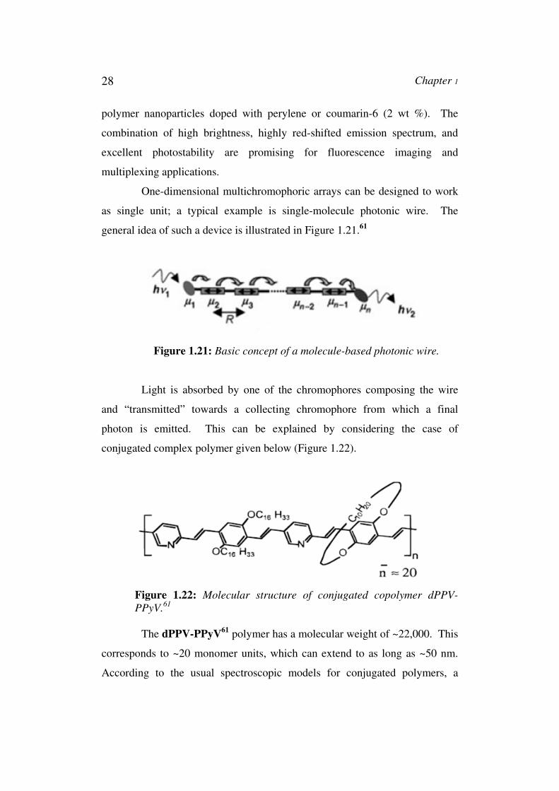
One-dimensional multichromophoric arrays can be designed to work
as single unit; a typical example is single-molecule photonic wire. The
general idea of such a device is illustrated in Figure 1.21.61
Figure 1.21: Basic concept of a molecule-based photonic wire.
Light is absorbed by one of the chromophores composing the wire
and “transmitted” towards a collecting chromophore from which a final
photon is emitted. This can be explained by considering the case of
conjugated complex polymer given below (Figure 1.22).
Figure 1.22: Molecular structure of conjugated copolymer dPPV-
PPyV.61
The dPPV-PPyV61 polymer has a molecular weight of ~22,000. This
corresponds to ~20 monomer units, which can extend to as long as ~50 nm.
According to the usual spectroscopic models for conjugated polymers, a
Chapter 1 29
polymer of this size would have multiple conjugated segments, implying that
the system is effectively multichromophoric. Accordingly, the absorption
spectrum of the polymer is due to overlapping absorption bands from different
conjugated segments of the polymer chain. In contrast, the emission may be
due to emission from a small fraction of the total polymer chain, specifically
the regions that correspond to local minima in the optical band structure. By
analogy to other conjugated polymers, these regions should be efficiently
populated on a picosecond time scale by intramolecular electronic energy
transfer. Due to the co-operative behaviour of the different conjugated
segments of the polymer, efficient intramolecular electronic energy transfer
takes place in such systems. This dramatically increases the fluorescence
intensity of multiple-chromophoric of the above conjugated polymer
molecule.
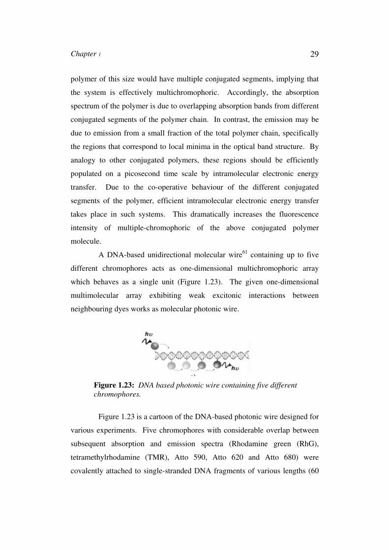
A DNA-based unidirectional molecular wire61 containing up to five
different chromophores acts as one-dimensional multichromophoric array
which behaves as a single unit (Figure 1.23). The given one-dimensional
multimolecular array exhibiting weak excitonic interactions between
neighbouring dyes works as molecular photonic wire.
Figure 1.23: DNA based photonic wire containing five different
chromophores.
Figure 1.23 is a cartoon of the DNA-based photonic wire designed for
various experiments. Five chromophores with considerable overlap between
subsequent absorption and emission spectra (Rhodamine green (RhG),
tetramethylrhodamine (TMR), Atto 590, Atto 620 and Atto 680) were
covalently attached to single-stranded DNA fragments of various lengths (60
30 Chapter 1
or 20 bases) using 6C linkers. Hybridisation of the labelled DNA fragments to
the complementary DNA strand (60 base pairs) results in a construct
containing five different chromophores positioned at well-defined distances.
The unique features of double-stranded DNA make it an ideally suited
building block for nanoscaled molecular devices. With a persistence length of
50 nm, DNA constitutes a stiff scaffold for the chromophores. In addition,
DNA offers many well-developed labelling strategies including hybridisation
of labelled short complementary oligonucleotides and dye intercalation.
Identical or different dyes can be easily incorporated at specific positions
along the DNA strand, tuning in this way the directionality of the energy flow.
1.5. Bischromophoric Systems
As model systems we have investigated properties of a few
bischromophoric systems. Bischromophoric systems are better-defined
models for the processes occurring in multichromophoric systems. Upon
excitation of a bischromophoric compound with conventional light source
several processes can occur depending on the nature of the chromophores.
However, it is recognized that considering a particular bischromophoric
molecule as having “isolated” chromophores is, at best, an approximation.
The degree of interaction can be expected to span a wide range, with the
“isolated” and “completely mixed” characterizations at the extremes of the
scale. Excitation of molecules containing more than one chromophores leads
to intramolecular interaction such as excimer, exciplex and product formation.
A number of workers have studied the photophysics and
photochemistry of bischromophoric molecules. In majority of these systems,
the chromophores are connected via tethers which allow a large number of
conformations. This study explores the photochemical and photophysical
studies of a new bischromophoric system, the anthracene diadduct, in which a
Chapter 1 31
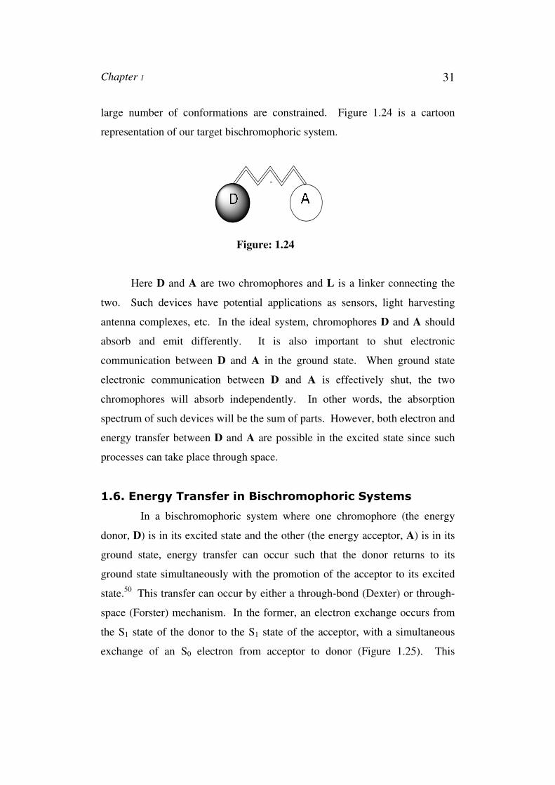
large number of conformations are constrained. Figure 1.24 is a cartoon
representation of our target bischromophoric system.
Figure: 1.24
Here D and A are two chromophores and L is a linker connecting the
two. Such devices have potential applications as sensors, light harvesting
antenna complexes, etc. In the ideal system, chromophores D and A should
absorb and emit differently. It is also important to shut electronic
communication between D and A in the ground state. When ground state
electronic communication between D and A is effectively shut, the two
chromophores will absorb independently. In other words, the absorption
spectrum of such devices will be the sum of parts. However, both electron and
energy transfer between D and A are possible in the excited state since such
processes can take place through space.
1.6. Energy Transfer in Bischromophoric Systems
In a bischromophoric system where one chromophore (the energy
donor, D) is in its excited state and the other (the energy acceptor, A) is in its
ground state, energy transfer can occur such that the donor returns to its
ground state simultaneously with the promotion of the acceptor to its excited
state.50 This transfer can occur by either a through-bond (Dexter) or through-
space (Forster) mechanism. In the former, an electron exchange occurs from
the S1 state of the donor to the S1 state of the acceptor, with a simultaneous
exchange of an S0 electron from acceptor to donor (Figure 1.25). This
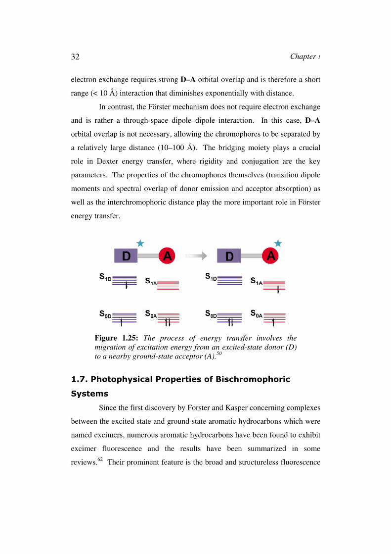
32 Chapter 1
electron exchange requires strong D–A orbital overlap and is therefore a short
range (< 10 Å) interaction that diminishes exponentially with distance.
In contrast, the Förster mechanism does not require electron exchange
and is rather a through-space dipole–dipole interaction. In this case, D–A
orbital overlap is not necessary, allowing the chromophores to be separated by
a relatively large distance (10–100 Å). The bridging moiety plays a crucial
role in Dexter energy transfer, where rigidity and conjugation are the key
parameters. The properties of the chromophores themselves (transition dipole
moments and spectral overlap of donor emission and acceptor absorption) as
well as the interchromophoric distance play the more important role in Förster
energy transfer.
Figure 1.25: The process of energy transfer involves the
migration of excitation energy from an excited-state donor (D)
to a nearby ground-state acceptor (A).50
1.7. Photophysical Properties of Bischromophoric
Systems
Since the first discovery by Forster and Kasper concerning complexes
between the excited state and ground state aromatic hydrocarbons which were
named excimers, numerous aromatic hydrocarbons have been found to exhibit
excimer fluorescence and the results have been summarized in some
reviews.62 Their prominent feature is the broad and structureless fluorescence
Chapter 1 33
band which is shifted 4000-6000 cm-1 to lower frequencies relative to the
normal molecular fluorescence. This fluorescence has been observed both in
fluid solutions and in solid state at atmospheric pressure, if the structure
allows close overlap between the molecular planes. So the aromatic molecules
which consist of two identical planar aromatic systems joined through a single
carbon-carbon bond may exhibit spectra which are either very different from
or virtually identical with the known spectrum of one half of the molecule.
The mode of interaction between the chromophores responsible for the
phenomena has largely, although not exclusively, through space interaction
(TSI) and thus to be limited to molecules that adopt a “folded” conformation,
which allows direct overlap between chromophores in the ground or excited
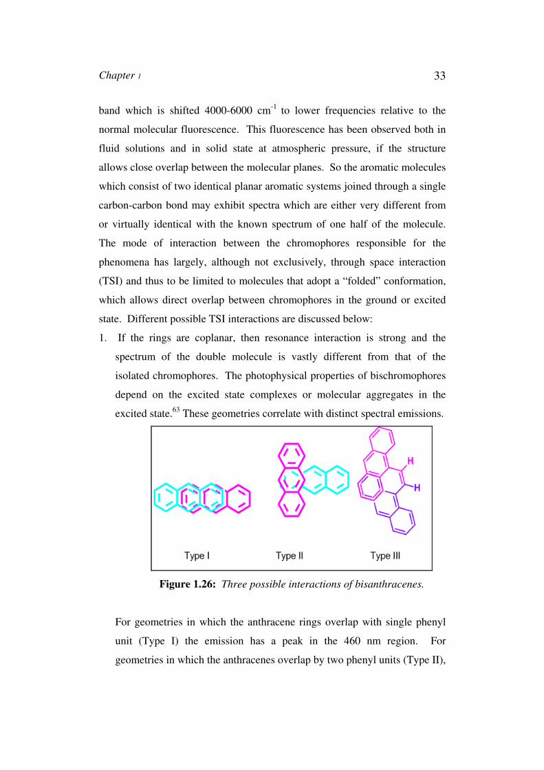
state. Different possible TSI interactions are discussed below:
1. If the rings are coplanar, then resonance interaction is strong and the
spectrum of the double molecule is vastly different from that of the
isolated chromophores. The photophysical properties of bischromophores
depend on the excited state complexes or molecular aggregates in the
excited state.63 These geometries correlate with distinct spectral emissions.
Figure 1.26: Three possible interactions of bisanthracenes.
For geometries in which the anthracene rings overlap with single phenyl
unit (Type I) the emission has a peak in the 460 nm region. For
geometries in which the anthracenes overlap by two phenyl units (Type II),

34 Chapter 1
then it is more stable than above type and emission occurs at 570 nm
region. For geometries in which the anthracenes overlap by three phenyl
units (type III), emission occurs at 620 nm region (Figure 1.26).63
2. If the molecular planes are perpendicular, the systems are essentially non-
interacting, within a π-electron approximation, and the double-molecule
spectrum does not appear different from that of one ring of the system.64 The
binaphthyls were studied by Friedel and Orchin and the ultraviolet spectra in
solution at room temperature indicated that resonance interaction was at a
minimum for 1,1’-binaphthyl whose spectrum was quite similar to that of
naphthalene. 65 2,2'-binaphthyl on the other hand exhibited a spectrum which
extended much further to the red than that of naphthalene, and in this case the
steric inhibition of resonance was presumed to be at a minimum. In the first
case steric hindrance made a perpendicular twist in the molecule, which was
responsible for little resonance interaction between the two naphthyls in it.
This twist was not present in the second case.
So the properties of absorption and luminescence emissions are
important in analytical techniques as spectroscopy.
1.7.1. Conformational Dependence on Spectra - Exciton
Coupling
In a system of N non interacting chromophores if one chromophore is
excited by light, the energies of the other N-1 non interacting molecules are
unchanged. Thus an array of N chromophores would have N-fold degeneracy,
as any chromophores could be equally excited by the light. In biopolymers,
however, the chromophores are in ordered arrays, and when electrons are
excited they do interact. For example, if one chromophore is excited, its
transition dipole may interact with the transition dipole of a neighbouring
unexcited molecule. The net result is an exchange of excitation. The energy
Chapter 1 35
of the transition will be perturbed by the potential which allows excitation
energy of one molecule to migrate to another.
If one had only two identical chromophores in a crystal, and one was
excited and the other was not, there could be resonance of the excitation
between the two chromophores. It would be unclear, at any given time, which
of the two chromophores was in the excited state. The exchange of excitation
energy between the chromophores leads to two states, one of which has higher
energy, and one of which has lower energy with respect to two isolated non
interacting chromophores. In the case of an ordered array with two
chromophores, the transition dipole of one of the isolated chromophores will
be split into two transitions, one of which having lower energy and one of
which having higher energy than the original transition. This splitting of one
transition into two is called "exciton" splitting.66
Here for the above D-A system, absorption of two photons by a
concentrated assembly of chromophores can lead to formation of a biexciton.
1.7.2. Effect of Conjugation on Spectra
Identical functional groups in different molecules will not necessarily
absorb at exactly the same wavelength. For example consider the absorption
spectra of butadiene. It gives absorption band at 217 nm (ε 21000). Here the
magnitude of molar extinction coefficient for a particular absorption is directly
proportional to the probability of the particular electronic transition; the more
probable a given transition (slightly less energy required for the transition), the
larger the extinction coefficient. The actual appearance of a simple conjugated
diene with the chromophore will be similar to the above one. There are a large
number of exceptions to the above rules, where special factors can operate.
Transitions of identical functional groups in different molecules will not have
exactly the same energy requirement because of different structural

36 Chapter 1
environments (examples shown in Figure 1.27). Distortion of chromophores
may lead to red or blue shifts, depending on the nature of the distortion.67
3 4 5
Figure 1.27: Conjugated dienes with different constraints.
The strained molecule verbenene 3 has an absorption maximum at
245.5 nm, whereas the usual calculation gives the value of 229 nm. The diene
4 might be expected to have a maximum at 273 nm; but the distortion of the
chromophore, presumably out of planarity with consequent loss of
conjugation, causes the maximum to be as low as 220 nm with a similar
intensity (ε 5500). The diene 5, in which coplanarity of the diene is more
likely, gives a maximum at 248 nm (ε 15800) showing that this is so, although
it still does not agree with the expected value.
It may be mentioned in this connection that the structural diversity
exhibited by cyclic compounds having two or more exocyclic double bonds
can provide an ideal platform for synthesizing several bischromphoric systems
with tunable relative geometry of the chromophore components. We propose
to utilize this possibility in the present investigation.
1.7.3. Effect of Cycloalkanone Ring Size in Spectra
S. A. Ahmed has reported that large cycloalkanone ring has more
electron donating methylene groups, which led to increase the electron density
of the carbonyl chromophore. So the absorption maximum of a particular
bischromophore with cyclooctanone is higher than with cyclopentanone.68,69
Chapter 1 37
1.7.4. Effect of Electronically Excited State in Spectra –
Excimer Emission (Dual Emission Spectra)
The concentration quenching of fluorescence is a phenomenon that
has been known for a long time. It was discovered only recently, on the other
hand, that instead of quenching, many fluorescent organic compounds exhibit
a change in the fluorescence spectrum; i. e. a new component becomes evident
in such a spectrum with increasing concentration. Since no corresponding
change is observed in the absorption spectrum, the new fluorescence
component must be ascribed to an associate formed only after absorption of
light in the electronically excited state. The term “excimers” has become
widely accepted for associates of this type. So excimer is a dimer which is
associated in the excited state and which is dissociative in the ground state.70
The role of the association of excited molecules as the cause of a
change in the fluorescence spectrum with concentration was first recognized in
the case of pyrene and some of its derivatives; and the formation of excimers
is still most easily demonstrated for pyrene. A large aromatic π system
exhibits dual emission. The formation of pyrene excimer requires encounter
of an electronically excited pyrene with the second pyrene in the ground state.
According to this definition the two pyrenes must be far enough away, so that
the incident radiation can cause “localised excitation”.69 These locally excited
molecules can cause “monomer” emission.
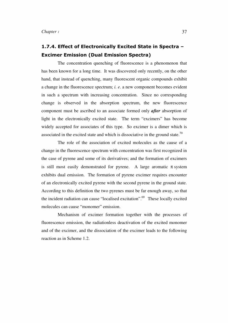
Mechanism of excimer formation together with the processes of
fluorescence emission, the radiationless deactivation of the excited monomer
and of the excimer, and the dissociation of the excimer leads to the following
reaction as in Scheme 1.2.
38 Chapter 1
Scheme 1.2: Mechanism of excimer formation.
It is a bimolecular mechanism for the formation of the excimer by
combination of an electronically excited molecule A*(in the lowest excited
singlet state) with an unexcited molecule A (in the singlet ground state). The
observation of excimer emission indicates that diffusive encounters between
pyrenes have occurred. There are instances that excimer emission is observed
although there is no evidence that the pyrenes are separated in the ground
state. These excited species are sometimes referred to as “static excimers”
while those behaving as discussed earlier as “dynamic excimers”. Forster and
co-workers first recognised the role of association of the excited molecules as
the cause of change in the fluorescence spectrum with concentration in the
case of pyrene and some of its derivatives. In 1964, Stevens and Ban
proposed a sandwich configuration of the excimer resulting from the mutual
approach of the two components with their molecular planes parallel.
1.7.5. Charge Transfer Emission Spectra
The structures of the excited states of carbonyl compounds such as
aldehydes and ketones have been studied most extensively.
Excited state reactions other than geometrical rearrangements can
result in spectral changes. In the ground state the structure of carbonyl group
contains an important contribution from the polar group. The position of
absorption that involves nonbonding electrons is particularly sensitive to the
Chapter 1 39
polarity of solvent used in the determination. If the group is more polar in the
ground state than in the excited state, the non-bonding electrons are stabilised
relative to the excited state by hydrogen bonding or electrostatic interaction
with the polar solvent; the absorption is shifted to shorter wavelength.
Conversely, if the group is more polar in the excited state, the absorption is
shifted to longer wavelength with increasing solvent polarity.
The α,β-unsaturated ketone such as mesityl oxide (4-methyl-3-
penten-2-one) shows λmax 230 mµ, ε 12600 and λ max 329 mµ, ε 41 in hexane
and λ max 243 mµ, ε 10,000 and λmax 305mµ, ε 60 in water. These data
indicate that the long wavelength n π∗ absorption is shifted to shorter
wavelength in the more polar solvent; thus the excited state would appear to
be less polar than the ground state. That is the excited state is having
contributions from rather than as in the ground state. The shift
to longer wavelength observed in the π π* transition of α,β-unsaturated
ketones with increasing solvent polarity indicates that the excited state in this
transition is more polar than in the ground state. For this transition the data
have been interpreted as the promotion of an ethylenic π electron to a carbonyl
π* orbital. Because the intramolecular effects such as these involve the
transfer of charge from one atom to another, they are frequently called as
charge-transfer spectra. Here A-B system with cycloalkenone linker can
undergo intermolecular charge transfer depending on the size of
cycloalkenone linkage.71
Same is the case with fluorescence emission spectra. Depending on
the structures in the excited state and medium, emission spectra vary. Mataga
and Murata reported fluorescence quantum yield and lifetime data for several
CT complexes. They explored the effects of donor structure, temperature and
solvent polarity on the emission and concluded that thermally equilibrated
structures for the ground and excited states are quite different for CT
complexes. They observed large Stokes shifts, which depend on structure and
40 Chapter 1
medium. Dipole-dipole interactions between the solute and solvent molecules
are proposed to be responsible for the large Stokes shift seen in polar solvents.
So such intramolecular charge transfer emission and absorption in
bischromophoric molecule is through-bond interaction.
1.8. Photochemical Properties of Bischromophoric
Systems
Light induced reactions are very important to life. So it is very
important to know factors affecting photochemical reactions to make them
useful for day to day life.
Photochemical reactions originate from the electronically excited
states of molecules. Those electronically excited states may be singlet or
triplet excited state. Normally a reaction occurs when a molecule gains the
necessary activation energy to undergo change. Here in the case of
photochemical reactions light provides the activation energy. This may lead
directly to products (e.g. in dissociations or isomerisations) or it can react with
another ground state molecule (e.g. in additions or substitutions) or more often
to unstable or reactive chemical species (e.g. free radicals or radical cations)
which then react further in secondary processes through dark reactions which
lead ultimately to the final photoproducts.72
The sequence of photochemical reactions can be given as a
succession of steps. It includes absorption of light, fluorescence, non-radiative
deactivation, intersystem crossing and chemical reactions. The processes
which occur between the absorption and emission of light can be illustrated by
a Jablonski diagram (Figure 1.28).
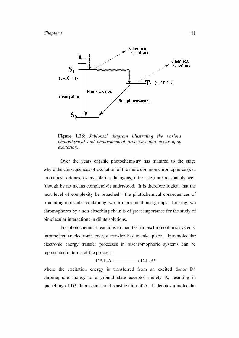
Chapter 1 41
Figure 1.28: Jablonski diagram illustrating the various
photophysical and photochemical processes that occur upon
excitation.
Over the years organic photochemistry has matured to the stage
where the consequences of excitation of the more common chromophores (i.e.,
aromatics, ketones, esters, olefins, halogens, nitro, etc.) are reasonably well
(though by no means completely!) understood. It is therefore logical that the
next level of complexity be broached - the photochemical consequences of
irradiating molecules containing two or more functional groups. Linking two
chromophores by a non-absorbing chain is of great importance for the study of
bimolecular interactions in dilute solutions.
For photochemical reactions to manifest in bischromophoric systems,
intramolecular electronic energy transfer has to take place. Intramolecular
electronic energy transfer processes in bischromophoric systems can be
represented in terms of the process:
D*-L-A D-L-A*
where the excitation energy is transferred from an excited donor D*
chromophore moiety to a ground state acceptor moiety A, resulting in
quenching of D* fluorescence and sensitization of A. L denotes a molecular

42 Chapter 1
spacer bridge connecting the two chromophores. This bridge may play a role
in promoting the transfer process. In all the EET processes, a resonance
matching between the energy of the initial state of the system and that of its
final state is required.
Another possibility is the intramolecular reaction of a component in
the excited state with another component in the ground state. Dimerization
arising through a [4+4] cycloaddition pathway in certain bisanthracenes is a
typical example for such intramolecular photochemical reactions of
bischromophoric systems.
The main photochemical reactions which have been reported in literature
are explained below. Here we particularly discuss photochemistry of
bisaromatic compounds connected through alkenone or alkene spacer.
Arylanthrylethylenes or alkenones containing methyl, phenyl, 1-naphthyl and
9-anthryl as one substituent and anthryl group as the other, form an interesting
group of bischromophoric systems. In these molecules, there is scope for
strong interactions between low-lying excited state associated with the
anthracene and arylethylene/arylalkenone moieties. Also anthracenes are
unique in combining the advantages of having easily accessible absorption
spectra, exhibiting monomer (and often excimer) fluorescence and high
photoreactivity; they also have fairly good solubility in organic solvents and,
although sometimes with difficulty, a variety of derivatives can be prepared.
Due to the above discussed peculiarities bisanthryls have been
extensively investigated, so particularly, described here the photochemical
reactions of bisanthryl alkenones. The observed reactions include geometrical
isomerization, cycloaddition reactions, rearrangement reactions and
dehydrocyclization reactions, which are described below.
Chapter 1 43
1.8.1. Fundamental Photochemical Reactions
1.8.1.1. Geometrical Isomerization Reactions
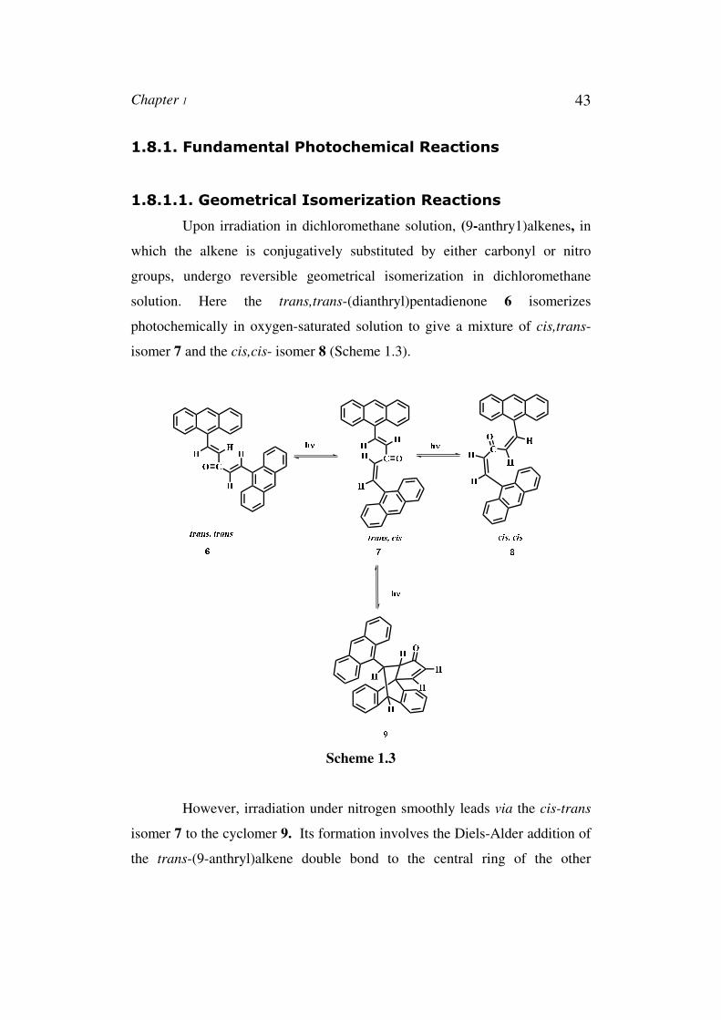
Upon irradiation in dichloromethane solution, (9-anthry1)alkenes, in
which the alkene is conjugatively substituted by either carbonyl or nitro
groups, undergo reversible geometrical isomerization in dichloromethane
solution. Here the trans,trans-(dianthryl)pentadienone 6 isomerizes
photochemically in oxygen-saturated solution to give a mixture of cis,trans-
isomer 7 and the cis,cis- isomer 8 (Scheme 1.3).
Scheme 1.3
However, irradiation under nitrogen smoothly leads via the cis-trans
isomer 7 to the cyclomer 9. Its formation involves the Diels-Alder addition of
the trans-(9-anthryl)alkene double bond to the central ring of the other
44 Chapter 1
anthracene moiety. The quenching by oxygen suggests that the [4π+2π]
cycloaddition proceeds as a triplet-state reaction, while the geometrical
isomerization involves the excited singlet state.73
1.8.1.2. Cycloaddition Reactions
The anthracene chromophore can undergo cycloaddition in which the
central ring represents a 4π electron system or, more rarely, one of the lateral
rings or any of the diene linkage acts as diene or dienophile. It can be either
[4π + 2π] or [4π + 4π] cycloaddition reaction.
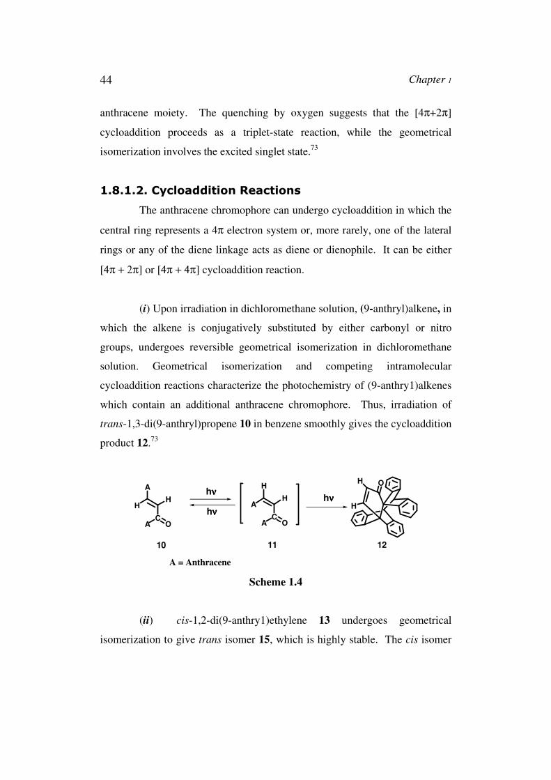
(i) Upon irradiation in dichloromethane solution, (9-anthryl)alkene, in
which the alkene is conjugatively substituted by either carbonyl or nitro
groups, undergoes reversible geometrical isomerization in dichloromethane
solution. Geometrical isomerization and competing intramolecular
cycloaddition reactions characterize the photochemistry of (9-anthry1)alkenes
which contain an additional anthracene chromophore. Thus, irradiation of
trans-1,3-di(9-anthryl)propene 10 in benzene smoothly gives the cycloaddition
product 12.73
A
H
C
H
OA
H
A
C
H
OA
OH
Hhνννν
hνννν
10 11 12
hνννν
A = Anthracene
Scheme 1.4
(ii) cis-1,2-di(9-anthry1)ethylene 13 undergoes geometrical
isomerization to give trans isomer 15, which is highly stable. The cis isomer
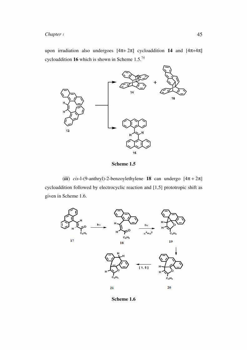
Chapter 1 45
upon irradiation also undergoes [4π+ 2π] cycloaddition 14 and [4π+4π]
cycloaddition 16 which is shown in Scheme 1.5.74
Scheme 1.5
(iii) cis-l-(9-anthryl)-2-benzoylethylene 18 can undergo [4π + 2π]
cycloaddition followed by electrocyclic reaction and [1,5] prototropic shift as
given in Scheme 1.6.
Scheme 1.6
46 Chapter 1
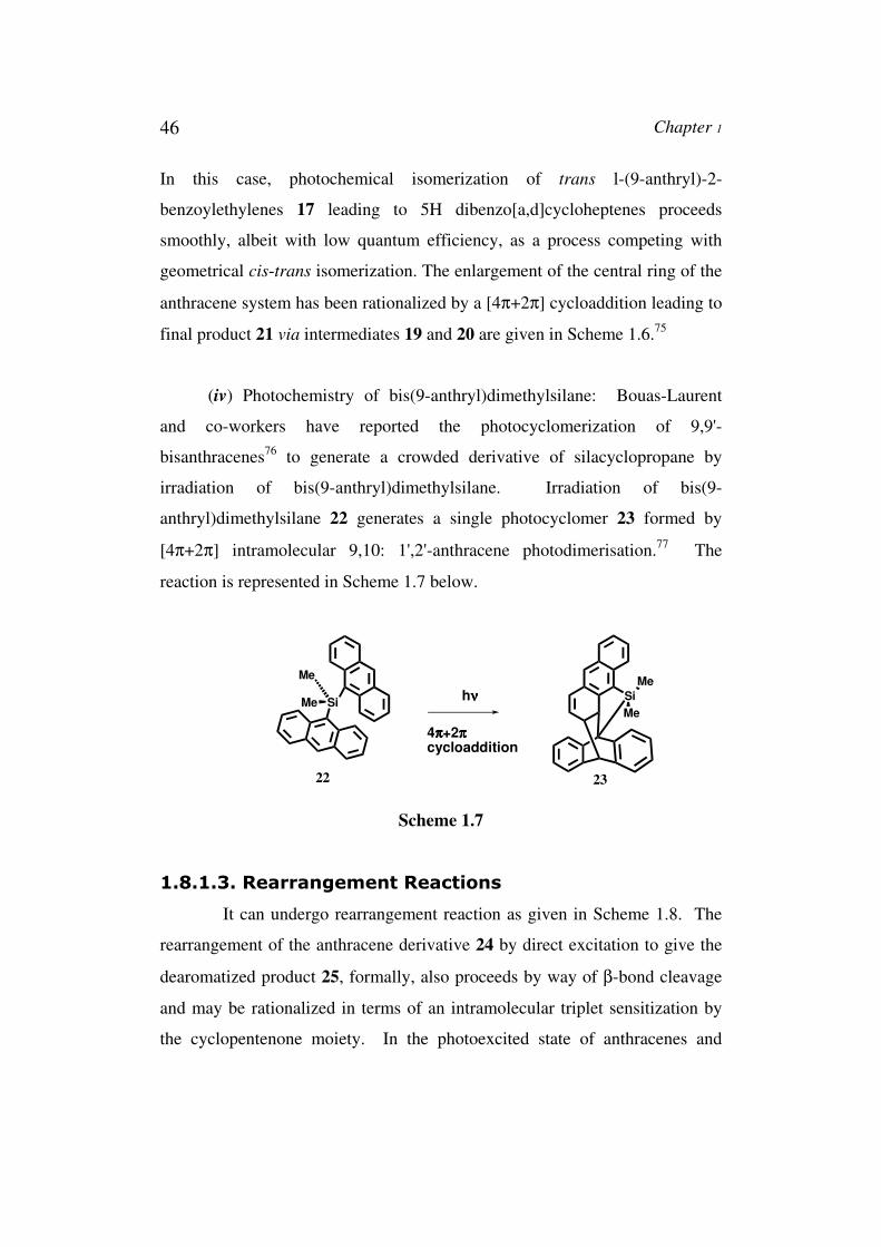
In this case, photochemical isomerization of trans l-(9-anthryl)-2-
benzoylethylenes 17 leading to 5H dibenzo[a,d]cycloheptenes proceeds
smoothly, albeit with low quantum efficiency, as a process competing with
geometrical cis-trans isomerization. The enlargement of the central ring of the
anthracene system has been rationalized by a [4π+2π] cycloaddition leading to
final product 21 via intermediates 19 and 20 are given in Scheme 1.6.75
(iv) Photochemistry of bis(9-anthryl)dimethylsilane: Bouas-Laurent
and co-workers have reported the photocyclomerization of 9,9'-
bisanthracenes76 to generate a crowded derivative of silacyclopropane by
irradiation of bis(9-anthryl)dimethylsilane. Irradiation of bis(9-
anthryl)dimethylsilane 22 generates a single photocyclomer 23 formed by
[4π+2π] intramolecular 9,10: 1',2'-anthracene photodimerisation.77 The
reaction is represented in Scheme 1.7 below.
Si
Me
Mehνννν Si
Me
Me
4ππππ+2ππππcycloaddition
22 23
Scheme 1.7
1.8.1.3. Rearrangement Reactions
It can undergo rearrangement reaction as given in Scheme 1.8. The
rearrangement of the anthracene derivative 24 by direct excitation to give the
dearomatized product 25, formally, also proceeds by way of β-bond cleavage
and may be rationalized in terms of an intramolecular triplet sensitization by
the cyclopentenone moiety. In the photoexcited state of anthracenes and
Chapter 1 47
aromatic ketones, intramolecular singlet energy transfer from the anthracene
chromophore to the cyclopentenone in 24 is energetically feasible because of
the favourable electron spectral overlap of the anthracene absorption around
380 nm, and the absorption due to the cyclopentenone n π* transition.
The subsequent intersystem-crossing step is assumed to be an efficient
process, as may be deduced from the relatively low fluorescence quantum
yield of 24.78
Scheme 1.8
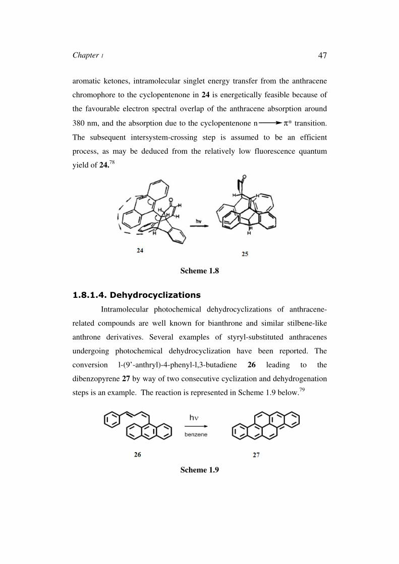
1.8.1.4. Dehydrocyclizations
Intramolecular photochemical dehydrocyclizations of anthracene-
related compounds are well known for bianthrone and similar stilbene-like
anthrone derivatives. Several examples of styryl-substituted anthracenes
undergoing photochemical dehydrocyclization have been reported. The
conversion l-(9’-anthryl)-4-phenyl-l,3-butadiene 26 leading to the
dibenzopyrene 27 by way of two consecutive cyclization and dehydrogenation
steps is an example. The reaction is represented in Scheme 1.9 below.79
Scheme 1.9
48 Chapter 1
1.9. Applications of above Systems
A-B substituted systems also have a number of potential applications.
These bischromophoric arrays serve as prototypical light-harvesting devices,
molecular photonic wires and optoelectronic gates by combining these
systems with various other systems. Concentrated assemblies of chromophore
centres in macromolecular superstructures offer ideal media for the absorption
of light - a principle that applies to both solar and laser radiation. The
development of molecular photonic devices requires the creation of molecular
arrays that absorb light of specific wavelengths and undergo excited-state
energy and/or charge transfer reactions.
Also the excitation of the above systems is usually associated with an
increase in dipole moment. The properties of such systems depend on solvent
polarity, and these can be exploited to determine the polarity of unknown
solvent mixtures. The emission characteristics of the CT fluorescence bands
are sensitive to the environment. Parameters such as fluorescence emission
maximum, intensity, lifetime, polarization, and excitation spectrum can act as
potential indicators of the features of probe surroundings. Molecules
exhibiting ICT fluorescence have been used as fluorescent probes for probing
the polarity of various micro heterogeneous systems and also as sensors.
In short, the substrates reviewed in this thesis serve to illustrate the
difficulties facing a photochemist who attempts to predict the excited-state
properties of multifunctional molecules using only a background of
monochromophoric chemistry. Conversely, the diversity of photophysical and
photochemical phenomena evident in even the small number of molecules
discussed confirms that multichromophoric compounds will provide a fruitful
area of investigation for photochemists, spectroscopists, and theoreticians for
some time to come.
Chapter 1 49
1.10. Objectives of the Present Work
The primary goal of the research presented in this thesis is to
investigate the photophysical and photochemical properties of the
bischromophoric arrays in relation to their structure and to determine a
systematic change in intermolecular orientation or conformation of these
influences their properties. Furthermore, it is aimed, based on the knowledge
gathered by studying these arrays, at the design, synthesis and characterization
of new multichromophoric structures, with anticipated properties and /or
combination of properties which might be useful in future functional materials
or molecular devices.
For this purpose several structurally and geometrically different
arrays have been synthesized, their conformational changes have been studied
in detail both experimentally and theoretically. The spatial arrangement of
chromophores is varied by changes in the connecting linkers, thus varying e.g.
the angle and/or distance between the chromophoric units. A systematic
change in the chromophoric unit, furthermore led to series of compounds from
which structure-properties relationships were derived.
Based on these considerations, we propose to develop efficient
synthetic procedures for the preparations of suitable bischromophoric arrays in
which the following constraints are built in: i) the two chromophores are
separated through a suitable linker that shuts electronic communication
between the two, ii) the procedure should provide the flexibility to tune the
geometry of resultant arrays and iii) the procedure should be scaleable.
The chromophores used in these studies consist of aromatic system of
varying sizes, which makes them, in principle, suitable for nonlinear optical
measurements.
50 Chapter 1
1.11. References
1. Lehn, J. M. Angew. Chem. Int. Ed. Engl. 1988, 27, 89.
2. Lehn, J. M. Supramolecular Chemistry: Concepts and Perspectives,
VCH Verlag, Weinheim, 1995.
3. Nicolaou, K. C.; Sorensen, E. J. Classics in Total Synthesis: Targets,
Strategies, Methods, VCH, Verlag, Weinheim, 1995.
4. McGimpsey, W. G.; Samaniego, W. N.; Chen, L.; Wang, Z. J. Phys.
Chem. A. 1998, 102, 8679.
5. (a) Hisda, K.; Tsuchida, A.; Ito, S.; Yamamoto, M. J. Phys. Chem. A.
1998, 102, 2640.
6. Levine, B. F.; Bethea, C. G. J. Chem. Phys. 1975, 63, 2666.
7. Singer, K. D.; Garito, A. F. J. Chem. Phys. 1981, 75, 3572.
8. Meredith, G. R. Rev. Sci. Instrum. 1982, 53, 48.
9. Clays, K.; Persoons, A. Phys. Rev. Lett. 1991, 66, 2980.
10. Clays, K.; Persoons, A.; Maeyer, L. De. Adv. Chem. Phys, 1994,
85(II), 45.
11. Clays, K.; Persoons, A. Rev. Sci. Instrum. 1992, 63, 3285.
12. de Haas, M. P.; Warman, J. M. Chem. Phys. 1982, 73, 35.
13. Warman, J. M.; de Hass, M. P. Pulse Radiolysis, Y Tabata Ed, CRC
Press, Boca Raton, 1991, ch.6.
14. (a) Closs, G. L.; Miller, J. R. Science. 1988, 240, 440. (b) Marcus, R.
A. Angew. Chem. Int. Ed. Engl. 1993, 32, 1111. (c) Marcus, R. A.;
Sutin, N. Biochim. Biophys. Acta. 1985, 811, 265.
15. Weller, A. Z. Phys. Chem. Neue Folge. 1982, 133, 93.
16. Marcus, R. A. J. Chem. Phys. 1965, 43, 679.
17. Ying, L.; Xie, X. S. J. Phys. Chem. B. 1998, 102, 10399.
18. Garcia-Parajo, M. F.; Koopman, M.; van Dijk, E. M. H. P.;
Subramaniam, V.; van Hulst, N. F. Proc. Natl. Acad. Sci. USA 2001,
98, 14392.
Chapter 1 51
19. (a) Wu, M.; Goodwin, P. M.; Ambrose, W. P.; Keller, R. A. J. Phys.
Chem. 1996, 100, 17406. (b) Bopp, M. A.; Jia, Y.; Li, L.; Cogdell, R.
J.; Hochstrasser, R. M. Proc. Natl. Acad. Sci. USA. 1997, 94, 10630.
(c) van Oijen, A. M.; Ketelaars, M.; Kçhler, J.; Aartsma, T. J.;
Schmidt, J. Science. 1999, 285, 400.
20. (a) Bout, D. A.; Yip, W.; Hu, D.; Fu, D.; Swager, T. M.; Barbara, P.
F. Science. 1977, 277, 1074. (b) Hu, D.; Yu, J.; Barbara, P. F. J. Am.
Chem. Soc. 1999, 121, 6936. (c) Huser, T.; Yan, M.; Rotherberg, L. J.
Proc. Natl. Acad. Sci. USA. 2000, 97, 11187. (d) Muller, J. G.;
Lemmer, U.; Raschke, D.; Anni, A.; Scherf, U.; Lupton, J. M.;
Feldmann, J. Phys. Rev. Lett. 2003, 91, 267.
21. (a) Kasha, M.; Rawls, H. R.; Ashaf El-Bayoumi, M. Pure Appl.
Chem. 1965, 11, 371.
22. Forster, T. in Modern Quantum Chemistry (Ed.: O. Sinanoglu),
Academic Press, New York, 1965.
23. Merrigan, J. A. Prospects for Solar Energy Conversion by
Photovoltaics, MIT Press, Cambridge, 1975.
24. Lehninger, A. L.; Nelson, D. L.; Cox, M. M. Principles of
Biochemistry, Worth Publishers Inc., New York, 1993.
25. X. Hu, X.; Damjanovic, A.; Ritz, T.; Schulten, K. Proc. Natl. Acad.
Sci. U.S.A. 1998, 95, 5935.
26. McDermott, G.; Prince, S. M.; Freer, A. A.; Hawthornthwaite-
Lawless, A. M.; Papiz, M. Z.; Cogdell, R. J.; Isaacs, N. W. Nature.
1995, 374, 517.
27. (a) Huber, R. Angew. Chem. Int. Ed. 1989, 28, 848. (b) Moser, C. C.;
Keske, J. M.; Warncke, K.; Farid, R. S.; Dutton, P. L. Nature. 1992,
355, 796.
28. (a) Jortner, J. J. Chem. Phys. 1976, 64, 4860. (b) Jortner, J. J. Am.
Chem. Soc. 1980, 102, 6676. (c) Franzen, S.; Goldstein, R. F.; Boxer,
52 Chapter 1
S. G. J. Phys. Chem. 1993, 97, 3040. (d) Page, C. C.; Moser, C. C.;
Chen, X.; Dutton, P. L. Nature. 1999, 402, 47.
29. (a) Wasielewski, M. R. Chem. Rev. 1992, 92, 435. (b) Gust, D.;
Moore, T. A.; Moore, A. L. Acc. Chem. Res. 1993, 26, 198. (c) Gray,
H. B.; Winkler, J. R. Ann. Rev. Biochem. 1996, 65, 537. (d) Gust, D.;
Moore, T. A.; Moore, A. L. Acc. Chem. Res. 2001, 34, 40.
30. (a) Holten, D.; Bocian, D. F.; Lindsey, J. S. Acc. Chem. Res. 2002,
35, 57. (b) Lindsey, J. S.; Wagner, R. W. J. Org. Chem. 1989, 54,
828. (c) Wagner, R.W.; Johnson, T.E.; Lindsey, J. S. J. Am. Chem.
Soc. 1996, 118, 11166.
31. Ambroise, A.; Li, J.; Yu, L.; Lindsey, J. S. Org. Lett. 2000, 2, 2563.
32. (a) Milgrom, L. R. J. Chem. Soc., Perkin Trans. 1. 1983, 2535. (b)
Davila, J.; Harriman, A.; Milgrom, L. R. Chem. Phys. Lett. 1987,
136, 427. (c) Wennerstrom, 0.; Ericsson, H.; Raston, I.; Svensson, S.;
Pimlott, W. Tetrahedron Lett. 1989, 30, 1129. (d) Nagata, T.; Osuka,
A.; Maruyama, K. J. Am. Chem. SOC. 1990, 112, 3054. (e)
Anderson, S.; Anderson, H. L.; Sanders, J. K. M. Angew. Chem. Inr.
Ed. Engl. 1992, 31, 907. (f) Dubowchik, G. M.; Hamilton, A. D. J.
Chem. Soc. Chem. Commun. 1987, 293.
33. Prathapan, S.; Johnson, T. E.; Lindsey, J. S. J. Am. Chem. Soc. 1993,
115, 7519.
34. Van der Boom, T; Hayes, R. T.; Zhao, Y.; Bushard, P. J.; Weiss, E.
A.; Wasielewski, M. R. J. Am. Chem. Soc. 2002, 124, 9582.
35. (a) Williams, R. M.; Zwier, J. M.; Verhoeven, J. W. J. Am. Chem.
Soc. 1995, 117, 4093. (b) Guldi, D. M. Chem. Soc. Rev. 2002, 31, 22.
(c) Fukuzumi, S. Org. Biomol. Chem. 2003, 1, 609.
36. (a) Wasielewski, M. R.; Niemczyk, M. P. J. Am. Chem. Soc. 1984,
106, 5043, (b) Paddon-Row, M. N. Acc. Chem. Res. 1994, 27, 18.
Chapter 1 53
37. (a) Guldi, D. M. Chem. Soc. Rev. 2002, 31, 22. (b) Imahori, H.; Mori,
Y.; Matano, J. Photochem. Photobiol. C. 2003, 4, 51.
38. (a) Kuciauskas, D.; Liddell, P. A.; Lin, S.; Johnson, T. E.; Weghorn,
S. J.; Lindsey, J. S.; Moore, A. L.; Moore, T. A.; Gust, D. J. Am.
Chem. Soc. 1999, 121, 8604. (b) Andersson, M.; Sinks, L. E.; Hayes,
R. T.; Zhao, Y.; Wasielewski, M. R. Angew. Chem. Int. Ed. 2003, 42,
3139.
39. (a) Chambron, J.-C.; Chardon-Noblat, S.; Harriman, A.; Heitz, V.;
Sauvage, J.-P. Pure Appl. Chem. 1993, 65, 2343. (b) Harriman, A.;
Sauvage, J.-P. Chem. Soc. ReV. 1996, 26, 41. (c) Blanco, M.-J.;
Jimenez, M. C.; Chambron, J.-C.; Heitz, V.; Linke, M.; Sauvage, J.-
P. Chem. Soc. Rev. 1999, 28, 293. (d) Kurreck, H.; Huber, M. Angew.
Chem. Int. Ed. Engl. 1995, 34, 849. (e) Balzani, V.; Juris, A.; Venturi,
M.; Campagna, S.; Serroni, S. Chem. Rev. 1996, 96, 759. (f)
Amabilino, D. B.; Stoddart, J. F. Chem. Rev. 1995, 95, 2725.
40. (a) Boyd, P. D. W.; Hodgson, M. C.; Rickard, C. E. F.; Oliver, A. G.;
Chaker, L.; Brothers, P. J.; Bolskar, R. D.; Tham, F. S.; Reed, C. A.
J. Am. Chem. Soc. 1999, 121, 10487. (b) Wang, Y.-B.; Lin, Z. J. Am.
Chem. Soc. 2003, 125, 6072. (c) Shirakawa, M.; Fujita, N.; Shinkai,
S. J. Am. Chem. Soc. 2003, 125, 9902. (d) Shoji, Y.; Tashiro, K.;
Aida, T. J. Am. Chem. Soc. 2004, 126, 6570. (e) Guldi, D. M.;
Gouloumis, A.; Vazquez, P.; Torres, T.; Georgakilas, V.; Prato, M. J.
Am. Chem. Soc. 2005, 127, 5811.
41. (a) Yamaguchi, T.; Ishii, N.; Tashiro, K.; Aida, T. J. Am. Chem. Soc.
2003, 125, 13934. (b) D'Souza, F.; Smith, P. M.; Zandler, M. E.;
McCarty, A. L.; Itou, M.; Araki, Y.; Ito, O. J. Am. Chem. Soc. 2004,
126, 7898. (c) Hasobe, T.; Kamat, P. V.; Troiani, V.; Solladie, N.;
Ahn, T. K.; Kim, S. K.; Kim, D.; Kongkanand, A.; Kuwabata, S.;
Fukuzumi, S. J. Phys. Chem. B. 2005, 109, 19.
54 Chapter 1
42. Zhang, X.; Xiao, Y.; Qian, X. Org. Lett. 2008, 10, 29.
43. Coskun, A.; Akkaya, E. U. J. Am. Chem. Soc. 2005, 127, 10464.
44. Hargland, R. P. Handbook of Fluorescent Probes and Research
Products, 9th ed.; Molecular Probes: Carlsbad, CA, 2002, Chapter 1.
45. Ziessel, R.; Ulrich, G.; Harriman, A. New J. Chem. 2007, 31, 496.
46. Li, F.; Yang, S.; Ciringh, Y.; Seth, J.; Martin, C.; Singh, D.; Kim, D.;
Birge, R.; Bocian, D. F.; Holten, D.; Lindsey, J. S. J. Am. Chem. Soc.
1998, 120, 10001.
47. O'Neil, M. P.; Niemczyk, M. P.; Svec, W. A.; Gosztola, D.; Gaines,
G. L.; Wasielewski, M. R. Science. 1992, 257, 63.
48. (a) Gvishi, R.; Reisfeld, R.; Burshtein, Z. Chem. Phys. Lett. 1993,
213, 338. (b) Langhals, H. Heterocycles. 1995, 40, 477. (c) Würthner,
F. Chem. Commun. 2004, 1564.
49. (a) De Schryver, F. C.; Vosch, T.; Cotlet, M.; Van der Auweraer, M.;
Müllen, K.; Hofkens, J. Acc. Chem. Res. 2005, 38, 514. (b) Gronheid,
R.; Stefan, A.; Cotlet, M.; Hofkens, J.; Qu, J.; Mullen, K.; Van der
Auweraer, M.; Verhoeven, J. W.; De Schryver, F. C. Angew. Chem.
Int. Ed. Engl. 2003, 42, 4209.
50. Adronov, A.; Frechet, J. M. J. Chem. Commun. 2000, 1701.
51. Freeman, A. W.; Frechet, J. M. J.; Koene, S. C.; Thompson, M. E.
Polym. Prepr. 1999, 40, 1246.
52. Odobel, F.; Zabri, H. Inorg. Chem. 2005, 44, 5600.
53. Wenz, G. Angew. Chem. Int. Ed. Engl. 1994, 33, 803.
54. Jullien, L.; Canceill, J.; Valeur, B.; Bardez, E.; Lefevre,J.-P.; Lehn,J.-
M.; Marchi-Artzner, V.; Pansu, R. J. Am. Chem. Soc. 1996, 118,
5432.
55. Berberan-Santos, M. N.; Canceill, J.; Brochon, J. C.; Jullien, L.;
Lehn, J. M.; Pouget, J.; Tauc, P.; Valeur, B. J. Am. Chem. Soc. 1992,
114, 6427.
Chapter 1 55
56. Vincent Souchon, V.; Maisonneuve, S.; David, O.; Leray, I.; Xie, J. ;
Valeur, B. Photochem. Photobiol. Sci. 2008, 7, 1323.
57. Valeur, B.; Leray, I. Inorg. Chim. Acta. 2007, 360, 765.
58. Van Wageningen, A. M. A.; Timmerman, P.; van Duynhoven, J. P.
M.; Verboom, W.; van Veggel, F. C. J. M.; Reinhoudt, D. N. Chem.
Eur. J. 1997, 3, 639.
59. Chattopadhyay, N.; Haldar, B.; Mallick, A.; Sengupta, S. Tetrahedron
Lett. 2005, 46, 3089.
60. Brasseur, N.; Ali, H.; Langlois, R.; van Lier, J. E. Photochem.
Photobiol. 1988, 47, 705.
61. Garcia-Parajo, M. F.; Hernando, J.; Mosteiro, G. S.; Hoogenboom, J.
P.; van Dijk, E. M. H. P.; van Hulst, N. F. Chem. Phys. Chem. 2005,
6, 819.
62 . Forster, Th.; Angew. Chem. 1969, 81, 364. (b) Birks, J. B.;
Photophysics of Aromatic Molecules, Wiley-Interscience, London,
1970, chap. 7.
63. Lin, Z.; Priyadarshy, S.; Bartko, A.; Waldeck, D. H. Photochem.
Photobiol. 1997, 110, 131.
64. Hochstrasser, R. M. Can. J. Chem. 1961, 39, 459.
65. Friedel, R. A.; Orchin, M.; Reggel, L. J. Am. Chem. Soc. 1948, 70,
199.
66. Greenfield, N.; Fasman, G. D. J. Am. Chem.Soc. 1970, 92, 177.
67. Williams, D. H.; Fleming, I.; Spectroscopic Methods in Organic
Chemistry, Fourth edition, 1986, p.8.
68. (a) Fromm, R.; Ahmed, S. A.; Hartmann, T.; Huch, V.; Abdel-
Wahab, A. A.; Durr, H. Eur. J. Org. Chem. 2001, 21, 4077. (b)
Weber, C.; Rustemeyer, F.; Durr, H. Adv. Mater. 1998, 10, 1348. (c)
Andreis, C.; Durr, H.; Wintgens, V.; Valat, P.; Kossanyi, J. Chem.
Eur. J. 1997, 3, 509.
56 Chapter 1
69. (a) Ahmed, S. A.; Abdel-Wahab, A. A.; Durr, H.; CRC Handbook of
Organic Photochemistry and Photobiology (2nd edn). Horspool WM,
Lenci F (eds). CRC press: New York, 2004, Chapter 96, 1. (b) Seto,
J. In Infrared Absorbing Dyes, Matsuoka M (ed). Plenum Press: New
York, 1990, 71.
70. (a) Leonhardt, H.; Weller, A. Ber. Bunsenges. Phys. Chem. 1963, 67,
791. (b) Mataga, N.; Okada, T.; Ezumi, K. Mol. Phys. 1966, 10, 203.
(c) Beens, H.; Weller, A. Acta Phys. Pol. 1968, 34, 593. (d) Weller,
A. in The Exciplex; Gordon, M., Ware, W. R., Eds.; Academic: New
York, 1976, p 23.
71. Dyer, J. R.; Applications of Absorption Spectroscopy of Organic
Compounds, (Ninth ed), Prentice-Hall of India Private Limited, New
Delhi, 1994, p-8.
72. Suppan, P.; Chemistry and Light; The Royal Society of Chemistry,
Thomas Graham House, The Science Park, Cambridge, 1994, p-92.
73. Becker, H.-D.; Andersson, K. J. Org. Chem. 1983, 48, 4542.
74. (a) Becker, H.-D.; Hansen, L.; Andersson, K. J. Org. Chem. 1981, 46,
5419. (b) Becker, H.-D.; Sandros, K.; Andersson, K. Angew. Chem.
Int. Ed. Engl. 1983, 22, 495.
75. Becker, H.-D.; Becker, H.-C.; Sandros, K.; Andersson, K.
Tetrahedron Lett. 1985, 26, 1589.
76. Bouas-Laurent, H.; Castellan, A.; Desvergne, J-P. Pure and Appl.
Chem. 1980, 52, 2633.
77. Daney, M.; Vanucci, C.; Desvergne, J-P.; Castellan, A.; Bouas-
Laurent, H. Tetrahedron Lett. 1985, 26, 1505.
78. Becker, H.-D.; Skelton, B. W.; White, A. H. Aust. J. Chem. 1986, 38,
1471.
79. Hayward, R. J.; Leznoff, C. C. Tetrahedron. 1971, 27, 2085.