Phospho-Akt (Ser473) (D9E) XP - Cell Signaling Technologymedia.cellsignal.com/pdf/4060.pdf · #4060...
2
#4060 Store at –20°C Phospho-Akt (Ser473) (D9E) XP ® Rabbit mAb rev. 05/22/17 W, IP, IHC-P, IHC-F, IF-IC, F Endogenous H, M, R, Mk, Hm, B, Dm, Z, (Pg, C, X, Dg) 60 kDa Background: Akt, also referred to as PKB or Rac, plays a critical role in controlling survival and apoptosis (1-3). This protein kinase is activated by insulin and various growth and survival factors to function in a wortmannin-sensitive pathway involving PI3 kinase (2,3). Akt is activated by phospholipid binding and activation loop phosphorylation at Thr308 by PDK1 (4) and by phosphorylation within the carboxy terminus at Ser473. The previously elusive PDK2 responsible for phosphorylation of Akt at Ser473 has been identified as mam- malian target of rapamycin (mTor) in a rapamycin-insensitive complex with rictor and Sin1 (5,6). Akt promotes cell survival by inhibiting apoptosis by phosphorylating and inactivating several targets, including Bad (7), forkhead transcription factors (8), c-Raf (9) and caspase-9. PTEN phosphatase is a major negative regulator of the PI3 kinase/Akt signaling path- way (10). LY294002 is a specific PI3 kinase inhibitor (11). Another essential Akt function is the regulation of glycogen synthesis through phosphorylation and inactivation of GSK-3α and β (12,13). Akt may also play a role in insulin stimulation of glucose transport (12). In addition to its role in survival and glycogen synthesis, Akt is involved in cell cycle regulation by preventing GSK-3β mediated phosphorylation and degradation of cyclin D1 (14) and by negatively regulating the cyclin dependent kinase inhibitors p27 Kip (15) and p21 Waf1/CIP1 (16). Akt also plays a critical role in cell growth by directly phosphorylating mTOR in a rapamycin-sensitive complex containing raptor (17). More importantly, Akt phosphorylates and inactivates tuberin (TSC2), an inhibitor of mTOR within the mTOR-raptor complex (18). Inhibition of mTOR stops the protein synthesis machinery by inactivating p70 S6 kinase and activating the eukaryotic initiation factor 4E binding protein 1 (4E-BP1), an inhibitor of translation (18,19). Storage: Supplied in 10 mM sodium HEPES (pH 7.5), 150 mM NaCl, 100 µg/ml BSA, 50% glycerol and less than 0.02% sodium azide. Store at –20°C. Do not aliquot the antibody. *Species cross-reactivity is determined by western blot. **Anti-rabbit secondary antibodies must be used to detect this antibody. Recommended Antibody Dilutions: Western blotting 1:2000 Immunoprecipitation 1:50 Immunohistochemistry (Paraffin) 1:100† Unmasking buffer: Citrate Antibody diluent: SignalStain ® Antibody Diluent #8112 Detection reagent: SignalStain ® Boost (HRP, Rabbit) #8114 †Optimal IHC dilutions determined using SignalStain ® Boost IHC Detection Reagent. Immunohistochemistry (Frozen) 1:100† Fixative: 3% Formaldehyde/MeOH Detection reagent: SignalStain ® Boost (HRP, Rabbit) #8114 †Optimal IHC dilutions determined using SignalStain ® Boost IHC Detection Reagent. Immunofluorescence (IF-IC) 1:400 Flow Cytometry 1:100 For application specific protocols please see the web page for this product at www.cellsignal.com. Please visit www.cellsignal.com for a complete listing of recommended companion products. Orders n 877-616-CELL (2355) [email protected] Support n 877-678-TECH (8324) [email protected] Web n www.cellsignal.com Applications Species Cross-Reactivity* Molecular Wt. Specificity/Sensitivity: Phospho-Akt (Ser473) (D9E) XP ® Rabbit mAb detects endogenous levels of Akt only when phosphorylated at Ser473. Source/Purification: Monoclonal antibody is produced by immunizing animals with a synthetic phosphopeptide corresponding to residues around Ser473 of human Akt. IMPORTANT: For western blots, incubate membrane with diluted antibody in 5% w/v BSA, 1X TBS, 0.1% Tween-20 at 4°C with gentle shaking, overnight. Rabbit IgG** Isotype kDa Phospho- Akt (Ser473) Akt wortmannin LY294002 PDGF PC-3 100 80 60 50 60 80 50 NIH/3T3 – – – + – – – + – + – – Western blot analysis of extracts from PC-3 cells, untreated or LY294002/wortmannin-treated, and NIH/3T3 cells, serum- starved or PDGF-treated, using Phospho-Akt (Ser473) (D9E) XP ® Rabbit mAb (upper) or Akt (pan) (C67E7) Rabbit mAb #4691 (lower). Events Phospho-Akt (Ser473) Flow cytometric analysis of Jurkat cells, untreated (green) or treated with LY294002, wortmannin and U0126 (blue), using Phospho-Akt (Ser473) (D9E) XP ® Rabbit mAb compared to a nonspecific negative control antibody (red). Entrez-Gene ID #207 Swiss-Prot Acc. #P31749 Immunohistochemical analysis of paraffin-embedded human lung carcinoma using Phospho-Akt (Ser473) (D9E) XP ® Rabbit mAb. Species Cross-Reactivity Key: H—human M—mouse R—rat Hm—hamster Mk—monkey Mi—mink C—chicken Dm—D. melanogaster X—Xenopus Z—zebrafish B—bovine Dg—dog Pg—pig Sc—S. cerevisiae Ce—C. elegans Hr—Horse All—all species expected Species enclosed in parentheses are predicted to react based on 100% homology. Applications Key: W—Western IP—Immunoprecipitation IHC—Immunohistochemistry ChIP—Chromatin Immunoprecipitation IF—Immunofluorescence F—Flow cytometry E-P—ELISA-Peptide Alexa Fluor ® is a registered trademark of Molecular Probes, Inc. DRAQ5 ® is a registered trademark of Biostatus Limited. © 2012 Cell Signaling Technology, Inc. XP ® and Cell Signaling Technology ® are trademarks of Cell Signaling Technology, Inc. For Research Use Only. Not For Use In Diagnostic Procedures.
Transcript of Phospho-Akt (Ser473) (D9E) XP - Cell Signaling Technologymedia.cellsignal.com/pdf/4060.pdf · #4060...

#406
0St
ore
at –
20°C
Phospho-Akt (Ser473) (D9E) XP® Rabbit mAb
rev. 05/22/17
W, IP, IHC-P, IHC-F, IF-IC, FEndogenous
H, M, R, Mk, Hm, B, Dm, Z, (Pg, C, X, Dg)
60 kDa
Background: Akt, also referred to as PKB or Rac, plays a critical role in controlling survival and apoptosis (1-3). This protein kinase is activated by insulin and various growth and survival factors to function in a wortmannin-sensitive pathway involving PI3 kinase (2,3). Akt is activated by phospholipid binding and activation loop phosphorylation at Thr308 by PDK1 (4) and by phosphorylation within the carboxy terminus at Ser473. The previously elusive PDK2 responsible for phosphorylation of Akt at Ser473 has been identified as mam-malian target of rapamycin (mTor) in a rapamycin-insensitive complex with rictor and Sin1 (5,6). Akt promotes cell survival by inhibiting apoptosis by phosphorylating and inactivating several targets, including Bad (7), forkhead transcription factors (8), c-Raf (9) and caspase-9. PTEN phosphatase is a major negative regulator of the PI3 kinase/Akt signaling path-way (10). LY294002 is a specific PI3 kinase inhibitor (11).
Another essential Akt function is the regulation of glycogen synthesis through phosphorylation and inactivation of GSK-3α and β (12,13). Akt may also play a role in insulin stimulation of glucose transport (12).
In addition to its role in survival and glycogen synthesis, Akt is involved in cell cycle regulation by preventing GSK-3β mediated phosphorylation and degradation of cyclin D1 (14) and by negatively regulating the cyclin dependent kinase inhibitors p27 Kip (15) and p21 Waf1/CIP1 (16). Akt also plays a critical role in cell growth by directly phosphorylating mTOR in a rapamycin-sensitive complex containing raptor (17). More importantly, Akt phosphorylates and inactivates tuberin (TSC2), an inhibitor of mTOR within the mTOR-raptor complex (18). Inhibition of mTOR stops the protein synthesis machinery by inactivating p70 S6 kinase and activating the eukaryotic initiation factor 4E binding protein 1 (4E-BP1), an inhibitor of translation (18,19).
Storage: Supplied in 10 mM sodium HEPES (pH 7.5), 150 mM NaCl, 100 µg/ml BSA, 50% glycerol and less than 0.02% sodium azide. Store at –20°C. Do not aliquot the antibody.
*Species cross-reactivity is determined by western blot.
** Anti-rabbit secondary antibodies must be used to detect this antibody.
Recommended Antibody Dilutions: Western blotting 1:2000 Immunoprecipitation 1:50 Immunohistochemistry (Paraffin) 1:100† Unmasking buffer: Citrate Antibody diluent: SignalStain® Antibody Diluent #8112 Detection reagent: SignalStain® Boost (HRP, Rabbit) #8114 †Optimal IHC dilutions determined using SignalStain® Boost IHC Detection Reagent. Immunohistochemistry (Frozen) 1:100† Fixative: 3% Formaldehyde/MeOH Detection reagent: SignalStain® Boost (HRP, Rabbit) #8114 †Optimal IHC dilutions determined using SignalStain® Boost IHC Detection Reagent. Immunofluorescence (IF-IC) 1:400 Flow Cytometry 1:100
For application specific protocols please see the web page for this product at www.cellsignal.com.
Please visit www.cellsignal.com for a complete listing of recommended companion products.
Orders n 877-616-CELL (2355)[email protected]
Support n 877-678-TECH (8324)[email protected]
Web n www.cellsignal.com
Applications Species Cross-Reactivity* Molecular Wt.
Specificity/Sensitivity: Phospho-Akt (Ser473) (D9E) XP® Rabbit mAb detects endogenous levels of Akt only when phosphorylated at Ser473.
Source/Purification: Monoclonal antibody is produced by immunizing animals with a synthetic phosphopeptide corresponding to residues around Ser473 of human Akt.
IMPORTANT: For western blots, incubate membrane with diluted antibody in 5% w/v BSA, 1X TBS, 0.1% Tween-20 at 4°C with gentle shaking, overnight.
Rabbit IgG**
Isotype
kDa
Phospho-Akt (Ser473)
Akt
wortmanninLY294002PDGF
PC-3100
80
60
50
60
80
50
NIH/3T3
– –– +– –– +– +– –
Western blot analysis of extracts from PC-3 cells, untreated or LY294002/wortmannin-treated, and NIH/3T3 cells, serum-starved or PDGF-treated, using Phospho-Akt (Ser473) (D9E) XP® Rabbit mAb (upper) or Akt (pan) (C67E7) Rabbit mAb #4691 (lower).
Even
ts
Phospho-Akt (Ser473)
Flow cytometric analysis of Jurkat cells, untreated (green) or treated with LY294002, wortmannin and U0126 (blue), using Phospho-Akt (Ser473) (D9E) XP® Rabbit mAb compared to a nonspecific negative control antibody (red).
Entrez-Gene ID #207 Swiss-Prot Acc. #P31749
Immunohistochemical analysis of paraffin-embedded human lung carcinoma using Phospho-Akt (Ser473) (D9E) XP® Rabbit mAb.
Species Cross-Reactivity Key: H—human M—mouse R—rat Hm—hamster Mk—monkey Mi—mink C—chicken Dm—D. melanogaster X—Xenopus Z—zebrafish B—bovine
Dg—dog Pg—pig Sc—S. cerevisiae Ce—C. elegans Hr—Horse All—all species expected Species enclosed in parentheses are predicted to react based on 100% homology.
Applications Key: W—Western IP—Immunoprecipitation IHC—Immunohistochemistry ChIP—Chromatin Immunoprecipitation IF—Immunofluorescence F—Flow cytometry E-P—ELISA-Peptide
Alexa Fluor® is a registered trademark of Molecular Probes, Inc.
DRAQ5® is a registered trademark of Biostatus Limited.
© 2
012
Cell
Sign
alin
g Te
chno
logy
, Inc
.XP
® a
nd C
ell S
igna
ling
Tech
nolo
gy® a
re tr
adem
arks
of C
ell S
igna
ling
Tech
nolo
gy, I
nc.
For Research Use Only. Not For Use In Diagnostic Procedures.
Orders n 877-616-CELL (2355) [email protected] Support n 877-678-TECH (8324) [email protected] Web n www.cellsignal.com© 2
012
Cell
Sign
alin
g Te
chno
logy
, Inc
.Background References: (1) Franke, T.F. et al. (1997) Cell 88, 435–7.
(2) Burgering, B.M. and Coffer, P.J. (1995) Nature 376, 599–602.
(3) Franke, T.F. et al. (1995) Cell 81, 727–36.
(4) Alessi, D.R. et al. (1996) EMBO J 15, 6541–51.
(5) Sarbassov, D.D. et al. (2005) Science 307, 1098–101.
(6) Jacinto, E. et al. (2006) Cell 127, 125–37.
(7) Cardone, M.H. et al. (1998) Science 282, 1318–21.
(8) Brunet, A. et al. (1999) Cell 96, 857–68.
(9) Zimmermann, S. and Moelling, K. (1999) Science 286, 1741–4.
(10) Cantley, L.C. and Neel, B.G. (1999) Proc Natl Acad Sci USA 96, 4240–5.
(11) Vlahos, C.J. et al. (1994) J Biol Chem 269, 5241–8.
(12) Hajduch, E. et al. (2001) FEBS Lett 492, 199–203.
(13) Cross, D.A. et al. (1995) Nature 378, 785–9.
(14) Diehl, J.A. et al. (1998) Genes Dev 12, 3499–511.
(15) Gesbert, F. et al. (2000) J Biol Chem 275, 39223–30.
(16) Zhou, B.P. et al. (2001) Nat Cell Biol 3, 245-52.
(17) Navé, B.T. et al. (1999) Biochem J 344 Pt 2, 427-31.
(18) Inoki, K. et al. (2002) Nat Cell Biol 4, 648–57.
(19) Manning, B.D. et al. (2002) Mol Cell 10, 151–62.
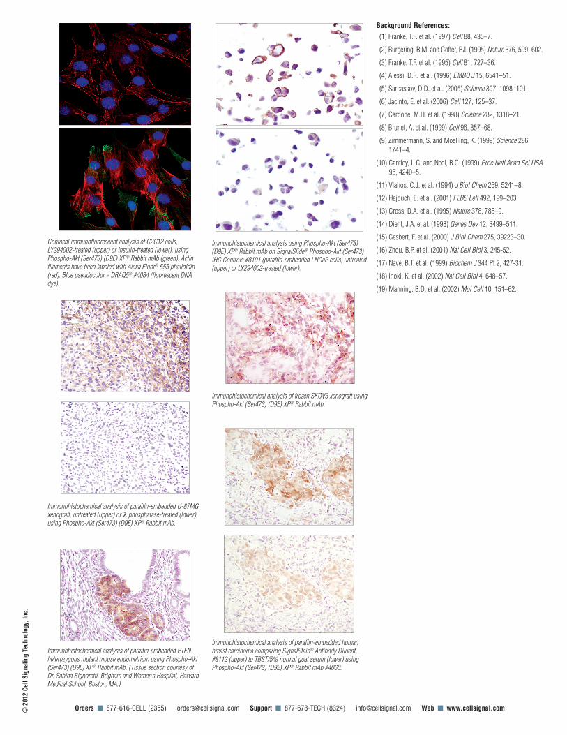
Confocal immunofluorescent analysis of C2C12 cells, LY294002-treated (upper) or insulin-treated (lower), using Phospho-Akt (Ser473) (D9E) XP® Rabbit mAb (green). Actin filaments have been labeled with Alexa Fluor® 555 phalloidin (red). Blue pseudocolor = DRAQ5® #4084 (fluorescent DNA dye).
Immunohistochemical analysis of paraffin-embedded U-87MG xenograft, untreated (upper) or l phosphatase-treated (lower), using Phospho-Akt (Ser473) (D9E) XP® Rabbit mAb.
Immunohistochemical analysis using Phospho-Akt (Ser473) (D9E) XP® Rabbit mAb on SignalSlide® Phospho-Akt (Ser473) IHC Controls #8101 (paraffin-embedded LNCaP cells, untreated (upper) or LY294002-treated (lower).
Immunohistochemical analysis of paraffin-embedded human breast carcinoma comparing SignalStain® Antibody Diluent #8112 (upper) to TBST/5% normal goat serum (lower) using Phospho-Akt (Ser473) (D9E) XP® Rabbit mAb #4060.
Immunohistochemical analysis of frozen SKOV3 xenograft using Phospho-Akt (Ser473) (D9E) XP® Rabbit mAb.
Immunohistochemical analysis of paraffin-embedded PTEN heterozygous mutant mouse endometrium using Phospho-Akt (Ser473) (D9E) XP® Rabbit mAb. (Tissue section courtesy of Dr. Sabina Signoretti, Brigham and Women’s Hospital, Harvard Medical School, Boston, MA.)