PhD Thesis · Marklund, Thomas Glott, Arve Opheim, Jesus Benito, Narda Murillo, Janneke Nachtegaal,...
136
UNIVERSITY OF COPENHAGEN FACULTY OF HEALTH AND MEDICAL SCIENCES Robotic exoskeleton training in rehabilitation after Spinal Cord Injury Exploratory studies on safety, feasibility, gait function, secondary complications following SCI and cardiometabolic changes during walking PhD Thesis Carsten Bach Baunsgaard
Transcript of PhD Thesis · Marklund, Thomas Glott, Arve Opheim, Jesus Benito, Narda Murillo, Janneke Nachtegaal,...

U N I V E R S I T Y O F C O P E N H A G E NFA C U LT Y O F H E A LT H A N D M E D I C A L S C I E N C E S
Robotic exoskeleton training in rehabilitation after Spinal Cord InjuryExploratory studies on safety, feasibility, gait
function, secondary complications following SCI
and cardiometabolic changes during walking
PhD ThesisCarsten Bach Baunsgaard

Academic supervisors
Professor Fin Biering-Sørensen, dr. med.
Clinic for Spinal Cord Injuries,
Rigshospitalet, University of Copenhagen, Denmark
Professor Mark S. Nash, PhD
The Miami Project to Cure Paralysis,
Department of Neurological Surgery and Physical Medicine & Rehabilitation,
University of Miami Miller School of Medicine, Miami, FL, USA
Assessment committee
Chairman: Professor John Vissing, dr. med.
Department of Clinical Medicine
University of Copenhagen, Denmark
Professor Jørgen Feldbæk Nielsen, dr. med.
Department of Clinical Medicine, University of Aarhus
Regionshospital Hammel Neurocenter, Denmark
Professor Jörgen Borg, MD
Department of Clinical Sciences, Karolinska Institute
Danderyds Hospital, Stockholm, Sweden
This thesis was submitted to the Graduate School of Health and Medical Sciences,
University of Copenhagen, on 4 October 2018 with public defence 27 February 2019

2
Table of contents
PREFACE ............................................................................................................................ 7
ACKNOWLEDGEMENTS ................................................................................................... 8
PAPERS INCLUDED IN THE THESIS ................................................................................ 9
Paper I ............................................................................................................................................................................... 9
Paper II .............................................................................................................................................................................. 9
Paper III ............................................................................................................................................................................ 9
Paper IV............................................................................................................................................................................. 9
ENGLISH SUMMARY ....................................................................................................... 10
DANSK RESUMÉ .............................................................................................................. 12
LIST OF ABBREVIATIONS .............................................................................................. 14
INTRODUCTION ............................................................................................................... 16
BACKGROUND ................................................................................................................ 17
Demographics, Incidence and Prevalence of Spinal Cord Injury .............................................................................. 17
Secondary Health Conditions and Exoskeleton Gait Training ................................................................................... 17
Gait Function and Prognoses Following Spinal Cord Injury ..................................................................................... 18
Physical activity and health in Spinal Cord Injury ..................................................................................................... 19
Energy expenditure in exoskeleton walking ................................................................................................................. 20
Cardiovascular drift effect ............................................................................................................................................. 21
Priorities and stakeholder’s priorities .......................................................................................................................... 21
Description of the exoskeleton from Ekso Bionics ....................................................................................................... 23
Exoskeletons and need for more research .................................................................................................................... 24
THE AIMS OF THIS THESIS ............................................................................................. 25

3
METHODS AND STUDY DESIGN .................................................................................... 27
General description ........................................................................................................................................................ 27
METHODS STUDY I-II ....................................................................................................... 28
Study Design .................................................................................................................................................................... 28
Participants ..................................................................................................................................................................... 28
Training Protocol ............................................................................................................................................................ 29
Assessments ..................................................................................................................................................................... 29
Safety, Skin Integrity and Feasibility of the Training Protocol .................................................................................. 30
Training characteristics ................................................................................................................................................. 30
Rating of Perceived Exertion, Borg Scale ..................................................................................................................... 30
Heart rate and Blood Pressure ...................................................................................................................................... 31
Neurological examination............................................................................................................................................... 31
Tests for gait function ..................................................................................................................................................... 31
Pain .................................................................................................................................................................................. 31
Spasticity.......................................................................................................................................................................... 32
Range of Motion .............................................................................................................................................................. 32
Spinal Cord Independence Measure III ....................................................................................................................... 32
Bowel, Lower Urinary Tract Function, and Quality of Life ....................................................................................... 32
METHODS STUDY III-IV ................................................................................................... 33
Study Design .................................................................................................................................................................... 33
Participants ..................................................................................................................................................................... 33
Study protocol, device description and habituation period ........................................................................................ 33
Expired respiratory gases VO2 and VCO2 .................................................................................................................... 34
Cardio-dynamic parameters .......................................................................................................................................... 34

4
Rating of Perceived Exertion ......................................................................................................................................... 34
Heart Rate Variability .................................................................................................................................................... 34
Blood Pressure ................................................................................................................................................................ 34
Head-Up-Tilt ................................................................................................................................................................... 35
Oral Glucose Tolerance Test ......................................................................................................................................... 35
Test days .......................................................................................................................................................................... 35
Peak exercise capacity test ........................................................................................................................................... 35
Test day 1: sitting ......................................................................................................................................................... 35
Test day 2: standing ..................................................................................................................................................... 35
Test day 3: walking ...................................................................................................................................................... 36
Test day 4: fasting ........................................................................................................................................................ 36
Data analysis ................................................................................................................................................................... 36
Total energy expenditure and whole-body carbohydrate and lipid oxidation .............................................................. 36
Walking economy analysis .......................................................................................................................................... 36
Statistical methods for paper I, II, III and IV .............................................................................................................. 36
RESULTS .......................................................................................................................... 38
RESULTS STUDY I-II ........................................................................................................ 38
Safety, Adherence and Feasibility of the Protocol ....................................................................................................... 38
Change in training characteristics over time ............................................................................................................... 40
Physical Exertion During Walking................................................................................................................................ 40
Gait function outside of the exoskeleton ....................................................................................................................... 41
Pain .................................................................................................................................................................................. 45
Spasticity.......................................................................................................................................................................... 46
Range of motion .............................................................................................................................................................. 47
Spinal Cord Independence Measure ............................................................................................................................. 47
Bowel function ................................................................................................................................................................. 48
Lower urinary tract function ......................................................................................................................................... 49
5
Quality of Life ................................................................................................................................................................. 49
RESULTS PAPER III-IV .................................................................................................... 51
Habituation period.......................................................................................................................................................... 52
Oxygen consumption at seated, standing and walking ................................................................................................ 52
Total energy expenditure while seated, standing and walking ................................................................................... 52
Walking economy............................................................................................................................................................ 55
Rate of Perceived Exertion............................................................................................................................................. 55
Cardiodynamic parameters ........................................................................................................................................... 55
Difference between timepoints within session .............................................................................................................. 58
Main effect of group and of condition (sitting, standing, walking) ............................................................................. 58
Changes during HUT and between test days (Figure 11) ............................................................................................ 58
DISCUSSION .................................................................................................................... 63
Safety and feasibility ....................................................................................................................................................... 64
Changes in gait function without the exoskeleton and lower extremity motor score ............................................... 65
Pain .................................................................................................................................................................................. 66
Spasticity.......................................................................................................................................................................... 66
Spinal Cord Independence Measure, bowel- and bladder function ........................................................................... 67
Quality of Life (QoL) ...................................................................................................................................................... 68
Physical activity and energy expenditure ..................................................................................................................... 69
Cardiodynamic outcomes ............................................................................................................................................... 70
Strengths and limitations of the methodology and study design ................................................................................ 72
Paper I-II ...................................................................................................................................................................... 72
Paper III-IV .................................................................................................................................................................. 72
CONCLUSION ................................................................................................................... 73

6
PERSPECTIVES AND SUGGESTIONS FOR FUTURE FOR RESEARCH ...................... 74
Other areas to explore .................................................................................................................................................... 74
Exoskeletons for use with other neurological disorders .............................................................................................. 75
Exoskeletons as mobility devices ................................................................................................................................... 75
REFERENCES .................................................................................................................. 77
APPENDICES ................................................................................................................... 86
Papers .............................................................................................................................................................................. 86

7
PREFACE
This thesis “Robotic exoskeleton training in rehabilitation after Spinal Cord Injury: exploratory
studies on safety, feasibility, gait function, secondary complications following SCI and cardio-
metabolic changes during walking” is based of two clinical studies. One was carried out in
collaboration between nine European spinal cord injury centres, Clinic for Spinal Cord Injuries,
Rigshospitalet, Denmark. I was participating in data collection as well as the Danish site was the
coordinator centre. The other study was carried out at the Miami Project to Cure Paralysis,
University of Miami, Florida, USA. The first study has been published in two papers (paper I-II)
and the other study are reported in two papers (paper III-IV) which are under submission. The
method and result sections for article I-II and article III-IV, are reported together, respectively.
The two clinical studies used an exoskeleton by the manufacturer Ekso Bionics®. The company
did not provide funding directly to this PhD but provided a grant for development of the protocol
for study one (paper I-II) and support for the study lead, thereby indirectly supporting this PhD.
The included papers are not part of any other person’s thesis. Published papers and figures have
been re-printed with permission from the publishers of Spinal Cord and Journal of
Rehabilitation Medicine.

8
ACKNOWLEDGEMENTS
I would like to acknowledge the many people that have made this PhD thesis possible. First, I would like
to express my deepest gratitude to my principal supervisor Fin Biering-Sørensen. From Fin I have learned
many skills, both professionally and personally. Fin has always reminded me of the importance of
clinically relevant research and to keep the patients in focus, which I truly inspiring. I am grateful that Fin
always have been generous and readily available with his help, guidance and support.
My colleagues at clinic for spinal cord injuries also deserves my a warmest thank and especially a warm
thank to Mette Skov Henriksen and Mats Christer Nilfyr who helped with data collection, provided their
clinical experience and was always very joyful to work with. Likewise, I would like to thank postdoc Ulla
Vig Nissen, co-author of two of the papers in this thesis. I am grateful for all our scientific discussions,
our collaboration on this and other projects and for her good company. Furthermore, I would also extend
my gratitude to all the other co-authors of the European Multicentre Study, paper I and II in the thesis.
Our good collaboration while conducting the study and writing the papers was remarkable.
I will also like to thank my supervisor Mark S. Nash for the opportunity to visit his lab for six months at
the Miami Project to Cure Paralysis, University of Miami, Florida, USA. This was a vibrant research
environment, I learned many skills while I was there, and I am truly grateful for that period as a PhD
student. I am grateful to the new friends and colleagues whom I met at the Miami Project: Jenn Maher,
Jan Gerven, Ann Palermo, Lasse Christiansen and Camilla Dalby Hansen.
A special thank goes to all those who participated in the studies. For taking their time to participate in the
training and patience so all the different tests could be performed. And, for sharing their personal
experiences of their lives, living with a spinal cord injury. From these persons I have truly learned many
important things and they have provided me with insights and perspectives, both professionally and
personally.
I would also like to extend my gratitude to Tine Alkjær and Tue Hvass Petersen for supervising two other
research projects that, however, not ended up being included in this thesis, gave me much experience and
knowledge within basic neuroscience. Likewise, would I like to thank my fellow PhD students at the PhD
office at Department of Neuroscience and Pharmacology, Faculty of Health and Medical Sciences at
University of Copenhagen for many fruitful discussions and their good company.
I would like to thank friends and family for always being there for me. My warmest thank you goes to my
sister Malene and my parents. A special appreciation goes to my good friend Lars Tang for all the support
through the whole period, for scientific discussions, new perspectives on rehabilitation and always good
humour.
And finally, my deepest gratitude goes to my beloved girlfriend Malin for always supporting me and for
filling my life with love and happiness. Thank you all.

9
PAPERS INCLUDED IN THE THESIS
Paper I
Gait training after spinal cord injury: safety, feasibility and gait function following 8 weeks
of training with the exoskeletons from Ekso Bionics. Carsten Bach Baunsgaard, Ulla Vig
Nissen, Anne Katrin Brust, Angela Frotzler, Cornelia Ribeill, Yorck-Bernhard Kalke, Natacha
León, Belén Gómez, Kersti Samuelsson, Wolfram Antepohl, Ulrika Holmström, Niklas
Marklund, Thomas Glott, Arve Opheim, Jesus Benito, Narda Murillo, Janneke Nachtegaal,
Willemijn Faber, Fin Biering-Sørensen. Spinal Cord 2018; 56: 106–116
Paper II
Exoskeleton gait training after spinal cord injury: an exploratory study on secondary
health conditions. Carsten Bach Baunsgaard, Ulla Vig Nissen, Anne Katrin Brust, Angela
Frotzler, Cornelia Ribeill, Yorck-Bernhard Kalke, Natacha León, Belén Gómez, Kersti
Samuelsson, Wolfram Antepohl, Ulrika Holmström, Niklas Marklund, Thomas Glott, Arve
Opheim, Jesus Benito, Narda Murillo, Janneke Nachtegaal, Willemijn Faber, Fin Biering-
Sørensen. Journal of Rehabilitation Medicine 2018;50(9): 806–13
Paper III
Effects of Acute Bionic Ambulation on Metabolism and Substrate Utilization in Persons
with Spinal Cord Injuries. Jennifer L Maher, Carsten Bach Baunsgaard, Jan van Gerven, Anne
Palermo, Fin Biering-Sørensen, Armando J Mendez, Robert W Irwin, Mark S Nash
Under submission
Paper IV
Cardio-dynamic and autonomic adaptations to exoskeleton walking in Persons with Spinal
Cord Injury. Carsten Bach Baunsgaard, Jennifer L Maher, Jan van Gerven, Anne Palermo, Fin
Biering-Sørensen, Armando J Mendez, Robert W Irwin, Mark S Nash.
Under submission

10
ENGLISH SUMMARY
An exoskeleton is a wearable, motorized orthosis that can facilitate ambulation for people with
limited or no walking ability, such as individuals with a spinal cord injury (SCI). Exoskeleton
gait training for rehabilitation of SCI is, in many regards, still in the early phase of development
despite being available on the market for some time. Many areas still need to be investigated
further to evaluate the potential benefits of this training modality and its place in the
rehabilitation setting for SCI.
Improvement in gait-function, on secondary health complications and cardiovascular benefits
have already been identified, but still with limited evidence. In addition, there is lack of
knowledge regarding which users are most likely to benefit from this training modality. Before
larger scale randomized controlled trials (RCTs) are to be conducted, more exploratory studies
are still needed to guide these RCTs.
The overall aim of the thesis was to investigate safety and feasibility of a training program of
exoskeleton gait training and explore areas of potential benefits on gait function, on secondary
health complications and acute adaptations of energy expenditure and cardiometabolic function
while walking with an exoskeleton.
These aims were explored in two clinical exploratory studies, using an exoskeleton from the
manufacture Ekso Bionics (’the Ekso’ and ’EksoGT’) and were reported in four articles. One
study (articles I-II) included nine European SCI centres and investigated safety, feasibility and
changes in gait function and secondary health complications including pain, spasticity, bowel
and bladder function, as well as quality of life over an eight-week gait training program with
three weekly exoskeleton gait training sessions. In total 52 participants completed the training
protocol and the study population included participants with recent as well as chronic injury,
with complete and incomplete injury and people with para- and tetraplegia.
The other study (articles III-IV) took place at the Miami Project to Cure Paralysis, University of
Miami, Florida, USA. Ten participants with SCI and ten non-disabled control persons performed
45 min of sitting, standing and walking on three separate days, as well as a peak test. Participants
with SCI learned to ambulate with the exoskeleton during a habituation period prior to the test
days. Outcome measures were energy expenditure (VO2) and cardiodynamic parameters,
measured as stroke volume, heart rate, and cardiac output.
The main findings in articles I-II were that the exoskeleton training was overall safe, but special
attention should be given to skin areas in contact with the device. It was feasible to complete the

11
training protocol which consisted of minimum 16 out of 24 training session to be completed.
Training parameters related to time spend walking and number of steps increased during the
training period. Participants with spared gait function improved on some gait-tests performed
without the exoskeleton. There was found reduced spasticity immediately after the training
session. Participants with pain seemed to tolerate the exoskeleton training well and indications of
benefits in activities of daily living.
The main findings from articles III-IV were that the energy expenditure while walking with the
exoskeleton was higher than when sitting and standing, but lower than when non-injured walked
without assistance. However, measured as percentage of peak energy expenditure, similar
percentage was found between the two groups. Energy expenditure while walking was lower
than the threshold where conditioning effects normally occur. Regarding cardiodynamic
outcomes there were indications of venous pooling while standing in the exoskeleton that was
mitigated during walking. There was no indication of cardiovascular drift while walking, which
is an increase in heart rate and fall in stroke volume at the same time. The rate of perceived
exertion showed a light to moderate intensity which was like findings in study one.
In conclusion showed the results that exoskeleton training was safe and feasible with indications
of health benefits. The results encourage further randomised, controlled studies to be performed
to compare to other training modalities.

12
DANSK RESUMÉ
Et exoskelet er en bærbart, motoriseret ortose som kan facilitere gang for personer med
begrænset gangfunktion, hvilket ofte er tilfældet for personer med rygmarvsskade. Exoskeletter
til rehabilitering efter rygmarvsskade er på mange måder fortsat i en tidlig fase i udviklingen,
selvom de har været på markedet i længere årrække. Mange områder mangler fortsat at blive
undersøgt yderligere for at kunne evaluere det potentialet af denne træningsmodalitet.
Forbedring i gangfunktion, sekundære helbredskomplikationer og kardiovaskulære fordele er
allerede blevet identificeret som områder hvor exoskelet træning kan have en positiv effekt, men
der stadig med begrænset evidens. Derudover er der begrænset viden om hvilke personer med
rygmarvsskade denne træningsmodalitet er bedst egnet til. Før der udføres større randomiserede
kontrollerede forsøg (Randomised Controlled Trial, RCT), er der stadig behov for explorative
studier til at guide sådanne RCT studier.
Det overordnede mål med denne afhandling var at undersøge sikkerheden og
gennemførbarheden af et gangtræningsprogram med et exoskelet og udforske områder med
potentielle fordele, herunder gangfunktion, sekundære helbredskomplikationer og adaptationer af
energiforbrug og kardiometabolisk funktion, mens man går med exoskelet.
Disse mål blev undersøgt i to kliniske explorative studier et exoskelet fra producenten Ekso
Bionics (’the Ekso’ og ’EksoGT’) blev anvendt og resultaterne blev rapporteret i fire artikler.
Første studie (artiklerne I-II) blev udført i et samarbejde mellem ni europæiske
rygmarvsskadecentre. Studiet undersøgte sikkerhed, gennemførbarhed, ændringer i
gangfunktion, samt sekundære helbredskomplikationer, herunder smerte, spasticitet, mave-
tarmfunktion og urinvejsfunktion, samt livskvalitet over et otte ugers gang-træningsprogram med
tre ugentlige træningssessioner i exoskelettet. I alt gennemførte 52 deltagere
træningsprotokollen. Disse deltagere havde nylig såvel som kronisk skade, bestod af personer
med komplet og inkomplet skade, samt personer med para- og tetraplegi.
Det andet studie (artiklerne III-IV) fandt sted på Miami Project for Cure Paralysis, University of
Miami, Florida, USA. Ti deltagere med rygmarvsskade og ti raske kontrolpersoner udførte tre
sessioner af 45 minutter i henholdsvis stillesiddende position, stående og under gang på tre
separate test dage, samt gennemførte en peak-test. Deltagere med rygmarvsskade lærte at gå med
exoskeletettet i en periode forud for testdagene. Endepunkterne var energiforbrug (VO2) og
kardiodynamiske mål som var slagvolumen, puls og kardielt output.

13
De vigtigste resultater fra artiklerne I-II var, at exoskeletetræningen blev vurderet sikker, men at
der bør udvises særlig opmærksomhed på hudområder der er i kontakt med enheden.
Forsøgsprotokollen, hvor der skulle deltages i mindst 16 ud af 24 træningssessioner, blev
vurderet at være gennemførbar. Træningsparametre relateret til tidsforbrug, gang og antal skridt i
exoskelettet steg i løbet af træningsperioden. Deltagere med bevaret gangfunktion forbedrede sig
desuden i nogle gangtest. Der blev fundet tegn på nedsat spasticitet umiddelbart efter træningen i
forhold til inden træningssessionen. Deltagere med smerte så ud til at toleremtræningen, og
slutteligt var der forbedringer i ADL funktioner (almindelig daglig levevis) på SCIM III skalaen.
De vigtigste resultater fra artiklerne III-IV var, at energiforbruget, mens man gik med
exoskeletettet, var højere end når man sad stille og stod op, men lavere end når raske
kontrolpersoner gik uden hjælpemiddel. Dog, målt som procent af de maksimale energiforbrug,
var der sammenlignelige procentsats mellem de to grupper. Energiforbrug under gang var lavere
end tærskelværdien, hvor konditioneringseffekt normalt forekommer. Med hensyn til
kardiodynamiske resultater var der tegn på venøs stase af blod, mens forsøgspersonerne stod op i
exoskeletetet, og at dette blev modgået af gang, hvor der ikke var tegn til venøs stase. Desuden
var der under gang ikke tegn på ’cardiovascular drift’ som er stigning i puls og et samtidigt fald i
slagvolumen. I begge studier var den selvoplevede anstrengelse målt på Borg skalaen under gang
i exoskelettet fra let til moderat i intensitet.
Samlet viste resultaterne at gangtræning med exoskelet var sikker, gennemførbar og med
indikationer på helbredsgevinster. Resultaterne tilskynder at der foretages yderligere
undersøgelser i randomiserede, kontrollerede studier hvor der sammenlignes med andre
træningsmetoder.

14
LIST OF ABBREVIATIONS
10 Meter Walk Test (10MWT)
American College of Sports Medicine (ACSM)
American Spinal Injury Association (ASIA)
American Spinal Injury Association impairment scale (AIS)
Berg Balance Scale (BBS)
Blood pressure (BP)
Body Mass Index (BMI)
Carbohydrates (CHO)
Cardiac Output (Q)
Cardiovascular (CV)
Confidence interval (CI)
Deep vein thrombosis (DVT)
Diastolic blood pressure (DBP)
Energy expenditure (EE)
Fast Fourier Transform (FFT)
Functional electrical stimulation (FES)
Follow-up (FU)
HAL (Hybrid Assistive Limb)
Heart rate (HR)
Heart Rate Variability (HRV)
Head-Up-Tilt (HUT)
International Standards for Neurological Classification of SCI (ISNCSCI)
Interquartile range (IQR)
Lower Extremity Motor Score (LEMS)
Lower urinary tract (LUT)
Modified Ashworth Scale (MAS)
Neurological level of injury (NLI)
Non-disabled controls (CON)
Oral Glucose Tolerance Test (OGTT)
Quality of Life (QoL)
Range of motion (ROM)

15
Rating of perceived exertion (RPE)
Randomised controlled trial (RCT)
Reciprocal gait orthosis (RGO)
Respiratory exchange ratio (RER)
Secondary health conditions (SHC)
Spinal Cord Injury (SCI)
Spinal Cord Independence Measure III (SCIM III)
Standard deviation (SD)
Stroke Volume (SV)
Systolic blood pressure (SBP)
Training session (TS)
Time since injury (TSI)
Timed Up and Go (TUG)
Total energy expenditure (TEE)
Upper extremity (UE)
Walking Index for Spinal Cord Injury II (WISCI II)
World Health Organization (WHO)

16
INTRODUCTION
A robotic exoskeleton is a wearable, motorized orthosis that can facilitate walking for people
with limited or no walking ability, such as individuals with a spinal cord injury (SCI).
Exoskeleton training is approved for the rehabilitation of people with SCI in the European Union
and the United States of America(1). Walking using a robotic exoskeleton is also sometimes
referred to as ‘bionic ambulation’ in the literature, but in this thesis the term ‘exoskeleton
walking’ will be used when referring to a person performing ambulation using an exoskeleton.
The terms ambulation, locomotion, gait and walking are in the context of this thesis referring to
the same bipedal walking, with or without the exoskeleton.
Standing regimens as well as walking with non-motorized orthoses have been claimed to
produce health benefits such as improving bone mineral density, enhancing cardiovascular (CV)
function, reducing the risks of sustaining bed sores, enhancing bowel and bladder function, and
lessening risks of spasms and joint contractures(2) (3). Evidence to support these claims is
limited(3). Likewise, other benefits of exoskeleton training such as gait simulation and mobility
have been conjectured. There is, however, now a growing body of evidence suggesting such
benefits, but more research is needed before they can be fully established(1).
The overall aim of this thesis was to investigate the use of a robotic exoskeleton in a
rehabilitation setting for people with SCI from a perspective of safety and feasibility, and to
explore areas of potential benefit of exoskeleton gait training on locomotor function, secondary
health complications (SHC) following SCI, and acute changes of cardiometabolic function while
performing exoskeleton walking. Two clinical studies were carried out, which were published in
four papers enclosed in this thesis.
Before describing the methodology and results from these studies, background will be described
on SCI, encompassing SHC, gait training with exoskeletons, energy expenditure (EE) and
cardiometabolic function.

17
BACKGROUND
Demographics, Incidence and Prevalence of Spinal Cord Injury
The estimated global prevalence of SCI was estimated in 2006 to be 485 per million, with large
variability between countries and an incidence from 10.4 to 83 persons per million(4). Another
study(5) estimated incidence rate of traumatic-SCI to be 23 cases per million per year. In the
United States of America (2017) the prevalence and incidence were estimated to be 288,000 and
57 per million, respectively(6). In Denmark, the incidence is 10-15 cases per million per year(7).
Approximately two-thirds of persons with SCI have paraplegia and one-third have tetraplegia
globally, but also here there is a significant difference in findings between studies(4). The
distribution is approximately equal between people with complete and incomplete SCI(4) (8) (6).
The global gender distribution is around 80% male and 20% female. For traumatic injuries, there
is a clear overrepresentation of males whereas the gender distribution is more even for non-
traumatic injuries(7).
A SCI can affect most body systems with paresis or paralysis, loss of sensory and autonomic
function, as well as a range of SHCs(9). When referring to the concept of SHCs, this thesis will
use the definition by Jensen et: “physical or psychological health conditions that are influenced
directly or indirectly by the presence of a disability or underlying physical impairment”(10).
These SHCs include pain(11) (12), spasticity(12) (13), contractures and decreased range of
motion (ROM) (13), bowel(14) and bladder dysfunction(15), and cardiovascular and respiratory
problems(9). These conditions affect activities of daily life, independence(16) and quality of life
(QoL)(9) (17) (18). Adriaansen et al.(9) reported that people with SCI have median number of
four different SHCs at any given time, with the most frequent being musculoskeletal pain,
neuropathic pain, oedema and urinary tract infection. Furthermore, the study found that 98.5% of
the participants had at least one SHC at any given time. Andresen et al.(19) documented that
73% of individuals report chronic pain of which 60% could be neuropathic and 71% reported
spasticity and that is was associated with lower quality of life.
Secondary Health Conditions and Exoskeleton Gait Training
Improvements in pain, spasticity, bowel and bladder function have been reported after
exoskeleton gait training. Kressler et al. reported improvements in pain, but not on spasticity(20)
following training with an exoskeleton from Ekso Bionics® (Ekso Bionics, Richmond, CA,
USA), while Esquenazi et al.(21) reported improvements in pain, bowel and bladder function,

18
and spasticity for some participants following exoskeleton training using a ReWalk™ (ReWalk
Robotics, Ltd, Yokneam, Israel). Cruciger et al.(22) reported two cases of improvements in
neuropathic pain and QoL following HAL® (Hybrid Assistive Limb; Cyberdyne Inc., Ibaraki,
Japan) training. Case reports of short-term transitory improvements for spasticity spanning a few
hours after training have also been published following exoskeleton training with the HAL
exoskeleton(23). Stampacchia(24) found a short-term decrease of spasticity from pre-to post-
training following one walking session in a study of 21 participants with SCI, as well as a
transitory reduction of self-reported pain on a 0-10 Numeric Rating Scale, however location and
type (neuropathic/nociceptive) was not described. Improvements of pain have also been reported
following Locomat® (Hocoma AG, Zurich, Switzerland) ambulation training(25). Bowel and
bladder function in relation to exoskeleton gait training has not been well investigated, but a
recent study by Hubscher et al.(26) found an increased bladder capacity, voiding efficiency and
detrusor contraction time, and decreased voiding pressure after exoskeleton gait training. The
study also found a decrease in nycturia and urinary incontinence, and decreased time for
defecation. The study sample was, however, only eight participants. Otherwise most reports have
been case studies that report improvements of bowel and bladder function, such as the study by
Kolakowsky-Hayner et al.(27). Otherwise, Raab et al. reported a case study with improvements
in QoL following ReWalk exoskeleton training(28).
Gait Function and Prognoses Following Spinal Cord Injury
Training of locomotor function is typically targeted towards strengthening of preserved
sensorimotor functions to stimulate neuronal plasticity and reorganization of neuronal circuits.
This is done by repetitive, task-specific gait-training(29) (30). Prognoses for recovery of
locomotor function is dependent on the etiology and severity of the SCI(31), making recovery of
gait function possible for some persons with incomplete SCI(32) but less likely with injuries
classified as complete(33) (34). Exoskeletons can perform multiple step repetitions in a pattern
simulating natural gait while keeping full mass bearing on the body; thereby being task-specific
for rehabilitation of gait function.
The term ‘gait velocity’ is often used instead of ‘gait speed’ and the term is also used in article
III and IV interchangeably. However, only the term ‘gait speed’ is used throughout this thesis.

19
Physical activity and health in Spinal Cord Injury
Life expectancy for people with SCI has greatly increased in the recent decades, but still remain
below the general population(35). There is a higher risk of all-cause cardiovascular disease,
increased insulin resistance and obesity(36) in people with SCI than the non-disabled population.
This is associated with the physical deconditioning, diminished active muscle contraction and
lower muscle mass, and a sedentary life that often follow SCI. The need for special equipment
when doing physical exercise and the presence of pain are further barriers for physical
activity(36) (37) (38).
There is a dose-response relationship of amount of physical activity and health benefits(39) and
replacing sedentary time with walking and moderate-to-vigorous physical activity is correlated
with decrease of all-cause mortality in the adult population(40). Moreover, there is increasing
evidence that reducing the sedentary time can result in health benefits that are additive to
exercise of higher intensity(39). Physical activity is therefore an important strategy for lowering
the risk of cardiometabolic SHC(36).
The Consortium for Spinal Cord Medicine have the following guidelines from 2018 for physical
activity for managing the risk of cardiovascular disease(38):
“Individuals with SCI should participate in at least 150 minutes of physical exercise per week,
according to their ability, beginning as soon as possible following acute spinal cord injury. The
150-minutes-per-week guideline can be satisfied by sessions of 30-60 minutes performed 3-5
days per week, or by exercising for at least three, 10-minute sessions per day. When individuals
with SCI are not able to meet these guidelines, they should engage in regular physical activity
according to their abilities and should avoid inactivity.”
These guidelines are aligned with the guidelines from the World Health Organization, WHO(41),
as well as the American College of Sports Medicine (ACSM)(39).
It is often mentioned that there is a lower threshold of the exercise intensity to stimulate a cardio-
respiratory conditioning effect. That minimum exercise intensity or lower threshold depends on
the persons current fitness level, age as well as a number of other factors(39). The threshold for
most adults have been stated to be 40-50% of VO2max(42), but for people with very low fitness
levels, the threshold seems to be much lower(39).
Walking at moderate intensity has many advantages for promoting physical activity. Walking
has low risk of injuries, easy accessibility and can be incorporated into activities of daily
living(43). Systematic reviews of walking interventions have shown to improve several
cardiovascular risk factors such as reduce blood pressure (BP), resting heart rate, waist
20
circumference, body mass and body fat, increase aerobic capacity and VO2max(43) (44) (45), and
lower the risk of type 2 diabetes mellitus(46).
Walking with the assistance of an exoskeleton for people with SCI might thus be a possible
option for maintaining and increase health.
Energy expenditure in exoskeleton walking
The EE of exoskeleton walking is important to characterize when investigating the usability both
for rehabilitation and as a potential device to assist activities of daily living and mobility as well
as on potential cardiovascular health benefits. If energy demand for walking with the device is
high it could be a limiting factor for longer training sessions and as a mobility device as seen
with unpowered orthoses and hybrid systems with incorporated functional electrical stimulation
(FES)(1).
Most studies of exoskeleton walking have focused on considerably short bouts of walking of
shorter duration than the recommended exercise guidelines of more than 30 minutes(38).
Kressler et al. investigated the EE of exoskeleton gait using an Ekso Bionics device, which
showed an energy consumption of 25-41% of VO2peak, which was a similar percentage as
walking for non-injured individuals(20). In a later study, Kressler et al.(47) documented that the
EE differed between people with different level and severities of incomplete SCI. That study also
explored different settings of the Ekso Bionics exoskeleton, where the ‘Fixed’ mode generally
elicited the highest energy levels, since this was set to the lowest possible level of assistance to
provide sufficient power to complete the motion, whereas ‘Adapt’ and ‘Max’ provided a higher
level of assistance and thereby lower demand of energy from the user. For reference to Fixed,
Adapt and Max please see the paragraph “Description of the exoskeleton from Ekso Bionics”
below.
The intensity reported by Kressler et al.(20) was equivalent to what was measured in a study by
Escalona et al.(48), both studies using the Ekso Bionics exoskeleton, where the Escalona study
found a VO2peak of 41.8% measured over a 10-meter distance. A study by Evans et al.(49)
reported EE during a 6-minute walk test using the Indego® (Parker Hannifin Corporation) up to
1.5-2.5 times greater than reported by Kressler and Escalona(20) (48). Asselin et al.(50) reported
EE for walk in ReWalk similar to the Evans study(49).
The recommended guidelines (38) for the length of the session mentioned previously, was 30-60
minutes. But most of the studies exploring the EE of exoskeleton walking have been of short
duration(50) (48) (49) (47), with just one study testing a longer session(20). It is therefore

21
important to investigate how a training session that fulfil the requirements of 30-60 minutes of
duration affects the person with SCI regarding the EE over the longer training session.
Furthermore, the mentioned studies had small study populations(20) (49) (47) and tests were
performed indoors in lab facilities, not reflecting potential use in community ambulation(50) (48)
(49) (47).
Cardiovascular drift effect
CV drift is a phenomenon characterized by an increase in heart rate (HR) and decrease in stroke
volume (SV), often resulting in decline of cardiac output (Q)(51), seen when performing
prolonged exercise. It can sometimes be detected already after approximately 10 min of
exercise(51). The phenomenon is described in non-disabled persons(52) as well as persons with
SCI(53). Factors believed to influence CV drift are venous blood pooling, fluid loss,
hyperthermia(52) (54), or by the increase in HR per se(52). Persons with SCI, depending on
level of injury, could be affected by loss of sympathetically mediated vasoconstriction below the
level of injury and diminished venous return(53). This could in turn potentially affect the CV
drift. A dysregulation of the autonomic system could also potentially affect CV drift following
orthostatic stress by the upright posture and a decrease autonomic control.
Priorities and stakeholder’s priorities
A systematic literature review was published by Simpson et al.(55) which investigated domains
of health and life priorities that people with SCI perceive as important in relation to QoL. Four
areas were found particularly important: bowel, bladder, sexual and motor function, the latter
category included both arm/hand function and walking function. According to that review there
was no clear priority among the above-mentioned functions across studies, but with the greatest
attention in literature to motor function and bladder function. The study also identified pain as an
important condition for people with SCI, however with lower priority than bowel and bladder
function.
In a survey by Wolff et al.(56) from 2014, wheelchair users (primarily following SCI) and
healthcare professionals working with wheelchair users in Canada and the United States of
America were asked about reasons to use exoskeletons. They were asked if they would use the
exoskeleton for health benefits, rehabilitation purposes, social interactions, and/or functional
day-to-day tasks. Both groups answered that the primary use of an exoskeleton would be for
health benefits and rehabilitation purposes. Specifically, reasons that was mentioned was

22
pressure relief, increased circulation, improved bone density, improved bowel and bladder
function, reduced risk of orthostatic hypotension and general benefits associated with standing
and walking. The most important design features from the questionnaire to implement was
minimising the risk of falling and of comfort using the device, followed by cost, battery life, the
ability to walk on uneven surfaces, ease of putting on and off, portability, amount of energy
needed for use and ability to carry out daily tasks while standing. Lowest on the list of
importance for wheelchair users was the length of training to become proficient and the overall
appearance, whereas these features was ranked of higher importance by healthcare professionals.
The findings that safety and comfort are important priorities with regards to exoskeletons are
well in line with what already is known from studies on mechanical orthoses and leg braces. A
follow-up study of the long-term use of long leg calipers showed that this was only used to a
very limited degree after discharge(57). The main reasons for giving up the use of calipers was
that it was too time consuming to put them on and take them off, fear of falling and that that the
calipers were impractical, as their hands had to be occupied in keeping balance and therefore
could not be used for other purposes, including carrying items(57). Another important limitation
is the high energy demand of the braces and poor mechanical gait parameters(58) which means
that people with a SCI only can walk 20-50 meter with an reciprocal gait orthosis (RGO) before
fatiguing which limited long term use(59).

23
Description of the exoskeleton from Ekso Bionics
The exoskeletons used for this study was the two versions
from Ekso Bionics, the first version, the Ekso™ and
second version, the EksoGT™ (picture 1). It is lower
extremity robotic exoskeletons with motors at the hip and
knee joints, and a passive spring at the ankle joint. The
motors actuate hip and knee flexion and extension in the
sagittal plane. The legs of the exoskeleton are connected
to a torso structure containing the computer and batteries.
The torso is aligned to the user’s lower back and held in
place by an abdominal binder and two straps over the
user’s shoulders, like wearing a backpack. The legs of the
exoskeleton are fastened to the user’s legs by straps.
Upper and lower leg length and hip width can be adjusted
to fit the user. The neutral position of hip and knee can be
adjusted if needed, for example in the case of
contractures, by adding some degrees of flexion to the
neutral position. The device is always attached to the user
in the sitting position. The following manoeuvres can be
performed: sit-to-stand, walk, turn and stand-to-sit.
Initiation of a new step is either cued manually by the
assisting therapist (step-mode) or by the user by pushing
a button (the setting FirstStep), or automatically by lateral
mass shift (the settings ProStep and ProStep+). The
amount of assistance (walk-mode) exerted by the motors can be varied from either full assistance
(Max), a predefined fixed percentage of assistance (Fixed), or dynamically from step to step by
the device (Variable Assist; Adapt). Description of the different modes have also been published
elsewhere(47). Mechanical updates on the EksoGT include stiffer and angled footplates to
improve mass shift, an option of free hip abduction, an increased maximum hip width, as well as
changes to ease adjustments.
Credits: Ekso Bionics®
Picture 1: EksoGT™

24
Exoskeletons and need for more research
The role of the exoskeleton as either a device for rehabilitation or for mobility is still to be
explored. For rehabilitation, it may facilitate locomotor training for neuro-rehabilitation of
locomotor function and improve health and lessen SHC. As a mobility-device it can be used to
supplement the wheelchair. Work towards making the exoskeletons future mobility devices are
evident from manufacturer’s claim’s(1) and is also a wish from people with SCI(56). With the
technological evolution this is likely to happen with future advancements. What needs to be
further investigated and established are the potential benefits on rehabilitation and health related
areas. As aforementioned, areas of potential benefit are gait function, SHC following SCI such as
pain, spasticity, contractures, bowel and bladder function, bone mineral density, activities of
daily life and QoL, and cardiometabolic changes associated with SCI. Many studies so far have
had the main focus on parameters of gait function(60) (61) (62). Many of the previous
investigations of safety and feasibility of exoskeletons were performed on relative small study
samples as well as an overrepresentation of participants with motor complete SCI. A systematic
review from 2016 reported 75% of participants had motor complete SCI(63). Therefore, there is
still a need to focus on safety and feasibility to include subgroups of the heterogenous SCI
population in studies as well as including the different models of exoskeletons available on the
market.
Randomised controlled trials (RCTs) where the new intervention, here exoskeletons gait training,
is compared to standard care are the gold standard to establish the causal relationship. However,
before embarking into those studies, many areas still need to be explored with focus on safety,
feasibility, physiological effects and areas of health benefits of this training to guide such RCTs.
This thesis should contribute to the area by increasing the knowledge and thereby help guide and
pave the way for future RCTs, to add knowledge to the area on the use of exoskeletons in
rehabilitation and explore potential health-related benefits. The areas this thesis cover, by the
enclosed studies, are safety and feasibility of an eight-week gait training protocol, changes in
gait function without the use of the exoskeleton and changes on complications following SCI and
acute cardiometabolic effects while using the exoskeleton.

25
THE AIMS OF THIS THESIS
The overall aim of this thesis was to explore exoskeleton training for people with SCI and
changes on a variety of parameters both longitudinally as well as acute changes during walking
and following a single bout of exoskeleton gait training.
The overall aim was based on the research questions:
1. Is it safe and feasible to complete an 8-week gait training program for people with SCI
across a population with different level and severity of injury as well as newly and
chronically injured persons?
2. Can changes in gait function without the use of the exoskeleton and changes on a variety of
SHC following SCI be detected?
3. Is the metabolic response to a bout of exoskeleton in the higher or lower range to elicit a
cardiorespiratory conditioning effect or not?
4. Is there a cardiovascular drift effect and an upward drift in metabolism during exoskeleton
walking and is the response different to walking in non-disabled healthy persons?
These questions were explored in four papers with the following objectives:
Paper I: Gait training after spinal cord injury: safety, feasibility and gait function
following 8 weeks of training with the exoskeletons from Ekso Bionics
• explore safety, hereunder adverse events, and the feasibility of an eight-week robotic
exoskeleton gait training program for persons with SCI. It should include a range of
participants to reflect different levels of severity and level of injury, as well as time since
injury. Feasibility testing included documentation of the number of participants who
completed the training protocol, changes in training characteristics during the exoskeleton
gait training and perceived rate of exertion during training.
• explore changes in gait function without the use of the exoskeleton where applicable
Paper II: Exoskeleton gait training after spinal cord injury: an exploratory study on
secondary health conditions
• explore changes in the secondary health complications pain, spasticity, range of motion
(ROM), Spinal Cord Independence Measure III (SCIM III), bowel and lower urinary tract
(LUT) function as well as potential changes in quality of life (QoL)

26
Paper III: Effects of Acute Bionic Ambulation on Metabolism and Substrate Utilization in
Persons with Spinal Cord Injuries
• explore potential differences in energy expenditure between participants with SCI and non-
disabled control persons (CON) in the three conditions sitting, standing and walking with a
robotic exoskeleton (for persons with SCI) or for CON undergoing unassisted ambulation at
self-selected pace. Hereunder, explore whether the standing position alone can explain
potential differences to the sitting condition or whether walking is different to standing
• explore if there is change or drift in metabolism over time during standing and walking with
the exoskeleton
Paper IV: Cardio-dynamic and autonomic adaptations of exoskeleton walking.
• characterize the cardiodynamic responses of HR, SV and Q during 45 minutes of
exoskeleton gait in a mixed indoor and outdoor setting and explore whether CV drift occurs
during this period. The walking condition is compared to sitting and standing, to assess if
changes are attributable to the upright posture alone
• explore acute cardiodynamic and autonomic adaptations following the exoskeleton walking
session during an orthostatic provocation test (tilt test) and explore potential differences in
the response to a tilt test before and after standing and walking.
Assessment of the use of an exoskeleton for mobility in supplement to the wheelchair was not
the aim of this thesis but will be discussed briefly where results from the enclosed studies
contribute with relevant data.
27
METHODS AND STUDY DESIGN
General description
The two studies presented in paper I-II and paper III-IV used different methodologies to explore
different perspectives of exoskeleton gait training. Both studies used the two exoskeletons, first
and second version from Ekso Bionics. The first study, a European multicentre study, was
prospective and had the primary focus on changes over eight weeks of training to assess the
safety, feasibility and exploratory analysis of gait function as well as many secondary
complications following SCI. The second study, conducted at the Miami Project to Cure
Paralysis at the University of Miami, USA, tested acute changes of cardiometabolic variables
during a training session, but did not test changes longitudinally. The two studies had unique
inclusion and exclusion criteria, as well as some criteria that were needed for participants to be
eligible for exoskeleton use.
Inclusion criteria in relation to using the exoskeleton: Participants was cleared by a physician
as medically stable and assessed to be able to perform locomotor training with full mass bearing.
The participant had sufficient functional upper extremity strength to use a front-wheeled walker
and sufficient range of motion to achieve a reciprocal gait pattern and to perform a sit-to-stand
transition in the device, to follow manufacturer recommendations for use of the exoskeleton.
Participants were tested for orthostatic tolerance to remain upright for 15 minutes in a fully
supported standing frame without unacceptable blood pressure drop. Body height was between
157−188 cm, hip width of a maximum 42 cm with upper leg length 51−61.4 cm, and lower leg
length of 48−63.4 cm. The maximum allowed body mass of 100 kg.
Exclusion criteria in relation to using the exoskeleton: Upper leg length discrepancy greater
than 1.3 cm or lower leg discrepancy greater than 1.9 cm, spinal instability, acute deep vein
thrombosis, severe, recurrent attacks of autonomic dysreflexia requiring medical intervention,
heterotopic ossification in the lower extremities resulting in restrictions of range of motion
(ROM) at the hip or knee, known hip subluxation, spasticity assessed with the Modified
Ashworth Scale (MAS) of 4 in lower extremity muscles, skin integrity issues in areas in contact
with the device, pregnancy, cognitive impairments resulting in motor planning or impulsivity
concerns or that would limit the participant to understand instructions and safely participate in a
training program, as evaluated by the investigator.

28
METHODS STUDY I-II
Study Design
The study was a prospective, observational, open-label, multi-centre study. The study was
conducted across nine SCI rehabilitation centres located in Denmark, Germany, the Netherlands,
Norway, Spain, Sweden and Switzerland. The study was coordinated from the participating site
in Danmark.
Data collection in the centres started in April 2014 and ended in March 2016. Data were
collected at each site after local ethical committee approval and data was pooled from the sites
after data collection had ended.
Investigators and therapists from the nine centres participated in a two-day kick-off meeting
before participants were enrolled in the study.
Recruitment was done at the discretion of the investigator at each site, referred to as a
convenience sample. Participants were eligible for participation if they fulfilled the inclusion and
exclusion criteria listed below. For the study to be regarded successful, as per protocol, it should
include participants with characteristics corresponding to all the following subgroups: people
with complete injury, people with incomplete injury, people with paraplegia, tetraplegia, as well
as participants who were recently injured and participants who were chronically injured. Each of
the nine centres were encouraged, but not required, to include participants from all the
subgroups.
Level and completeness of injury were assessed according to International Standards for
Neurological Classification of SCI (ISNCSCI) including the American Spinal Injury Association
(ASIA) impairment scale (AIS). With regards to time since injury (TSI), the following two
groups were defined: ‘recently injured’ were defined as TSI≤1yr and ‘chronically injured’ was
defined as TSI>1yr group. The rationale for dividing based on TSI was that the degree of
neurological recovery in the early phase after injury was expected to be larger than in the later
chronic phase(33) (34) (64). Additional participant characteristics were collected according to
the International SCI Core Data Set Version 1.1(65) (66).
Participants
To be eligible, the participants met the criteria to use the exoskeleton as stated above as well as
met the following criteria:

29
Inclusion criteria were a SCI of either traumatic or non-traumatic etiology. The eligible
neurological level of injury (NLI) and severity was the following: motor complete injured (AIS
A and B) and level C7 to L2 or motor incomplete (AIS C and D) with level of injury from C1 to
L2. TSI was more than 30 days. Age limitation was 15−65 years, though some centres had age
limitation of 18−65 years due to local legislation. However, no centres included participants
under the age of 18.
Exclusion criteria were previous training with an exoskeleton and other types of robotic assisted
gait training; Two or more pathological fractures in the last 48 months in a major mass bearing
bone in the lower extremity (femur or tibia); and no concurrent neurological injury that in the
opinion of the investigator would confound the results.
Training Protocol
The training period of exoskeleton gait training was eight weeks with three weekly training
sessions. To complete the training protocol, at least 16 out of the 24 training sessions (TSs) had
to be attended. For each TS the investigators allowed up to one hour of training plus time to
mount and unmount (also referred to as don and doff) the exoskeleton.
The choice of training frequency and duration of the TSs was based on clinical experience from
gait training in rehabilitation centres, and similar exoskeleton studies using a training frequency
of three times per week(63). The rationale was also based on recommendations of frequency and
duration for endurance training for people with SCI(67).
All centres used the exoskeletons manufactured by Ekso Bionics; seven centres used the second
version, the EksoGT (n=44 participants) and two centres used the first generation, the Ekso (n=8
participants). All the walk-modes available at the time of the study (Max, Fixed or Adapt) could
be chosen by the therapist. An assistive device of either a front wheeled walker or crutches were
always used.
The study did not control for other types of training that individuals attended.
Assessments
Different outcomes were documented over the course of the study period: safety and feasibility,
characteristics of the training time, assessments of skin, Rate of Perceived Exertion (RPE), BP
and HR, gait function without the exoskeleton, pain, spasticity, activities of daily living, bowel
and urinary bladder function, and Quality of Life (QoL).

30
The assessments were done at different time points for the different tests, described below under
each assessment. All tests were performed at baseline before start of exoskeleton training. This
was done before the first training session, either on the first training day (training session 1, TS1)
or a few days prior. The baseline tests will be referred to as TS1.
Safety, Skin Integrity and Feasibility of the Training Protocol
Outcome measures for safety were the number of adverse events with special focus on skin
injuries. The skin at injury-prone locations ere the contact points with the exoskeleton that was
checked before and after each TS. It was documented whether there had been any adverse events
during the TS. Skin ulcers were categorized according to The US National Pressure Ulcer
Advisory Panel and European Pressure Ulcer Advisory Panel pressure ulcer classification
system(68): Category I, Non-blanchable Erythema; Category II, Partial Thickness Skin Loss;
Category III, Full Thickness Skin Loss; Category IV, Full Thickness Tissue Loss; Unstageable,
Depth Unknown.
The outcome measures of feasibility were number of included participants who completed
minimum 16 out of 24 training sessions and did not discontinue of their own will, or, were
removed by the investigator. In addition, we explored if participants from all the SCI sub-
categories completed the study protocol.
Training characteristics
To describe the changes over time, the following descriptive parameters were documented after
each TS: the total time standing and walking in the exoskeleton, called ‘up-time’, the time in
walk motion, called ‘walk-time’ and the number of steps, all recorded by the on-board computer
in the exoskeleton after the TS. Gait speed during training was not used as an outcome measure
since the step length could vary and be changed and thereby making it an unprecise measure.
Furthermore, it was documented which walk-mode and assistive device was primarily used
during the TS.
Rating of Perceived Exertion, Borg Scale
To document the participants’ subjective experience of how strenuous the TS was, the Rating of
Perceived Exertion (RPE) on the 6-20 Borg Categorical Scale (6=very easy to 20=very
exhaustive)(69) (70) was recorded after each TS. The Borg scale is a psychometric index of

31
work intensity where the corresponding HR can be approximated by multiplying the participant-
selected RPE value by a factor of 10.
Heart rate and Blood Pressure
HR and BP were measured as an outcome measure of physical strain. This was done three times
during the study; at TS1, TS12 and TS24. These outcomes were recorded before walking in a
sitting position and after 10 min of exoskeleton walking.
Neurological examination
To test changes of neurological status over time, the ISNCSCI(71) (72) was performed at
baseline (TS1) and at end of the training period, (TS24) as well as at follow-up (FU). Since the
training program was a gait training program, the focus of the neurological examination was on
the Lower Extremity Motor Score (LEMS)(73). Previously, it has also been shown that the
LEMS is a better predictor of gait function than the total upper and LEMS(74).
Tests for gait function
The participants who had spared gait function at time of study inclusion or acquired gait function
during the training period, performed gait tests without the use of the exoskeleton. They were
allowed the use of a walker or crutches and the same device were used at all the following
assessments. Only participants with LEMS≥1 were included in the analysis of gait function. The
following gait tests were performed: 10 Meter Walk Test (10MWT)(73) (75) (76) (77), Timed
Up and Go (TUG)(75) (76), The Berg Balance Scale (BBS)(78) (79) and Walking Index for
Spinal Cord Injury II (WISCI II)(73) (77). The 10MWT was performed with flying start and the
participant was instructed to perform at comfortable speed. The tests were performed at TS1,
TS12, at TS24 and at FU.
Pain
Pain assessments evaluated potential longitudinal changes during the training period, as well as
evaluate pain experienced during the exoskeleton training session. Pain was assessed by
questionnaire, using the International SCI Pain Basic Data Set (version 2.0)(80). Participants
were asked to rate their pain overall in the previous seven days, not including the training
sessions of exoskeleton gait. Pain experienced during the training session was asked for on a
separate questionnaire. The type of pain was classified as either neuropathic or nociceptive.

32
Spasticity
Spasticity was assessed by the Modified Ashworth Scale (MAS)(81). The following six muscle
groups were tested bilaterally: hip flexors and extensors, knee flexors and extensors, ankle dorsi-
flexors and plantar flexors. Assessments were performed with participants lying down, and
movement of the joint was done while counting ‘one second’ without using a metronome. MAS-
assessments were performed immediately before training at TS1, TS12, TS24, and FU. These
were used for analysis of changes over time. Testing was also performed immediately after the
training at TS12 and TS24 to assess changes before and after a single TS.
For statistical analysis, MAS was considered an ordinal scale. Each muscle group was evaluated
individually, but for statistical analysis, the sum score of all 12 muscle groups (0-60 points in
total, as each joint movement was tested on a 0-5 scale) was calculated for each participant at
each time point as a measure of the level of the overall spasticity. A similar approach with a sum
score of spasticity has previously been described(24), but the method has not been validated. If
the MAS assessment triggered clonus, the measure was treated as a missing value, since the
MAS scale does not include clonus. For calculation of the MAS sum-score, in case of clonus, all
measures at other time points for the same muscle were removed (list-wise deleting) to avoid an
unbalanced sum-score when comparing at different time points.
Range of Motion
ROM was measured by goniometry and assessed at TS1, TS24 and FU, bilaterally for the
following lower extremity joint-movements: hip flexion, hip extension, knee flexion, knee
extension, ankle dorsiflexion, ankle plantarflexion. ROM was included, to assess potential
mobility changes over time, as well as in conjunction with spasticity measures.
Spinal Cord Independence Measure III
SCIM III(82) (83) was assessed for all participants at TS1, TS24 and FU. The subtotal-scores of
Self-Care (0-20), Respiration and Sphincter Management (0-40), Mobility (0-40) and the total
SCIM III-score (0-100) were used for analyses.
Bowel, Lower Urinary Tract Function, and Quality of Life
The International SCI Basic Data Sets were used to assess bowel function(84), lower urinary
tract (LUT) function(85) and QoL(86). All the International SCI Basic Data Sets were translated

33
into the local languages, according to recommendations(87). The International SCI Basic Data
Sets can be found on the ISCoS homepage (http://www.iscos.org.uk/international-sci-data-sets).
METHODS STUDY III-IV
Study Design
The study was an open label, sample of convenience, repeated measures design between two-
groups. Data collection was performed from December 2015 to April 2016 at the Miami Project
to Cure Paralysis, University of Miami, USA.
Participants
Participants with SCI and non-injured control participants (CON) were eligible to participate if
they were aged 18 to 60 and had no history of CV disease. For SCI participants, the injury
should be from C7 or below resulting in paresis or paralysis and the time since injury (TSI)
should be ≥1 year. Furthermore, eligibility criteria for using an exoskeleton as stated previously
should be met.
Study protocol, device description and habituation period
The EksoGT robotic exoskeleton was used for all participants with SCI in this study. The
assistance level was for all participants set to ‘Max’ and initiation of a new step was performed
with the ‘ProStep’ mode.
Participants with SCI went through a period of habituation to learn to ambulate with the
exoskeleton before the testing days began. The participants were considered ready to proceed to
the testing days, described below, when they could walk 45 minutes with minimal assistance, as
judged by the assisting therapist. There were four testing days, which were completed within a
10-day period. The first day measured during sitting rest and peak test by arm-ergometer was
performed after that on the same day. On the second day was standing tested, on third day was
walking tested and on the fourth day the participant was fasting and walked followed by an Oral
Glucose Tolerance test (OGTT). The order of the days was the same for all participants.
Participants did not drink water during any of the test procedures.
Data was recorded during the session at each of the four testing days at four time-points, at
minutes 0-1, 14-15, 29-30 and 44-45. At the day of standing and walking there was in addition

34
performed a head-Up-Tilt (HUT) test before and after the standing or walking intervention to test
responses to orthostatic repositioning as described below. In the following are each of the
assessments described.
Expired respiratory gases VO2 and VCO2
Expired respiratory gases were collected and analysed using the open-circuit method (Oxycon
Mobile, VIASYS, Inc.).
Cardio-dynamic parameters
HR, SV and Q were measured using noninvasive impedance cardiography (PhysioFlow PF07
Enduro, Manitec Biomedical, Macharen, France). For further description of the technique, please
refer to article IV.
Rating of Perceived Exertion
RPEs were assessed using the 0-10 categorical-ratio scale of Borg(69). The 0-10 scale was
Introduced by Borg following his 6-20 scale (see methods paper I-II) to express RPE on a range
scale that allowed use of decimals(69).
Heart Rate Variability
Electrical activity of the heart was recorded with a Polar heart rate monitor (Polar Electro Oy,
Kempele, Finland). Kubios HRV Analysis Software 2.0 for Windows (The Biomedical Signal
and Medical Imaging Analysis Group, Department of Applied Physics, University of Kuopio,
Finland) was used to analyse the data.
HRV in the frequency-domain was used as a surrogate for autonomic tone, like in the previously
mentioned article by Ditor et al.(88). The ratio between LF and HF is hypothesised to reflect the
balance between sympathetic and parasympathetic tone(89). An increased LF/HF was
operationally defined as a shift towards greater sympathetic nervous system activity.
Blood Pressure
Systolic blood pressure (SBP) and diastolic blood pressure (DBP) were measured by automated
sphygmomanometry during the HUT manoeuvre, as described below.

35
Head-Up-Tilt
The tilt test was a progressive tilt in 5 min intervals from supine to 30, 60, and 90 degrees, or as
tolerated. At 90 degrees was the participant standing erect. To achieve steady-state resting
conditions, participants lay down in supine position for a 10 minutes period. Participants
remained awake during data collection. Data collection was ZCG, HRV and BP averaged in 5-
minute intervals.
Oral Glucose Tolerance Test
Participants with SCI underwent a 2-hour OGTT. Details of the method are described in
Methods of paper III.
Test days
Peak exercise capacity test
Participants underwent a continuous progressive exercise test on an arm ergometer (Angio, Lode
BV, Groningen, Netherlands) to test the peak values of cardiorespiratory level, cardiodynamics,
and RPE. Participants with SCI and CON performed the same peak test. Different test protocols
exist. For this study we used a protocol with stages of one minute duration and progressive
increase in resistance, as further described in a publication by Maher and Cowan, 2016(90). The
test stopped when the participants could not maintain a cadence of >55 revolutions per minute on
the arm-ergometer, or the participant chose to stop for another reason. The highest 20-second
averages were defined as peak values. The test was performed on test day 1 after sitting.
Test day 1: sitting
Quiet sitting for 45 minutes was tested on Day 1 to record baseline resting data. After the sitting
rest, the peak test was performed.
Test day 2: standing
Standing in upright position for 45 minutes was tested on Day 2. Participants with SCI wore the
exoskeleton, and CON stood without assistance. Immediately before and after the standing
session a HUT performed, referred to as HUT 1 and HUT 2 respectively.

36
Test day 3: walking
Walking was tested on Day 3. Participants with SCI ambulated in the exoskeleton for 45 minutes
in a mixed indoor and outdoor session. CON underwent the same procedures but ambulated
without the exoskeleton. HUT 3 and HUT 4 were performed before and after the walking session
Test day 4: fasting
An OGTT was performed on Day 4 after a 45-min walking exoskeleton walking session. Only
participants with SCI were tested on Day 4.
Data analysis
Total energy expenditure and whole-body carbohydrate and lipid oxidation
From the respiratory gasses VO2 and VCO2 the respiratory exchange ratio (RER) calculated as
VCO2/VO2.
Since the ratio between produced VCO2 and VO2 and the consumed O2 are different when
carbohydrates (CHO) and fat is metabolised, this will change the RER. Energy expenditure (EE)
derived from total CHO and lipid oxidation was calculated as described by Frayn(91): For
details, please refer to paper III.
Walking economy analysis
Walking economy was calculated as kcal/meter, and gives a representation of the metabolic cost
of the walking to compare between participants and study groups(92).
Statistical methods for paper I, II, III and IV
Median and interquartile range (IQR) were used for descriptive statistics in paper I-II. A Linear
Mixed Model was used to analyse repeated measures in paper I. The Mixed Model analysis was
chosen due to its robustness to handle missing data points for the repeated measures in paper I.
This was useful since there was repeated measured over 24 training sessions and as well as gait
tests that at some test days had uneven number of participants completing the tests.
In paper II were repeated measures of intensity of pain, MAS, ROM, SCIM III and QoL
analysed with Wilcoxon Signed Rank Test of the repeated measures. Non-parametric tests of the
repeated measures were chosen since most of the outcomes were considered ordinal scales.
McNemar’s test was used for paired nominal data.

37
In paper III-IV were all descriptive statistics expressed as mean and standard deviation (SD). In
paper III were assessments of between-group differences done using unpaired t-tests and
repeated measures by ANOVA with the Least Significant Difference test for post hoc analyses.
In paper IV repeated measures analysed with a Linear Mixed Model analysis for changes within
test days and between test days, as well as between groups. Post-hoc pairwise comparisons were
Bonferroni corrected. In case of non-normal distributed data, logarithmic transformation was
performed. Normality was judged visually by histogram distribution.
Statistical significance was set a priori at α = 0.05 in all the studies. Statistical analyses were
performed with IBM SPSS Statistics version 22 (IBM Corp. Released 2013. Armonk, NY,
USA).

38
RESULTS
Results study I-II
Safety, Adherence and Feasibility of the Protocol
Eligibility criteria were met by 60 participants who were included in the study. The study was
completed by 52 participants (87%). The reasons for discontinuation were: time constraint
(N=2), surgery not related to the training (N=1) that prohibited gait training, adverse events with
ankle swelling (N=3), and concurrent medical conditions (N=2, resulting in excessive spasticity
during walking at the first TS
Participant’s characteristics are listed in Table 1.
Table 1. Participant characteristics, n=52, paper I-II
Completed (n=52) Discontinued (n=8)
Age, mean (SD); [years] 38.9 (13.3) 46.9 (14.7)
Sex, n (%)
Men 36 (69.2%) 6 (75%)
Women 16 (30.8%) 2 (25%)
Height 175.3 (8.6) 174.0 (6.8)
Weight 72.8 (12.3) 70.5 (8.8)
BMI; mean (SD) [m/kg2] 23.6 (2.9) 23.3 (3.0)
Time since injury, n (%); mean (SD); [years]
Recently injured (TSI≤1yr) 25 (48%); 0.3 (0.2) 2 (25%); 0.5 (0.3)
Chronically injured (TSI>1yr) 27 (52%); 7.2 (5.7) 6 (75%); 17.5
(15.1)
Spinal Cord Injury Etiology n (%) n (%)
Sport/Leisure 16 (30.8%) 1 (12.5%)
Assault 2 (3.8%) 0 (0%)
Transport 17 (32.7%) 2 (25%)
Fall 7 (13.5%) 1 (12.5%)
Other traumatic cause 1 (1.9%) 0 (0%)
Non-traumatic spinal cord dysfunction 9 (17.3%) 4 (50%)

39
NLI and severity of injury at baseline n (%) n (%)
C1–C4 AIS A, B, C 0 (0%) 0 (0%)
C5–C8 AIS A, B, C 4 (7.7%) 2 (25%)
T1–S5 AIS A, B, C 29 (55.8%) 4 (50%)
All AIS D 19 (36.5%) 2 (25%)
Grouping of Neurological Injury n (%) n (%)
Motor complete tetraplegia, 3 (5.8%) 1 (12.5%)
C7-C8, AIS A and B
Motor incomplete tetraplegia 11 (21.2%) 1 (12.5%)
C2-C8, AIS A and B
Motor complete paraplegia 22 (42.3%) 3 (37.5%)
T1-L2, AIS A and B
Motor incomplete paraplegia 16 (30.8%) 3 (37.5%)
T1-L2, AIS C and D
Abbreviations: neurological level of injury, NLI; Body Mass Index, BMI; American Spinal Injury
Association Impairment Scale, AIS; interquartile range, IQR; time since injury, TSI
There were no serious adverse events and no falls. The reported adverse events and skin injuries
are listed here:
Adverse events that lead to exclusion (n=3; AIS A-B, all chronically injured)
• Swelling of the ankle joint (n=3):
o The swelling/joint effusion occurred the day after training, unilaterally. Nothing
unexpected had occurred during the training session. In two of the cases was the
joint effusion was preceded by pain or spasticity in the hip of the same leg following
the training session. Standard X-rays of the 3 ankles showed no fracture and with
rest the swelling disappeared.
Time since injury was 9.9, 17.4 and 23.2 years respectively for the three
participants
Adverse events that did not lead to exclusion or drop-out (n=19)
• Dizziness or syncope (n=9, NLI C7-T12, AIS A-C)
o Dizziness was reported by eight participants on one or more occasions and the
session was stopped
o One participant experienced a syncope (T3-A) while walking. The person was
helped to sit without problems and regained full consciousness when sitting down.
No injuries occurred.
• Neurological symptoms (n=3, NLI T6-T11, AIS A-D)
o Sensory discomfort in hand while walking with crutches. No discomfort when
walking with walker
• Mechanical errors and pain in relation to sit-to-stand (n=5):
o Device error causing cancellation of the training (n=2) and episodes during stand-to-
sit procedure that caused pain around the ribs or sacrum (n=3)
40
Skin injuries (n=9), (NLI C2-T12, AIS A-D).
• Blanching erythema or non-blanching Category I PU (n=5)
o Locations: thigh, tibia, foot (instep of the foot or at the heal)
• Category II PU (n=4)
o Location shoulder at contact point with backpack strap (n=1). The strap was too tight
and scratched the skin, causing a blister.
o Location thigh (n=2). The strap caused skin abrasion. Resolved by new straps from
the manufacturer.
o Location: foot (instep of the foot) (n=1).
Change in training characteristics over time
The median number of completed TS was 21 (88%) (IQR 20–23.5, range 17–24). The median
(IQR) up-time was 31.5 (24.5-39.0) minutes during the eight weeks of training. Up-time, walk-
time, ratio between the two and number of steps are shown in Figure 1 A-C. All training
characteristics increased significantly from TS1 to TS24 in a linear mixed model of repeated
measures analysis (up-time: p<0.001; walk-time: p<0.001; steps: p<0.001). There was a trend
that up-time, walk-time and steps were higher for people with chronic injury compared to
recently injury, higher for people with paraplegia compared to tetraplegia, and higher for people
with motor incomplete injury compared to motor complete injury.
Figure 1D shows the distribution of gait modes for the EksoGT: Max, Fixed and Adapt.
Figure 1E shows the distribution at each TS of participants using the assistive devices: walker or
crutches.
Physical Exertion During Walking
The RPE at each TS is shown in Figure 1F. The median Borg values of 11-13 range from ‘light’
to ‘somewhat hard’ exertion. In a mixed model analysis there was a significant decrease in RPE
over the eight weeks training (p=0.001). Furthermore, there were no significant group
differences in RPE between participants with recently vs. chronically injuries (p=0.833),
participants with tetra- vs. paraplegia (p=0.983) or complete vs. incomplete SCI (p=0.948).
HR and BP did not change over time at time points TS1, TS12 and TS24 (p>0.05). Change in
HR from sitting position to walking, after 10 min training increased by 15-21% at the three time-
points but corresponding BP did not change significantly.

41
Figure 1: Training characteristics at each training session while walking with the exoskeleton
Figure 1A: median up-time and walk-time in minutes, Error bars representing IQR. Figure 1B: ratio of
walk time and up time; Figure 1C: median number of steps; Figure 1D: walk-modes (Max, Fixed, Adapt);
Figure 1E: distribution of participants using either walker or crutches as assistive devices at the training
session; Figure 1F: Median RPE on the Borg Scale (6-20).
Abbreviations: TS, Training session; RPE, Rate of Perceived Exertion; IQR, Interquartile range.
Gait function outside of the exoskeleton
Figure 2 shows the changes in gait function over time. Recently injured and chronically injured
participants are shown separately. At TS1 had five participants (20%) gait function in the
recently injured group. This number increased to 14 (56%) at TS24, (p=0.004 and to 15
participants (60%) at FU (p=1.00). At TS1 had 11 participants (41%) gait function in the
chronically injured group. One chronically injured participant acquired gait function at T24 and

42
had retained this function at FU. The participant had a 13-year-old T12 injury, AIS D. LEMS
changed from 36 to 37 from screening to TS24 and at FU.
Figure 2A-B. Distribution of participants with and without gait function over time
Figure 2: Abbreviations: TS, Training Session; FU, Follow-up
Figure 3 shows as individual plots and Table 2 group statistics of the results of the gait tests
without the use of the exoskeleton for recently and chronically injured participants.
The recently injured participants significantly improved TUG, 10MWT, BBS and LEMS but not
WISCI II from TS1 to TS24. The chronically injured participants significantly improved TUG
and BBS and changes were retained at FU. There were no significant changes in 10MWT,
WISCI II or LEMS from TS1 to TS24. It can be added that two chronically injured participants
with complete paraplegia reported a subjective experience of improved sitting balance after the
two months of training, but these did not perform the gait tests.

43
Figure 3. Gait tests, balance tests and lower extremity motor score over time.
Figure 3: Group statistics are shown in Table 4.
Abbreviations: TUG, Timed Up and Go; 10MWT, 10 Meter Walk Test; BBS, Berg Balance Scale; WISCI II,
Walking Index for Spinal Cord Injury II; LEMS, Lower Extremity Motor Score; TS, Training Session; FU,
Follow-up.

44
Table 2. Gait and balance tests of participants with gait function
Estimated marginal means (95% CI)
TS1 TS12 TS24 Follow-up p-value
Rec
entl
y i
nju
red
(n
=1
5)
TUG [s] 38.3
(30.5-46.1)
36.6
(29.0-44.2)
31.3
(23.8-38.8)
28.3
(20.8-35.8)
<0.01
10MWT [s] 35.3
(26.5-44.1)
35.8
(27.1-44.4)
28.6
(20.0-37.1)
26.0
(17.5-34.4)
<0.01
BBS 25.4
(18.2-32.5)
28.8
(21.8-35.8)
31.5
(24.6-38.4)
33.3
(26.4-40.2)
<0.01
WISCI II 12.5
(10.5-14.4)
12.6
(10.7-14.6)
12.8
(11.0-14.7)
13.7
(11.8-15.5)
0.18
LEMS 19.4
(11.6-27.3)
- 24.1
(16.3-32.0)
24.0
(16.1-31.8)
0.01
Ch
ron
icall
y i
nju
red
(n
=12)
TUG [s] 35.0
(21-49.0)
31.4
(17.4-45.4)
27.2
(13.2-41.2)
28.7
(14.7-42.6)
0.01
10MWT [s] 33.8
(20.8-46.8)
33.2
(20.2-46.2)
27.3
(14.3-40.3)
27.0
(14.0-40.0)
0.07
BBS 25.0
(12.4-37.6)
27.8
(15.2-40.4)
28.9
(16.3-41.5)
29.0
(16.4-41.6)
0.01
WISCI II 14.0
(10.7-17.3)
14.3
(11.0-17.6)
13.7
(10.4-17.0)
13.9
(10.6-17.2)
0.71
LEMS 14.5
(7.7-21.3)
- 14.8
(8.0-21.6)
14.7
(7.8-21.5)
0.74
Individual plots are shown in Figure 3.
Abbreviations: CI, Confidence interval; TUG, Timed Up and Go; 10MWT, 10 Meter Walk Test; BBS,
Berg Balance Scale; WISCI II, Walking Index for Spinal Cord Injury II; LEMS, Lower Extremity Motor
Score; TS, Training Session.
For mobility score of SCIM III, please see section on SCIM III.
Adapted from paper I (93), for further details on pairwise comparisons, please see paper I

45
Pain
Figure 4 shows the number of participants who reported presence of pain, nociceptive or
neuropathic, when asked for pain in the last 7 days, before starting the gait training session
(Figure 4A) and presence of pain during the exoskeleton gait training session (Figure 4B). Worth
noting is that pain in the previous week is referring to pain at some point during the previous 7
days and not necessarily all days. Results showed that 17 participants (33%) reported pain in the
previous week at all three time points TS1, TS12 and TS24. Presence of pain the previous week
at one or two of the three time points was reported by 14 participants (27%). The remaining 17
participants (33%) did not have pain in the previous week at any of the three time points. When
asked for pain during the exoskeleton training had two participants (4%) experienced pain at all
three time points TS1, TS12 and TS24, 17 (33%) had pain at one or two of the time points and
33 (63%) did not have pain at any of the three time points.
Looking individually at the participants who reported pain during training, the pain reported
during training had the same location and was of the same type, as the pain reported for the
previous week. The only exceptions to this were two participants, who reported pain in the hip
and shoulder during their first training session but did not report pain in the previous week.
Figure 4: Presence of pain in the last week and pain during exoskeleton training, n=52
Figure 4. (A) pain in the previous week; (B) pain during the training session.
Abbreviations: TS, Training session; FU, Follow-up

46
There were no significant longitudinally change regarding pain during the previous week for the
questions: pain on day-to-day activities, overall mood, and ability to get a good night’s sleep or
in the number of pain problems experienced in the previous week. It was tested separately for the
groups recently and chronically injured participants.
For pain reported during training the distribution between neuropathic and nociceptive pain
showed that seven participants reported neuropathic pain during training at TS1, TS12 and TS24,
while 15 reported nociceptive pain, with four participants experiencing both types of pain. The
following locations were reported for nociceptive pain: lower back (n=7), upper back (n=1),
shoulder (n=3), hip (n=2) and knee (n=2). The locations of neuropathic pain during training
were: thigh and lower extremity (n=4), lower back and hip (n=3).
Spasticity
Spasticity on the MAS was tested for change longitudinally and from before to after a single TS.
No significant longitudinally difference was found in MAS from TS1, TS12 and TS24 or follow-
up, in either the recently, or the chronically injured group.
The MAS scores for each of the 12 muscle groups were listed as rank scores and compared
before and after a training session. This was done at TS12 and TS24. There were more muscles
scores that decreased than increased for all muscle groups at the two assessed TSs and the sum-
score of these muscle groups decreased significantly from before to after the TS, p<0.01. Figure
5 shows these MAS sum score results (TS12: median (IQR) decrease from 4 (0-16) to 2 (0-10)
and at TS24: from 5 (0-14) to 2 (0-9)). Spasticity with a value >0 was documented 65% to 71%
of participants throughout the study period, from screening to follow up.
The MAS assessment caused clonus in the ankle plantar flexor muscle group for eight
participants and the data points were excluded from the SUM score analysis.

47
Figure 5. MAS sum-score before and after a single training session, n=51
Figure 5. Abbreviations: MAS, Modified Asworth Scale; TS, Training session; CI, Confidence interval.
Range of motion
There was no difference in ROM values over time between TS1, TS24 and FU in the assessed
joint movements: hip flexion, hip extension, knee flexion, knee extension, ankle dorsiflexion,
ankle plantarflexion.
Spinal Cord Independence Measure
Table 3 shows results of SCIM III longitudinally. Results are divided by the two groups recently
injured and chronically injured participants.
The recently injured group improved significantly on the total SCIM III score, as well as on all
sub-categories. The chronically injured group improved significantly by one point on the total
score. The sub-score ‘Respiration and Sphincter Management’ increased significantly on the
rank score (Wilcoxon signed rank score), however the median value did not change. Within that
sub-category, ‘Use of Toilet’ had the largest increase. The mobility sub-score did not change in
median value or significantly in the rank score. With special attention on gait function and
mobility for the chronically injured participants, was the mobility sub-score further split between
incomplete and complete injured participants. This showed a median (IQR) increase for
chronically incomplete injured participants from 20.0 (17.5-29.0) at TS1 to 22.0 (19.0-28.5) at
TS24 to 22.0 (18.0-27.0), which however was not significant (TS1 to TS24, p=0.98 and TS1 to
follow up, p=0.65).

48
Table 3. Spinal Cord Independence Measure III
TS1 TS24 Follow- up TS1-
TS24,
p-value
TS -
FU,
p-value
Recently injured (n=25)
Self-care 18
(14.5-19.5)
18
(16.0-20.0)
18
(16.0-19.0)
<0.01 0.03
Respiration and
sphincter
management
30
(23.5-35.5)
33
(30.5-36.0)
34
(31.5-36.0)
<0.01 <0.01
Mobility 16
(13.0-18.5)
19
(16.5-24.5)
19
(15.5-26.5)
<0.01 <0.01
Total score 62
(51.0-73.0)
70
(65.0-77.0)
73
(67.5-78.0)
<0.01 <0.01
Chronically injured (n=27)
Self-care 18
(17.0-19.0)
18
(17.0-19.0)
18
(17.0-20.0)
0.28 0.19
Respiration and
sphincter
management
35
(30-37.0)
35
(33-38.0)
36
(33-37.0)
0.03 0.01
Mobility 19
(16-23)
19
(17.0-25.0)
19
(17.0-23.0)
0.06 0.30
Total score 73
(64.0-77.0)
74
(68.0-81.0)
74
(71.0-78.0)
<0.01 0.01
Descriptive statistics are the median (IQR).
Abbreviations: Interquartile range, IQR; Training session, TS, Follow up, FU
Bowel function
The item ‘awareness of the need to defecate’, improved in 6 out of 25 participants of the recently
injured group and none worsened which was a significant difference, p=0.02. The chronically
injured did not change on this item. Three recently injured participants improved defecation
method from needing some type of stimulation to having normal defecation and there were also
three participants who stopped using laxatives. These changes were, however, not significant.

49
The chronically injured group did not change on defecation method, but two stopped using
laxatives. Time for defecation or frequency of defecation showed no clear pattern of
improvements or worsening for neither of the groups. On the item faecal incontinence, there
were 7 participants who improved and 3 that worsened in the recently injured group and in the
chronically injured group there were 4 that improved and 1 that worsened. These changes were
not significant.
Lower urinary tract function
There were no significant differences over time, nor did there seem to be any clear trends, in the
data on the LUT function in neither the recently injured group nor the chronically injured group.
One item worth mentioning is urinary incontinence in the chronic injured group. There were
improvements for 5 participants who had monthly, weekly or daily incontinence at TS1 and
haven’t had incontinence in the last 4 weeks at TS24. There were on the contrary 3 who
worsened in the same period (2 who at TS1 did not have incontinence but experienced it at TS24
and one who had more frequent incontinence). The difference was not significant.
Quality of Life
Results for recently and chronically injured participants are shown in Table 4.
QoL in the recently injured group increased on ‘satisfaction of life as a whole’ and ‘satisfaction
with physical health’, but decreased on ‘satisfaction of psychological health, emotions and
mood’ from TS1 to TS24, to increase on follow-up again. However, none of these changes were
significantly.
In the chronically injured group there was a significant increase on ‘satisfaction of life as a
whole’.

50
Table 4. Quality of Life
TS1 TS24 FU TS1-
TS24,
p-value
TS1 -
FU,
p-value
Recently injured (n=25)
Satisfaction of life as a
wholea
5
(3.0-7.0)
6
(4.5-8.0)
6
(4.0-8.0)
0.10 0.08
Satisfaction of physical
healthb
6
(3.5-7.5)
7
(4.5-8.0)
6
(4.0-7.0)
0.13 0.36
Satisfaction of
psychological health,
emotions and moodc
7
(5.0-8.0)
5
(4.0-8.0)
7
(4.0-8.0)
0.07 0.64
Chronically injured (n=27)
Satisfaction of life as a
wholea
6
(5.0-7.0)
7
(6.0-9.0)
8
(7.0-8.0)
0.04 0.01
Satisfaction of physical
healthb
7
(6.0-8.0)
7
(6.0-8.0)
7
(4.0-8.0)
0.28 0.33
Satisfaction of
psychological health,
emotions and moodc
7
(6.0-9.0)
8
(7.0-8.0)
7.5
(5.8-8.0)
0.08 0.27
Descriptive statistics is median and interquartile range (IQR).
Abbreviations: Training Session 24, TS24; Follow Up, FU
a ‘Satisfaction of life as a whole’ refers to the question “Thinking about your own life and personal
circumstances, how satisfied are you with your life as a whole in the past four weeks? Please use a
scale ranging from 0 (completely dissatisfied) to 10 (completely satisfied)”.
b Satisfaction of physical health refers to the question “How satisfied are you with your physical
health in the past four weeks? Please use a scale ranging from 0 (completely dissatisfied) to 10
(completely satisfied)”.
c Satisfaction of psychological health, emotions and mood refers to the question “How satisfied are
you with your psychological health, emotions and mood in the past four weeks? Please use a scale
ranging from 0 (completely dissatisfied) to 10 (completely satisfied)“.

51
Results paper III-IV
Participant characteristics including peak test values are shown in Table 5. All participants were
male and the SCI participants were all wheel chair users. Three of the ten SCI participants had
injury above the level of T6.
Ten individuals with SCI and 10 non-injured controls completed the testing (Table 5). Data for
HUT was available for 9 participants.
Table 5: Participant characteristics
SCI (n = 10)
Mean (SD)
CON (n = 10)
Mean (SD)
Age [years] 32.9 (11.4) 29.5 (7.7)
Height [cm] 176 (6) 183 (7)
Weight [kg] 67.6 (7.8) 80.8 (9.7)
BMI [m/kg2] 21.5 (2.7) 24.3 (3.1)
Time since injury [years] 23.8 (7.5) NA
Level of injury [range] C7 - L1 NA
Severity of injury [range] AIS A-C NA
Etiology Traumatic NA
Peak test, Watt 105.6 (24.2) 131.5 (18.2)
VO2peak [mL/kg/min] 23.5 (7.1) 31.7 (4.6)
SV [mL/beat] 102.2 (13.3) 122.8 (23.9
HR [beat/min] 173.4 (10.6) 170.6 (9.4)
Q [L/min] 17.7 (2.3) 20.8 (4.0)
Peak test was performed as arm-crank ergometer test.
Abbreviations: Spinal Cord Injury, SCI; non-disabled control participants,
CON; Body mass index, BMI; American Spinal Injury Association
(ASIA) Impairment Scale, AIS; Stroke Volume, SV; Heart Rate, HR;
Cardiac Output, Q.
The VO2peak values of the participants with SCI was significantly lower than then CON (p <
0.01). Simmons et al.(94) have published reference values for VO2peak among a group of
untrained people with chronic SCI. In comparison to these reference values were the participants
with SCI in the study in the upper 80-100% percentile of the reference values, i.e. the ‘Excellent’
fitness category.

52
Habituation period
The average number of sessions in the habituation period was 15 and a total up-time (standing
and walking time) of 14.5 hours. There was a walk-time of 8 hours and approximately 14,000
steps on average per participant in the habituation period.
Oxygen consumption at seated, standing and walking
Table 6 shows the mean VO2 and TEE for the three conditions sitting, standing and walking for
SCI and CON and figure 6 shows it as percentage of peak VO2. Walking elicited the highest
VO2 for both SCI and CON which was significant higher than sitting and walking (p<0.01). The
CON group had a higher mean VO2 than the SCI group during the walking (P<0.01).
During sitting and standing there were no significant different between time points within the
session (rest, 14-15, 29-30, 44-45) (Figure 7 A-B), but during walking there was a significant
increase of time (Figure 7C) and incremental increase at the following time points. (P < .001)
(Figure 7). There was a significant main effect of time in seated (Figure 7A) condition: rest >
minutes 29-30 and 44-45 (P < 0.05) and a significant effect of time in walking (Figure 7C):
minutes 14-15, 29-30, 44-45 > rest (P < 0.05). Between the groups SCI and CON there was a
significant difference in the walking condition (Figure 7C): CON > SCI at all time points (P <
0.01).
Total energy expenditure while seated, standing and walking
TEE was greater in the CON group than in the SCI group when standing (P<0.05) and walking
(P <0.01) but there were no differences in the seated condition (Table 6). TEE while walking
was significantly higher than when standing or sitting in both CON and SCI (P <0.01). Like for
VO2, TEE was higher at the succesive time points within the walking session (P < 0.01) (Figure
8).
Figure 7 and 8 have the first data point as ‘rest’, which is the first data point of the sitting rest,
used for reference.

53
Figure 6
Figure 6. Oxygen consumption in SCI (■) and CON (□) during seated, standing and walking conditions,
expressed as percentage of peak oxygen consumption. *Significantly greater than seated condition (P <
0.01). †Significantly greater than standing condition (P < 0.01). Dashed line indicates significance
between SCI and CON (P < 0.01). Values are mean (SD). SCI, n = 10; CON, n = 10. Abbreviations:
Spinal Cord Injury, SCI; non-disabled healthy control, CON
Table 6. Mean VO2 (mL/kg/min) and total energy expenditure (kcal/min) at seated,
standing and walking Variable VO2 (mL/kg/min) TEE (kcal/min)
SCI CON SCI CON
Seated 4.3 (1.12) 4.2 (0.44) 1.38 (0.30) 1.65 (0.30)
Standing
4.7 (0.58) 4.8 (0.45)* 1.52 (0.09)*# 1.87 (0.37)*
Walking 8.5 (0.90)*†# 11.3 (1.30)*† 2.81 (0.35)*†# 4.46 (0.98)*†
Values are mean (SD), SCI, n = 10; CON, n = 10. *Significantly different from seated in same group (p
< 0.01). †Significantly different from standing in same group (p <0.0001). #Significantly different from
CON at same time of evaluation (p < 0.01).
Abbreviations: SCI, Spinal Cord Injury; CON, Non-injured control participants

54
Figure 7 and 8. A-C
Figure 7 and 8. Oxygen consumption (mL/kg/min) and TEE (kcal/min) in SCI (●) and CON (○)at rest and
minutes 14-15, 29-30 and 44-45 during (A) seated, (B) standing and (C) walking. Values are mean (SD).
SCI, n = 10; CON, n = 10. Spinal Cord Injury, SCI; non-disabled healthy controls, CON

55
Walking economy
Walking economy was better for CON than SCI and the CON group walking multiple times
longer that SCI and faster (Table 7).
There was no significant difference between exoskeleton gait speed indoor or outdoor (p=0.635).
Outside temperature was in average 24.4(4.6)⁰C with a humidity of 63.6 (19.3) %.
Rate of Perceived Exertion
There were no significant differences in the participant’s reported RPE in the rest condition over
45 min. (Figure 9). In the standing condition, SCI reported significantly higher RPE values than
CON at time point 14-15, 29-30 and 44-45. In the walk condition there was a significant
increase in RPE over the 45 min and higher RPE for SCI than CON at each time point (Figure
9C).
Cardiodynamic parameters
Table 7 shows the values for SV, HR and Q for change within the sitting, standing and walking
session, for SCI and CON. Figure 10 show the data presented as percentage change of SV, HR
and Q relative to the first minute of the test day
Table 7. Mean distance walked, speed and movement economy
Variable SCI CON
Distance (m) 604.3
(278.4)*
3,511.2 (445.8)
Speed (m/s)
0.23 (0.1)* 1.3 (0.18)
Movement Economy
(kcal/m)
0.25 (0.02)* 0.06 (0.01)
Table 7. Values are mean (SD), SCI, n = 10; CON, n = 10. *Significantly different
from CON (p < 0.01). Spinal Cord Injury, SCI; non-disabled healthy control
person, CON
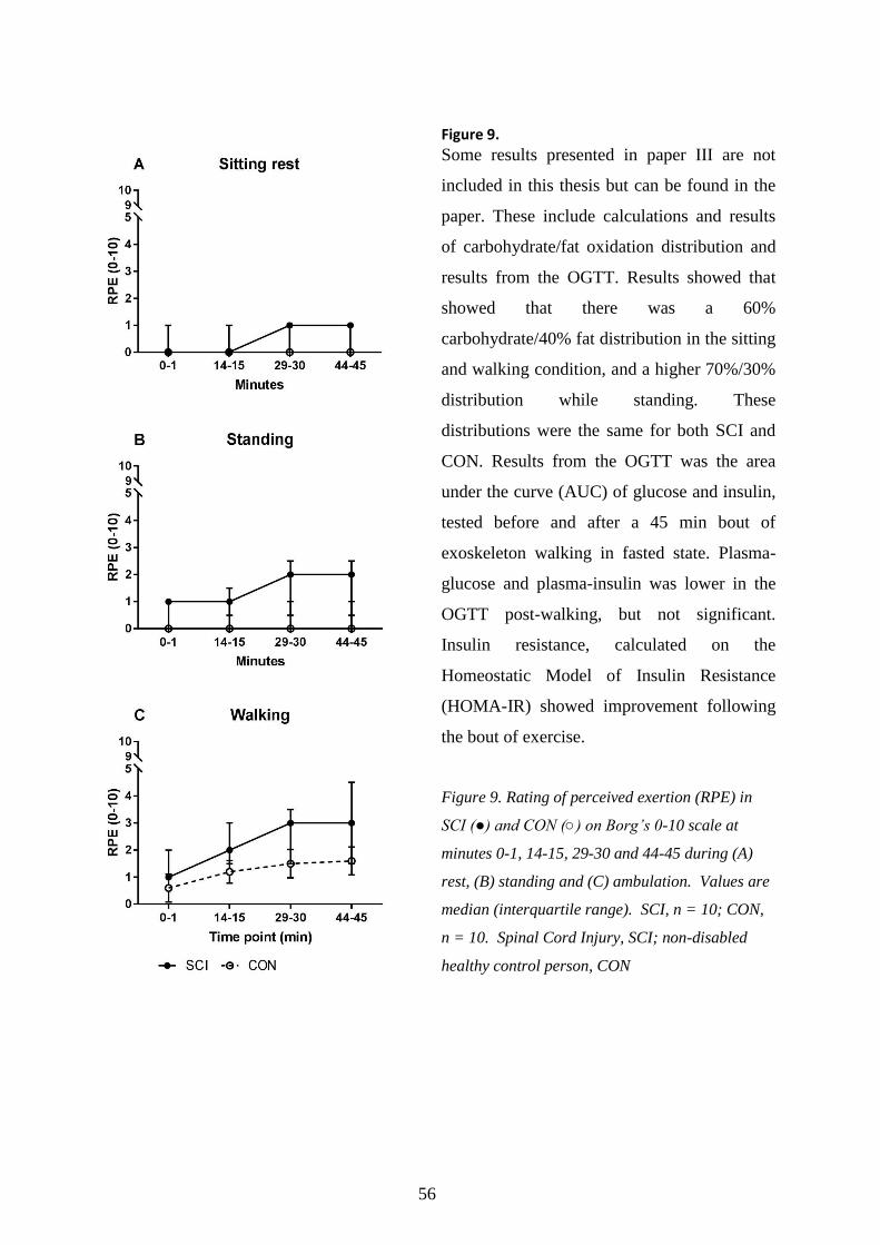
56
Figure 9.
Some results presented in paper III are not
included in this thesis but can be found in the
paper. These include calculations and results
of carbohydrate/fat oxidation distribution and
results from the OGTT. Results showed that
showed that there was a 60%
carbohydrate/40% fat distribution in the sitting
and walking condition, and a higher 70%/30%
distribution while standing. These
distributions were the same for both SCI and
CON. Results from the OGTT was the area
under the curve (AUC) of glucose and insulin,
tested before and after a 45 min bout of
exoskeleton walking in fasted state. Plasma-
glucose and plasma-insulin was lower in the
OGTT post-walking, but not significant.
Insulin resistance, calculated on the
Homeostatic Model of Insulin Resistance
(HOMA-IR) showed improvement following
the bout of exercise.
Figure 9. Rating of perceived exertion (RPE) in
SCI (●) and CON (○) on Borg’s 0-10 scale at
minutes 0-1, 14-15, 29-30 and 44-45 during (A)
rest, (B) standing and (C) ambulation. Values are
median (interquartile range). SCI, n = 10; CON,
n = 10. Spinal Cord Injury, SCI; non-disabled
healthy control person, CON

57
Table 7. Cardiodynamic parameters across tests and time points for participants with spinal cord
injury (SCI) and controls (CON). (See also Figure 10).
Time point
0-1 min 14-15 min 29-30 min 44-45 min p-value
SCI,
n=10
SV
(mL/beat)
Sitting 65.9 (11.9) 63.2 (11.4) 64.1 (11.1) 62.8 (11.3) 0.11
Standing 62.1 (12.7) 62.4 (11.2) 60.6 (10.3) 57.6 (9.3) <0.01
walking 73.9 (14.8) 73.8 (13.6) 74.7 (15.3) 75.7 (16.0) 0.62
HR (bpm) Sitting 81.8 (8.3) 79.1 (10.9) 77.2 (11.7) 79.0 (10.5) 0.13
Standing 87.1 (13.8) 92 (17.3) 91.4 (17.0) 93.2 (17.3) 0.10
walking 94.6 (18) 102 (20.2) 107.8 (23.6) 107.6 (21.5) <0.01
Q (L/min) Sitting 5.4 (1.1) 5.0 (1.1) 4.9 (1.1) 5.0 (1.4) 0.06
Standing 5.5 (1.6) 5.8 (1.6) 5.6 (1.6) 5.4 (1.4) 0.21
walking 7.0 (2.3) 7.7 (2.6) 8.2 (3.1) 8.2 (2.8) <0.01
CON,
n=10
SV
(mL/beat)
Sitting 102.9 (13.8) 105.8 (15.7) 103.4 (13.2) 102.5 (14.2) 0.37
Standing 98.6 (17) 99.6 (18.1) 98.8 (14) 98.0 (14.8) 0.80
walking 107.8 (15.2) 119.8 (23.5) 124.3 (25.8) 121.2 (23.4) <0.01
HR (bpm) Sitting 66.0 (11.8) 66.3 (10.4) 67.1 (11.3) 65.5 (10.4) 0.50
Standing 72.0 (10.2) 77.0 (12.0) 77.2 (12.2) 78.5 (12.8) <0.01
walking 77.8 (10.7) 86.3 (12.6) 87.4 (11.4) 87.4 (12.4) <0.01
Q (L/min) Sitting 6.8 (1.4) 6.9 (0.9) 6.9 (1.1) 6.7 (0.9) 0.56
Standing 7.1 (1.5) 7.6 (1.6) 7.6 (1.6) 7.7 (1.6) 0.02
walking 8.3 (1.1) 10.3 (2.2) 10.8 (2.0) 10.5 (1.9) <0.01
Descriptives are mean (s.d.)
Abbreviations: Stroke Volume, SV; Heart Rate, HR; Cardiac Output, Q. Test-day, T; beats-per-minute,
bpm; Spinal Cord Injury, SCI; non-injured control participants, CON.

58
Difference between timepoints within session
Table 7 shows the table the values of SV, HR and Q and p-values repeated measures between the
15 minutes time points within each of the three test days, for SCI and CON respectively and
significance level for main effect of time. Figure 1 show the same data presented as percentage
change of SV, HR and Q relative to the first minute of the test day to see the relative changes.
Summarising the results from Table 7 and Figure 10 was that while standing day there was a
significant drop for SCI in SV while standing, and no significant change in HR or Q for SCI. In
CON there was no change in SV, but a significant increase in HR and Q on the standing day.
When SCI participants were walking, there were, unlike at standing, there was no change for
SCI, but HR and Q increased significantly. In CON there were significant increases in SV, HR
and Q over 45 min of walking
Main effect of group and of condition (sitting, standing, walking)
The SCI group had significant higher HR and lower SV and Q than CON on all three testing
days (main effect of group, p<0.01). The three participants with NLI over T6 was separated in
the analysis and by visual inspection. The cardiodynamic response followed the rest of the SCI
participants.
HR was significant higher on the walking day compared to standing, which in turn was
significant higher than sitting (main effect of condition, p<0.05). Neither SV and Q was
significant different between sitting or standing for either SCI or CON, but walking was
significant higher than both sitting and standing for both groups SCI and CON (main effect of
condition, p<0.01).
Changes during HUT and between test days (Figure 11)
With increasing HUT tilt angle there was a significantly (p=0.02) drop in SV for whereas HR
and Q increased (p<0.001) with increasing angle both SCI and CON. The SCI group had overall
lower SV during HUT than CON, (main effect of group, p<0.01), whereas SCI had higher HR
than CON during HUT, (main effect group, p=0.01). There was no main effect of group on Q
between SCI and CON. The HRV measure LF/HF increased also with increasing tilt angles for
both SCI (p<0.001) and CON (p<0.001) for the SCI group had significant higher values in
LF/HF compared to CON (main effect group, p=0.034). For SCI there was higher value of the
second HUT after sitting or standing (HUT 2 and HUT4) compared to HUT before standing or
walking (HUT1 and HUT3) (main effect for both days 0.014). For CON there was no significant
difference between the HUT. .BP (Figure 3) was largely unaffected during tilt, except for one
participant with SCI with a neurological level of injury at C7 who experienced large drop in BP
during the tilt test.

59
Figure 10. Stroke Volume, Heart Rate and Cardiac Output during 45 minutes of sitting,
standing and walking
Figure 10. All descriptive statistics are mean (s.d.). Plots showing percentage-change from initial value
of 100% over 45 minutes at three test sessions, sitting, standing, walking.
Abbreviations: Spinal Cord Injury, SCI, non-injured controls, CON; Stroke Volume, SV; Heart Rate, HR;
Cardiac Output, Q.

60
Figure 11. Stroke Volume, Heart Rate and Cardiac Output during HUTs before and after
standing day 2 and walking day 3

61
Figure 11. All descriptive statistics are mean (s.d.). Plots showing change in HR, SV, Q and
LF/HF at four tilt-angles on each of the four HUT tests performed.
Abbreviations: Head-Up-Tilt, HUT; Spinal Cord Injury, SCI, non-injured controls, CON; Stroke
Volume, SV; Heart Rate, HR; Cardiac Output, Q; Low Frequency, LF; High Frequency, HF

62
Figure 12. Blood pressure during HUTs, day 2 and day 3.
Figure 12. All descriptive statistics are mean (s.d.). Plots showing change SBP and DBP at four
tilt-angles on each of the four HUT tests performed.
Abbreviations: Head-Up-Tilt, HUT; Spinal Cord Injury, SCI, non-injured controls, CON;
Systolic Blood Pressure, SBP; Diastolic Blood Pressure, DBP

63
DISCUSSION
This thesis consists of four papers from two clinical exploratory studies evaluating safety and
feasibility, changes in gait parameters, changes in SHC, amount of energy expended and changes
in cardiodynamic parameters during sitting, standing and walking, as well as adaptations in
autonomic response following a bout of walking in the exoskeleton.
The primary findings of paper I-II showed that there were no serious adverse events, but some
skin issues were documented, it showed that it was feasible for all included SCI-subgroups to
complete the training protocol and to increase walk-time and number of steps and there were
indications of improvements in gait function without the use of the exoskeleton for some
participants with incomplete SCI. Furthermore, the findings showed that there were short-term
reductions in lower extremity spasticity and for a subset of participants there were improvements
over time in SCIM III and QoL and the training did not seem to elicit new pain sensations, but
pain did not seem to change over time. On bowel and bladder function there did, overall, not
seem to be any major changes over time.
Main findings from paper III-IV were that the EE was light to moderate i.e. in the lower end of
the range and intensities that would elicit a cardiorespiratory conditioning effect. EE while
walking was greater than sitting and standing and no metabolic drift was seen during 45 min
walking and the response to walking was not only attributable to the upright position alone.
There were not detected cardio-vascular drift effects during the exoskeleton walking session, as
opposed to standing where signs of venous pooling were indicated by a decreasing SV over time
during standing in the SCI group. During exoskeleton walking increased Q in both SCI and
CON. In SCI this was primarily driven by an increase in HR and not SV, where there in CON
were seen an increase in both HR and SV during walking. During the HUT there was a drop in
SV with increasing tilt-angle but an increase in HR and Q, as well as increase in the LF/HR ratio
in both SCI and CON, whereas the BP was overall stabile during the tilting.
One type of exoskeleton was used for the studies in this thesis. The exoskeleton has different
modes where the level of assistance can be changed. The European multicentre study used all
available modes at the discretion of the therapist and the study performed at the Miami Project
only used the mode of maximum assistance. The different modes were not investigated
separately in this thesis. Likewise, clinical studies that thoroughly compare models between
different manufactures are not available in the literature and it was not the aim of this thesis to
evaluate this aspect either. Based on the referred studies performed on different exoskeletons it

64
seems reasonable, however, that some generalization between models from the different
manufactures can be done.
Safety and feasibility
No specific subgroup seemed to be more prone to adverse events than others, there were
documented adverse events for participants with high and low level of injury as well as complete
and incomplete SCI. In paper I was described that three participants experienced a swollen joint
the day following training. They all had a chronic SCI and it was concluded that the injuries
were due to overuse. Benson et al. (2016)(95) reported in a study using ReWalk a swollen ankle.
The same pattern of joint effusion the day after training was described. The risk of overuse
injuries should be considered when an extensive gait training program is introduced to
individuals with chronic SCI who has not walked or stood up regularly for several years.
Overall, it was concluded that training was safe, but extra attention should be given to prone skin
areas. This is in line with the results of previous studies(63). Also, persons with chronic injury
might in risk of overuse injuries of foot and ankle, since this was reported in three cases in this
study. A suggestion would be to initiate standing training prior to gait training and initiate more
slowly in the first weeks. The training program is time consuming which is important to consider
and match expectations with the participants and concurrent medical conditions are of
importance when evaluating if this training is the right for the individual participants.
In paper I, 88% of the TSs were completed: equivalent to 2.6 completed sessions per week. This
was sufficient to lead to progression in walk time and steps for all subgroups. There was no
difference in up-time between SCI subgroups and only minor differences between subgroups in
the outcomes of these training characteristics. Transition from walker to crutches, which could
reflect an improved level of control, happened primarily within the first four weeks of training.
In paper III-IV it took in average 15 training sessions in the habituation period to reach the
desired level of walking independence. This time period is similar to what have been described
previously(20) (93) (63) and is also in line with the level of walking that was reached within the
first 4 weeks of walking in paper I-II.

65
Changes in gait function without the exoskeleton and lower extremity motor
score
As descriptive statistics for the gait tests, was the estimated marginal means chosen. The reason
for this was that some participants acquired ambulatory function at different timepoints through
the eight-week training period and to incorporate this in a mean value was this measure chosen.
As shown in the result section experienced the recently injured group most of the improvements,
but interestingly there was also improvement in the chronically injured group on the TUG and
BBS and indication that the improvement was retained at follow up.
The recently injured group had a median of less than 4 months (median (IQR) 0.3 (0.2-0.4) year,
Table 5). Since the largest degree of neurological recovery is expected to take place in the early
phase following an SCI, this would explain the improvement that the recently injured
experienced over the chronically injured, also further indicated by the increase LEMS in the
recently injured group that was not evident among chronically injured(33) (34) (64).
Improvements in relation to exoskeleton training have previously been reported, for example in a
study by Aach et al. showed improved 10MWT, TUG and LEMS after three months using the
HAL exoskeleton(23) and another study by Wirz et al.(96) showed improvements on 10MWT
and TUG using the Lokomat. That study(96) also found WISCI II to be less sensitive to detect
changes in gait function in complete SCI compared to the tests 10MWT and TUG. This could be
an explanation that WISCI II parameters did not change in our study as well, when the other gait
and balance test did.
The chronically injured participants seemed to improve most on TUG and BBS which reflects
balance function more than gait function. Likewise, were there also case reports of improved
sitting balance in two complete injured participants which could support this hypothesis. It seems
plausible that mastering the exoskeleton walking using the Ekso Bionics exoskeleton, rely more
on balance over correct movement of once own legs and that the balance function is being
trained by performing a mass shift between the legs when a new step is initiated. Future studies
could have increased focus on assessing balance function when testing exoskeleton gait training.
When evaluating changes, it is important to take into account if a significant change also is
clinical significant, for example using ‘the minimal clinically important difference’ (MCID) as
well as ‘the minimal detectable difference’ (MDD). Lam et al.(61) found for people with SCI a
MCID for TUG to be 10.8 seconds and 0.13m/s for the 10MWT and MDD for TUG of 3.9 s and
0.05m/s for the 10MWT, which was were calculated on distribution scores measured over a
three-month period. Wu et al.(97) reported similar values. In our study we found improvement

66
on TUG of 7.0s for recently injured and 7.8s for chronically injured from TS1 to TS24 and an
average gait speed increase on the 10MWT by 0.07 m/s for both groups, based on the averaged
marginal means. These values are above MDD but below MCID. It could, however, be
hypothesized that larger improvements could be seen following a longer or more intense training
program and worth exploring in future studies.
Pain
Approximately half of the participants had presence of pain (nociceptive and neuropathic) at
some point during the previous week when asked (Figure 4A) and these frequencies were similar
to what has been previous reported by Adriaansen et al.(9), but lower than reported by Andresen
et al.(19). Relative few persons experienced pain during training session. The participants who
did reported pain during the training session, overall speaking, had also reported the same type
and location of pain also in the previous week to the training session. The questionnaire did not
explicatively ask that question, but an interpretation of the results could be that the training did
not provoke a new pain sensation and that the training was well tolerated. In hindsight could it
however have been beneficial to ask the question more explicitly. Kolakowsky-Hayner(27)
reported lower back pain during training with Ekso walking but also reported that it did not
affect participation in the study. In our study, although reports of low back pain were
documented as the most frequent location of pain and like previous reported like in the
Kolakowsky-Hayner study(27), no participants and no dropped out as a result of this. Case
reports on improvements of pain after exoskeleton training have previously been reported in
studies using exoskeletons from Ekso Bionics, ReWalk and HAL(20) (21) (22), as well as
reports of decrease in pain sensation over time following training using the Lokomat®(25). We
did, however, not see changes over time in pain sensation in our study.
Spasticity
Spasticity was documented in 65% to 71% of participants which is similar to prevalence of self-
reported spasticity in the study by Andresen et al.(19). Spasticity for each muscle group was
evaluated as rank scores and there was overall a decrease in spasticity for all the muscle groups.
A sum-score of MAS from all the 12 muscles were created for the purpose of an overall measure
of spasticity, following the logic of other compound scores such as the SCIM III and was also
inspired by a similar approach in a study by Stampacchia et al. with a sum score of
spasticity(24).The spasticity sum-score is, however, not validated. Since the MAS scale has 5
67
levels, the sum-score maximum value is thus 60. However, since many muscles had no
spasticity, i.e. a MAS of 0, there was an uneven distribution and floor effect and thereby a bias.
The highest value of MAS 4 (the fifth level on the score) was an exclusion criterion and thereby
also adds to a shift towards lower values. It must also be mentioned that this sum-score has the
assumption of MAS being an ordinal scale. Under this assumption, we found in a previous study
that the MAS was reliable(98), but the assumption of the scale being ordinal as also been
challenged(98) as well having several limitations(99). Limitations aside, it is recommended by
the National Institute of Neurological Disorders and Stroke (NINDS) Common Data Elements
(CDE)(100) and is widely used in the clinical setting(13). Future studies assessing spasticity in
exoskeleton training could consider adding other scales of spasticity as well.
The previous mentioned study by Stampacchia et al.(24) also found a decrease of spasticity after
a training session, as well as reduction of pain. There has also been case reports of lowered
spasticity in the hours after training in the HAL exoskeleton(23).The results from our study are
in line with the previous findings.
That the spasticity decreased by the training is useful for people with spasticity who wants to use
exoskeletons, since spasticity is problematic using the exoskeleton as well as can be
uncomfortable for the user. If used regularly or even daily, it could be incorporated as part of a
spasticity relief program alongside other types of physical activity.
Spinal Cord Independence Measure, bowel- and bladder function
SCIM III, bowel and bladder function results are discussed together in this paragraph. The
recently injured participants improved on SCIM III, where this was could be considered
attributable to early phase improvements following SCI as referred to previously for other
parameters. There were improvements on the respiration and sphincter management sub-score
and on bowel-related items in the recently injured group on the SCIM III sub-score as well as
and in one item on the International SCI Bowel Function Basic Data Set but not significantly on
the LUT dataset.
Interestingly were there also found improvements for chronically injured on the total SCIM III
score, however to a smaller degree than the recently injured. On the sub-item ‘Use of Toilet’ on
the SCIM III score were there indications of improvements. If this is reflecting a true change, it
could be speculated if this was related to an improved balance function, since these participants
also improved their results on TUG and BBS over the same period(93), as previous described.
This could be an included in future research to investigate.

68
There has previous been published case reports on changes in bowel and bladder function
following exoskeleton training(27) (21). Hubscher et al.(26) published recently a study where
they documented improvements of urinary incontinence and time needed for defecation, after 80
training sessions of body mass supported treadmill training, done daily. The study used the same
assessment questionnaires as in our study thereby being comparable. The study by Hubscher et
al. had a more intense training protocol and it a hypothesis for not detecting changes in our study
on bowel and LUT function could be that the volume, intensity and duration of the study was too
low and short to cause detectable changes.
Quality of Life (QoL)
The participants with chronic injury showed a significant improved QoL on the ‘satisfaction of
life as a whole’ question over the study period and at follow up. The values of QoL in our study
were similar to a study be Adriaansen et al.(9), including 282 participants, where a median value
of 7 on the item ‘satisfaction of life as a whole’ was reported, a value of 6 on ‘satisfaction with
physical health’ and 7 on the item ‘satisfaction with psychological health’ in a study on
individuals with chronic SCI. Improvements in QoL have previous been reported in two case
studies with chronically injured participants using the exoskeletons ReWalk(28) and HAL(22).
QoL in our study was, however, not asked specifically in relation to the exoskeleton training, but
was asked for QoL in general.
There was in increase ‘satisfaction of life as a whole’ and ‘satisfaction of physical health’ of one
point in the recently injured group, and a drop in ‘satisfaction of psychological health’ through
the training period and return to baseline values at follow up (Table 4), but neither was
statistically significant. There was not in this study further questions on the QoL and thereby is
it not possible to further evaluate what these values are reflecting. On a speculative note, if these
values are reflecting no change over the study period, an explanation could be that QoL has been
shown to be better years after the injury compared to those who were recently injured reflecting
an adaptive process, as reported by Westgren and Levi(17).
The study by Westgren and Levi(17) also documented that several SHC such as neurogenic pain,
spasticity, and neurogenic bladder and bowel problems were associated with lower QoL scores.
We did not in our study correlate these SHC to QoL, but this could be considered in future
studies evaluating QoL in relation to exoskeleton training. Here it could also be considered to
include specific QoL questions related directly to the training intervention.

69
Physical activity and energy expenditure
Recommendations for physical activity to achieve health benefits include regularly exercise of
moderate to vigorous intensity and to reduce sedentary time(39). A strategy to reduce sedentary
time can be to include physical activity of lower intensity(39) (36). The intensity found in paper
III was overall light to moderate in intensity and would for many people not qualify as intense
enough to promote cardiorespiratory conditioning (increase VO2max). It should be noted that the
settings of the exoskeleton was set to ‘max’ mode so the exoskeleton performed all the work to
move the legs which likely contributed to the lower intensities during walking compared to
modes where the participant contribute more, as shown in a previous study by Kressler(47).
Results from paper I indicated similar intensities of RPE and change in HR from sitting to
walking, also in line with previous findings of walking with an exoskeleton(101) (63). In the
study in paper I all walking modes could be used, but the walking mode that gave the best
quality of walking, judged by the therapist, was selected and therefore likely also the one that
required less energy. Interestingly, we did not find major differences between subgroups in RPE
in paper I. The reason could be that different levels of assistance from exoskeleton were allowed
and therefore the individual optimal mode could be selected. A study by Lewis et al.(102)
showed that the RPE did increase with increasing work load, but showed inconsistent
associations between RPE and physiological measures like HR, VO2 in people with paraplegia
and tetraplegia when performing arm ergometer training. This limitation should be kept in mind
when the scale is used in people with SCI(102).
The results from paper III showed that the absolute energy cost was lower for the participants
with SCI walking with the exoskeleton than for the non-disabled walking at self-selected pace
without the exoskeleton. When looking at the values relative to peak VO2 of the peak ergometer
test, the opposite was the case. Exoskeleton walking had a several folds lower walking economy
compared to non-disabled walking without the exoskeleton, which could explain the higher
relative intensity of exoskeleton walking of the peak VO2. Gait speed of able-bodied was
multiple times faster than SCI (1.3 m/s vs. 0.23 m/s), which was comparable to walking speeds
reported by Escalona(48) and Evans(49) as well as what was found in a review by Louie(103),
where exoskeleton gait speeds were estimated to an average of 0.26 m/s. However, this review
included exoskeleton gait speeds from different devices and during different conditions and gait
tests such as the 10 Meter Walk Test, 6 Minute Walk Test, whereas the reported gait speed in
this study is the average over a 45 min session. In the study by Kressler(20), using the Ekso, the
three participants had mean VO2-peak test of 20.7 mL/kg/min and mean VO2 during walking on

70
6.1 mL/kg/min. A study by Asselin et al.(50) reported a VO2 consumption of 35% of VO2peak
during 6-minute bouts of exoskeleton walking and an average RPE representing ‘very light to
fairly light’ effort. In a study by Evans(49), using the Indego exoskeleton, showed a VO2 during
walk of 9.5 (0.8) to 11.5 (1.4) mL/kg/min on two testing days which was higher than in our
study, which showed 8.5 (0.90). The participants in the Evans study has also a lower peak VO2
in an arm crank peak test, yielding an even higher percentage of peak during walking of VO2
was (51.5 % to 63.2 %) than in our study. Asselin study(50) had averaged VO2 while walking of
mean (SD) 11.1 (1.7) mL/kg/min using ReWalk and an estimation of 35% VO2-reserve capacity.
The above-mentioned studies reported averaged values over relative short bouts of walking. In
our study we also assessed the trends over 45 min of walking and standing to detect potential
upwards drift in metabolism. Our results from passive standing were similar to a previous
report(104), where standing used 20% of VO2peak. Despite that the walk mode of the exoskeleton
was set to max, there was a significantly higher VO2 and TEE as well as RPE during exoskeleton
walking than during standing. During exoskeleton walking must the persons with SCI use the
arms for support and likely some postural muscles, which is the likely explanation for the higher
EE. The difference between the reports from different exoskeleton models is likely also due to
differences in demand of postural and upper extremity muscles.
All in all, suggested the results that the exoskeleton training should not be the primary method
for cardiovascular conditioning training, but could be used as a strategy to implement lower
intensity training and decrease sedentary time. The gait speed is however not fast enough for
functional community ambulation, where the speed for example needed to cross a road
intersection previously has been estimated to be 1.22m/s(105). The daily use of non-motorised
orthoses were often limited because of high energy demand and fatigue experienced after
walking short distances(58) and have resulted in limited long-term use by individuals(59). This
seems to have been mitigated with the motorized exoskeletons.
Cardiodynamic outcomes
Cardiac output can be increased by the Frank-Starlings mechanism by increased venous return
and end-diastolic ventricular volume that could arise from a peripheral vasoconstrictor
response(106). However, Kinzer(107) found that persons with paraplegia exhibited a higher HR
and lower SV during arm-crank exercise compared both to persons with leg amputation and non-
injured persons. Persons with paraplegia had greater accumulation of fluids in the legs compared
to their non-injured counterparts whereby the legs seemed to act as a reservoir. Similar findings

71
have been reported by Jacobs et al.(53) and Hotettler(108). Hjeltnes(109) attributed the
decreased venous return to a loss of sympathetic vasomotor regulation below the level of lesion
in thoracic SCI. higher HR in persons with paraplegia and tetraplegia compared to non-injured
persons have been described by Dela et al.(110) which were documented during electrically
induced leg ergometer training.
Our findings suggest a similar pattern, where SCI had higher HR and lower SV and Q than CON.
As a matter of fact, was it seen in all test conditions; peak test, sitting, standing, walking and
during tilt test. CV-drift effects, defined as both increase in HR and drop in SV were not seen
during walking with exoskeleton in SCI despite the prolonged period of 45 min of walking. The
outdoor temperature lack of fluid replacement could also have pushed towards CV drift(54)
(111) (112) but was not documented. This is could be due to a relative low intensity of
exoskeleton walking, below a threshold to elicit this phenomenon. Likely it also reflects
selection of the study population. The results suggest that the movement of the legs during
exoskeleton walking could have mitigated the venous pooling with reference to the drop in SV
seen in the SCI group in the standing session (Figure 10).
Results from the tilt tests showed an increase in the LF/HF ratio which implies a shift towards
higher sympathetic nervous system input relative to parasympathetic autonomic tone(89). This is
what would be expected with the orthostatic stress induced by tilting to erect position. Judging
by the Figure 11 it looked like there was a parallel shift from the first to the second HUT after
standing and walking (HUT2 and HUT4) and thereby suggest a carry-over effect from the
walking and standing intervention for the SCI group. That the CON group did not show signs of
this is not surprising since this non-injured group will be making multiple orthostatic
repositioning during the day. The apparent trend in SCI would be relevant to follow up in future
studies. Also, it could be interesting to test this in a longitudinal study to test for potential
longitudinal changes. Lastly, suggest the results that the LF/HF-ratio can be used as a proxy for
autonomic tone.
The SCI group reached a higher peak HR during the peak test than CON, despite the max effect
(watt) was lower than for CON. An explanation could be the limitation of the arm-crank test, not
being able to elicit the max HR of the CON group. The SCI group would likely have been be
more conditioned for upper body exercise than the CON group, since the SCI participants were
wheelchair user and thus the peak test would underestimate the maximum effect in the CON
group. Studies on able bodied persons have shown that individuals who have not trained the arm-

72
crank ergometer test will achieve a lower output compared to a leg ergometer test due to a
reduced stress on the CV system during the arm-crank test(113).
Strengths and limitations of the methodology and study design
Paper I-II
One strengths of the study is that it included a heterogeneous sample of participants with SCI
including participants with complete and incomplete injury, tetra- and paraplegia, recent and
chronic injury and both men and women, and included different rehabilitation centres and
different countries, to reflecting the heterogenicity of the SCI population(114). This, however, is
also a limitation since it can mask potential changes only seen in a subgroup of the study
population and in combination with a relative modest study sample, it was not feasible to
perform thorough sub-group analysis. The study explored many different outcomes with increase
the risk of type 1 errors.
The participants were included as a convenience sample at the discretion of the investigators and
required that the participants could commit with a thrice weekly training program with the time
restrictions of their life that this protocol encompassed. There has likely been a bias in the
selection of the participants.
Two centres, n=8 participants, used the first-generation exoskeleton, the Ekso, whereas the other
used the second generation, EksoGT. The difference between the two versions were considered
only to be minor and not to confound the training substantially and thereby the results of the
study.
The study was a non-controlled, non-randomised study with a sample of convenience, which
does not allow conclusions to be drawn on causality of the findings in relation to the intervention
of exoskeleton gait training. The results should be interpreted as indicators and can be used only
as directions of future studies.
Paper III-IV
The sample size was relative small with the risk of lack of statistical power for analysis and sub-
group analysis was not feasible. Three included participants had neurological level of injury
above T6 and potential loss of cardiac sympathetic drive and could then exhibiting different
responses compared to participants with injuries below T6(115). Whereas many persons with
SCI suffer from orthostatic hypotension, the present study population was selected to have
orthostatic tolerance as per eligibility criteria. This selection was chosen to reflect the potential

73
users of the exoskeleton and is a common inclusion criterion in exoskeleton studies but does
thereby not reflect the whole SCI population. Furthermore, all participants were male and were
more fit than the average SCI population(94).
The three test days were not randomized and there could be a conditioning effect of the repeated
HUTs that is not accounted for in the study. The non-disabled group performed unassisted
walking of self-selected pace and not in the exoskeleton as the SCI participants which makes
direct comparisons between the two groups difficult. However, all comparisons between two
such different groups are with limitations. The same is the case when comparing two such
different walking conditions, where exoskeleton walking largely require an effort from the upper
body exercise compared to a lower body performance as well as the difference in trunk control
between the two conditions. For the explorative purpose of this study, we believed that this
design was the most appropriate, all things considered.
CONCLUSION
The overall aim of this thesis was to explore exoskeleton training on a variety of parameters both
longitudinally and acute during a single training session.
It is concluded in paper I that training was safe and feasible when special attention was focused
on areas such as prone skin and due diligence is performed. There was not detected specific
groups that was more prone to safety issues, except for the potential overuse injuries of the ankle
reported in three participants who were injured many years ago. Proper precautions should also
be taken for this group of participants in the future.
The results of paper I-II indicated that both recently injured and chronically injured can benefit
of exoskeleton training on several SHC, and possibly on balance function, but final conclusions
cannot be drawn because of the study design not being randomised or controlled.
Work intensity described in paper III was below the threshold to promote cardiorespiratory
conditioning, but above that observed for seated rest and standing and could help decrease
sedentary time and potentially also provide health benefits like has been shown on walking in
non-injured persons. The low energy cost during exoskeleton walking is consistent with a low
self-reported RPE that was found in both paper I and III and in paper III also found modest EE
during walking allow participants to tolerate longer ambulation without experiencing fatigue
which would be desired if the device is to be used as a mobility device.

74
There did not seem to be a drift in metabolism since the VO2 and TEE graph plateaued after
some time. CON used more energy while walking compared to SCI measured in absolute values,
but similar when measured in percentage of peak performance, however with the limitations of
the design considered.
The exoskeleton walking seemed to mitigate the drop in SV that was seen during the 45 min
standing session, possibly due to mechanical effects of lower limb movements. Increase in Q
during walking was in SCI driven by increase in HR, where in CON it was driven by both SV
and HR increase, in line with previous reporting in the literature. Changes during walking both
for energy expenditure and cardiodynamics was not only attributable to the standing position.
HUTs elicited the same cardiodynamic response pattern in SCI and CON with decrease in SV
and increase in HR and Q. However, did SCI have a higher HR and lower SV than CON, like
what was seen during walking. Increase in the HRV parameter LF/HF ratio with increasing tilt
angle suggests a higher sympathetic drive during the HUTs, which is what would be expected.
There was a pattern towards a parallel shift on LF/HF values on all cardio dynamic of the HUT
test that following standing and walking compared to the HUT before the standing or walking
intervention and a heretofore-unreported carry-over effect from exoskeleton walking to
responses experience during head up tilt, worth exploring in future studies.
PERSPECTIVES AND SUGGESTIONS FOR FUTURE
FOR RESEARCH
Future studies could investigate whether more frequent and intense training protocols would
elicit changes in e.g. pain, bowel and LUT function. Comparator in randomized trials has often
been body mass supported treadmill training, but also comparison to daily clinical routine
treatment or even functional electric stimulation would be worth exploring since the latter can be
incorporated into exoskeletons.
Other areas to explore
Changes in bone mineral density was not investigated in this thesis since it was believed that the
duration of the training intervention was too short to detect any potential changes. This would
however be an interesting outcome to access in future studies of longer duration. Of many other
relevant areas of exoskeleton gait training worth exploring are combinations of functional
electrical stimulation while ambulating in the exoskeleton. This has been done for example with

75
the Lokomat(116) or it could be even more integrated systems for multiple muscle groups, such
as FES systems.
Exoskeletons for use with other neurological disorders
The powered exoskeleton from Ekso Bionics is also approval for stroke rehabilitation, but
studies including this patient group have yet to be published. A systematic review of the HAL
found beneficial effects on gait function and walking independence in a mixed population of
neurological disorders, including stroke(117). Treadmill-based robotic exoskeletons, like the
Lokomat have been introduced in stroke rehabilitation and a recent Cochrane review(118)
concluded that electromechanical-assisted gait training in combination with physiotherapy
increased the odds of participants becoming independent in walking, but mainly in sub-acute
stroke(118) and that it was equivalent to traditional gait therapy for chronic stroke patients(119).
Apart from stroke, robotic gait training could be used in persons with multiple sclerosis,
traumatic brain injury, cerebral palsy, and other neurological disorders that lead to lower
extremity paresis(120).
Exoskeletons as mobility devices
Evaluation of exoskeleton technology was not part of the primary aim of this thesis. However,
the results of the studies still provide some insight into this aspect.
From a safety point of view, it seems that skin issues at the contact points of the exoskeleton, as
well as on the feet in case of problems with the footwear, are one of the most common of the
safety concerns. This comes as no surprise as pressure ulcers is a very well-known issue in
people with SCI. If exoskeletons are to be used as mobility device, this would suggest daily use
of up to several hours which increases the importance to keep focusing on this area. Sitting in
wheelchair is also associated with risk of pressure ulcers and there could be benefit of increasing
the time spend in upright position by decreasing the sitting time in the wheelchair and thereby
decrease the risk of pressure ulcers from the sitting position.
There were reported a few cases of shoulder pain during training. The exoskeleton does require
the use of a walker or crutches and thereby imposing pressure on the shoulder girdle which is
already an injury prone joint in persons with SCI. Focus on good technique during walking is an
important mitigation and minimizing the amount of stress on the shoulder while walking with the
exoskeleton remains an important area to focus on going forward.

76
Another area that was investigated was the energy demand. The high energy demand of non-
motorized orthoses has been mentioned as a limitation for their use for training and as mobility
devices(3) (58) (59). Results from this study indicate that the studied exoskeleton require a lower
energy demand and could be used for a longer period. Thereby seems the issue of the non-
motorized orthoses in that respect to have be solved.
On average, the habituation period study 2 (paper III-IV) required 15 sessions in to reach a level
of walking with minimal assistance in exoskeleton walking. This time period is similar to what
have been described previously(20) (93) (63). The exoskeleton ambulation speedy on the
walking day in study 2 was 0.23 m/s, which is similar to what was described in a review by
Louie(103) where average exoskeleton speeds were estimated at 0.26 m/s. However, this value
includes varying devices, conditions and gait tests such as the 10 Meter Walk Test, 6 Minute
Walk Test, whereas the reported gait speed in our study is the average of a 45-minute bout of
exoskeleton walking. As a side note can be mentioned that the gait speed is below the value that
has been estimated to be the minimum for functional community ambulation(105). This is based
on gait speed needed for example to cross an road intersection, which has been estimated to be
1.22m/s(105).
As final remarks on barriers to overcome for exoskeletons to be used as community devices can
be mentioned a long list that, however, is not based on data from the enclosed studies of this
thesis. Worthy of mentioning are the ability to safely walk on different surfaces, walk on stairs,
ramps (uphill and downhill), improved safety against falls without the use of an assistant and be
able to accommodate persons of different mass and body size than what is currently possible(1).
Further technological advancements are desired including increased battery life, increasing the
degrees of motion in the joints and finally decreasing the time to put the exoskeleton on and off.
As technology improves over time, these devices hold great potential for people with SCI and for
rehabilitation in general.

77
REFERENCES
1. Palermo AE, Maher JL, Baunsgaard CB, Nash MS. Clinician-Focused Overview of Bionic
Exoskeleton Use After Spinal Cord Injury. Top Spinal Cord Inj Rehabil. 2017 Jun
1;23(3):234–44.
2. Walter JS, Sola PG, Sacks J, Lucero Y, Langbein E, Weaver F. Indications for a home
standing program for individuals with spinal cord injury. J Spinal Cord Med.
1999;22(3):152–8.
3. Karimi MT. Evidence-Based Evaluation of Physiological Effects of Standing and Walking
in Individuals with Spinal Cord Injury. Iran J Med Sci. 2011 Dec;36(4):242–53.
4. Wyndaele M, Wyndaele J-J. Incidence, prevalence and epidemiology of spinal cord injury:
what learns a worldwide literature survey? Spinal Cord. 2006 Jan 3;44(9):523–9.
5. Lee BB, Cripps RA, Fitzharris M, Wing PC. The global map for traumatic spinal cord
injury epidemiology: update 2011, global incidence rate. Spinal Cord. 2014 Feb;52(2):110–
6.
6. National Spinal Cord Injury Statistical Center (NSCISC). Spinal Cord Injury (SCI) Facts
and Figures at a Glance. [cited 2018 Jul 9]; Available from: https://www.nscisc.uab.edu/
7. Rygmarvsskade - Lægehåndbogen på sundhed.dk [Internet]. [cited 2018 Jun 5]. Available
from: https://www.sundhed.dk/sundhedsfaglig/laegehaandbogen/neurologi/tilstande-og-
sygdomme/rygmarv/rygmarvsskade/
8. Marino RJ, Ditunno JF, Donovan WH, Maynard F. Neurologic recovery after traumatic
spinal cord injury: data from the model spinal cord injury systems. Arch Phys Med Rehabil.
1999 Nov 1;80(11):1391–6.
9. Adriaansen JJE, Ruijs LEM, van Koppenhagen CF, van Asbeck FWA, Snoek GJ, van
Kuppevelt D, et al. Secondary health conditions and quality of life in persons living with
spinal cord injury for at least ten years. J Rehabil Med. 2016 Nov 11;48(10):853–60.
10. Jensen MP, Molton IR, Groah SL, Campbell ML, Charlifue S, Chiodo A, et al. Secondary
health conditions in individuals aging with SCI: terminology, concepts and analytic
approaches. Spinal Cord. 2012 May;50(5):373–8.
11. Anke AG, Stenehjem AE, Stanghelle JK. Pain and life quality within 2 years of spinal cord
injury. Paraplegia. 1995 Oct;33(10):555–9.
12. Haisma JA, van der Woude LH, Stam HJ, Bergen MP, Sluis TA, Post MW, et al.
Complications following spinal cord injury: occurrence and risk factors in a longitudinal
study during and after inpatient rehabilitation. J Rehabil Med. 2007 May;39(5):393–8.
13. Sköld C, Levi R, Seiger A. Spasticity after traumatic spinal cord injury: nature, severity,
and location. Arch Phys Med Rehabil. 1999 Dec;80(12):1548–57.

78
14. Krassioukov A, Eng JJ, Claxton G, Sakakibara BM, Shum S. Neurogenic bowel
management after spinal cord injury: a systematic review of the evidence. Spinal Cord.
2010 Oct;48(10):718–33.
15. Wyndaele J-J. The management of neurogenic lower urinary tract dysfunction after spinal
cord injury. Nat Rev Urol. 2016 Dec;13(12):705–14.
16. Richards C, MacKenzie N, Roberts S, Escorpizo RD. People with Spinal Cord Injury in the
United States. J Phys Med. 2017 Feb;96(2).
17. Westgren N, Levi R. Quality of life and traumatic spinal cord injury. Arch Phys Med
Rehabil. 1998 Nov 1;79(11):1433–9.
18. Jörgensen S, Ginis KA, Iwarsson S, Lexell J. Depressive symptoms among older adults
with long-term spinal cord injury: Associations with secondary health conditions, sense of
coherence, coping strategies and physical activity. J Rehabil Med. 2017 Aug 31;49(8):644–
51.
19. Andresen SR, Biering-Sørensen F, Hagen EM, Nielsen JF, Bach FW, Finnerup NB. Pain,
spasticity and quality of life in individuals with traumatic spinal cord injury in Denmark.
Spinal Cord. 2016 Nov;54(11):973–9.
20. Kressler J, Thomas CK, Field-Fote EC, Sanchez J, Widerström-Noga E, Cilien DC, et al.
Understanding Therapeutic Benefits of Overground Bionic Ambulation: Exploratory Case
Series in Persons With Chronic, Complete Spinal Cord Injury. Arch Phys Med Rehabil.
2014 Oct;95(10):1878–87.
21. Esquenazi A, Talaty M, Packel A, Saulino M. The ReWalk powered exoskeleton to restore
ambulatory function to individuals with thoracic-level motor-complete spinal cord injury.
Am J Phys Med Rehabil Assoc Acad Physiatr. 2012 Nov;91(11):911–21.
22. Cruciger O, Schildhauer TA, Meindl RC, Tegenthoff M, Schwenkreis P, Citak M, et al.
Impact of locomotion training with a neurologic controlled hybrid assistive limb (HAL)
exoskeleton on neuropathic pain and health related quality of life (HRQoL) in chronic SCI:
a case study. Disabil Rehabil Assist Technol. 2014 Nov 10;1–6.
23. Aach M, Cruciger O, Sczesny-Kaiser M, Höffken O, Meindl RC, Tegenthoff M, et al.
Voluntary driven exoskeleton as a new tool for rehabilitation in chronic spinal cord injury:
a pilot study. Spine J Off J North Am Spine Soc. 2014 Dec 1;14(12):2847–53.
24. Stampacchia G, Rustici A, Bigazzi S, Gerini A, Tombini T, Mazzoleni S. Walking with a
powered robotic exoskeleton: Subjective experience, spasticity and pain in spinal cord
injured persons. NeuroRehabilitation. 2016 Jan 1;39(2):277–83.
25. Labruyère R, van Hedel HJA. Strength training versus robot-assisted gait training after
incomplete spinal cord injury: a randomized pilot study in patients depending on walking
assistance. J NeuroEngineering Rehabil. 2014 Jan 9;11:4.
26. Hubscher CH, Herrity AN, Williams CS, Montgomery LR, Willhite AM, Angeli CA, et al.
Improvements in bladder, bowel and sexual outcomes following task-specific locomotor

79
training in human spinal cord injury. PLoS ONE [Internet]. 2018 Jan 31;13(1). Available
from: https://www.ncbi.nlm.nih.gov/pmc/articles/PMC5791974/
27. Kolakowsky-Hayner SA. Safety and Feasibility of using the EksoTM Bionic Exoskeleton
to Aid Ambulation after Spinal Cord Injury. J Spine [Internet]. 2013 [cited 2016 Jun 10];
Available from: http://www.omicsgroup.org/journals/safety-and-feasibility-of-using-the-
ekso-bionic-exoskeleton-to-aid-ambulation-after-spinal-cord-injury-2165-7939.S4-
003.php?aid=18534
28. Raab K, Krakow K, Tripp F, Jung M. Effects of training with the ReWalk exoskeleton on
quality of life in incomplete spinal cord injury: a single case study. Spinal Cord Ser Cases.
2016 Jan 7;2:15025.
29. Dietz V. Neuronal Plasticity After Spinal Cord Injury: Significance for Present and Future
Treatments. J Spinal Cord Med. 2006;29(5):481–8.
30. Smith AC, Knikou M. A Review on Locomotor Training after Spinal Cord Injury:
Reorganization of Spinal Neuronal Circuits and Recovery of Motor Function. Neural Plast
[Internet]. 2016 [cited 2017 Mar 8];2016. Available from:
http://www.ncbi.nlm.nih.gov/pmc/articles/PMC4879237/
31. Van Kammen K, Boonstra A, Reinders-Messelink H, den Otter R. The Combined Effects
of Body Weight Support and Gait Speed on Gait Related Muscle Activity: A Comparison
between Walking in the Lokomat Exoskeleton and Regular Treadmill Walking. PLoS ONE.
2014 Sep 16;9(9):e107323.
32. Burns SP, Golding DG, Rolle WA, Graziani V, Ditunno JF. Recovery of ambulation in
motor-incomplete tetraplegia. Arch Phys Med Rehabil. 1997 Nov;78(11):1169–72.
33. Mehrholz J, Kugler J, Pohl M. Locomotor training for walking after spinal cord injury.
Cochrane Database Syst Rev. 2012;11:CD006676.
34. Waters RL, Yakura JS, Adkins RH, Sie I. Recovery following complete paraplegia. Arch
Phys Med Rehabil. 1992 Sep;73(9):784–9.
35. Krause JS, Saunders LL. Health, secondary conditions, and life expectancy after spinal cord
injury. Arch Phys Med Rehabil. 2011 Nov;92(11):1770–5.
36. Maher JL, McMillan DW, Nash MS. Exercise and Health-Related Risks of Physical
Deconditioning After Spinal Cord Injury. Top Spinal Cord Inj Rehabil. 2017;23(3):175–87.
37. Rimmer JH, Schiller W, Chen M-D. Effects of disability-associated low energy expenditure
deconditioning syndrome. Exerc Sport Sci Rev. 2012 Jan;40(1):22–9.
38. Clinical Practice Guidelines: Spinal Cord Medicine. Identification and Management of
Cardiometabolic Risk after Spinal Cord Injury 2018 [Internet]. Available from:
https://www.pva.org/CMSPages/GetFile.aspx?guid=f3c29b7e-e201-4392-b241-
9933dc620e40
39. American College of Sports Medicine, Riebe D, Ehrman JK, Liguori G, Magal M. ACSM’s
guidelines for exercise testing and prescription. 2018.

80
40. Stamatakis E, Rogers K, Ding D, Berrigan D, Chau J, Hamer M, et al. All-cause mortality
effects of replacing sedentary time with physical activity and sleeping using an isotemporal
substitution model: a prospective study of 201,129 mid-aged and older adults. Int J Behav
Nutr Phys Act [Internet]. 2015 Sep 30;12. Available from:
http://www.ncbi.nlm.nih.gov/pmc/articles/PMC4589071/
41. World Health Organization. WHO | Physical Activity and Adults [Internet]. WHO. [cited
2018 Jul 1]. Available from: http://www.who.int/dietphysicalactivity/factsheet_adults/en/
42. Swain DP, Franklin BA. VO(2) reserve and the minimal intensity for improving
cardiorespiratory fitness. Med Sci Sports Exerc. 2002 Jan;34(1):152–7.
43. Murtagh EM, Nichols L, Mohammed MA, Holder R, Nevill AM, Murphy MH. The effect
of walking on risk factors for cardiovascular disease: an updated systematic review and
meta-analysis of randomised control trials. Prev Med. 2015 Mar;72:34–43.
44. Oja P, Kelly P, Murtagh EM, Murphy MH, Foster C, Titze S. Effects of frequency,
intensity, duration and volume of walking interventions on CVD risk factors: a systematic
review and meta-regression analysis of randomised controlled trials among inactive healthy
adults. Br J Sports Med. 2018 Jun 1;52(12):769–75.
45. Hanson S, Jones A. Is there evidence that walking groups have health benefits? A
systematic review and meta-analysis. Br J Sports Med. 2015 Jun;49(11):710–5.
46. Knowler WC, Barrett-Connor E, Fowler SE, Hamman RF, Lachin JM, Walker EA, et al.
Reduction in the incidence of type 2 diabetes with lifestyle intervention or metformin. N
Engl J Med. 2002 Feb 7;346(6):393–403.
47. Kressler J, Wymer T, Domingo A. Respiratory, cardiovascular and metabolic responses
during different modes of overground bionic ambulation in persons with motor-incomplete
spinal cord injury: A case series. J Rehabil Med. 2018 Feb 13;50(2):173–80.
48. Escalona MJ, Brosseau R, Vermette M, Comtois AS, Duclos C, Aubertin-Leheudre M, et
al. Cardiorespiratory demand and rate of perceived exertion during overground walking
with a robotic exoskeleton in long-term manual wheelchair users with chronic spinal cord
injury: A cross-sectional study. Ann Phys Rehabil Med. 2018 Jan 31;
49. Evans N, Hartigan C, Kandilakis C, Pharo E, Clesson I. Acute Cardiorespiratory and
Metabolic Responses During Exoskeleton-Assisted Walking Overground Among Persons
with Chronic Spinal Cord Injury. Top Spinal Cord Inj Rehabil. 2015 Mar 1;21(2):122–32.
50. Asselin P, Knezevic S, Kornfeld S, Cirnigliaro C, Agranova-Breyter I, Bauman WA, et al.
Heart rate and oxygen demand of powered exoskeleton-assisted walking in persons with
paraplegia. J Rehabil Res Dev. 2015;52(2):147–58.
51. Ekelund L-G. Circulatory and Respiratory Adaptation during Prolonged Exercise of
Moderate Intensity in the Sitting Position. Acta Physiol Scand. 1967 Apr 1;69(4):327–40.
52. Coyle EF, González-Alonso J. Cardiovascular drift during prolonged exercise: new
perspectives. Exerc Sport Sci Rev. 2001 Apr;29(2):88–92.

81
53. Jacobs PL, Mahoney ET, Robbins A, Nash M. Hypokinetic circulation in persons with
paraplegia. Med Sci Sports Exerc. 2002 Sep;34(9):1401.
54. Wingo JE, Ganio MS, Cureton KJ. Cardiovascular Drift During Heat Stress: Implications
for Exercise Prescription. Exerc Sport Sci Rev. 2012 Apr;40(2):88–94.
55. Simpson LA, Eng JJ, Hsieh JTC, Wolfe DL. The health and life priorities of individuals
with spinal cord injury: A systematic review. J Neurotrauma. 2012 May 20;29(8):1548–55.
56. Wolff J, Parker C, Borisoff J, Mortenson WB, Mattie J. A survey of stakeholder
perspectives on exoskeleton technology. J NeuroEngineering Rehabil. 2014 Dec
19;11(1):169.
57. Hawran S, Biering-Sørensen F. The use of long leg calipers for paraplegic patients: a
follow-up study of patients discharged 1973-82. Spinal Cord. 1996 Nov;34(11):666–8.
58. Bernardi M, Canale I, Castellano V, Di Filippo L, Felici F, Marchetti M. The efficiency of
walking of paraplegic patients using a reciprocating gait orthosis. Paraplegia. 1995
Jul;33(7):409–15.
59. Sykes L, Edwards J, Powell ES, Ross ER. The reciprocating gait orthosis: long-term usage
patterns. Arch Phys Med Rehabil. 1995 Aug;76(8):779–83.
60. Tefertiller C, Pharo B, Evans N, Winchester P. Efficacy of rehabilitation robotics for
walking training in neurological disorders: A review. J Rehabil Res Dev. 2011;48(4):387.
61. Lam T, Noonan VK, Eng JJ. A systematic review of functional ambulation outcome
measures in spinal cord injury. Spinal Cord. 2008 Apr;46(4):246–54.
62. Schwartz I, Sajina A, Neeb M, Fisher I, Katz-Luerer M, Meiner Z. Locomotor training
using a robotic device in patients with subacute spinal cord injury. Spinal Cord. 2011
Oct;49(10):1062–7.
63. Miller LE, Zimmermann AK, Herbert WG. Clinical effectiveness and safety of powered
exoskeleton-assisted walking in patients with spinal cord injury: systematic review with
meta-analysis. Med Devices Auckl NZ. 2016;9:455–66.
64. Waters RL, Adkins RH, Yakura JS, Sie I. Motor and sensory recovery following
incomplete tetraplegia. Arch Phys Med Rehabil. 1994 Mar;75(3):306–11.
65. International SCI Core Data Sets [Internet]. [cited 2018 Apr 15]. Available from:
http://www.iscos.org.uk/international-sci-core-data-sets
66. DeVivo M, Biering-Sørensen F, Charlifue S, Noonan V, Post M, Stripling T, et al.
International Spinal Cord Injury Core Data Set. Spinal Cord. 2006 Sep;44(9):535–40.
67. Jacobs PL, Nash MS. Exercise recommendations for individuals with spinal cord injury.
Sports Med Auckl NZ. 2004;34(11):727–51.
68. National Pressure Ulcer Advisory Panel, European Pressure Ulcer Advisory Panel and Pan
Pacific Pressure Injury Alliance. Prevention and Treatment of Pressure Ulcers: Quick
Reference Guide. Emily Haesler (Ed.). Cambridge Media: Osborne Park, Western

82
Australia; 2014. [Internet]. [cited 2016 Aug 15]. Available from: http://www.npuap.org/wp-
content/uploads/2014/08/Quick-Reference-Guide-DIGITAL-NPUAP-EPUAP-PPPIA-
Jan2016.pdf
69. Borg GA. Psychophysical bases of perceived exertion. Med Sci Sports Exerc.
1982;14(5):377–81.
70. Anon. Perceived Exertion (Borg Rating of Perceived Exertion Scale) | Physical Activity |
CDC [Internet]. [cited 2017 Apr 26]. Available from:
https://www.cdc.gov/physicalactivity/basics/measuring/exertion.htm
71. Kirshblum SC, Burns SP, Biering-Sorensen F, Donovan W, Graves DE, Jha A, et al.
International standards for neurological classification of spinal cord injury (revised 2011). J
Spinal Cord Med. 2011 Nov;34(6):535–46.
72. Itzkovich M, Gelernter I, Biering-Sorensen F, Weeks C, Laramee MT, Craven BC, et al.
The Spinal Cord Independence Measure (SCIM) version III: reliability and validity in a
multi-center international study. Disabil Rehabil. 2007 Dec 30;29(24):1926–33.
73. Alexander MS, Anderson K, Biering-Sorensen F, Blight AR, Brannon R, Bryce T, et al.
Outcome Measures in Spinal Cord Injury. Spinal Cord. 2009 Aug;47(8):582–91.
74. Marino RJ, Graves DE. Metric properties of the ASIA motor score: Subscales improve
correlation with functional activities1. Arch Phys Med Rehabil. 2004 Nov;85(11):1804–10.
75. Poncumhak P, Saengsuwan J, Kamruecha W, Amatachaya S. Reliability and validity of
three functional tests in ambulatory patients with spinal cord injury. Spinal Cord. 2013
Mar;51(3):214–7.
76. van Hedel HJ, Wirz M, Dietz V. Assessing walking ability in subjects with spinal cord
injury: validity and reliability of 3 walking tests. Arch Phys Med Rehabil. 2005
Feb;86(2):190–6.
77. Jackson AB, Carnel CT, Ditunno JF, Read MS, Boninger ML, Schmeler MR, et al.
Outcome Measures for Gait and Ambulation in the Spinal Cord Injury Population. J Spinal
Cord Med. 2008;31(5):487–99.
78. Wirz M, Müller R, Bastiaenen C. Falls in persons with spinal cord injury: validity and
reliability of the Berg Balance Scale. Neurorehabil Neural Repair. 2010 Jan;24(1):70–7.
79. Lemay J-F, Nadeau S. Standing balance assessment in ASIA D paraplegic and tetraplegic
participants: concurrent validity of the Berg Balance Scale. Spinal Cord. 2010
Mar;48(3):245–50.
80. Widerström-Noga E, Biering-Sørensen F, Bryce TN, Cardenas DD, Finnerup NB, Jensen
MP, et al. The International Spinal Cord Injury Pain Basic Data Set (version 2.0). Spinal
Cord. 2014 Apr;52(4):282–6.
81. Bohannon RW, Smith MB. Interrater reliability of a modified Ashworth scale of muscle
spasticity. Phys Ther. 1987 Feb;67(2):206–7.

83
82. Anderson KD, Acuff ME, Arp BG, Backus D, Chun S, Fisher K, et al. United States (US)
multi-center study to assess the validity and reliability of the Spinal Cord Independence
Measure (SCIM III). Spinal Cord. 2011 Aug;49(8):880–5.
83. Anderson K, Aito S, Atkins M, Biering-Sørensen F, Charlifue S, Curt A, et al. Functional
Recovery Measures for Spinal Cord Injury: An Evidence-Based Review for Clinical
Practice and Research. J Spinal Cord Med. 2008;31(2):133–44.
84. Krogh K, Perkash I, Stiens SA, Biering-Sørensen F. International bowel function basic
spinal cord injury data set. Spinal Cord. 2009 Mar;47(3):230–4.
85. Biering-Sørensen F, Craggs M, Kennelly M, Schick E, Wyndaele J-J. International lower
urinary tract function basic spinal cord injury data set. Spinal Cord. 2008 May;46(5):325–
30.
86. Charlifue S, Post MW, Biering-Sørensen F, Catz A, Dijkers M, Geyh S, et al. International
Spinal Cord Injury Quality of Life Basic Data Set. Spinal Cord. 2012 Sep;50(9):672–5.
87. Biering-Sørensen F, Alexander MS, Burns S, Charlifue S, DeVivo M, Dietz V, et al.
Recommendations for translation and reliability testing of international spinal cord injury
data sets. Spinal Cord. 2011 Mar;49(3):357–60.
88. Ditor DS, Kamath MV, MacDonald MJ, Bugaresti J, McCartney N, Hicks AL. Effects of
body weight-supported treadmill training on heart rate variability and blood pressure
variability in individuals with spinal cord injury. J Appl Physiol. 2005 Apr 1;98(4):1519–
25.
89. Malmqvist L, Biering-Sørensen T, Bartholdy K, Krassioukov A, Welling K-L, Svendsen
JH, et al. Assessment of autonomic function after acute spinal cord injury using heart rate
variability analyses. Spinal Cord. 2015 Jan;53(1):54–8.
90. Maher JL, Cowan RE. Comparison of 1- Versus 3-Minute Stage Duration During Arm
Ergometry in Individuals With Spinal Cord Injury. Arch Phys Med Rehabil.
2016;97(11):1895–900.
91. Frayn KN. Calculation of substrate oxidation rates in vivo from gaseous exchange. J Appl
Physiol. 1983 Aug 1;55(2):628–34.
92. Womack CJ, Sieminski DJ, Katzel LI, Yataco A, Gardner AW. Oxygen uptake during
constant-intensity exercise in patients with peripheral arterial occlusive disease. Vasc Med
Lond Engl. 1997;2(3):174–8.
93. Baunsgaard CB, Nissen UV, Brust AK, Frotzler A, Ribell C, Kalke Y-B, et al. Gait training
after spinal cord injury: safety, feasibility and gait function following 8 weeks of training
with the exoskeletons from Ekso Bionics (in press). Spinal Cord. 2017 in press;
94. Simmons OL, Kressler J, Nash MS. Reference Fitness Values in the Untrained Spinal Cord
Injury Population. Arch Phys Med Rehabil. 2014 Dec;95(12):2272–8.
95. Benson I, Hart K, Tussler D, Middendorp JJ van. Lower-limb exoskeletons for individuals
with chronic spinal cord injury: findings from a feasibility study. Clin Rehabil. 2016 Jan
1;30(1):73–84.

84
96. Wirz M, Zemon DH, Rupp R, Scheel A, Colombo G, Dietz V, et al. Effectiveness of
automated locomotor training in patients with chronic incomplete spinal cord injury: A
multicenter trial. Arch Phys Med Rehabil. 2005 Apr;86(4):672–80.
97. Wu X, Liu J, Tanadini LG, Lammertse DP, Blight AR, Kramer JLK, et al. Challenges for
defining minimal clinically important difference (MCID) after spinal cord injury. Spinal
Cord. 2015 Feb;53(2):84–91.
98. Baunsgaard CB, Nissen UV, Christensen KB, Biering-Sørensen F. Modified Ashworth
scale and spasm frequency score in spinal cord injury: reliability and correlation. Spinal
Cord [Internet]. 2016 Feb 9 [cited 2016 Mar 30]; Available from:
http://www.nature.com/sc/journal/vaop/ncurrent/full/sc2015230a.html
99. Pandyan AD, Johnson GR, Price CI, Curless RH, Barnes MP, Rodgers H. A review of the
properties and limitations of the Ashworth and modified Ashworth Scales as measures of
spasticity. Clin Rehabil. 1999 Oct;13(5):373–83.
100. NIH commen data elements. Available from:
http://www.commondataelements.ninds.nih.gov/SCI.aspx#tab=Data_Standards
101. Kozlowski AJ, Bryce TN, Dijkers MP. Time and Effort Required by Persons with Spinal
Cord Injury to Learn to Use a Powered Exoskeleton for Assisted Walking. Top Spinal Cord
Inj Rehabil. 2015;21(2):110–21.
102. Lewis JE, Nash MS, Hamm LF, Martins SC, Groah SL. The relationship between perceived
exertion and physiologic indicators of stress during graded arm exercise in persons with
spinal cord injuries. Arch Phys Med Rehabil. 2007 Sep;88(9):1205–11.
103. Louie DR, Eng JJ, Lam T. Gait speed using powered robotic exoskeletons after spinal cord
injury: a systematic review and correlational study. J NeuroEngineering Rehabil.
2015;12:82.
104. Jacobs PL, Johnson B, Mahoney ET. Physiologic responses to electrically assisted and
frame-supported standing in persons with paraplegia. J Spinal Cord Med. 2003;26(4):384–
9.
105. Lapointe R, Lajoie Y, Serresse O, Barbeau H. Functional community ambulation
requirements in incomplete spinal cord injured subjects. Spinal Cord. 2001 Jun;39(6):327–
35.
106. Teasell RW, Arnold JMO, Krassioukov A, Delaney GA. Cardiovascular consequences of
loss of supraspinal control of the sympathetic nervous system after spinal cord injury. Arch
Phys Med Rehabil. 2000 Apr;81(4):506–16.
107. Kinzer SM, Convertino VA. Role of leg vasculature in the cardiovascular response to arm
work in wheelchair-dependent populations. Clin Physiol Oxf Engl. 1989 Dec;9(6):525–33.
108. Hostettler S, Leuthold L, Brechbühl J, Mueller G, Illi SK, Spengler CM. Maximal cardiac
output during arm exercise in the sitting position after cervical spinal cord injury. J Rehabil
Med. 2012 Feb;44(2):131–6.

85
109. Hjeltnes N. Oxygen uptake and cardiac output in graded arm exercise in paraplegics with
low level spinal lesions. Scand J Rehabil Med. 1977;9(3):107–13.
110. Dela F, Mohr T, Jensen CMR, Haahr HL, Secher NH, Biering-Sørensen F, et al.
Cardiovascular control during exercise: insights from spinal cord-injured humans.
Circulation. 2003 Apr 29;107(16):2127–33.
111. Zacharakis ED, Kounalakis SN, Nassis GP, Geladas ND. Cardiovascular drift in trained
paraplegic and able-bodied individuals during prolonged wheelchair exercise: effect of fluid
replacement. Appl Physiol Nutr Metab Physiol Appliquée Nutr Métabolisme. 2013
Apr;38(4):375–81.
112. Lafrenz AJ, Wingo JE, Ganio MS, Cureton KJ. Effect of ambient temperature on
cardiovascular drift and maximal oxygen uptake. Med Sci Sports Exerc. 2008
Jun;40(6):1065–71.
113. Larsen RT, Christensen J, Tang LH, Keller C, Doherty P, Zwisler A-D, et al. A systematic
review and meta-analysis comparing cardiopulmonary exercise test values obtained from
the arm cycle and the leg cycle respectively in healthy adults. Int J Sports Phys Ther. 2016
Dec;11(7):1006–39.
114. Ijzerman MJ, Baardman G, Hermens HJ, Veltink PH, Boom HBK, Zilvold G. Comparative
trials on hybrid walking systems for people with paraplegia: An analysis of study
methodology. Prosthet Orthot Int. 1999 Dec 1;23(3):260–73.
115. Biering-Sørensen F, Biering-Sørensen T, Liu N, Malmqvist L, Wecht JM, Krassioukov A.
Alterations in cardiac autonomic control in spinal cord injury. Auton Neurosci [Internet].
[cited 2017 Mar 13]; Available from:
http://www.sciencedirect.com/science/article/pii/S1566070217300449
116. Laursen CB, Nielsen JF, Andersen OK, Spaich EG. Feasibility of Using Lokomat
Combined with Functional Electrical Stimulation for the Rehabilitation of Foot Drop. Eur J
Transl Myol. 2016 Jun 13;26(3):6221.
117. Wall A, Borg J, Palmcrantz S. Clinical application of the Hybrid Assistive Limb (HAL) for
gait training—a systematic review. Front Syst Neurosci [Internet]. 2015 Mar 25 [cited 2016
Sep 22];9. Available from: http://www.ncbi.nlm.nih.gov/pmc/articles/PMC4373251/
118. Mehrholz J, Elsner B, Werner C, Kugler J, Pohl M. Electromechanical-assisted training for
walking after stroke. In: Cochrane Database of Systematic Reviews. John Wiley & Sons,
Ltd; 2013.
119. Louie DR, Eng JJ. Powered robotic exoskeletons in post-stroke rehabilitation of gait: a
scoping review. J NeuroEngineering Rehabil [Internet]. 2016 Jun 8 [cited 2016 Sep 22];13.
Available from: http://www.ncbi.nlm.nih.gov/pmc/articles/PMC4898381/
120. Schwartz I, Meiner Z. Robotic-Assisted Gait Training in Neurological Patients: Who May
Benefit? Ann Biomed Eng. 2015 Feb 28;43(5):1260–9.


Spinal Cordhttps://doi.org/10.1038/s41393-017-0013-7
ARTICLE
Gait training after spinal cord injury: safety, feasibility and gaitfunction following 8 weeks of training with the exoskeletons fromEkso Bionics
Carsten Bach Baunsgaard 1● Ulla Vig Nissen1 ● Anne Katrin Brust2 ● Angela Frotzler2 ● Cornelia Ribeill3 ●
Yorck-Bernhard Kalke3 ● Natacha León4 ● Belén Gómez4 ● Kersti Samuelsson5 ● Wolfram Antepohl5 ●
Ulrika Holmström6● Niklas Marklund 6
● Thomas Glott7 ● Arve Opheim7,8● Jesus Benito9 ● Narda Murillo9 ●
Janneke Nachtegaal10 ● Willemijn Faber10 ● Fin Biering-Sørensen1
Received: 22 February 2017 / Revised: 15 August 2017 / Accepted: 15 August 2017© International Spinal Cord Society 2017
AbstractStudy design Prospective quasi-experimental study, pre- and post-design.
Objectives Assess safety, feasibility, training characteristics and changes in gait function for persons with spinal cord injury(SCI) using the robotic exoskeletons from Ekso Bionics.
Setting Nine European rehabilitation centres.
Methods Robotic exoskeleton gait training, three times weekly over 8 weeks. Time upright, time walking and steps in thedevice (training characteristics) were recorded longitudinally. Gait and neurological function were measured by 10 MetreWalk Test (10 MWT), Timed Up and Go (TUG), Berg Balance Scale (BBS), Walking Index for Spinal Cord Injury (WISCI)II and Lower Extremity Motor Score (LEMS).
Results Fifty-two participants completed the training protocol. Median age: 35.8 years (IQR 27.5–52.5),men/women: N= 36/16, neurological level of injury: C1-L2 and severity: AIS A–D (American Spinal Injury AssociationImpairment Scale). Time since injury (TSI)< 1 year, N= 25; > 1 year, N= 27.
No serious adverse events occurred. Three participants dropped out following ankle swelling (overuse injury). Four parti-cipants sustained a Category II pressure ulcer at contact points with the device but completed the study and skin normalized.Training characteristics increased significantly for all subgroups. The number of participants with TSI< 1 year and gait functionincreased from 20 to 56% (P= 0.004) and 10MWT, TUG, BBS and LEMS results improved (P< 0.05). The number ofparticipants with TSI> 1 year and gait function, increased from 41 to 44% and TUG and BBS results improved (P< 0.05).
Conclusions Exoskeleton training was generally safe and feasible in a heterogeneous sample of persons with SCI. Resultsindicate potential benefits on gait function and balance.
Introduction
Recovery of gait and related functions such as balance andmobility are important priorities for persons with spinalcord injury (SCI) [1, 2]. Recovery of gait function is pos-sible for some persons with an incomplete SCI [3], but lesslikely if the injury is complete [4, 5]. A central paradigm inrehabilitation is task-specific training aimed at recovery ofan identified and desired function [6].
Wearable robotic exoskeletons are motorized orthosesthat facilitate untethered standing and walking over ground.
Shared first authorship
Carsten Bach Baunsgaard and Ulla Vig Nissen contributed equally tothis work.
* Carsten Bach [email protected]@regionh.dk
Extended author information available on the last page of the article
Electronic supplementary material The online version of this article(https://doi.org/10.1038/s41393-017-0013-7) contains supplementarymaterial, which is available to authorized users.

These devices can support multiple step repetitions whilehaving full weight bearing on the body thus, being task-specific for rehabilitation of gait function.
Rationales for engaging in exoskeleton training, in caseswhere rehabilitation of gait function is not the aim, could bea desire to decrease time in sitting position [7], to test effectson secondary complications following SCI, such as pain,spasticity, bowel and bladder function [8–11] or use of theexoskeleton as a potential future mobility device.
Previous studies of wearable exoskeletons have investi-gated safety and changes in training characteristics withinsmall study samples with an overall 75% majority of par-ticipants with complete SCI [12]. There is a need to assessrobotic wearable exoskeleton gait training in the differentsubgroups of the heterogeneous SCI population, i.e., per-sons with recent and chronic injury, paraplegia and tetra-plegia as well as persons with complete and incompleteSCI. There is also a need for studies representing the dif-ferent models of wearable exoskeletons on the market. Todate, only few studies using the exoskeletons from EksoBionics (Ekso Bionics, Richmond, CA) have been pub-lished [8, 10, 13, 14].
The primary objective of this study was to assess safetyand feasibility during an eight week training programmewith the robotic exoskeletons from Ekso Bionics for per-sons with SCI, while representing a broad range of parti-cipants based on severity and level of injury, as well as time
since injury. Feasibility testing included documentation ofthe number of participants who completed the trainingprotocol, changes in training characteristics during theexoskeleton gait training and perceived rate of exertionduring training. The secondary objective was to assesschanges in gait function outside of the exoskeleton in thesubgroup of participants who were able to walk without theexoskeleton.
Methods
Study design
The study was designed as an open-label, prospective quasi-experimental study with a pre- and post-design. The datacollection was conducted as a multicentre study across nineEuropean SCI rehabilitation centres located in Denmark,Germany, the Netherlands, Norway, Spain, Sweden andSwitzerland.
Participants
The study population was a convenience sample recruited ateach of the nine centres. Eligibility criteria are listed inTable 1. Data collection was completed between April 2014and March 2016. Recruitment was done by verbal
Table 1 Eligibility criteria
Inclusion criteria Exclusion Criteria
A traumatic or non-traumatic SCI with either motor complete (AIS A or B)with NLI from C7 to L2 (inclusive) or motor incomplete (AIS C and D)with NLI from C1 to L2 (inclusive), as determined by the ISNCSCI18
Previous training with an exoskeleton and other types of roboticassisted gait training
Age 15−65 years at time of entry to the trial (some centres 18−65 years) Spinal instability
More than 30 days of time since injury (TSI) Acute deep vein thrombosis
Body height 157−188 cm OR max hip width 42 cm, upper leg length51−61.4 cm and lower leg length 48−63.4 cm
Severe, recurrent attacks of autonomic dysreflexia requiring medicalintervention
Maximum body weight of 100 kg Heterotopic Ossification in the lower extremities resulting inrestrictions of ROM at the hip or knee
Sufficient upper extremity strength to use a front-wheeled walker Two or more pathological fractures in the last 48 months in a majorweight bearing bone in the lower extremity (femur or tibia)
Sufficient range of motion to achieve a reciprocal gait pattern and toperform sit-to-stand transition in the device
Hip subluxation
Medically stable and cleared by a physician for full weight bearinglocomotor training
Cognitive deficits
Standing orthostatic tolerance trial by standing 15-minute, fully supportedin a standing-frame, while measuring blood pressure regularly
Spasticity assessed with the Modified Ashworth Scale of ≤4 inlower extremity muscles
Skin integrity issues in areas in contact with the device
Concurrent neurological injury or any other issue that in the opinionof the investigator would confound the results
Pregnancy
AIS American Spinal Injury Association (ASIA) impairment scale, ISNCSCI International Standards for Neurological Classification of SCI,NLI neurological level of injury
C. Bach Baunsgaard et al.

communication at in- and outpatient facilities, as well aswritten advertisement for the study at each centre and inconsumer magazines.
The study protocol and all tests were systematicallyreviewed at two kick-off meetings prior to study start. Allresearchers and therapists involved in the study attended inorder to ensure consistency in training and testingprocedures.
Training protocol
This study used the two exoskeletons manufactured by EksoBionics, the Ekso (n= 8 participants) and the Ekso GT(n= 44 participants). Control of the walk-mode was doneby the three settings Max, Fixed or Adapt. Either a front-wheeled walker or crutches were used as assistive devices.At least one Ekso-certified physiotherapist assisted andguided the participant while walking.
The training protocol consisted of gait training threetimes per week for eight weeks and the training protocolwas considered completed if, at least, 16 out of the 24training sessions (TS) were attended. The training inter-vention was given as an “add on” to existing training. Thestudy did not control for other types of training the parti-cipants participated in.
Assessments
Training characteristics of the gait training were describedby the outcome variables total up time (time standingplus time walking), walk time (time in walk motion) andnumber of steps, recorded by the device during the TS,alongside the walk-mode and the assistive device used.After each TS the participants’ Rate of Perceived Exertion(RPE) on the Borg Scale (6= very easy to 20= veryexhaustive) [15] was recorded by asking the question: “whatwas your perceived exertion for the whole TS on a scalefrom a 6 to 20?” [16].
Skin integrity at contact points with the device wasinvestigated before and after each TS and any adverseevents were documented. Skin ulcers were categorizedaccording to The US National Pressure Ulcer AdvisoryPanel and European Pressure Ulcer Advisory Panel pressureulcer classification system [17]: Category I, Non-blanchableErythema; Category II, Partial Thickness Skin Loss; Cate-gory III, Full Thickness Skin Loss; Category IV, FullThickness Tissue Loss; Unstageable, Depth Unknown.
A neurological examination was performed, according tothe ISNCSCI [18, 19] at baseline, at end of the trainingperiod (TS24) and at a follow-up (FU) session 4 weeks afterthe last TS. Lower Extremity Motor Score (LEMS) [20] wasused as an outcome measure instead of the total upper andlower extremity motor score since LEMS has shown to be a
better predictor of gait function than the total score [21].SCI-subgroups were defined by three dichotomizations:Tetraplegia (neurological level of injury, NLI C1–C8) orparaplegia (NLI T1–L2), recently injured (time since injury,TSI <1 year) or chronically injured (TSI ≥ 1 year), andMotor complete SCI (American Spinal Injury AssociationImpairment Scale, AIS A and B) or motor incomplete (AISC and D).
Participants who had or acquired gait function during thetraining period performed the following tests: 10 MetreWalk Test (10MWT) [20, 22–24], Timed Up and Go (TUG)[22, 23], Berg Balance Scale (BBS) [25, 26] and WalkingIndex for Spinal Cord Injury II (WISCI II) [20, 24]. Thesetests were recorded at baseline, midway (TS12), at end(TS24) and at FU. The 10MWT was performed at com-fortable speed and with flying start. Gait function withoutthe exoskeleton was defined as the ability to complete the10MWT independently without an assistant, with or with-out a walker or crutches and/or with a brace. Only partici-pants with LEMS ≥1 were included in the analysis of gaitfunction.
Heart rate (HR) and blood pressure (BP) measurementswere recorded in sitting position before walking and after10 min of walking. Change from sitting to walking HR andBP was used as an outcome measure of physical strain andrepeated measures were performed at TS1, TS12 and TS24to test change over time.
Statistical methods
Repeated measures were analysed with a linear MixedModel analysis, using compound symmetry for the repeatedcovariance type, group differences were assessed as afixed factor and pairwise comparisons were adjusted withBonferroni correction. Changes in number of persons withgait function was analysed with McNemar’s test fordichotomous variables. Statistical significance was set at α= 0.05. Statistical analyses were performed with IBM SPSSStatistics version 22 (IBM Corp., Released 2013. Armonk,NY, USA).
Statement of ethics
All applicable institutional and governmental regulationsconcerning the ethical use of human volunteers were fol-lowed during the course of this research. All the necessaryapprovals were obtained in each centre. All participantsreceived oral as well as written information about the studybefore written consent was obtained. The study followed theHelsinki Declaration guidelines. The study was registered atClinicalTrials.gov with identifier: NCT02132702.
Exoskeleton gait training after SCI

Results
Population characteristics are shown in Table 2 and detailson centres are listed as Supplementary Material 1.
Feasibility and adherence to the protocol and adverseevents
Eligibility criteria were met by 60 participants. Of these,completed 52 participants (87%) the training protocol and 8dropped out. Reasons for drop out were time constraint(N= 2), surgery not related to the training (N= 1), adverseevents with ankle swelling (N= 3), and concurrent medicalconditions (N= 2, one participant had a previous minortraumatic brain injury and one had a previous minor stroke).The two concurrent medical conditions were at thescreening session evaluated not to affect training or con-found the results, but spasticity was too prominent during
the first TS and the participants were excluded. There wereno serious adverse events and no falls. The reported adverseevents and skin issues are listed in Table 3.
Change in training characteristics over time
The group that adhered to the training protocol (N= 52) hada median number of 21 (88%) completed sessions (IQR20–23.5, range 17–24). The training characteristics up time,walk time, ratio between the two and number of steps areshown in Fig. 1a–c. All training characteristics increasedsignificantly from TS1 to TS24 in a mixed model analysis(up time: F= 2.168, P< 0.001; walk time: F= 10.988, P<0.001; steps: F= 15.556, P< 0.001), including all sub-groups: recently and chronically injured, paraplegia andtetraplegia and incomplete and complete injury (P< 0.001).There were no differences in up time between groups (nointeractions, no main effects). Walk time was longer for
Table 2 Study populationcharacteristics
Included (n= 52) Excluded (n= 8)
Age, median (IQR) (years) 35.8 (27.5–52.6) 47 (32.5–61.8)
Gender n (%) n (%)
Men 36 (69.2%) 6 (75%)
Woman 16 (30.8%) 2 (25%)
BMI, median (IQR) (m/kg2) 24.1 (22.0–26.2) 22.5 (21.0–24.4)
Time since injury n (%); median (IQR) (years) n (%); median (IQR) (years)
Recently injured (TSI ≤1 year) 25 (48%); 0.3 (0.2–0.4) 2 (25%); 0.5 (0.4–0.6)
Chronically injured (TSI >1 year) 27 (52%); 5.5 (2.1–10.8) 6 (75%); 13.7 (7.2–28.5)
Spinal cord injury aetiology n (%) n (%)
Sport/Leisure 16 (30.8%) 1 (12.5%)
Assault 2 (3.8%) 0 (0%)
Transport 17 (32.7%) 2 (25%)
Fall 7 (13.5%) 1 (12.5%)
Other traumatic cause 1 (1.9%) 0 (0%)
Non-traumatic spinal cord dysfunction 9 (17.3%) 4 (50%)
NLI and severity of injury at baseline n (%) n (%)
C1–C4 AIS A, B, C 0 (0%) 0 (0%)
C5–C8 AIS A, B, C 4 (7.7%) 2 (25%)
T1–S5 AIS A, B, C 29 (55.8%) 4 (50%)
All AIS D 19 (36.5%) 2 (25%)
Grouping of neurological injury n (%) n (%)
Motor complete tetraplegia (C1–C8, AISA and B)
3 (5.8%) 1 (12.5%)
Motor incomplete tetraplegia C7–C8, AISA and B
11 (21.2%) 1 (12.5%)
Motor complete paraplegia T1–L2, AIS Aand B
22 (42.3%) 3 (37.5%)
Motor incomplete paraplegia T1–L2, AISC and D
16 (30.8%) 3 (37.5%)
AIS American Spinal Injury Association Impairment Scale, BMI body mass index, IQR interquartile range,NLI neurological level of injury, TSI time since injury
C. Bach Baunsgaard et al.

chronically injured participants compared to recentlyinjured participants with a mean difference of 5.3 min (95%confidence interval (CI) 0.8–9.8; P= 0.22), but there wasno difference in the number of steps in a TS. There were nogroup differences between participants with paraplegiaversus tetraplegia in walk time (F= 2.821, P= 0.100), ornumber of steps in a session (F= 1.358, P= 0.250). Therewere no differences in walk time for participants withincomplete, versus complete injuries (F= 1.532, P=0.222). The participants with incomplete injuries had moresteps per session than the group with complete SCI, themean difference was 335 steps (95% CI 112–558, P=0.004).
Figure 1e shows the distribution at each TS of partici-pants using the two assistive devices: walker, or crutches.Figure 1f shows the distribution of participants using eachof the three walk-modes: Max, Fixed and Adapt.
Physical exertion during walking
The RPE at each TS is shown in Fig. 1d. The median Borgvalues correspond to “light” to “somewhat hard” exertion. Amixed model analysis showed a significant decrease overtime of RPE (F= 2.269, P= 0.001). There were no sig-nificant group differences in RPE between participants withrecently vs. chronically injuries (F= 0.45, P= 0.833),participants with tetra- vs. paraplegia (F= 0.000, P=0.983) or complete vs. incomplete SCI (F= 0.004, P=0.948).
There were no significant differences over time for HR,or BP between time points TS1, TS12 and TS24 (P> 0.05).Within the session, HR increased 15–21% (P< 0.001) fromsitting to walking at the three time points but correspondingBP did not change significantly.
Number of persons with gait function outside of theexoskeleton
Figure 2 shows the changes in number of participants withgait function over time, split between recently injured andchronically injured participants. In the recently injuredgroup, five participants (20%) had gait function at baselinewhich increased to 14 (56%) at TS24, (test statistic= 7.11,P= 0.004) and to 15 participants (60%) at follow up (teststatistics= 0, P= 1.00). In the chronically injured group,11 participants (41%) had gait function at baseline. Onechronically injured participant acquired gait function duringthe training period and retained this at follow up, making atotal of 12 (44%). The participant had a 13 years old T12injury, AIS D and changed LEMS from 36 to 37 fromscreening to TS24 and at follow up.
Figure 3 shows the individual plots for the subgroup ofparticipants who had gait function outside of the exoske-leton. Table 4 illustrate the estimated marginal means andtest statistics from a mixed model analysis of the gait tests,at each time point for the same participants. The recentlyinjured participants significantly improved TUG, 10MWT,BBS and LEMS but not WISCI II from baseline to TS24.The chronically injured participants significantly improvedTUG and BBS but not 10MWT, WISCI II or LEMS frombaseline to TS24. These changes were retained at follow upin both groups. Two chronically injured participants withcomplete SCI reported a subjective experience of improvedsitting balance after the two months of training.
Discussion
This study assessed safety, feasibility, changes in trainingcharacteristics, RPE and changes in gait function of an eightweek training programme with the exoskeletons from Ekso
Table 3 Adverse events and skin issues
Adverse events that lead to exclusion (n= 3; chronic AIS A–B)
Swelling of the ankle joint
Occurred the day after training, unilaterally. Nothing unexpectedoccurred during the training session. In two of the cases, the jointeffusion was preceded by pain or spasticity in the hip of the same legafter training. Standard X-rays of the 3 ankles showed no fractureand with rest the swelling disappeared.
Adverse events that did not lead to exclusion or drop out (n= 19)
Dizziness or syncope (n= 9)
Dizziness was reported by eight participants at one or moreoccasions and the session was stopped (participants NLI-AIS: C7-B,T4-A, T4-A, T9-B, T10-A, T10-A, T11-C and T12-C)
One participant experienced a syncope (T3-A) while walking.Was helped to sit without problems. No injuries occurred.
Neurological symptoms (n= 3)
Sensory disturbances in hand while walking with crutches—resolved by switching walker with crutches.
Mechanical errors and pain in relation to sit-to-stand (n= 5):
Device error that cancelled the training (n= 2) and episodesduring stand-to-sit procedure that caused pain around the ribs orsacrum (n= 3)
Skin issues (n= 9)
Blanching erythema or non-blanching Category I PU (n= 5)
Locations thigh, tibia, instep of foot at contact points with thedevice or at the heal
Category II PU (n= 4)
Location shoulder at contact point with backpack strap (n= 1).The strap was too tight and scratched the skin, causing a blister.
Location thigh (n= 2). The strap caused skin abrasion. Resolvedby new straps from the manufacturer.
Location instep of the foot (n= 1). Contact point of foot strap.Resolved by new footwear.
AIS American Spinal Injury Association Impairment Scale,NLI neurological level of injury, PU pressure ulcer
Exoskeleton gait training after SCI
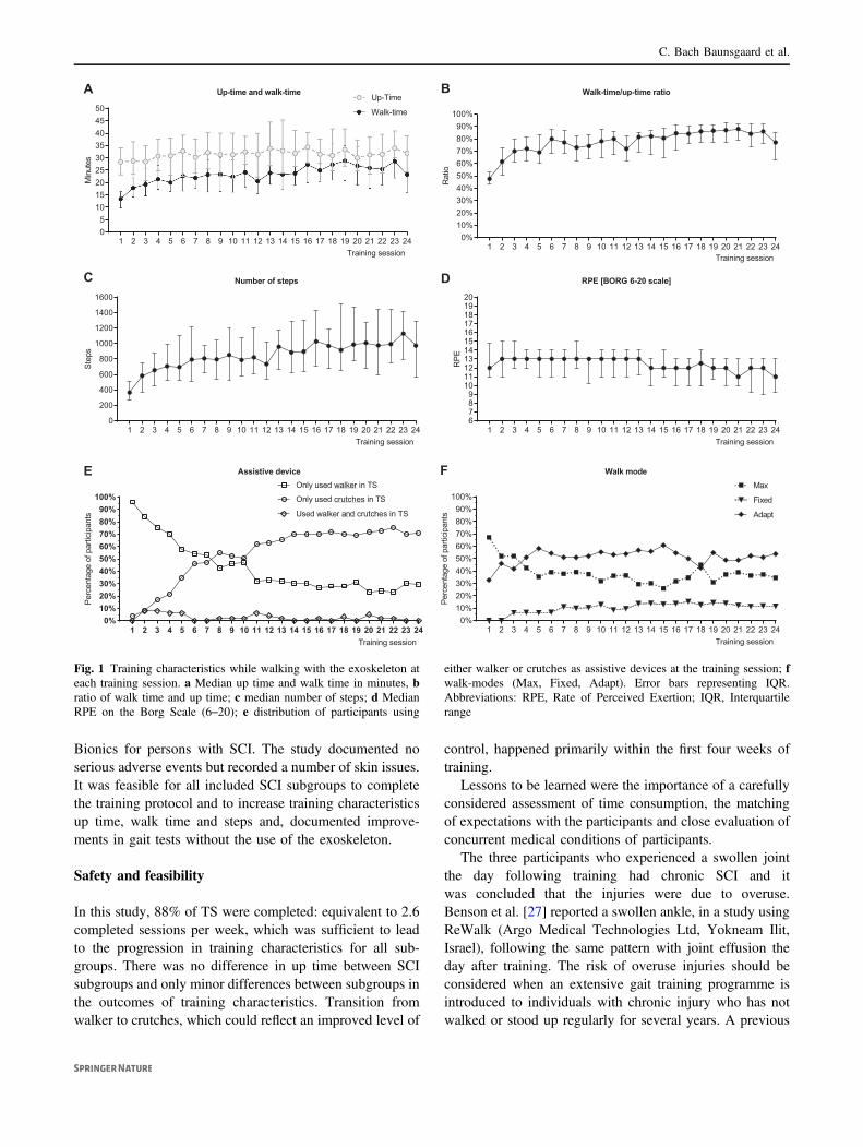
Bionics for persons with SCI. The study documented noserious adverse events but recorded a number of skin issues.It was feasible for all included SCI subgroups to completethe training protocol and to increase training characteristicsup time, walk time and steps and, documented improve-ments in gait tests without the use of the exoskeleton.
Safety and feasibility
In this study, 88% of TS were completed: equivalent to 2.6completed sessions per week, which was sufficient to leadto the progression in training characteristics for all sub-groups. There was no difference in up time between SCIsubgroups and only minor differences between subgroups inthe outcomes of training characteristics. Transition fromwalker to crutches, which could reflect an improved level of
control, happened primarily within the first four weeks oftraining.
Lessons to be learned were the importance of a carefullyconsidered assessment of time consumption, the matchingof expectations with the participants and close evaluation ofconcurrent medical conditions of participants.
The three participants who experienced a swollen jointthe day following training had chronic SCI and itwas concluded that the injuries were due to overuse.Benson et al. [27] reported a swollen ankle, in a study usingReWalk (Argo Medical Technologies Ltd, Yokneam Ilit,Israel), following the same pattern with joint effusion theday after training. The risk of overuse injuries should beconsidered when an extensive gait training programme isintroduced to individuals with chronic injury who has notwalked or stood up regularly for several years. A previous
1 2 3 4 5 6 7 8 9 10 11 12 13 14 15 16 17 18 19 20 21 22 23 2405
101520253035404550
Up-time and walk-time
Training session
Min
utes
Up-Time
Walk-time
1 2 3 4 5 6 7 8 9 10 11 12 13 14 15 16 17 18 19 20 21 22 23 240
200
400
600
800
1000
1200
1400
1600
Number of steps
Training session
Step
s
1 2 3 4 5 6 7 8 9 10 11 12 13 14 15 16 17 18 19 20 21 22 23 240%
10%20%30%40%50%60%70%80%90%
100%
Assistive device
Training session
Perc
enta
geof
parti
cipa
nts
Only used walker in TS
Only used crutches in TS
Used walker and crutches in TS
1 2 3 4 5 6 7 8 9 10 11 12 13 14 15 16 17 18 19 20 21 22 23 240%
10%20%30%40%50%60%70%80%90%
100%
Walk mode
Training session
Perc
enta
geof
parti
cipa
nts
Max
Fixed
Adapt
1 2 3 4 5 6 7 8 9 10 11 12 13 14 15 16 17 18 19 20 21 22 23 246789
1011121314151617181920
RPE [BORG 6-20 scale]
Training sessionR
PE
1 2 3 4 5 6 7 8 9 10 11 12 13 14 15 16 17 18 19 20 21 22 23 240%
10%20%30%40%50%60%70%80%90%
100%
Walk-time/up-time ratio
Training session
Rat
io
A B
C D
E F
Fig. 1 Training characteristics while walking with the exoskeleton ateach training session. a Median up time and walk time in minutes, bratio of walk time and up time; c median number of steps; d MedianRPE on the Borg Scale (6–20); e distribution of participants using
either walker or crutches as assistive devices at the training session; fwalk-modes (Max, Fixed, Adapt). Error bars representing IQR.Abbreviations: RPE, Rate of Perceived Exertion; IQR, Interquartilerange
C. Bach Baunsgaard et al.

study [10] on the Ekso reported skin issues of category I incontact areas with the skin and similar findings have alsobeen reported with the ReWalk [27].
Overall, we conclude that training with the exoskeletonsfrom Ekso Bionics is safe, in line with the results of pre-vious studies [12]. However, it is important that particularattention is given to prone skin.
Physical strain and RPE
Robotic exoskeletons have been designed for gait training inrehabilitation facilities, but can potentially be used asmobility devices for persons with paralysis. The devices canbe seen as a further development of mechanical orthoses,such as the reciprocating gait orthosis. The daily use of thesewere often limited because of high energy demand andfatigue experienced after walking short distances [28] andhave resulted in limited long-term use by individuals [29].Self-perceived exertion was in the range from “fairly light” to“somewhat hard”, which was supported by the relative lowincrease in HR measure of 15–21% from sitting to standing.This result is also in line with previous findings of walkingwith an exoskeleton [8, 12]. Interestingly, we did not findsubgroup differences in RPE. The reason could be that dif-ferent levels of assistance from exoskeleton were allowed. Itshould also be noted that the subjective Borg RPE may notbe accurate in SCI [30]. However, all taken together, theresults indicate that walking can be performed for anextended period of time by persons with a variety of SCI.
Changes in gait function without the exoskeleton andLEMS
The recently injured participants who had gait functionwithout the exoskeleton improved in TUG, 10MWT, BBSand LEMS from baseline to TS24 and to follow up.Chronically injured participants improved TUG and BBS inthe same periods. The significant changes from baseline to
follow up indicate that these changes were sustained overtime. Some degree of neurological recovery would beexpected in the early phase after injury whereas this is lesslikely to happen in the chronic phase [4, 5, 31]. The increaseseen in LEMS in the recently injured group but not in thechronically injured group could be a reflection of that.
Improvements in 10MWT, TUG and LEMS have beenreported after three months of training with the exoskeletonHAL (Hybrid Assistive Limb; Cyberdyne, Inc., Japan) [32]and on 10MWT and TUG using the Lokomat [33]. Thelatter study [33] also found WISCI II to be less sensitive tochanges in gait function in complete SCI compared to the10MWT and TUG. The same could be suggested to be thecase in our study.
The minimal clinically important difference (MCID) forthe timed tests was reported by Lam et al. [34] to be 10.8 s(s) for the TUG test and 0.13 m/s for the 10MWT and theminimal detectable difference (MDD) was 3.9 s for theTUG test and 0.05 m/s for 10MWT. These were calculatedon distribution scores measured over a three month period.Similar values were reported by Wu et al. [35]. In our studyTUG improved by 7.0 and 7.8 s from baseline to TS24 forrecently and chronically injured respectively. Gait speed inthe 10MWT increased by 0.07 m/s for both groups, basedon the marginal means. These values are above MDD butbelow MCID. However, considering the relatively limitedtraining period of two months and the fact that the testswere performed at comfortable speed, these results couldindicate a potential for clinically relevant changes.
The improvements in the gait tests TUG and BBS in thechronically injured group could reflect improvements pri-marily in balance function, also supported by case reports inthis study of improved sitting balance. This seems plausible,since walking with the device relies on correct weight shiftfrom one leg to the other for initiating the next step.Training weight shift should even facilitate the training ofbalance skills. This could be a direction for future studies toexplore.
Baseli
neTS12
TS24 FU0%
10%20%30%40%50%60%70%80%90%
100%
Recently injured, N=25
Per
cent
age
ofpa
rtici
pant
s
A
Time point
Baseli
neTS12
TS24 FU0%
10%20%30%40%50%60%70%80%90%
100%
Chronically injured, N=27
Per
cent
age
ofpa
rtici
pant
s
Participantswith gaitfunction
Participantswithout gaitfunction
B
Time point
Fig. 2 Distribution ofparticipants with and withoutgait function over time amongrecently injured a andchronically injured b. LEMSLower Extremity Motor Score,TS training session, TSI timesince injury
Exoskeleton gait training after SCI

Strengths and limitations
A strength of this study is that we included both participantswith para- and tetraplegia, recent (<1 year) and chronic (>1year) injuries and motor complete as well as incompleteSCI. This reflects the nature of the SCI population [36] andincreases external validity. However, there is a selectionbias of the study population, since the study was non-blinded and non-controlled and only participants who couldcommit to the training protocol were included. This wouldnot reflect the SCI population as a whole. The heterogeneityof the study population resulted in relatively small samplesizes for the subgroups and lowered the statistical power incalculations. The differences between the two versions ofdevices from Ekso Bionics, the Ekso and the Ekso GT wereconsidered minor and not to confound the results withregards to the objective of the study. Hence, both versionswere allowed in the study. Participants from the age of 15were allowed according to the protocol but none under theage of 18 were included.
To our knowledge, no other studies have tested gaitfunction outside of the device with this or a similar overground walking exoskeleton. However it should be stressedthat the documented changes in the gait tests cannot beassigned to the exoskeleton training intervention with thiscurrent study design.
Conclusion
In conclusion, this multicentre study showed that theexoskeletons from Ekso Bionics is safe and feasible for useby persons with SCI, including persons with paraplegia aswell as tetraplegia, recent and chronic injury as well ascomplete and incomplete SCI. Likewise, it was feasible forall the subgroups to increase training characteristics duringtraining. Self-reported RPE, supported by HR measure-ments, was relatively low and indicates that the device canbe used for longer periods of time. For those participantswith gait function outside of the exoskeleton, results indi-cate improvements primarily in balance. This stronglysupports continued research in future randomized controlledtrials to test wearable exoskeleton gait training in compar-ison to other types of gait training.
Data archiving
All relevant data are within this manuscript and raw data arearchived by the authors.
Acknowledgements We thank Linda Jones, PT, MS, Consultant, forwriting the initial protocol. We would also like to thank all participantswith SCI for being part of the study and to express our gratitude to thefollowing therapists who took part in the training protocol and datacollection:Mette Skov Henriksen and Mats Christer Nilfyr (Clinic forSpinal Cord Injuries, Rigshospitalet, University of Copenhagen,
Fig. 3 Gait tests, balance tests and lower extremity motor score(LEMS) over time. Group statistics are shown in Table 4. TUG, TimedUp and Go, 10MWT, 10 Metre Walk Test, BBS, Berg Balance Scale,
WISCI II, Walking Index for Spinal Cord Injury II, LEMS, LowerExtremity Motor Score
C. Bach Baunsgaard et al.

Hornbaek, Denmark); Michael Baumberger and Ines Bersch-Porada(Swiss Paraplegic Centre (SPC), Nottwil, Switzerland); SebastianStallasch and Sebastian Lux (Universitäts- und Rehabilitationskliniken(RKU), Ulm, Germany); Esperanza Moreno and German Novillo(Fundación Lesionado Medular (FLM), Madrid, Spain); NiklasFransson, Anna-Karin Hansson, Anna Granström and MikaelLundgren (Clinical Department of Rehabilitation Medicine at Lin-köping University Hospital, Jönköping hospital and Västervik hospi-tal, Sweden); Margareta Arnell and Peter Svensson (Spinal CordRehabilitation Unit, Uppsala University Hospital, Sweden); OleChristian Andersen, Anna-Beth Netteland, Trygve Danielsen, Gyri S.Ingebretsen (Sunnaas Sykehus HF, Oslo, Norway); Eloy Opisso,Raquel Lopez, Josep Medina, Manel Ochoa and Eva Morales (InstitutGuttmann, Neurorehabilitation Hospital, Barcelona, Spain); DavidGobets, Mark van de Mijll Dekker, Ruth Sijsma (Heliomare Rehabi-litation Center, Wijk aan Zee, The Netherlands). All authors weresupported by their institutions’ internal funds. Ekso Bionics™ pro-vided an unconditional grant for protocol writing, salary for the clin-ical study lead, and expenses related to the shared database to compiledata across the nine centres.
Funding Each centre was funded independently by grants or internalfunds required for data collection on site. Department of RehabilitationMedicine and Department of Medical and Health Sciences, LinköpingUniversity, Linköping, Sweden were funded by Stiftelsen Promobilia,Region Östergötland and Medical Research Council of SoutheastSweden. Spinal Cord Rehabilitation Unit, Uppsala University Hospi-tal, Sweden was funded by Uppsala University Hospital ALF funds.Sunnaas Rehabilitation Hospital, Nesoddtangen, Norway was fundedby Sunnaas Stiftelsen (The Sunnaas Foundation). Remaining centres
were funded by internal funds. Ekso Bionics provided an uncondi-tional grant for protocol writing, salary for the clinical study lead, andexpenses related to the shared database to compile data across the ninecentres.
Compliance with ethical standards
Conflict of interest The authors declare that they have no competinginterests.
References
1 Kirshblum SC, Priebe MM, Ho CH, Scelza WM, Chiodo AE,Wuermser L-A. Spinal Cord Injury Medicine. 3. RehabilitationPhase After Acute Spinal Cord Injury. Arch Phys Med Rehabil.2007;88:S62–70.
2 Ditunno PL, Patrick M, Stineman M, Ditunno JF. Who wants towalk? Preferences for recovery after SCI: a longitudinal and cross-sectional study. Spinal Cord. 2008;46:500–6.
3 Burns SP, Golding DG, Rolle WA, Graziani V, Ditunno JF.Recovery of ambulation in motor-incomplete tetraplegia. ArchPhys Med Rehabil. 1997;78:1169–72.
4 Mehrholz J, Kugler J, Pohl M. Locomotor training for walkingafter spinal cord injury. Cochrane Database Syst Rev. 2012;11:CD006676.
5 Waters RL, Yakura JS, Adkins RH, Sie I. Recovery followingcomplete paraplegia. Arch Phys Med Rehabil. 1992;73:784–9.
6 Van Kammen K, Boonstra A, Reinders-Messelink H, den Otter R.The Combined Effects of Body Weight Support and Gait Speed on
Table 4 Gait and balance tests of participants with gait function
Estimated marginal means (95% CI) Univariate teststatistics
Pairwise comparisons (P-value)
Baseline TS12 TS24 Follow up F-test P-value
Baseline vs.TS12
Baseline vs.TS24
Baselinevs. FU
Recently injured(n= 15)
TUG [s] 38.3(30.5–46.1)
36.6(29.0–44.2)
31.3(23.8–38.8)
28.3(20.8–35.8)
11.030 <0.001 1.000 0.007 <0.001
10MWT[s]
35.3(26.5–44.1)
35.8(27.1–44.4)
28.6(20.0–37.1)
26.0(17.5–34.4)
9.127 <0.001 1.000 0.041 0.002
BBS 25.4(18.2–32.5)
28.8(21.8–35.8)
31.5(24.6–38.4)
33.3(26.4–40.2)
9.846 <0.001 0.183 0.001 <0.001
WISCI II 12.5(10.5–14.4)
12.6(10.7–14.6)
12.8(11.0–14.7)
13.7(11.8–15.5)
1.667 0.183 1.000 1.000 0.365
LEMS 19.4(11.6–27.3)
24.1(16.3–32.0)
— 24.0(16.1–31.8)
5.082 0.01 — 0.022 0.028
Chronically injured(n= 12)
TUG [s] 35.0(21–49.0)
31.4(17.4–45.4)
27.2(13.2–41.2)
28.7(14.7–42.6)
5.558 0.004 0.555 0.005 0.028
10MWT[s]
33.8(20.8–46.8)
33.2(20.2–46.2)
27.3(14.3–40.3)
27.0(14.0–40.0)
2.582 0.072 1.000 0.322 0.268
BBS 25.0(12.4–37.6)
27.8(15.2–40.4)
28.9(16.3–41.5)
29.0(16.4–41.6)
4.582 0.009 0.177 0.021 0.017
WISCI II 14.0(10.7–17.3)
14.3(11.0–17.6)
13.7(10.4–17.0)
13.9(10.6–17.2)
0.466 0.708 1.000 1.000 1.000
LEMS 14.5(7.7–21.3)
14.8(8.0–21.6)
— 14.7(7.8–21.5)
0.310 0.735 — 1.000 1.000
Individual plots are shown in Fig. 3. Pairwise comparisons are Bonferroni corrected
BBS Berg Balance Scale, CI confidence interval, LEMS Lower Extremity Motor Score, TUG timed up and go, TSI time since injury, WISCI IIWalking Index for Spinal Cord Injury II, 10MWT 10 metre walk test
Exoskeleton gait training after SCI

Gait Related Muscle Activity: A Comparison between Walking inthe Lokomat Exoskeleton and Regular Treadmill Walking. PLoSONE. 2014;9:e107323.
7 Stamatakis E, Rogers K, Ding D, Berrigan D, Chau J, Hamer M,et al. All-cause mortality effects of replacing sedentary time withphysical activity and sleeping using an isotemporal substitutionmodel: a prospective study of 201,129 mid-aged and older adults.Int J Behav Nutr Phys Acta. 2015;12:121.
8 Kozlowski AJ, Bryce TN, Dijkers MP. Time and effort required bypersons with spinal cord injury to learn to use a powered exos-keleton for assisted walking. Top Spinal Cord Inj Rehabil.2015;21:110–21.
9 Zeilig G, Weingarden H, Zwecker M, Dudkiewicz I, Bloch A,Esquenazi A. Safety and tolerance of the ReWalk exoskeleton suitfor ambulation by people with complete spinal cord injury: a pilotstudy. J Spinal Cord Med. 2012;35:96–101.
10 Kolakowsky-Hayner SA. Safety and Feasibility of using theEksoTM Bionic Exoskeleton to Aid Ambulation after Spinal CordInjury. J Spine 2013. https://doi.org/10.4172/2165-7939.S4-003
11 Esquenazi A, Talaty M, Packel A, Saulino M. The ReWalkpowered exoskeleton to restore ambulatory function to individualswith thoracic-level motor-complete spinal cord injury. Am J PhysMed Rehabil Assoc Acad Physiatr. 2012;91:911–21.
12 Miller LE, Zimmermann AK, Herbert WG. Clinical effectivenessand safety of powered exoskeleton-assisted walking in patientswith spinal cord injury: systematic review with meta-analysis. MedDevices Auckl NZ. 2016;9:455–66.
13 Sale P, Russo EF, Russo M, Masiero S, Piccione F, Calabrò RS,et al. Effects on mobility training and de-adaptations in subjectswith Spinal Cord Injury due to a Wearable Robot: a preliminaryreport. BMC Neurol. 2016;16:12.
14 Kressler J, Thomas CK, Field-Fote EC, Sanchez J, Widerström-Noga E, Cilien DC, et al. Understanding Therapeutic Benefits ofOverground Bionic Ambulation: Exploratory Case Series in Per-sons With Chronic, Complete Spinal Cord Injury. Arch Phys MedRehabil. 2014;95:1878–87.
15 Borg GA. Psychophysical bases of perceived exertion. Med SciSports Exerc. 1982;14:377–81.
16 Anon. Perceived Exertion (Borg Rating of Perceived ExertionScale) | Physical Activity | CDC. https://www.cdc.gov/physicalactivity/basics/measuring/exertion.htm (accessed 26 Apr2017).
17 National Pressure Ulcer Advisory Panel, European Pressure UlcerAdvisory Panel and Pan Pacific Pressure Injury Alliance. Preven-tion and Treatment of Pressure Ulcers: Quick Reference Guide.Emily Haesler. Osborne Park, Western Australia: CambridgeMedia; 2014. Available at: http://www.npuap.org/wp-content/uploads/2014/08/Quick-Reference-Guide-DIGITAL-NPUAP-EPUAP-PPPIA-Jan2016.pdf (accessed on 15 Aug 2016).
18 Kirshblum SC, Burns SP, Biering-Sorensen F, Donovan W,Graves DE, Jha A, et al. International standards for neurologicalclassification of spinal cord injury (revised 2011). J Spinal CordMed. 2011;34:535–46.
19 Itzkovich M, Gelernter I, Biering-Sorensen F, Weeks C, LarameeMT, Craven BC, et al. The Spinal Cord Independence Measure(SCIM) version III: reliability and validity in a multi-centerinternational study. Disabil Rehabil. 2007;29:1926–33.
20 Alexander MS, Anderson K, Biering-Sorensen F, Blight AR,Brannon R, Bryce T, et al. Outcome Measures in Spinal CordInjury. Spinal Cord. 2009;47:582–91.
21 Marino RJ, Graves DE. Metric properties of the ASIA motor score:subscales improve correlation with functional activities. Arch PhysMed Rehabil. 2004;85:1804–10.
22 Poncumhak P, Saengsuwan J, Kamruecha W, Amatachaya S.Reliability and validity of three functional tests in ambulatorypatients with spinal cord injury. Spinal Cord. 2013;51:214–7.
23 van Hedel HJ, Wirz M, Dietz V. Assessing walking ability insubjects with spinal cord injury: validity and reliability of 3walking tests. Arch Phys Med Rehabil. 2005;86:190–6.
24 Jackson AB, Carnel CT, Ditunno JF, Read MS, Boninger ML,Schmeler MR, et al. Outcome measures for gait and ambulation inthe spinal cord injury population. J Spinal Cord Med.2008;31:487–99.
25 Wirz M, Müller R, Bastiaenen C. Falls in persons with spinal cordinjury: validity and reliability of the Berg Balance Scale. Neuror-ehabil Neural Repair. 2010;24:70–7.
26 Lemay J-F, Nadeau S. Standing balance assessment in ASIA Dparaplegic and tetraplegic participants: concurrent validity of theBerg Balance Scale. Spinal Cord. 2010;48:245–50.
27 Benson I, Hart K, Tussler D, Middendorp van JJ. Lower-limbexoskeletons for individuals with chronic spinal cord injury:findings from a feasibility study. Clin Rehabil. 2016;30:73–84.
28 Bernardi M, Canale I, Castellano V, Di Filippo L, Felici F,Marchetti M. The efficiency of walking of paraplegic patientsusing a reciprocating gait orthosis. Paraplegia. 1995;33:409–15.
29 Sykes L, Edwards J, Powell ES, Ross ER. The reciprocating gaitorthosis: long-term usage patterns. Arch Phys Med Rehabil.1995;76:779–83.
30 Lewis JE, Nash MS, Hamm LF, Martins SC, Groah SL. Therelationship between perceived exertion and physiologic indicatorsof stress during graded arm exercise in persons with spinal cordinjuries. Arch Phys Med Rehabil. 2007;88:1205–11.
31 Waters RL, Adkins RH, Yakura JS, Sie I. Motor and sensoryrecovery following incomplete tetraplegia. Arch Phys Med Reha-bil. 1994;75:306–11.
32 Aach M, Cruciger O, Sczesny-Kaiser M, Höffken O, Meindl RC,Tegenthoff M, et al. Voluntary driven exoskeleton as a new toolfor rehabilitation in chronic spinal cord injury: a pilot study. SpineJ Off J North Am Spine Soc. 2014;14:2847–53.
33 Wirz M, Zemon DH, Rupp R, Scheel A, Colombo G, Dietz V,et al. Effectiveness of automated locomotor training in patientswith chronic incomplete spinal cord injury: A multicenter trial.Arch Phys Med Rehabil. 2005;86:672–80.
34 Lam T, Noonan VK, Eng JJ. A systematic review of functionalambulation outcome measures in spinal cord injury. Spinal Cord.2008;46:246–54.
35 Wu X, Liu J, Tanadini LG, Lammertse DP, Blight AR, KramerJLK, et al. Challenges for defining minimal clinically importantdifference (MCID) after spinal cord injury. Spinal Cord.2015;53:84–91.
36 Ijzerman MJ, Baardman G, Hermens HJ, Veltink PH, Boom HBK,Zilvold G. Comparative trials on hybrid walking systems forpeople with paraplegia: An analysis of study methodology. Pros-thet Orthot Int. 1999;23:260–73.
Affiliations
Carsten Bach Baunsgaard 1 ● Ulla Vig Nissen1 ● Anne Katrin Brust2 ● Angela Frotzler2 ● Cornelia Ribeill3 ● Yorck-Bernhard Kalke3 ●
Natacha León4 ● Belén Gómez4 ● Kersti Samuelsson5 ● Wolfram Antepohl5 ● Ulrika Holmström6 ● Niklas Marklund 6 ● Thomas Glott7 ●
Arve Opheim7,8 ● Jesus Benito9 ● Narda Murillo9 ● Janneke Nachtegaal10 ● Willemijn Faber10 ● Fin Biering-Sørensen1
C. Bach Baunsgaard et al.

1 Clinic for Spinal Cord Injuries, Rigshospitalet, University ofCopenhagen, Copenhagen, Denmark
2 Swiss Paraplegic Centre (SPC), Nottwil, Switzerland
3 SCI Centre Orthopaedic Department Ulm University,Ulm, Germany
4 Fundación Lesionado Medular (FLM), Madrid, Spain
5 Department of Rehabilitation Medicine and Department ofMedical and Health Sciences, Linköping University,Linköping, Sweden
6 Spinal Cord Rehabilitation Unit, Uppsala University Hospital,Uppsala, Sweden
7 Sunnaas Rehabilitation Hospital, Nesoddtangen, Norway
8 Rehabilitation Medicine, Institute of Neuroscience andPhysiology, Sahlgrenska Academy, University of Gothenburg,Gothenburg, Sweden
9 Institute Guttmann, Neurorehabilitation Hospital, Barcelona, Spain
10 Heliomare Rehabilitation Center, Wijk aan Zee, The Netherlands
Exoskeleton gait training after SCI

JRM
JRM
Jour
nal o
f R
ehab
ilita
tion
Med
icin
eJR
MJo
urna
l of R
ehab
ilita
tion
Med
icin
e
Journal Compilation © 2018 Foundation of Rehabilitation Information. ISSN 1650-1977This is an open access article under the CC BY-NC license. www.medicaljournals.se/jrm
doi: 10.2340/16501977-2372
ORIGINAL REPORTJ Rehabil Med 2018; 50: 806–813
EXOSKELETON GAIT TRAINING AFTER SPINAL CORD INJURY: AN EXPLORATORY STUDY ON SECONDARY HEALTH CONDITIONS
Carsten Bach BAUNSGAARD, MD1, Ulla Vig NISSEN, PhD1, Anne Katrin BRUST, MSc2, Angela FROTZLER, PhD2, Cornelia RIBEILL, Dr. Med3, Yorck-Bernhard KALKE, Dr. Med3, Natacha LEÓN, MD4, Belén GÓMEZ, BSc4, Kersti SAMUELSSON, PhD5, Wolfram ANTEPOHL, Dr. Med5, Ulrika HOLMSTRÖM, PhD6, Niklas MARKLUND, PhD6, Thomas GLOTT, Dr. Med7, Arve OPHEIM, PhD7–9, Jesus BENITO PENALVA, PhD10, Narda MURILLO, PhD10, Janneke NACHTEGAAL, PhD11, Willemijn FABER, MD11 and Fin BIERING-SØRENSEN, Dr. Med1
From the 1Clinic for Spinal Cord Injuries, Rigshospitalet, University of Copenhagen, Denmark, 2Swiss Paraplegic Centre (SPC), Nottwil, Switzerland, 3SCI Centre Orthopaedic Department Ulm University, Germany, 4Fundación Lesionado Medular (FLM), Madrid, Spain, 5Department of Rehabilitation Medicine and Department of Medical and Health Sciences, Linköping University, Linköping, 6Spinal Cord Rehabilitation Unit, Uppsala University Hospital, Uppsala, Sweden, 7Sunnaas Rehabilitation Hospital, Nesoddtangen, Norway, 8Rehabilitation Medicine, Institute of Neuroscience and Physiology, Sahlgrenska Academy, University of Gothenburg, 9Habilitation & Health, Region Västra Götaland, Gothenburg, Sweden, 10Institute Guttmann, Neurorehabilitation Hospital, Barcelona, Spain, and 11Heliomare Rehabilitation Center, Wijk aan Zee, The Netherlands
LAY ABSTRACTThis report from a European multicentre, prospective, observational, open-label, exploratory study examines the effects of gait training using an exoskeleton (Ekso Bionics) after spinal cord injury on spasticity, pain, range of motion, bowel and lower urinary tract function, acti-vities of daily living and quality of life. Exoskeleton gait training seemed to be well-tolerated in spinal cord injury participants with neuropathic and nociceptive pain, but pain did not change during 8 weeks of training. Compa-red with testing prior to a single training session, spas-ticity decreased after training when calculating a sum-score for multiple lower extremity muscle groups. The results indicate a benefit in terms of independence mea-sure SCIM III as well as quality of life over time. Bowel and lower urinary tract function did not change overall. Future studies could investigate these parameters com-pared with traditional gait training for spinal cord injury, as well as testing more intensive training protocols.
Objective: To explore changes in pain, spasticity, range of motion, activities of daily living, bowel and lower urinary tract function and quality of life of in-dividuals with spinal cord injury following robotic exoskeleton gait training. Design: Prospective, observational, open-label mul-ticentre study.Methods: Three training sessions per week for 8 weeks using an Ekso GT robotic exoskeleton (Ekso Bionics). Included were individuals with recent (<1 year) or chronic (>1 year) injury, paraplegia and te-traplegia, complete and incomplete injury, men and women. Results: Fifty-two participants completed the train-ing protocol. Pain was reported by 52% of partici-pants during the week prior to training and 17% during training, but no change occurred longitudinal-ly. Spasticity decreased after a training session com-pared with before the training session (p < 0.001), but not longitudinally. Chronically injured partici-pants increased Spinal Cord Independence Measure (SCIM III) from 73 to 74 (p = 0.008) and improved life satisfaction (p = 0.036) over 8 weeks of training. Recently injured participants increased SCIM III from 62 to 70 (p < 0.001), but no significant change occurred in life satisfaction. Range of motion, bowel and lower urinary function did not change over time.Conclusion: Training seemed not to provoke new pain. Spasticity decreased after a single training ses-sion. SCIM III and quality of life increased longitudi-nally for subsets of participants.
Key words: exoskeleton; spinal cord injury; rehabilitation; pain; spasticity; SCIM III.
Accepted May 30, Epub ahead of print Sep 5, 2018
J Rehabil Med 2018; 50: 806–813
Correspondence address: Carsten Bach Baunsgaard, Clinic for Spinal Cord Injuries, Rigshospitalet, University of Copenhagen, Havnevej 25, DK-3100 Hornbaek, Denmark. E-mail: [email protected], [email protected]
Spinal cord injury (SCI) gives rise to a large number of secondary health conditions, including pain (1,
2), spasticity (2, 3) decreased range of motion (ROM) (3), decreased independence (4), bowel (5) and bladder dysfunction (6), and decreased quality of life (QoL) (7). Among individuals with SCI, these issues should be addressed with high priority (8, 9), as many of the secondary health conditions have been shown to be independently associated with decreased QoL (10, 11).
Exoskeleton training has been developed as a reha-bilitation tool and is approved for the rehabilitation of individuals with SCI. Gait function has often been the focus of studies, but indications of improvements in pain, spasticity, bowel and bladder function have also been reported (12–14). However, further studies are needed. Several commercial exoskeletons are currently available (15). The exoskeleton used for this study was the Ekso GT robotic (Ekso Bionics, Richmond, CA,

JRM
JRM
Jour
nal o
f R
ehab
ilita
tion
Med
icin
eJR
MJo
urna
l of R
ehab
ilita
tion
Med
icin
e
807Exoskeleton gait training after SCI
USA) exoskeleton. A characteristic of this exoskeleton is, among others, that initiation of a new step can be controlled by the user via sensors in the foot-plates that capture the weight transfer between the left and right legs. Power from the on-board motors controlling leg movement can be adjusted to accommodate the user’s needs. An assistive device, walker or crutches, are always used for balance.
The safety and feasibility of the training protocol for this study and changes in outcomes of gait and balance have been published separately (16). The initial publication documented that the training was safe with no serious adverse events and that the protocol was feasible for all of the subgroups of included partici-pants, i.e. recently and chronically injured individuals with paraplegia and tetraplegia, as well as those with complete and incomplete injury. There was progression over the 8 weeks in the time walking in the exoskeleton and the number of steps taken during the training ses-sions. The training was light to moderate in intensity. Lastly, there were indications of improvements in gait function and balance outside of the exoskeleton for individuals with incomplete SCI.
The objective of the present study was to explore changes in pain, spasticity, range of motion (ROM), Spinal Cord Independence Measure (SCIM III), bowel and lower urinary tract (LUT) function, and quality of life (QoL) in individuals with SCI following robotic ex-oskeleton gait training with Ekso GT, in order to guide future randomized controlled trials (RCT) and explore areas of potential benefit from exoskeleton training.
METHODS This study followed individuals with SCI over an 8-week train-ing period with an exoskeleton from Ekso GT (Ekso Bionics, Richmond, CA, USA) as an adjunct to therapy. Changes in pain, spasticity, ROM, SCIM III, bowel and LUT function and QoL, over the training period and at a follow-up were documented, in addition to spasticity pre- and post-training.
Study design
The study was a prospective, observational, open-label, multi-centre study. The 9 European SCI rehabilitation centres were located in Denmark, Germany, the Netherlands, Norway, Spain, Sweden and Switzerland.
Training protocol and participants
The training protocol consisted of 24 robotic exoskeleton gait training sessions (TSs) of 1 h, 3 times per week for 8 conse-cutive weeks. Prior to commencing the study, researchers and therapists from the 9 centres joined a 2-day kick-off meeting. The study protocol and all tests were reviewed systematically, to ensure that all therapists agreed on performance of the tests and data collection. Participants were recruited as a convenience sample at each centre and screened and included according to the eligibility criteria listed in Table I (16).
Participant characteristics were collected according to the International SCI Core Data Set Version 1.1 (17, 18). The fol-lowing 2 groups were defined; “recently injured” (Time since injury, TSI ≤1 year) and “chronically injured” (TSI >1 year). The rationale for this division in analysis was that some degree of neurological recovery would be expected in the early phase after injury, whereas this is less likely to happen in the chronic phase (19–21).
The training frequency of 3 times per week was based on general recommendations of training frequency and duration frequencies of (3–5 times per week with duration of 20–60
Table I. Eligibility criteria (16)
Inclusion criteria Exclusion criteria
A traumatic or non-traumatic SCI with either motor complete (AIS A or B) with NLI from C7 to L2 (inclusive) or motor incomplete (AIS C and D) with NLI from C1 to L2 (inclusive), as determined by the ISNCSCI
Previous training with an exoskeleton and other types of robotic assisted gait training
Age 15–65 years at time of entry to the trial (some centres 18–65 years) Spinal instabilityMore than 30 days since injury (TSI) Acute deep vein thrombosisBody height 157–188 cm OR max hip width 42 cm, upper leg length 51–61.4 cm and lower leg length 48–63.4 cm
Severe, recurrent attacks of autonomic dysreflexia requiring medical intervention
Maximum body weight 100 kg Heterotopic ossification in the lower extremities resulting in restrictions of ROM at the hip or knee
Sufficient upper extremity strength to use a front-wheeled walker Two or more pathological fractures in the last 48 months in a major weight-bearing bone in the lower extremity (femur or tibia)
Sufficient range of motion to achieve a reciprocal gait pattern and to perform sit-to-stand transition in the device
Hip subluxation
Medically stable and cleared by a physician for full weight bearing locomotor training
Cognitive deficits that limit the participant to understand instructions and safely participate in a training programme, evaluated by investigator
Standing orthostatic tolerance trial by standing for 15 min, fully supported in a standing-frame, while measuring blood pressure regularly
Spasticity assessed with the Modified Ashworth Scale = 4 in lower extremity muscles Skin integrity issues in areas in contact with the deviceConcurrent neurological injury or any other issue that in the opinion of the investigator would confound the resultsPregnancy
AIS: American Spinal Injury Association [ASIA] impairment scale; SCI: spinal cord injury; NLI: Neurological Level of Injury; ISNCSCI: International Standards for Neurological Classification of SCI.
J Rehabil Med 50, 2018

JRM
JRM
Jour
nal o
f R
ehab
ilita
tion
Med
icin
eJR
MJo
urna
l of R
ehab
ilita
tion
Med
icin
e
808 C. B. Baunsgaard et al.
min) (22) and has commonly been used in similar exoskeleton studies (23). A 1-h session was estimated to be feasible from clinical experience at the included sites.
The study did not control for other types of training that individuals attended.
Assessments
The included tests and time-points are specified below. All tests for changes over time were performed before training at baseline and at the end of the 8-week training period, i.e. training session 24 (TS24). Pain and spasticity were, in addition, also assessed midway before training at session 12 (TS12). Assessments were repeated, at a follow-up session 4 weeks after the final training session, in order to assess whether potential changes were retained. Pain and spasticity were furthermore assessed immediately after the training session (details below).
Pain. Pain was assessed with the International SCI Pain Basic Data Set (version 2.0) (24) in 2 versions. The original version asked about levels of pain experienced during the last 7 days, prior to the training. The second version was modified by asking about pain experienced during the training, assessed imme-diately after the session. Every episode of pain was recorded, according to the International SCI Pain Basic Data Set, in terms of location, type and intensity. Participants could report up to 3 types of pain at each time-point. The type of pain was classified as either neuropathic or nociceptive.
Spasticity. The Modified Ashworth Scale (MAS) was used to assess spasticity. The following muscle groups were tested bi-laterally: hip flexors and extensors, knee flexors and extensors, ankle dorsi-flexors and plantar flexors, 12 muscle groups in total. MAS assessments were performed immediately before training at baseline, TS12, TS24, and follow-up to assess changes over time. At TS12 and TS24, MAS was also tested immediately after the training session to assess changes pre-post a single training session. For statistical analysis, MAS was considered an ordinal scale. As a measure of overall spasticity, the sum-score of all 12 muscle groups was calculated for each person, at each time-point (0–60 scale). The calculated sum-score of MAS used in the analysis has not been validated, but a similar approach with a sum-score of spasticity has been described and used previously (25). If the MAS assessment triggered clonus, the measure was treated as a missing value, since the MAS scale does not include clonus, but rather describes increased tone or resistance against a passive movement. For calculation of the MAS sum-score, in case of clonus, all measures at other time-points for the same muscle were removed (list-wise deleting) to avoid an unbalanced sum-score when comparing different time-points.
Range of motion. ROM was measured by goniometry and assessed at baseline, TS24 and follow-up, bilaterally on the following lower extremity joint-movements: hip flexion, hip extension, knee flexion, knee extension, ankle dorsiflexion, and ankle plantarflexion. ROM was included, in order to assess potential mobility changes over time, as well as in conjunction with spasticity measures.
Spinal Cord Independence Measure. SCIM III (26, 27) was as-sessed for all participants at baseline, TS24 and follow-up. The subtotal-scores of Self-Care (0–20), Respiration and Sphincter Management (0–40), Mobility (0–40) and the total SCIM III-score (0–100) were used for analyses.
Bowel, lower urinary tract function and quality of life. The International SCI Basic Data Sets were used to assess bowel function (28), LUT function (29) and QoL (30).
All the International SCI Basic Data Sets were translated into the local language, according to recommendations (31). The International SCI Basic Data Sets can be found on the ISCoS homepage (http://www.iscos.org.uk/international-sci-data-sets).
Statistical analysis
Median and interquartile ranges (IQR) were used for descriptive statistics. Changes in paired, nominal data were analysed with McNemar’s test. Repeated measures of intensity of pain, MAS, ROM, SCIM III and QoL were analysed with Wilcoxon signed-rank test and Bonferroni corrected for multiple comparisons of the repeated measures. Non-parametric statistics (McNemar’s test and Wilcoxon signed-rank test) were also used for the analysis of change in MAS before and after a single training session at TS12 and TS24, the International SCI Bowel and Lower Urinary Tract Function Basic Data Sets. Significance level was set to α = 0.05. Statistical analysis was performed with IBM SPSS Statistics version 22 (IBM Corp. Released 2013. Armonk, NY, USA).
Statement of ethics
All applicable institutional and governmental regulations concerning the ethical use of human volunteers were followed during the course of this research. All the necessary approvals were obtained in each centre, including local ethics committee approvals. All participants received oral, as well as written information about the study, before written consent was ob-tained. The study followed the guidelines of the Declaration of Helsinki. The study was registered at ClinicalTrials.gov (identifier: NCT02132702).
RESULTS
The study was performed from April 2014 to March 2016. A total of 52 participants completed the training. Participant characteristics are shown in Table II. A training session was up to 1 h and the median (IQR) was 31.5 min (24.5–39.0 min) over the 8 weeks of training (16).
PainFig. 1A shows the number of participants who reported presence of pain in the week prior to training and Fig. 1B shows the number of participants who reported presence of pain during the exoskeleton training ses-sion. Pain in the week prior to training and during training (TS1, TS12 and TS24) was not always present, and thus was not reported by the same participants. Presence of pain in the previous week, at all 4 time-points (TS1, TS12, TS24, follow-up), was reported by 21 participants (40%), pain at 1, 2 or 3 time-points was reported by 15 (29%) and no pain at any time was
www.medicaljournals.se/jrm

JRM
JRM
Jour
nal o
f R
ehab
ilita
tion
Med
icin
eJR
MJo
urna
l of R
ehab
ilita
tion
Med
icin
e
809Exoskeleton gait training after SCI
reported by 16 (31%). Pain during all TSs was reported by 2 participants (4%), 4 participants (8%) had pain at 2 of the TSs and 13 (25%) reported pain at 1 of the 3 TSs where pain was surveyed.
There was no statistical difference, in either recently or chronically injured participants, from TS1 to TS24, or to follow-up of interference of pain on day-to-day activities, overall mood, ability to get a good night’s sleep or in the number of pain problems experienced in the previous week.
Seven participants reported neuropathic pain during training at TS1, TS12 and TS24, while 15 reported nociceptive pain, with an overlap of 4 participants who experienced both types of pain. Locations for nocicep-
tive pain were; lower back (n = 7), upper back (n = 1), shoulder (n = 3), hip (n = 2) and knee (n = 2). Locations of neuropathic pain during training were; thigh and lower extremity (n = 4), lower back and hip (n = 3).
When analysing the participants individually, the pain reported during training had the same location and was of the same type as pain reported between TSs in the previous week. The only exceptions to this were 2 participants who reported pain in the hip and shoulder during their first training session, but did not report pain in the previous week.
Spasticity Fig. 2 shows the MAS sum-score before and after a single training session at TS12 and at TS24. There was a significant reduction in spasticity on the MAS sum-score from pre- to post-training at both TS12 and TS24 sessions after Bonferroni correction (p = 0.05/2=0.025). The results at TS12 were a median (IQR) decrease from 4 (0–16) to 2 (0–10), p < 0.001 and at TS24 a decrease from 5 (0–14) to 2 (0–9), p < 0.001. In 8 participants, the movement of the ankle plantar flexors triggered clonus; thus, as it was not possible to rate on the MAS, the data-points were excluded from analysis. Statistical testing was done only on the MAS sum-score of the 12 muscle groups (6 muscle groups bilaterally), but all of the 12 muscle groups decreased on the MAS sum-score from before to after the training session at TS12 and TS24, calculated as rank scores.
No significant difference was found in MAS longi-tudinally from baseline to TS24 or follow up, in either the recently, or the chronically injured group.
Range of motion No differences in ROM were detected between base-line, TS24 and follow-up.
Spinal Cord Independence Measure
The results of the SCIM III for recently injured and chronically injured participants are shown in Table III.
Table II. Participant characteristics, n = 52
Characteristics
Age, years, median (IQR) 35.8 (27.5–52.6)Sex, n (%) Men 36 (69.2) Woman 16 (30.8)BMI, kg/m2, median (IQR) 24.1 (22.0–26.2)Time since injury, year, n (%); median (IQR)Recently injured (TSI ≤ 1 year) 25 (48); 0.3 (0.2–0.4)Chronically injured (TSI > 1 year) 27 (5); 5.5 (2.1–10.8)Spinal cord injury aetiology, n (%) Sport/leisure 16 (30.8) Assault 2 (3.8) Transport 17 (32.7) Fall 7 (13.5) Other traumatic cause 1 (1.9) Non-traumatic spinal cord dysfunction 9 (17.3)NLI and severity of injury at baseline, n (%) C1–C4 AIS A, B, C 0 (0) C5–C8 AIS A, B, C 4 (7.7) T1–S5 AIS A, B, C 29 (55.8) All AIS D 19 (36.5)Grouping of neurological injury, n (%)Motor complete tetraplegia C7–C8, AIS A and B 3 (5.8)Motor incomplete tetraplegia C2–C8, AIS A and B 11 (21.2)Motor complete paraplegia T1–L2, AIS A and B 22 (42.3)Motor incomplete paraplegia T1–L2, AIS C and D 16 (30.8)
IQR: interquartile range; NLI: neurological level of injury; BMI: body mass index; AIS: American Spinal Injury Association Impairment Scale; TSI: time since injury.
Fig. 1. Presence of pain in the last week and pain during exoskeleton training, n = 52. TS: training session; FU: follow-up.
Fig. 2. Modified Ashworth Scale (MAS) before and after a single training session, n = 51. CI: confidence interval.
J Rehabil Med 50, 2018

JRM
JRM
Jour
nal o
f R
ehab
ilita
tion
Med
icin
eJR
MJo
urna
l of R
ehab
ilita
tion
Med
icin
e
810 C. B. Baunsgaard et al.
injured group did not change significantly over time, whereas, satisfaction with life as a whole was found to increase significantly for the chronically injured group from baseline to TS24 (p = 0.03), as well as to follow-up (p = 0.01).
DISCUSSIONThis study aimed to assess changes in pain, spasti-city, ROM, SCIM III, QoL, bowel and LUT function, during a period of gait training using an exoskeleton. The main findings were short-term reductions in lower extremity spasticity and, for a subset of participants, there were improvements over time in SCIM III and QoL. There were no significant longitudinal changes on the other outcomes. The importance of early interven-tion is well established and there are no results from this study indicating that this training modality could not be initiated in the early phase of rehabilitation. To ensure this recommendation, further studies need to be carried out. Furthermore, also, the chronically injured participants (TSI > 1 year) seemed to benefit on some parameters from the training, indicating that the exoskeleton training might be well-suited, both for persons with recent, and those with chronic, injuries.
The recently injured group improved significantly on the total SCIM III score, as well as on all sub-cate-gories. The “Respiration and Sphincter Management” sub-category did not change on the bladder score, but increased on the bowel and use of toilet score.
The chronically injured group improved signifi-cantly on the total SCIM III score, with a median increase of 1 point. Out of all sub-categories scores, the “Respiration and Sphincter Management” was the only one with a significant increase. Within that sub-category, “Use of Toilet” had the largest change.
Bowel function and lower urinary tract function on the International SCI Basic Data SetsWithin the item “awareness of the need to defecate”, improvements were seen in 6 out of 25 participants of the recently injured group and none worsened. The chronically injured did not change on this item. No sig-nificant differences over time were found on outcomes measuring either bowel or LUT function.
Quality of life Results for recently and chronically injured parti-cipants are shown in Table IV. QoL in the recently
Table III. Spinal cord independence measure III
BaselineMedian (IQR)
TS24 Median (IQR)
Follow-up Median (IQR)
Baseline–TS24 p-value
Baseline–Follow-up p-value
Recently injured (n = 25) Self-care 18 (14.5–19.5) 18 (16.0–20.0) 18 (16.0–19.0) 0.006 0.025 Respiration and sphincter management 30 (23.5–35.5) 33 (30.5–36.0) 34 (31.5–36.0) 0.001 < 0.001 Mobility 16 (13.0–18.5) 19 (16.5–24.5) 19 (15.5–26.5) < 0.001 < 0.001 Total score 62 (51.0–73.0) 70 (65.0–77.0) 73 (67.5–78.0) < 0.001 < 0.001Chronically injured (n = 27) Self-care 18 (17.0–19.0) 18 (17.0–19.0) 18 (17.0–20.0) 0.280 0.189 Respiration and sphincter management 35 (30.0–37.0) 35 (33.0–38.0) 36 (33.0–37.0) 0.027 0.010 Mobility 19 (16.0–23.0) 19 (17.0–25.0) 19 (17.0–23.0) 0.064 0.297 Total score 73 (64.0–77.0) 74 (68.0–81.0) 74 (71.0–78.0) 0.008 0.014
IQR: interquartile range; TS24: training session 24.
Table IV. Quality of life
Baseline Median (IQR)
TS24 Median (IQR)
Follow-up Median (IQR)
Baseline–TS24 p-value
Baseline–Follow-up p-value
Recently injured (n = 25) Satisfaction with life as a wholea 5 (3.0–7.0) 6 (4.5–8.0) 6 (4.0–8.0) 0.101 0.079 Satisfaction with physical healthb 6 (3.5–7.5) 7 (4.5–8.0) 6 (4.0–7.0) 0.129 0.363 Satisfaction with psychological health, emotions and moodc 7 (5.0–8.0) 5 (4.0–8.0) 7 (4.0–8.0) 0.073 0.636Chronically injured (n = 27) Satisfaction with life as a wholea 6 (5.0–7.0) 7 (6.0–9.0) 8 (7.0–8.00) 0.036 0.010 Satisfaction with physical healthb 7 (6.0–8.0) 7 (6.0–8.0) 7 (4.0–8.0) 0.279 0.329 Satisfaction with psychological health, emotions and moodc 7 (6.0–9.0) 8 (7.0–8.0) 7.5 (5.75–8.0) 0.080 0.273
aSatisfaction with life as a whole refers to the question “Thinking about your own life and personal circumstances, how satisfied are you with your life as a whole in the past 4 weeks? Please use a scale ranging from 0 (completely dissatisfied) to 10 (completely satisfied)”. bSatisfaction with physical health refers to the question “How satisfied are you with your physical health in the past 4 weeks? Please use a scale ranging from 0 (completely dissatisfied) to 10 (completely satisfied)”. cSatisfaction with psychological health, emotions and mood refers to the question ”How satisfied are you with your psychological health, emotions and mood in the past 4 weeks? Please use a scale ranging from 0 (completely dissatisfied) to 10 (completely satisfied)”.TS24: Training Session 24.
www.medicaljournals.se/jrm

JRM
JRM
Jour
nal o
f R
ehab
ilita
tion
Med
icin
eJR
MJo
urna
l of R
ehab
ilita
tion
Med
icin
e
811Exoskeleton gait training after SCI
PainThe prevalence of pain in the week prior to training (Fig. 1) was similar to frequencies reported in the literature (10). The pain experienced in the week prior to training and during training did not differ regarding type and location of pain. Relatively few participants experienced pain during training. This could indicate that the training did not provoke a new pain sensation and that the training was well tolerated. Kolakowsky-Hayner (13) reported lower back pain during training with Ekso during walking, but this did not affect par-ticipation. In our study, although reports of low back pain were documented as the most frequent location of pain, no individuals dropped out as a result of this (16). Case reports on improvements of pain after exoskeleton training have been published (12, 14, 32), as well as decrease in pain over time after training in the Lokomat (Hocoma AG, Zurich, Switzerland) (33), but changes over time in pain between training session were not found in our study. Differences in the findings in this study from previous findings could, however, be related to the method of pain assessment.
SpasticitySpasticity did not change over the 8-week period, but decreased from before to after a training session at both TS12 and TS24. The sum-score values were in the lower end of the 0–60 sum-score scale. The reason for this was a high number of MAS ratings having a value of 0, i.e. no spasticity, since all values of the 12 muscle groups were included in the sum-score. This indicates a floor effect, which biased the analysis of the training effect on spasticity. However, when listed as rank scores, all of the individual 12 muscle groups tested had more ranks that decreased than increased in MAS score at both TS12 and TS24.
Decreased spasticity post-training could be useful for individuals with SCI who would like to participate in exoskeleton training or use it as a potential, future mobility device. If used on a daily basis, it could, po-tentially, be incorporated as part of a spasticity relief programme. Stampacchia et al. (25) found decreased spasticity, from pre- to post-training in 1 walking ses-sion in a study of 21 participants. Case reports of short-term improvements in spasticity of a few hours after training have also been published on the exoskeleton HAL (Hybrid Assistive Limb), Cyberdyne, Inc., Ibraki, Japan) (34). Thus, the results from the present study are in line with previous findings.
The MAS was chosen as assessment method for spasticity. The MAS has several limitations as a measure of spasticity (35), but is still the most-used
scale in the clinical setting (3). Furthermore, its use is recommended by the National Institute of Neurological Disorders and Stroke (NINDS) Common Data Element (CDE) (36). In a previous study, we found the MAS to be reliable, assuming that the scale is considered as an ordinal scale (37). The sum-score of MAS used in the analysis is not validated, but a similar approach with a sum-score of spasticity has been used previously (25). We argue that the sum-score could give an overall picture of the degree of lower limb spasticity. Future studies could consider adding other scales of spasticity alongside MAS to capture other aspects of spasticity.
ROM was included as an end-point, in conjunction to the spasticity tests. In case there would have been changes in ROM, this could potentially confound the MAS assessment, but no change in ROM was found.
Spinal Cord Independence Measure, bowel and bladder function
The improvements seen in SCIM III for the recently injured group could probably be attributed to early phase improvements following SCI, especially con-sidering that this group was relatively newly injured, with a median (IQR) time since injury of 0.3 (0.2–0.4) years (Table II). Interestingly, the results also indicated improvements for chronically injured participants on the total SCIM III score, although to a smaller degree.
Respiration and sphincter management sub-score on SCIM III improved for both recently and chronically injured participants. There were signs of improvement in bowel function in the recently injured group on the SCIM III sub-score and in one item on the International SCI Bowel Function Basic Data Set, but not signifi-cantly, within the chronically injured group. It can be speculated that a reason for not capturing a potential effect on bowel and LUT function was that the amount of training and intensity was too low to elicit changes. A recently published study by Hubscher et al. (38) documented improvements in urinary incontinence and time needed for defecation, assessed by the same questionnaires as in our study, following 80 daily loco-motor training sessions using body-weight-supported treadmill training.
There were, however, improvements on the SCIM III subscale “Use of Toilet”. We speculate whether this could be related to an improved balance function, since these participants also improved the Timed Up and Go test and the Berg Balance Scale in the same study period, as described previously (16). This could be a direction for future research. Previous studies on chan-ges in bowel and bladder function after exoskeleton training has been documented in case reports (13, 14).
J Rehabil Med 50, 2018

JRM
JRM
Jour
nal o
f R
ehab
ilita
tion
Med
icin
eJR
MJo
urna
l of R
ehab
ilita
tion
Med
icin
e
812 C. B. Baunsgaard et al.
Furthermore, the QoL score increased in chronically injured participants. No significant changes were detected in bowel and LUT function. Future studies assessing bowel and LUT function could investigate a longer training period and higher frequency of training.
ACKNOWLEDGEMENTSThe authors would like to express their gratitude to all parti-cipants in the study and thank the following therapists who took part in the training protocol and data collection: Mette Skov Henriksen and Mats Christer Nilfyr (Clinic for Spinal Cord Injuries, Rigshospitalet, University of Copenhagen, Hornbaek, Denmark); Michael Baumberger and Ines Bersch-Porada (Swiss Paraplegic Centre (SPC), Nottwil, Switzerland); Sebastian Stallasch and Sebastian Lux (Universitäts- und Rehabilitationskliniken (RKU), Ulm, Germany); Esperanza Moreno and German Novillo (Fundación Lesionado Medular (FLM), Madrid, Spain); Niklas Fransson, Anna-Karin Hansson, Anna Granström and Mikael Lundgren (Clinical Department of Rehabilitation Medicine at Linköping University Hospital, Jönköping hospital and Västervik hospital, Sweden); Margareta Arnell and Peter Svensson (Spinal Cord Rehabilitation Unit, Uppsala University Hospital, Sweden); Ole Christian Andersen, Anna-Beth Netteland, Trygve Danielsen, Gyri S. Ingebretsen (Sunnaas Sykehus HF, Oslo, Norway); Eloy Opisso, Raquel Lopez, Josep Medina, Manel Ochoa and Eva Morales (Institut Guttmann, Neurorehabilitation Hospital, Barcelona, Spain); David Gobets, Mark van de Mijll Dekker, Ruth Sijsma (Helio-mare Rehabilitation Center, Wijk aan Zee, The Netherlands). Finally, the authors would also like to thank Linda Jones, PT, MS, Consultant, for writing the initial protocol.
Funding. Expenses at each of the participating centres were funded independently. External funds were provided by Stif-telsen Promobilia, Region Östergötland and Medical Research Council of Southeast Sweden, which funded the Department of Rehabilitation Medicine and Department of Medical and Health Sciences, Linköping University, Linköping, Sweden. Uppsala University Hospital ALF funds funded the Spinal Cord Rehabilitation Unit, Uppsala University Hospital, Sweden. Sunnaasstiftelsen (The Sunnaas Foundation) funded Sunnaas Rehabilitation Hospital, Nesoddtangen, Norway. The remaining centres were funded by internal funds. Ekso Bionics provided an unconditional grant for protocol writing, salary for the Clini-cal Study Lead and expenses related to the shared database to compile data across the 9 centres.
Disclosure. Ekso GT provided an unconditional grant for protocol writing, salary for the Clinical Study Lead, Ulla Vig Nissen and expenses related to the shared database to compile data across the 9 centres. All other authors were supported by their institutions’ internal funds or independent funding grants.
Conflicts of interest. UVN received funding for salary in the study period from Ekso GT. All other authors have no conflicts of interest to declare.
REFERENCES1. Anke AG, Stenehjem AE, Stanghelle JK. Pain and life
quality within 2 years of spinal cord injury. Paraplegia 1995; 33: 555–559.
2. Haisma JA, van der Woude LH, Stam HJ, Bergen MP, Sluis TA, Post MW, et al. Complications following spinal cord injury: occurrence and risk factors in a longitudinal study
Quality of lifeChronically injured participants experienced an impro-ved QoL “satisfaction with life as a whole” over the study period. This result was maintained throughout the follow-up period. Adriaansen et al. (10) reported a median value of 7 on the item “satisfaction with life as a whole”, a value of 6 on “satisfaction with physical health” and 7 on the item “satisfaction with psycho-logical health” in a study on individuals with chronic SCI. Similar values on QoL were found in our study. Improvements in QoL have previous also been reported in case studies using the exoskeletons ReWalk (ReWalk Robotics Ltd, Yokneam, Israel (39) and HAL (32).
The recently injured group did not show any changes in QoL values in the present study. One explanation could be that QoL is known to improve in the years following the injury (7) and the recently injured group was only a few months post-injury.
The strengths of the current study include the hete-rogeneous sample of participants with SCI, including participants with complete and incomplete injury, tetraplegia and paraplegia, recently and chronically injury and both sexes, across different rehabilitation centres and countries, reflecting the heterogeneous nature of the SCI population. However, this also limits the possibility to detect changes in the subgroups, since this would need a larger study population to fully explore all subgroup differences. Future studies could investigate whether more frequent and intense training protocols would elicit changes in, for example, pain, bowel and LUT function. The comparator in ran-domized trials has often been body-weight-supported treadmill training, but also comparison with daily clinical routine treatment or even functional electric stimulation would be worth exploring, since the latter can be incorporated into exoskeletons.
The study was a non-controlled, non-randomized stu-dy with a sample of convenience, which does not allow conclusions to be drawn on causality of the findings in relation to the intervention of exoskeleton gait training. The results should be interpreted as indicators and can be used only to guide the direction of future studies.
Conclusion
The results of this study indicate that both recently injured and chronically injured participants can benefit from exoskeleton training. The exoskeleton training seemed to be well-tolerated in participants with pain and did not provoke new types of pain sensations. Spasticity decreased from before to after a training session, but did not change over time and there was no change in ROM. There was increase on SCIM III for both recently and chronically injured participants.
www.medicaljournals.se/jrm

JRM
JRM
Jour
nal o
f R
ehab
ilita
tion
Med
icin
eJR
MJo
urna
l of R
ehab
ilita
tion
Med
icin
e
813Exoskeleton gait training after SCI
during and after inpatient rehabilitation. J Rehabil Med 2007; 39: 393–398.
3. Sköld C, Levi R, Seiger A. Spasticity after traumatic spinal cord injury: nature, severity, and location. Arch Phys Med Rehabil 1999; 80: 1548–1557.
4. Richards C, MacKenzie N, Roberts S, Escorpizo RD. People with spinal cord injury in the United States. Am J Phys Med 2017; 96: S124-S126.
5. Krassioukov A, Eng JJ, Claxton G, Sakakibara BM, Shum S. Neurogenic bowel management after spinal cord injury: a systematic review of the evidence. Spinal Cord 2010; 48: 718–733.
6. Wyndaele J-J. The management of neurogenic lower uri-nary tract dysfunction after spinal cord injury. Nat Rev Urol 2016; 13: 705–714.
7. Westgren N, Levi R. Quality of life and traumatic spinal cord injury. Arch Phys Med Rehabil 1998; 79: 1433–1439.
8. Anderson KD. Targeting recovery: priorities of the spi-nal cord-injured population. J Neurotrauma 2004; 21: 1371–1383.
9. Simpson LA, Eng JJ, Hsieh JTC, Wolfe DL. The health and life priorities of individuals with spinal cord injury: a systematic review. J Neurotrauma 2012; 29: 1548–1555.
10. Adriaansen JJE, Ruijs LEM, van Koppenhagen CF, van As-beck FWA, Snoek GJ, van Kuppevelt D, et al. Secondary health conditions and quality of life in persons living with spinal cord injury for at least ten years. J Rehabil Med 2016; 48: 853–860.
11. Jörgensen S, Ginis KA, Iwarsson S, Lexell J. Depressive symptoms among older adults with long-term spinal cord injury: associations with secondary health conditions, sense of coherence, coping strategies and physical activity. J Rehabil Med 2017; 49: 644–651.
12. Kressler J, Thomas CK, Field-Fote EC, Sanchez J, Wider-ström-Noga E, Cilien DC, et al. Understanding therapeutic benefits of overground bionic ambulation: exploratory case series in persons with chronic, complete spinal cord injury. Arch Phys Med Rehabil 2014; 95: 1878–1887.
13. Kolakowsky-Hayner SA, Crew J, Moran S, Shah A. Safety and Feasibility of using the Ekso™ Bionic Exoskeleton to Aid Ambulation after Spinal Cord Injury. J Spine 2013; S4:003.
14. Esquenazi A, Talaty M, Packel A, Saulino M. The ReWalk powered exoskeleton to restore ambulatory function to individuals with thoracic-level motor-complete spinal cord injury. Am J Phys Med Rehabil Assoc Acad Physiatr 2012; 91: 911–921.
15. Palermo AE, Maher JL, Baunsgaard CB, Nash MS. Clinician-focused overview of bionic exoskeleton use after spinal cord injury. Top Spinal Cord Inj Rehabil 2017; 23: 234–244.
16. Baunsgaard CB, Vig Nissen U, Katrin Brust A, Frotzler A, Ribeill C, Kalke Y-B, et al. Gait training after spinal cord injury: safety, feasibility and gait function following 8 weeks of training with the exoskeletons from Ekso Bionics. Spinal Cord 2018; 56: 106–116.
17. International SCI Core Data Sets. 2016 [cited 2018 Apr 15]. Available from: http://www.iscos.org.uk/internatio-nal-sci-core-data-sets.
18. DeVivo M, Biering-Sørensen F, Charlifue S, Noonan V, Post M, Stripling T, et al. International Spinal Cord Injury Core Data Set. Spinal Cord 2006; 44: 535–540.
19. Mehrholz J, Kugler J, Pohl M. Locomotor training for walking after spinal cord injury. Cochrane Database Syst Rev 2012; 11: 1–49.
20. Waters RL, Yakura JS, Adkins RH, Sie I. Recovery follo-wing complete paraplegia. Arch Phys Med Rehabil 1992; 73: 784–789.
21. Waters RL, Adkins RH, Yakura JS, Sie I. Motor and sensory recovery following incomplete tetraplegia. Arch Phys Med Rehabil 1994; 75: 306–311.
22. Jacobs PL, Nash MS. Exercise recommendations for indivi-
duals with spinal cord injury. Sports Med Auckl NZ 2004; 34: 727–751.
23. Miller LE, Zimmermann AK, Herbert WG. Clinical effective-ness and safety of powered exoskeleton-assisted walking in patients with spinal cord injury: systematic review with meta-analysis. Med Devices Auckl NZ 2016; 9: 455–466.
24. Widerström-Noga E, Biering-Sørensen F, Bryce TN, Carde-nas DD, Finnerup NB, Jensen MP, et al. The International Spinal Cord Injury Pain Basic Data Set (version 2.0). Spinal Cord 2014; 52: 282–286.
25. Stampacchia G, Rustici A, Bigazzi S, Gerini A, Tombini T, Mazzoleni S. Walking with a powered robotic exoskeleton: Subjective experience, spasticity and pain in spinal cord injured persons. NeuroRehabilitation 2016; 39: 277–283.
26. Anderson KD, Acuff ME, Arp BG, Backus D, Chun S, Fisher K et al. United States (US) multi-center study to assess the validity and reliability of the Spinal Cord Independence Measure (SCIM III). Spinal Cord 2011; 49: 880–885.
27. Anderson K, Aito S, Atkins M, Biering-Sørensen F, Charlifue S, Curt A et al. Functional recovery measures for spinal cord injury: an evidence-based review for clinical practice and research. J Spinal Cord Med 2008; 31: 133–144.
28. Krogh K, Perkash I, Stiens SA, Biering-Sørensen F. Inter-national bowel function basic spinal cord injury data set. Spinal Cord 2009; 47: 230–234.
29. Biering-Sørensen F, Craggs M, Kennelly M, Schick E, Wyn-daele J-J. International lower urinary tract function basic spinal cord injury data set. Spinal Cord 2008; 46: 325–330.
30. Charlifue S, Post MW, Biering-Sørensen F, Catz A, Dijkers M, Geyh S, et al. International Spinal Cord Injury Quality of Life Basic Data Set. Spinal Cord 2012; 50: 672–675.
31. Biering-Sørensen F, Alexander MS, Burns S, Charlifue S, DeVivo M, Dietz V, et al. Recommendations for translation and reliability testing of international spinal cord injury data sets. Spinal Cord 2011; 49: 357–360.
32. Cruciger O, Schildhauer TA, Meindl RC, Tegenthoff M, Schwenkreis P, Citak M, et al. Impact of locomotion training with a neurologic controlled hybrid assistive limb (HAL) exoskeleton on neuropathic pain and health related quality of life (HRQoL) in chronic SCI: a case study. Disabil Rehabil Assist Technol 2016; 11: 529–534.
33. Labruyère R, van Hedel HJA. Strength training versus robot-assisted gait training after incomplete spinal cord injury: a randomized pilot study in patients depending on walking assistance. J NeuroEngineering Rehabil 2014; 11: 4.
34. Aach M, Cruciger O, Sczesny-Kaiser M, Höffken O, Meindl RC, Tegenthoff M, et al. Voluntary driven exoskeleton as a new tool for rehabilitation in chronic spinal cord injury: a pilot study. Spine J Off J North Am Spine Soc 2014; 14: 2847–2853.
35. Pandyan AD, Johnson GR, Price CI, Curless RH, Barnes MP, Rodgers H. A review of the properties and limitations of the Ashworth and modified Ashworth Scales as measures of spasticity. Clin Rehabil 1999; 13: 373–383.
36. NIH commen data elements. 2018 [cited 2018 May 14]. Available from: http://www.commondataelements.ninds.nih.gov/SCI.aspx#tab=Data_Standards.
37. Baunsgaard CB, Nissen UV, Christensen KB, Biering-Sørensen F. Modified Ashworth scale and spasm frequency score in spinal cord injury: reliability and correlation. Spinal Cord 2016; 54: 702–708.
38. Hubscher CH, Herrity AN, Williams CS, Montgomery LR, Willhite AM, Angeli CA, et al. Improvements in bladder, bowel and sexual outcomes following task-specific loco-motor training in human spinal cord injury. PLoS ONE 2018; 13: 1–26.
39. Raab K, Krakow K, Tripp F, Jung M. Effects of training with the ReWalk exoskeleton on quality of life in incomplete spinal cord injury: a single case study. Spinal Cord Ser Cases 2016; 2: 15025.
J Rehabil Med 50, 2018

Appendix – Paper III
1
Acute Metabolic and Glycemic Responses to Bionic Ambulation in Spinal Cord
Injured Humans and Controls
Jennifer L. Maher, PhD1, Carsten Bach Baunsgaard, MD2, Jan van Gerven, BSc3, Anne E Palermo, PT, DPT 1, Fin Biering-Sorensen, MD, DMSc2, Armando Mendez, PhD4, Robert W. Irwin, MD5, Mark S. Nash,
PhD1, 6 1The Miami Project to Cure Paralysis, University of Miami, Miller School of Medicine, Miami, FL, USA, 2Clinic for Spinal Cord Injuries, Rigshospitalet, Copenhagen, 3 Radboud University, Nijmegen Medical
Center 4 Division of Endocrinology, Diabetes and Metabolism, Diabetes Research Institute, University of
Miami Miller School of Medicine, 5Department of Physical Medicine and Rehabilitation, University of Miami
Miller School of Medicine, 6 Department of Neurological Surgery and Physical Medicine & Rehabilitation,
University of Miami Miller School of Medicine, Miami, FL
This study was funded by The Miami Project to Cure Paralysis.
There are no conflicts of interest to report.
Abstract
Objectives: 1) Compare energy expenditure during seated rest, standing, and prolonged bionic ambulation
(BA) or bipedal ambulation in participants with SCI and non-injured controls (CON), respectively, and 2)
test effects on post-BA glycemia in SCI.
Design: Two independent group comparison of SCI and CON.
Setting: Academic Medical Center.
Participants: 10 chronic SCI (C7-T1, AIS A-C) and 10 CON.
Interventions: A commercial bionic exoskeleton.
Main Outcome measures: Absolute and relative (to peak) oxygen consumption, perceived exertion,
carbohydrate/fat oxidation, energy expenditure, post-BA plasma glucose/insulin.
Results: Average work intensity accompanying 45 minutes of outdoor BA was <40% VO2peak, with
negligible drift after reaching steady-state. RPE did not differ between groups and reflected low exertion.
Absolute energy costs for BA and non-BA were not different between groups, despite a 565% higher
ambulation velocity in CON and 3.3x higher kilocalorie/meter in SCI. Fuel partitioning was similar between
groups and the same within groups for carbohydrate and fat oxidation. Non-significant (9%) lowering of the
area under a glucose tolerance curve following BA required 20% less insulin than at rest.
Conclusion: Work intensity during prolonged BA for this bionic is below a threshold for cardiorespiratory
conditioning but above seated rest and passive standing. BA metabolism is consistent with low RPE and
unchanged fuel partitioning from seated rest. BA did not promote beneficial effects on glycemia in well-
conditioned, euglycemic participants. These findings may differ in less fit individuals with SCI, or those with
impaired glucose tolerance. Observed trends favoring this benefit suggest they are worthy of testing.
Keywords: robotic exoskeleton; spinal cord injury; acute exercise; metabolism

Appendix – Paper III
2
Walking is described as “the ideal physical activity intervention to improve health…”1, and adopted as the
preferred activity in trials of obesity and diabetes management2. Two recent reviews3,4 describing
intervention studies in non-disabled individuals reported that moderate intensity walking increases maximal
oxygen consumption (V̇O2max) and physical functioning. Sustained walking further reduces resting heart
rate, blood pressure, waist circumference, body mass and enhances body composition3,4.
Spinal cord injury (SCI) resulting in complete motor deficits of the trunk and lower limbs restricts
independent walking, and is associated with lifelong physical deconditioning5,6 that contributes to accelerated
rate all-cause cardiovascular disease, insulin resistance, and obesity5. Regular physical activity after SCI is
therefore an effective strategy for lessening hazards imposed by these secondary conditions, and maintaining
physical capacity throughout the lifespan5. Walking by persons with SCI might thus be attractive option for
maintaining health and reserve capacity for daily functions.
Bionic exoskeletons are an evolving assistive technology, allowing individuals with SCI to walk over
relatively level and barrier-free surfaces. They offer a therapeutic option as a countermeasure to secondary
complications after SCI, and may provide an opportunity for home and community ambulation. Exoskeletons
have incorporated small motors into the hip and knee joints to initiate stepping without the need for user-
initiated muscle activation. However, exoskeletons remain challenged in their ability to simulate normal gait,
and typically require an upper extremity assistive device for stability. Thus, energy efficiency provided by
motors is counterbalanced by masses of the bionics and onboard batteries, mechanical inefficiency of gait,
and need for upper body support.
The energy demand of bionic ambulation (BA) plays an essential role in defining its practical use. If energy
demand is low, BA may be practical for home and community ambulation7. Conversely, if energy demand
is high enough to satisfy physical conditioning criteria, there may be a narrow utility for these devices in
exercise reconditioning after SCI7. However, limited data address the metabolic demand of BA in individuals
with SCI when tested under conditions that simulate typical use. A recent case series explored
cardiometabolic responses to a 6-week BA training program in individuals with motor-complete SCI. A 60-
minute BA session elicited energy expenditures and intensities similar to those observed during walking by
non-disabled individuals (1.4-2.6 kcal/min and 25-41% of peak oxygen consumption (V̇O2peak),
respectively)8. The American College of Sports Medicine (ACSM) classifies this work as ‘light to very light,'
and unlikely to improve cardiorespiratory fitness9. Another study exploring 6-minute bouts of BA described
mild efforts (35% V̇O2peak), and an average Rating of Perceived Exertion (RPE)10 representing “very light to
fairly light” effort11. These intensities were equivalent to 41.8% V̇O2peak reported in a study investigating
bouts of BA over a 10-meter distance12. By contrast, a recent case series13 reported work during a 6-minute
bout of BA within the ‘moderate intensity’ exercise category as defined by the ACSM9, and up to 1.5-2.5
times greater than reported8.
Unique study methods and different exoskeletons place distinctive demands on BA users and may account
for divergent metabolic and performance characteristics. Thus, the ability of BA devices to improve fitness
and attenuate secondary complications remains unknown. Only one study8 has investigated BA bouts of a
duration likely to be applied in clinical practice (45-60 minutes) or prescribed for exercise (30-60 minutes).
Most studies of BA have focused on considerably shorter bouts of walking11-14, which fall below the
recommended exercise guidelines15. Additionally, previous research has examined very small test
populations and case reports8,13,14 and tested BA in controlled laboratory conditions of temperature and
surface friction11-14. To more realistically assess the performance characteristics and potential benefits of
these devices requires untethered ambulation under ambient conditions of temperature and humidity, walking
on higher friction surfaces, and testing during longer walks. Thus, this investigation: 1) examined potential
differences in the energy cost and fuel substrate utilization measured in persons with SCI and non-injured
healthy controls while seated, standing and self-paced walking in a bionic exoskeleton (SCI) or bipedal
ambulation (CON); 2) examined whether there was an upward drift in metabolism while standing and
ambulating and; 3) tested effects of a single bout of bionic ambulation on post-challenge glycemia in persons
with SCI.

Appendix – Paper III
3
Methods
Participants
Fourteen individuals with SCI and 11 non-injured controls provided written consent to participate in this
study, which was approved by institutional ethical authorities. Participants were 18-60 years old without a
history of cardiovascular disease and, (for SCI participants) met inclusion and exclusion criteria in Table 17.
Bionic exoskeleton
The Ekso™ GT robotic exoskeletona (EKSO Bionics, CA, USA) was studied. The device has been described
elsewhere16,17.
Bionic ambulation habituation period
Participants with SCI underwent a 2-4 week BA habituation period. Training began indoors on smooth
linoleum and progressed to an outdoor, limited grade (<1%) parcourse over concrete pavers. Training
targeted 20 minutes/session and increased to 45 minutes of continuous activity, as tolerated. Participants
walked as long as possible, but rested if necessary. Participants used a walker or Lofstrand crutches for
balance and began in the FirstStep mode, wherein the investigator controlled the pace and trigger of steps.
When balance and ambulation control were satisfactory, participants progressed to the ProStep mode where
the user triggered a step by shifting their hips forward and laterally.
Peak exercise capacity
Once habituated to outdoor BA (range: 8-12 sessions), participants underwent a continuous progressive
exercise test on an arm ergometer (Angio, Lode BV, Groningen, Netherlands)b. The one-minute stage testing
protocol has been described18. Expired respiratory gases were collected and analyzed using the open-circuit
method (Oxycon Mobile, VIASYS, Inc.)c. Participants exercised until fatigue, indicated by verbal request to
stop or an inability to maintain cadence >55 rpm. The highest 20-second average was operationalized as
V̇O2peak. Non-injured controls underwent identical testing.
Baseline Oral Glucose Tolerance Test (OGTT)
Participants with SCI underwent a 2-hour oral glucose tolerance test using a 75-g glucose load (Trutol; NERL
Clinical Diagnostics, East Providence, RI)d ingested over 5-minutes. Participants fasted overnight (8 hours)
and were prepared under antiseptic conditions with a 21g antecubital venous catheter (Jelco) having a multi-
sample Leur cap. The catheter was kept patent with sterile 0.9% saline. Blood samples were collected at 0,
30, 60, 90, and 120 minutes into separate tubes containing clot lysis activator and sodium fluoride (glycolytic
inhibitor), which were centrifuged for 20 minutes at 1500xg. The plasma fraction was assayed for glucose
and insulin on a calibrated auto-analyzer (Cobas 6000 analyzer; Roche Diagnostics)e using manufacturer
reagents. Insulin resistance was assessed using the Homeostatic Model-2 Assessment (HOMA2-IR)
calculated as glucose (mg/dl) x insulin (mIU/L)/40519. (www.OCDEM.ox.ac.uk).
Experimental exercise trials
Testing was performed on four non-randomized days within a ten-day period.
Day 1
Participants remained seated with both feet flat on the floor. Respiratory measures were assessed as described
above. Blood pressure was measured by a calibrated electronic sphygmomanometer (A&D Engineering,
Inc.). Ratings of perceived exertion (RPE) were assessed using the Categorical-Ratio (0-10) scale of Borg10.
All measurements were recorded at rest and, to reflect a steady-state, at minutes 14-15, 29-30, and 44-45.
Cardiorespiratory outcomes measures were averaged over 20-second intervals.
Day 2
Testing on day 2 evaluated metabolic responses to the upright (standing) posture. Participants with SCI
donned the bionic exoskeleton and came to stand using the assistive device (i.e., rolling walker or Lofstrand
crutches) used to assist ambulation. Control participants stood without an assistive device. Participants
remained standing for 45 minutes during which data were collected at the same time-points as measured
during day one testing.
Day 3
Testing on day 3 examined responses to ambulation in the bionic exoskeleton (SCI) or by bipedal ambulation
(CON). Participants with SCI donned the bionic exoskeleton and walked for 45 minutes on the outdoor par

Appendix – Paper III
4
course, with data sampled at the same time-points as measured on day 2 testing. Control participants
underwent the same procedures but walked at a comfortable self-selected pace without assistance.
Day 4
Testing on day 4 examined responses to ambulation in the bionic exoskeleton under fasted conditions (SCI
only). Data sampling mirrored previous testing days. Upon completion of the 45-minute ambulation,
participants underwent a repeat OGTT. Blood glucose and insulin levels at time point 0 were considered
fasting levels.
Whole body carbohydrate and lipid oxidation and total energy expenditure
Substrate oxidation rates were derived from expired gas concentrations (V̇O2 and volume of carbon dioxide
[VCO2]) averaged at rest and over minutes 14-15, 29-30 and 44-45 of the 45 min seated, standing, and
walking bouts. The proportion of energy expenditure (EE) derived from carbohydrate (CHO) and fat, and
rates of whole-body CHO and fat oxidation were calculated using stoichiometric equations assuming
negligible protein oxidation:20
%EE from CHO = [(RER – 0.71)/0.29] x 100
%EE from fat = 100 - %EE from CHO
Whole body CHO oxidation (g/minute) = [(%EE from CHO V̇O2 ) x 5.05 kcal/ l/O2] / 4.2kcal/g CHO
Whole body fat oxidation (g/minute) = [(%EE from fat V̇O2 ) x 4.7 kcal/ l/O2] / 9kcal/g lipid,
Where V̇O2 is expressed in l/min and RER is Respiratory Exchange Ratio (RER= V̇CO2/V̇O2). Total energy
expenditure (TEE) summed the energy expended from fat and CHO oxidation.
Walking economy
Walking economy, a representation of the metabolic cost of exercise21, was computed as kcal/meter travelled.
Statistics
Data were expressed as means ± SD. Unpaired t-tests assessed the significance of difference between SCI
and CON in subject characteristics, mean distance walked, velocity, and movement economy. Significance
of differences between groups and across time for V̇O2, RPE, TEE, substrate oxidation, glucose, and insulin
were analyzed using a mixed-model two-factor (group x time) ANOVA, with repeated measures as the time
variable. Where statistically significant effects were observed, post hoc analyses used the Least Significant
Difference test (SPSS V22.x; SPSS). The area under the glucose and insulin curves (AUC) were calculated
for 120-min of the OGTT using the trapezoid rule (GraphPad Prism v5, GraphPad Software, La Jolla, CA,)f
to assess glucose tolerance. For all analyses, significance was established a priori and accepted if p was <
0.05.
Results
Subject characteristics and V̇O2peak
Ten individuals with SCI and ten CON participants completed testing (Table 2.). Fasting glucose and insulin
were available for 8 participants. Participants with SCI had lower V̇O2peak than CON (p < 0.01), however, a
V̇O2peak of 23.5 ± 7.1 ml/kg/min ranked them as ‘excellent’ for fitness based on published ratings22.
Oxygen consumption at seated, standing and walking
Walking elicited a higher mean V̇O2 than sitting or standing in both SCI (P < .001) and CON (P < .001).
However, seated and standing conditions were different in CON only (P < .05) (Table 3). The CON group
achieved a higher mean V̇O2 than the SCI group during the walking condition only (P < .001, Table 3).
Oxygen consumption in each trial was not different at any time point (rest, 14-15, 29-30, 44-45) in seated or
standing testing (Figure 1 A-B). However, there was a main effect of time on the walking condition (Figure
1C) in which V̇O2 was lower at rest than all successive timepoints (P < .001) (Figure 1). When comparing
the V̇O2peak, there was a significant main effect of time in which each time point produced a greater peak
percentage than the preceding time point (P < .001) (Figure 2.)
Total energy expenditure and whole-body substrate oxidation rates while seated, standing and walking
TEE was greater in CON than SCI when standing (P < .05) and walking (P < .001) with no differences in the
seated condition (Table 3A-C). Walking elicited significantly greater TEE than standing or sitting in both
CON and SCI (P < .001). TEE in each trial was not different at any time point (rest, 14-15, 29-30, 44-45)

Appendix – Paper III
5
while sitting or standing, although during walking TEE was lower at rest than all other time points in both
SCI (P < .05) and CON (P < .001) (Figure 3). There were no differences in substrate partitioning across trials
(Figure 4A), however, walking increased rates of CHO oxidation (Figure 4B) (P < .01) and fat oxidation
(Figure 4C) (P < .01) above rates for sitting and standing in both SCI and CON (P < .05).
Walking economy
CON walked nearly six times farther (P < .001) and faster (P < .001) than SCI with lower kcal/m (P < .01).
Fasting glucose and insulin
Figure 5 shows the 2-hour OGTT at baseline and immediately post-BA. The area under the curve (AUC)
represents changes in post-load plasma glucose and insulin. We observed a 9% and 21% reduction in post-
BA glucose and insulin AUC, respectively (Table 5), although these trends were not significant.
Discussion
Our key finding was that energy expended during prolonged BA was below guideline intensities used to
promote cardiorespiratory conditioning, but greater than expended while sitting and standing (Figure 2). To
our knowledge, this is the first study to characterize extended BA under ambient testing conditions, and
compare outcomes with a non-SCI control group. Our primary goal was to identify the energy cost of
prolonged BA and determine whether it was primarily suited to conditioning or community ambulation. The
exercise intensity during 45 minutes of outdoor BA qualified as “light”, which was verified by RPE values
reflecting “light to moderate” effort. The ACSM recommends moderate to vigorous intensity activity for
most adults to achieve and maintain health and fitness benefits9. While some deconditioned individuals with
SCI might benefit from light-moderate exercise9, it is unclear whether BA, as performed in the present study,
would be sufficiently intense to improve cardiorespiratory fitness. Previous research has reported that
individuals walking in bionic exoskeletons “work harder” than non-disabled individuals walking at similar
speeds23. Our findings indicate that the absolute energy cost is significantly lower during extended BA than
walking at a self-selected pace without a SCI, although when expressed relative to peak capacity this scenario
is reversed. BA resulted in a 4-fold lower walking economy compared to CON. This exaggerated walking
economy may explain the higher relative intensity achieved during BA. CON also walked nearly 6-times
faster than SCI (1.3 m/s v. 0.23 m/s), which is also consistent with a previous literature8. Thus, our study
produced comparable walking speeds and intensities to those observed during considerably shorter walking
distances13 12. In contrast, one study reported BA energy expenditure 2.5-times greater than those observed
in this study13. These differences can be explained in part by notable differences in exercise duration, which
were all-out 6-minute BA efforts13, compared to the longer bouts reported in this study.
Substrate partitioning was not altered across conditions in either group, however, this is not surprising given
that the intensity of walking was below the threshold beyond which a shift in fuel source from fat to
carbohydrate is commonly observed24. Nonetheless, absolute rates of carbohydrate and fat oxidation during
walking were elevated significantly above seated and standing trials in both groups.
The standing test condition revealed whether physiological responses to bionic ambulation in persons with
SCI were explained by assuming upright posture alone, or if an upward drift in metabolism occurred as a
function of time while standing and ambulating in the exoskeleton. This was important as energy costs for
BA are usually expressed as either instantaneous measurements or extended averages, and not trends. During
passive standing, the lower extremities are braced in the exoskeleton, with the only active contribution to the
upright posture from the upper extremities. Here, the energy cost of passive standing was similar to previous
report25 examining a 30-minute bout of standing in a commercial frame. It is likely that the significantly
greater TEE and RPE achieved during BA reflected the increased energy cost of propulsion rather than
postural stability. During BA, persons with SCI must use the upper limbs for support, which may explain
higher relative energy expenditure and variability between devices.
A single bout of cardiorespiratory exercise improves insulin sensitivity and glucose tolerance in healthy
persons without disability26-28, as well as those with insulin resistance and diabetes29. This observation
remains inconsistent across studies and is dependent on pre-exercise impairments in insulin sensitivity and
exercise intensity. Indeed, a single bout of aerobic exercise at 50% V̇O2peak did not improve insulin sensitivity

Appendix – Paper III
6
and glucose tolerance in healthy young men30. Conversely, some studies report improved insulin sensitivity
following conditioning where fitness is not enhanced31. More substantial improvements in metabolic
regulation occur in individuals with existing glycemic impairment, and appear exercise intensity-
dependent32. Here, the area under the glucose curve was lower after BA by 9%, requiring 20% less insulin
to achieve normoglycemia. While this represents a trend for a relative improvement in insulin sensitivity, it
was non-significant. Also, non-insulin-mediated glucose disposal during BA may partially explain the lower
post-exercise glucose concentrations, as glucose uptake may be used to support the energy cost of exercise.
Our participants were well-conditioned men without impaired glucose tolerance (HOMA2-IR), which was
established at baseline (Table 5). We may more likely observe significant differences following BA in an
SCI population with impaired glucose tolerance or type 2 diabetes, and when undergoing more intense
activity than BA.
Study limitations
Despite a larger sample than previous investigations, the sample size was still modest. Additionally, study
participants with SCI were 42% more fit than the population average22. All participants were males despite
an open inclusion for gender. We likely observed flooring effects of exercise on post-exercise glycemia by
studying euglycemic individuals. The CON group did not perform ambulation in an exoskeleton, thereby
limiting some direct comparisons between SCI and CON for walking. However, it is unrealistic to simulate
their non-assisted lower limb motion in the exoskeleton, compensate for their full trunk control, and estimate
metabolism from upper limb effort in a manner equivalent to BA with SCI.
Conclusion
Work intensity observed during prolonged BA was below a threshold targeted for cardiorespiratory
conditioning, but above that observed for seated rest and standing. The low energy cost during BA is
consistent with reports of low RPE, and fuel homeostasis that did not significantly differ from seated rest.
Modest energy expenditure during BA allowed participants to tolerate prolonged ambulation without
experiencing fatigue. BA did not improve glycemia in well-conditioned, euglycemic participants. This
finding may differ in less fit individuals with SCI, or those with impaired glucose tolerance.

Appendix – Paper III
7
References
1. Oja P, Kelly P, Murtagh EM, Murphy MH, Foster C, Titze S. Effects of frequency, intensity, duration
and volume of walking interventions on CVD risk factors: a systematic review and meta-regression analysis
of randomised controlled trials among inactive healthy adults. British journal of sports medicine.
2018;52(12):769-775.
2. Knowler WC, Barrett-Connor E, Fowler SE, et al. Reduction in the incidence of type 2 diabetes with
lifestyle intervention or metformin. New Engl J Med. 2002;346(6):393-403.
3. Hanson S, Jones A. Is there evidence that walking groups have health benefits? A systematic review
and meta-analysis. British journal of sports medicine. 2015;49(11):710-715.
4. Murtagh EM, Nichols L, Mohammed MA, Holder R, Nevill AM, Murphy MH. The effect of walking
on risk factors for cardiovascular disease: an updated systematic review and meta-analysis of randomised
control trials. Prev Med. 2015;72:34-43.
5. Maher JL, McMillan DW, Nash MS. Exercise and Health-Related Risks of Physical Deconditioning
After Spinal Cord Injury. Topics in spinal cord injury rehabilitation. 2017;23(3):175-187.
6. Rimmer JH, Schiller W, Chen MD. Effects of disability-associated low energy expenditure
deconditioning syndrome. Exerc Sport Sci Rev. 2012;40(1):22-29.
7. Palermo AE, Maher JL, Baunsgaard CB, Nash MS. Clinician-Focused Overview of Bionic
Exoskeleton Use After Spinal Cord Injury. Topics in spinal cord injury rehabilitation. 2017;23(3):234-244.
8. Kressler J, Thomas CK, Field-Fote EC, et al. Understanding therapeutic benefits of overground
bionic ambulation: exploratory case series in persons with chronic, complete spinal cord injury. Archives of
physical medicine and rehabilitation. 2014;95(10):1878-1887 e1874.
9. American College of Sports Medicine, Riebe D, Ehrman JK, Liguori G, Magal M. ACSM's
guidelines for exercise testing and prescription. Tenth edition. ed. Philadelphia, PA: Wolters Kluwer Health;
2018.
10. Borg GA. Psychophysical bases of perceived exertion. Medicine and science in sports and exercise.
1982;14(5):377-381.
11. Asselin P, Knezevic S, Kornfeld S, et al. Heart rate and oxygen demand of powered exoskeleton-
assisted walking in persons with paraplegia. Journal of rehabilitation research and development.
2015;52(2):147-158.
12. Escalona MJ, Brosseau R, Vermette M, et al. Cardiorespiratory demand and rate of perceived
exertion during overground walking with a robotic exoskeleton in long-term manual wheelchair users with
chronic spinal cord injury: A cross-sectional study. Ann Phys Rehabil Med. 2018.
13. Evans N, Hartigan C, Kandilakis C, Pharo E, Clesson I. Acute Cardiorespiratory and Metabolic
Responses During Exoskeleton-Assisted Walking Overground Among Persons with Chronic Spinal Cord
Injury. Topics in spinal cord injury rehabilitation. 2015;21(2):122-132.
14. Kressler J, Wymer T, Domingo A. Respiratory, cardiovascular and metabolic responses during
different modes of overground bionic ambulation in persons with motor-incomplete spinal cord injury: A
case series. J Rehabil Med. 2018;50(2):173-180.
15. Clinical Practice Guidelines: Spinal Cord Medicine. Identification and Management of
Cardiometabolic Risk after Spinal Cord Injury 2018;
https://www.pva.org/CMSPages/GetFile.aspx?guid=f3c29b7e-e201-4392-b241-9933dc620e40.
16. Strausser KA, Swift TA, Zoss AB, Kazerooni H, Bennett BC. Mobile Exoskeleton for Spinal Cord
Injury: Development and Testing. 2011(54761):419-425.
17. Strausser KA, Kazerooni, H. . The development and testing of a human machine interface for mobile
medical exoskeleton. 2011 IEEE/RSJ International Conference of Intelligent Robots and Systems.
2011:4911-4916.
18. Maher JL, Cowan RE. Comparison of 1- Versus 3-Minute Stage Duration During Arm Ergometry
in Individuals With Spinal Cord Injury. Archives of physical medicine and rehabilitation. 2016;97(11):1895-
1900.
19. Wallace TM, Levy JC, Matthews DR. Use and abuse of HOMA modeling. Diabetes care.
2004;27(6):1487-1495.
20. Jacobs KA, Casazza GA, Suh SH, Horning MA, Brooks GA. Fatty acid reesterification but not
oxidation is increased by oral contraceptive use in women. Journal of applied physiology. 2005;98(5):1720-
1731.

Appendix – Paper III
8
21. Womack CJ, Sieminski DJ, Katzel LI, Yataco A, Gardner AW. Oxygen uptake during constant-
intensity exercise in patients with peripheral arterial occlusive disease. Vasc Med. 1997;2(3):174-178.
22. Simmons OL, Kressler J, Nash MS. Reference fitness values in the untrained spinal cord injury
population. Archives of physical medicine and rehabilitation. 2014;95(12):2272-2278.
23. Esquenazi A, Talaty M, Packel A, Saulino M. The ReWalk powered exoskeleton to restore
ambulatory function to individuals with thoracic-level motor-complete spinal cord injury. Am J Phys Med
Rehabil. 2012;91(11):911-921.
24. Jacobs KA, Burns P, Kressler J, Nash MS. Heavy reliance on carbohydrate across a wide range of
exercise intensities during voluntary arm ergometry in persons with paraplegia. The journal of spinal cord
medicine. 2013;36(5):427-435.
25. Jacobs PL, Johnson B, Mahoney ET. Physiologic responses to electrically assisted and frame-
supported standing in persons with paraplegia. The journal of spinal cord medicine. 2003;26(4):384-389.
26. Bogardus C, Thuillez P, Ravussin E, Vasquez B, Narimiga M, Azhar S. Effect of muscle glycogen
depletion on in vivo insulin action in man. J Clin Invest. 1983;72(5):1605-1610.
27. Heath GW, Gavin JR, 3rd, Hinderliter JM, Hagberg JM, Bloomfield SA, Holloszy JO. Effects of
exercise and lack of exercise on glucose tolerance and insulin sensitivity. Journal of applied physiology:
respiratory, environmental and exercise physiology. 1983;55(2):512-517.
28. Richter EA, Mikines KJ, Galbo H, Kiens B. Effect of exercise on insulin action in human skeletal
muscle. Journal of applied physiology. 1989;66(2):876-885.
29. Devlin JT, Horton ES. Effects of prior high-intensity exercise on glucose metabolism in normal and
insulin-resistant men. Diabetes. 1985;34(10):973-979.
30. Numao S, Kawano H, Endo N, et al. Effects of a single bout of aerobic exercise on short-term low-
carbohydrate/high-fat intake-induced postprandial glucose metabolism during an oral glucose tolerance test.
Metabolism. 2013;62(10):1406-1415.
31. Bird SR, Hawley JA. Update on the effects of physical activity on insulin sensitivity in humans. BMJ
Open Sport Exerc Med. 2016;2(1):e000143.
32. Hayashi Y, Nagasaka S, Takahashi N, et al. A single bout of exercise at higher intensity enhances
glucose effectiveness in sedentary men. The Journal of clinical endocrinology and metabolism.
2005;90(7):4035-4040.
Appendix – Paper III
9
Supply List: a Ekso™ GT robotic exoskeleton (EKSO Bionics, CA, USA)
bArm ergometer (Angio, Lode BV, Groningen, Netherlands)
cMetabolic cart (Oxycon Mobile, VIASYS, Inc.) d75-g Glucose load (Trutol; NERL Clinical Diagnostics, East Providence, RI)
eCalibrated auto-analyzer (Cobas 6000 analyzer; Roche Diagnostics)
fGraphPad Prism v5, GraphPad Software, La Jolla, CA,)f
Figures Legends
Figure 1. Oxygen consumption in SCI (●) and CON (○) expressed as ml/kg/min at rest and minutes14-15,
29-30 and 44-45 during (A) seated, (B) standing and (C) walking. Values are mean ± SD. SCI, n = 10; CON,
n = 10. Significant main effect of time in seated (A) condition: rest > minutes 29-30 and 44-45 (P < 0.05).
Significant effect of time in walking (C): minutes 14-15, 29-30, 44-45 > rest (P < 0.05). Significant effect of
group in walking (C) condition: CON > SCI at all time points (P < 0.01).
Figure 2. Oxygen consumption in SCI (■) and CON (□) during seated, standing and walking conditions,
expressed as percentage of peak oxygen consumption. *Significantly greater than seated condition (P =
0.001). †Significantly greater than standing condition (P < 0.001). Dashed line indicates significance
between SCI and CON (P < 0.01). Values are mean ± SD. SCI, n = 10; CON, n = 10.
Figure 3. Total energy expenditure in SCI (●) and CON (○) expressed as kcal/min at rest and minutes 14-15,
29-30 and 44-45 during (A) seated, (B) standing and (C) walking. Values are mean ± SD. SCI, n = 10;
CON, n = 10. Significant effect of time in walking (C): minutes 14-15, 29-30, 44-45 > rest (P < 0.05).
Significant effect of group in walk (C) condition: CON > SCI at all time points (P < 0.002).
Figure 4. (A) Whole body carbohydrate (CHO) (■) and fat oxidation (□) expressed as percent of total energy
expenditure during seated, standing and ambulation. (B) Carbohydrate oxidation rate in SCI (■) and CON
(□) during seated, standing and walking conditions, expressed in grams/min. (C) Fat oxidation rate in SCI
(■) and CON (□) during seated, standing and walking conditions, expressed in grams/min. *Significantly
different from seated and standing in same group (p < 0.01). Values are mean ± SD. SCI, n = 10; CON, n =
10.
Figure 5. (A) Plasma glucose concentration (mg/dL) and (B) plasma insulin concentration (uIU/mL) during
a 75-g OGTT at prior to (●) and immediately following a 45-min bout of ambulation (○).

Appendix – Paper III
10
Figure 1. A-C
Figure 1. Oxygen consumption in SCI (●) and CON
(○) expressed as ml/kg/min at rest and minutes14-15,
29-30 and 44-45 during (A) seated, (B) standing and
(C) walking. Values are mean ± SD. SCI, n = 10;
CON, n = 10. Significant main effect of time in
seated (A) condition: rest > minutes 29-30 and 44-45
(P < 0.05). Significant effect of time in walking (C):
minutes 14-15, 29-30, 44-45 > rest (P < 0.05).
Significant effect of group in walking (C) condition:
CON > SCI at all time points (P < 0.01).

Appendix – Paper III
11
Figure 2.
Figure 2. Oxygen consumption in SCI (■) and CON (□)
during seated, standing and walking conditions, expressed as
percentage of peak oxygen consumption. *Significantly
greater than seated condition (P = 0.001). †Significantly
greater than standing condition (P < 0.001). Dashed line
indicates significance between SCI and CON (P < 0.01).
Values are mean ± SD. SCI, n = 10; CON, n = 10.
Figure 3. A-C
Figure 3. Total energy expenditure in SCI (●) and CON (○)
expressed as kcal/min at rest and minutes 14-15, 29-30 and
44-45 during (A) seated, (B) standing and (C) walking.
Values are mean ± SD. SCI, n = 10; CON, n = 10.
Significant effect of time in walking (C): minutes 14-15, 29-
30, 44-45 > rest (P < 0.05). Significant effect of group in
walk (C) condition: CON > SCI at all time points (P <
0.002).

Appendix – Paper III
12
Figure 4. A-C
Figure 4. (A) Whole body carbohydrate (CHO) (■) and fat
oxidation (□) expressed as percent of total energy
expenditure during seated, standing and ambulation. (B)
Carbohydrate oxidation rate in SCI (■) and CON (□) during
seated, standing and walking conditions, expressed in
grams/min. (C) Fat oxidation rate in SCI (■) and CON (□)
during seated, standing and walking conditions, expressed in
grams/min. *Significantly different from seated and
standing in same group (p < 0.01). * Values are mean ± SD.
SCI, n = 10; CON, n = 10.

Appendix – Paper III
13
Figure 5. A and B
Figure 5. Plasma glucose concentration (mg/dL) (A) and
plasma insulin concentration (uIU/mL) (B) during a 75-g
OGTT at prior to (●) and immediately following a 45-min
bout of ambulation (○).

Appendix – Paper III
14
Table 1. Study inclusion and exclusion criteria for spinal cord injury only
Table 2. Subject Characteristics
Values are mean ± SD, SCI, n = 10; CON, n = 10. SCI, spinal cord injury; CON, control; cm, centimeter;
kg, kilogram; C, cervical; L, lumbar; VO2, oxygen consumption, mL, milliliter; min, minute.
*Significantly different from SCI (P < .05).
Inclusion Criteria Exclusion Criteria
1. Lower extremity weakness or paralysis
resulting from spinal cord injury for ≥1 year.
2. Stable, motor complete paralysis at C7 or
below, or, incomplete paralysis with functional
bilateral upper extremity strength (triceps 3/5 or
greater), or, the functional strength of 1 upper
extremity and 1 lower extremity.
3. Screened and cleared by a physician before
physical therapy evaluation.
4. Screened and cleared by a licensed
Physical Therapist using standards mandated by
the Ekso Bionic Protocol.
5. Previous use of a standing frame and
orthostatic stability.
6. Ability to stand for ≥ 15 minutes.
7. Weight ≤ 220 pounds (100 kilograms).
8. Height between 60-74 inches (152-191
cm).
9. Standing hip width of 18 inches (46 cm) or
less.
10. Near normal range of motion in hips,
knees, and ankles.
1. Range of motion restrictions that would
prevent a participant from achieving a normal,
reciprocal gait pattern, or would restrict a
participant from completing normal sit-to-stand or
stand-to-sit transitions
2. Fracture, dislocation, or malformations of
lower extremity bones (assessed by radiographs of
the lower extremity bones and joints in
anteroposterior and lateral views)
3. Spinal instability
4. Positional hypotension resulting in the
inability to maintain an upright posture without
signs or symptoms of syncope
5. Unresolved deep vein thrombosis
6. Uncontrolled spasticity; Modified
Ashworth ≥ 3
7. Uncontrolled Autonomic Dysreflexia
8. Pressure ulcers or skin integrity issues on
contact surfaces of the device or on surfaces that
would prohibit sitting
9. Upper leg length discrepancy greater than
half an inch (>.5”) or lower leg discrepancy greater
than three-quarters of an inch (>.75”)
10. Cognitive impairments resulting in motor
planning or impulsivity concerns
11. Pregnancy
Variable SCI CON
Age (years) 32.9 ± 11.4 29.5 ± 7.7
Age Range (years) 18-54 24-49
Height (cm) 176 ± 6 183 ± 7*
Weight (kg) 67.6 ± 7.8 80.8 ± 9.7*
Duration of injury (years) 23.8 ± 7.5 NA
Level of injury (range) C7 - L1 NA
V̇O2peak (mL/kg/min) 23.5 ± 7.1 31.7 ± 4.6*
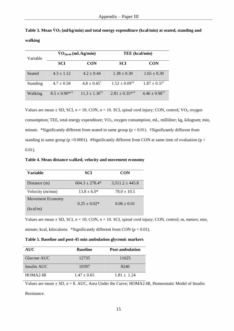
Appendix – Paper III
15
Table 3. Mean V̇O2 (ml/kg/min) and total energy expenditure (kcal/min) at seated, standing and
walking
Values are mean ± SD, SCI, n = 10; CON, n = 10. SCI, spinal cord injury; CON, control; VO2, oxygen
consumption; TEE, total energy expenditure; VO2, oxygen consumption, mL, milliliter; kg, kilogram; min,
minute. *Significantly different from seated in same group (p < 0.01). †Significantly different from
standing in same group (p <0.0001). #Significantly different from CON at same time of evaluation (p <
0.01).
Table 4. Mean distance walked, velocity and movement economy
Values are mean ± SD, SCI, n = 10; CON, n = 10. SCI, spinal cord injury; CON, control; m, meters; min,
minute; kcal, kilocalorie. *Significantly different from CON (p < 0.01).
Table 5. Baseline and post-45 min ambulation glycemic markers
AUC Baseline Post-ambulation
Glucose AUC 12735 11625
Insulin AUC 10397 8240
HOMA2-IR
1.47 ± 0.65 1.81 ± 1.24
Values are mean ± SD, n = 8. AUC, Area Under the Curve; HOMA2-IR, Homeostatic Model of Insulin
Resistance.
Variable
V̇O2peak (mL/kg/min) TEE (kcal/min)
SCI CON SCI CON
Seated 4.3 ± 1.12 4.2 ± 0.44 1.38 ± 0.30 1.65 ± 0.30
Standing
4.7 ± 0.58 4.8 ± 0.45* 1.52 ± 0.09*# 1.87 ± 0.37*
Walking 8.5 ± 0.90*†# 11.3 ± 1.30*† 2.81 ± 0.35*†# 4.46 ± 0.98*†
Variable SCI CON
Distance (m) 604.3 ± 278.4* 3,511.2 ± 445.8
Velocity (m/min)
13.8 ± 6.0* 78.0 ± 10.5
Movement Economy
(kcal/m)
0.25 ± 0.02* 0.06 ± 0.01

Appendix – Paper IV
1
Cardio-dynamic and autonomic adaptations to exoskeleton walking in Persons with Spinal Cord Injury
Authors Carsten Bach Baunsgaard1; Jennifer L. Maher2; Jan van Gerven3; Anne Palermo2; Fin Biering-Sorensen1; Armando J Mendez4; Robert W Irwin5; Mark S. Nash2,6
1Department of Spinal Cord Injuries, Rigshospitalet, University of Copenhagen; 2The Miami Project to Cure Paralysis, University of Miami, Miller School of Medicine, Miami, FL, USA; 3Radboud University, Nijmegen Medical Center; 4Division of Endocrinology, Diabetes and Metabolism, Diabetes Research Institute, University of Miami Miller School of Medicine; 5Department of Physical Medicine and Rehabilitation, University of Miami Miller School of Medicine; 6Department- of Neurological Surgery and Physical Medicine & Rehabilitation, University of Miami Miller School of Medicine, Miami, FL
Abstract Objective: First, to characterize cardiodynamic responses of heart rate (HR), stroke volume (SV) and cardiac output (Q) during 45 minutes of Bionic Ambulation (BA) in a mixed indoor and outdoor setting and explore whether cardiovascular (CV) drift occurs. Second, to explore acute cardiodynamic and autonomic adaptations with heart rate variability (Low Frequency/High Frequency (LF/HF)-ratio) following the exoskeleton walking session during a head-up-tilt test (HUT) and explore potential differences in the response to a tilt test before and after standing and walking.
Design: A controlled comparison of acute responses to walking in a bionic exoskeleton for persons with spinal cord injury (SCI) and non-injured control persons (CON).
Setting: Academic Medical Center.
Participants: 10 individuals with chronic (≥ 1 year) SCI (C7-T1, AIS A-C) and 10 individuals without neurological impairment (CON).
Interventions: A commercially available bionic exoskeleton.
Main Outcome measures: HR, SV, Q, LF/HF-ratio.
Results: There was seen a drop in SV during standing, but not in walking. SCI had similar response of HR, SV and Q during HUT, but SCI had higher LF/HF-ratio than CON. A second HUT after, compared to before standing or walking, seemed to be higher than the first HUT.
Conclusion: BA seemed to mitigate venous pooling during walking and no CV drift was documented. SCI increased HR, but not SV during walking. LF/HF increased during HUT as expected. There was a trend towards a parallel shift of LF/HF during HUT after standing and walking for SCI but not for CON.
Keywords: robotic exoskeleton; bionic ambulation; spinal cord injury; cardiodynamics; cardiovascular drift, heart rate variability

Appendix – Paper IV
2
Introduction Robotic exoskeletons are devices that have been developed and tested to maintain the upright posture and facilitate power-driven stepping by persons with spinal cord injury (SCI), referred to as Bionic Ambulation (BA). These devices are used in rehabilitation facilities for gait training and may ultimately serve as routinely used mobility or community ambulation devices. There are also suggested to be physiological benefits to time spent in the upright posture including enhancing cardiovascular function, enhancing bowel and bladder function, and lessening risks of spasms and joint contractures, reducing the risks of sustaining bed sores (1), (2). There is growing evidence to support the claims (3), however is still limited(2). BA could show as a desirable tool for early rehabilitation and long-term use after SCI if there is benefits of BA for persons with SCI. Previous studies(4) (5) (6) (7) have tested bionic ambulation (BA) in short, indoor training sessions, although cardiodynamic responses to BA in outdoor ambient temperatures and over uneven surface conditions have not been previously reported.
The upright posture challenges blood pressure regulation because of increasing orthostatic stress. Dysregulation of the integrated cardiovascular (CV) system is a concern for persons with SCI, particularly those with SCI at or above the T5 spinal level, as there is a loss of sympathetically mediated vasoconstriction below the level of injury and thus a chronically diminished venous return(8). When challenged by time spend in the upright position this dysregulation may lead to a CV drift, which is characterized by a decrease in stroke volume (SV) and a compensatory increase in heart rate (HR) to often a prevent decline of cardiac output (Q) observed over time for a given workload(9). This HR drift is seen with prolonged exercise starting after approximately 10 min of exercise(9) and is described in non-disabled persons(10) as well as persons with SCI(8). Factors believed to influence CV drift are venous blood pooling, fluid loss , hyperthermia(10) (11), or loss of active muscle pumps in the foot, calf thigh, and abdomen/thorax(10).
Analysis of heart rate variability (HRV) is a non-invasive method for assessing relative shifts in autonomic cardiac control(12) (13). The frequency domain measure of the ratio between low frequency (LF) power and high-frequency (HF) power is a commonly used surrogate measure for the sympathetic-parasympathetic balance(14). Ditor et al(15) showed that six months of body weight supported treadmill training elicited positive changes to the CV autonomic control, measured on HR and HRV, in persons with tetraplegia. A similar positive effects of exoskeleton gait training could therefore be hypothesised.
The aim of this study was thus two-fold: 1) to characterize the cardiodynamic responses of HR, SV and Q during 45 minutes of BA in a mixed indoor and outdoor setting, and to observe possible occurrence of CV drift during this period. As the CV responses to BA might be attributable to the upright position and not ambulation, we examined the same responses to seated rest and standing. 2) to investigate if autonomic adaptations explain the cv responses to BA and the extent to which they might benefit orthostatic tolerance.
Methods
Study Design The study was as an open-label, sample of convenience, repeated measures design that compared two-groups, one with SCI and another a non-disabled control. Data collection was performed from December 2015 to July 2016 at the Miami Project to Cure Paralysis, University of Miami, USA.
Participants Participants with SCI and non-injured control participants (CON) were eligible if they were aged 18 to 60 years, had no history of CV disease, and for SCI participants, met the eligibility criteria detailed in Table 1.

Appendix – Paper IV
3
Table 1. Study eligibility criteria only for participant with spinal cord injury (SCI)
Inclusion Criteria Exclusion Criteria
Lower extremity paresis or paralysis resulting from SCI for ≥1 year.
Range of motion (ROM) restrictions that would prevent a participant from achieving a normal, reciprocal gait pattern, or would restrict a participant from completing normal sit-to-stand or stand-to-sit transitions
Stable, motor-complete paralysis at the C7 or lower level, or, incomplete paralysis with functional bilateral upper extremity (UE) strength (triceps 3/5 or greater)
Fracture, dislocation, or malformations of LE bones (assessed by radiographs of the LE bones and joints in anteroposterior and lateral views)
Screened and cleared by a physician followed by a licensed Physical Therapist using standards mandated by the manufacturer
Spinal instability
Ability to stand for ≥ 15 minutes. Positional hypotension resulting in the inability to maintain an upright posture without signs of symptoms of syncope
Weight ≤ 100 kilograms (220 pounds). Unresolved deep vein thrombosis
Height between 152-191 cm (60-74 inches). Uncontrolled spasticity; Modified Ashworth ≥ 3 in LE
Standing hip width of 46 cm (18 inches) or less. Autonomic Dysreflexia requiring medical intervention
Near normal range of motion (ROM) in hips, knees, and ankles that would accommodate the bionic and safe BA
Pressure ulcers or skin integrity issues on contact surfaces of the device, or on body surfaces that would prohibit sitting
Upper leg length discrepancy greater than 1.3 cm (half an inch) or lower leg discrepancy greater than 1.9 cm (three-quarters of an inch)
Cognitive impairments resulting in motor planning or impulsivity concerns
Pregnancy
Study protocol, device description and outcome measures The Ekso™ GT (EKSO Bionics, CA, USA) was used as the robotic exoskeleton for this study. The exoskeleton has the option of changing the level of assistance, and for this study was set to the maximal level, meaning that the device performed the maximum possible workload. Initiation of a new step was done using the ProStep mode. In this mode, the user performs an anterolateral weight shift between legs to activate sensors in the foot plates, which in turn actuate the onboard motors. All participants used either a front wheeled walker (FWW) or crutches for balance. An assistant provided light-moderate contact guarding always and other assistance as needed.
Exoskeleton ambulation habituation period Participants with SCI underwent a habituation period of BA. Training began indoors in a thermoneutral environment and over a smooth flooring surface and progressed to include outdoor walking on a pre-specified par course of concrete pavers with slight change in grade. The participant was considered ready to perform the described testing procedures when they could walk for 45 minutes of mixed indoor and outdoor walking with minimal assistance, as judged by the assisting therapist.
Cardiodynamic parameters HR, SV and Q were measured using noninvasive impedance cardiography (PhysioFlow PF07 Enduro, Manitec Biomedical, Macharen, France). By convention, electrical impedance is designated by the letter Z, and the endpoint represents the first derivative of impendence with respect to time (ZCG; dz/dt). The PhysioFlow device emits a low-amperage (3.6 mA), high frequency (75 KHz) alternating electrical signal between two sets of electrodes. One was placed on the supraclavicular fossa on the left side of the neck, the other set on the middle of the back at the level of the xiphoid process. SV was calculated from the measurement of changes in transthoracic electrical impedance during the cardiac cycle. Synchronized ECG activity (V1 and V6) provide the HR. Q was calculated as the product of SV and HR. The coefficient of variations for SV

Appendix – Paper IV
4
and Q from the PhysioFlow across repeated cycle ergometer peak tests in healthy, non-disabled conditioned men was reported to be 3.6 and 3.4%, respectively(16).
Heart Rate Variability (HRV) A Polar heart rate monitor (Polar Electro Oy, Kempele, Finland) with elastic chest strap sensor recorded the electric activity of the heart. Beat-to-beat variability in the frequency spectrum by Fast Fourier Transform (FFT) was analyzed using Kubios HRV Analysis Software 2.0 for Windows (The Biomedical Signal and Medical Imaging Analysis Group, Department of Applied Physics, University of Kuopio, Finland).
HRV in the frequency-domain was used as a surrogate for autonomic tone, like in the previously mentioned article by Ditor et al(15). A shift in the relationship between LF and HF reflects the balance between sympathetic and parasympathetic tone(13). An increased LF/HF was operationally defined as a shift towards greater sympathetic nervous system activity.
Blood Pressure Systolic blood pressure (SBP) and diastolic blood pressure (DBP) were measured during HUT as described below.
Test days, study protocol Testing was performed on three days within a ten-day period; one day each for the three conditions; sitting, standing and walking. In addition, a peak test was performed on the first test day, after the sitting condition. Furthermore, was ahead-Up-Tilt (HUT) tests were performed to examine responses to orthostatic repositioning. The HUT procedure was performed as described below. CV hemodynamics from the ZCG were recorded at four timepoints on the three days of quiet sitting, standing and walking at minutes 0-1, 14-15, 29-30 and 44-45. During the HUT, recordings from ZCG, blood pressure (BP) and HR for HRV analysis were recorded. Participants did not consume fluids during the test procedures.
Sitting and peak test The first day of testing consisted of a 45-minute quiet sitting procedure to record baseline data, recorded every 15 minutes. Participants with SCI remained seated in their wheelchair and CON sat in a chair with both feet placed flat on the floor.
To establish the peak values of cardio-dynamics, participants underwent a continuous progressive exercise test on an arm ergometer (Angio, Lode BV, Groningen, Netherlands). Participants with SCI and CON performed the same peak test. In the study we used a protocol with progressive increase of the resistance in stages of one minute increments, as further described in a publication by Maher and Cowan, 2016(17). The test stopped when the participant was unable to maintain a cadence of >55 revolutions per minute of the arm-ergometer or the participant chose to stop for another reason. The highest 20s average was used as peak values. The peak test was performed following the quiet sitting procedure.
Standing The second test day tested responses to upright standing. Participants with SCI wore the exoskeleton and stood using an assistive device for balance (i.e., FWW or crutches). CON participants stood without the exoskeleton. Participants remained standing for 45 minutes during which data sampling every 15 min like in the sitting condition. A HUT was performed before the 45 min of sitting (called HUT1) and a second HUT was performed immediately after again (called HUT2).
Standing Responses to ambulation were tested on day three. Participants with SCI ambulated in the exoskeleton for 45 minutes on the outdoor par course, with data sampling every 15 minutes as with the sitting and standing condition. CON underwent the same procedures but ambulated without an assistive device. Like on the second test day, there was performed a HUT before (called HUT3) and after (called HUT4) the 45 min of walking.
Head-Up-Tilt (HUT) The participants transferred to a motorized tilt-table and were supported by Velcro straps. Participants did not use an abdominal binder or anti-embolic stockings during the test and had emptied their bladder or urine bag before testing. To achieve steady-state resting conditions, participants underwent a 10-minute period of supine rest, followed by progressive tilt in 5 min intervals to 30, 60, and 90 degrees, or as tolerated. Participants remained awake during data collection. Data

Appendix – Paper IV
5
from ZCG and HRV were sampled and averaged over 5 min intervals in the tilt angles: supine, 30, 60, and 90 degrees, where 90 degrees meant standing fully erect. BP was measured in the 10s after the new angle was completed to document the immediate changes when repositioning, and in the last 30s of each angle to reflect the BP stability at each tilt-angle.
Statistical analysis Repeated measures of changes for the 15 minutes intervals while sitting, standing and walking and during the progressive tilt test was analyzed in a Linear Mixed Model of fixed effects with compound symmetry as covariance matrix (assumption of equal correlation and variance at each time point). Post-hoc pairwise comparisons were Bonferroni corrected. In case of non-normal distributed data, logarithmic transformation was performed. A linear mixed model was also used to test between group (SCI and CON) and between test conditions (sitting, standing, walking) as well as between the HUT test performed on the same day (HUT1 vs HUT2 and HUT3 vs HUT4). Normality was judged visually by histogram distribution. Statistical analysis was performed by IBM SPSS Statistics version 22 (IBM Corp., Released 2013. Armonk, NY, USA).
Heart rate variability was analysed with Kubios HRV Analysis Software 2.0 for Windows (The Biomedical Signal and Medical Imaging Analysis Group, Department of Applied Physics, University of Kuopio, Finland).
Statement of ethics All participants received oral as well as written information about the study before written consent was obtained. The study was approved by the University of Miami Institutional Review Board (#20150704).
Results Participant characteristics are shown in Table 2. All participants were males and the SCI participants were all wheelchair users. Three SCI participants had injury above T6 level, but otherwise satisfied inclusion/exclusion criteria.
Table 2: Participant characteristics SCI (n = 10) CON (n = 10)
Age (years) 32.9 (11.4) 29.5 (7.7)
Height (cm) 176 (6) 183 (7)
Weight (kg) 67.6 (7.8) 80.8 (9.7)
Time since injury (years) 23.8 (7.5) NA
Level of injury (range) C7 - L1 NA
Severity of injury (range) AIS A-C NA
Etiology Traumatic NA
Peak test, Watt 105.6 (24.2) 131.5 (18.2)
SV 102.2 (13.3) 122.8 (23.9
HR 173.4 (10.6) 170.6 (9.4)
Q 17.7 (2.3) 20.8 (4.0)
Descriptives are expressed as mean (s.d.). Peak test was performed as arm-crank ergometer test. Abbreviations: Spinal Cord Injury, SCI; Able-bodied control participants, CON; American Spinal Injury Association (ASIA) Impairment Scale, AIS; Stroke Volume, SV; Heart Rate, HR; Cardiac Output, Q.
Normality testing SV and Q were normally distributed for sitting, standing and walking. HR, SBP and DBP as well as LF/HF was normally after log10 transformation.

Appendix – Paper IV
6
Habituation period The average number of sessions in the habituation period was 15, and a total upright -time (standing and walking time) was 14.5 hours. Total habituation walk time was approximately 8 hours, and on average used 14,000 steps.
Walking speed and temperature During the walking test day, the average gait speed of exoskeleton walking over the whole training session was 0.23m/s. There was no significant difference between exoskeleton gait speed indoor or outdoor (p=0.635). CON gait speed was 1.3 m/s. Outside temperature was in average 24.4(4.6)⁰C / 75.9 (8.3)⁰F with a humidity of 63.6 (19.3) %.
Repeated measures during sitting, standing and walking Table 3 shows the values of SV, HR and Q and p-values repeated measures between the 15 minutes time points within each test day, for SCI and CON respectively. Significance test are for main effect of time. Figure 1 show the same data presented as percentage change of SV, HR and Q relative to the first minute of the test day to see the relative changes.
Table 3 (and Figure 1): On the sitting day there were no main effect of time either SCI nor CON. On the standing day there was a significant drop for SCI in SV within session over time, and no significant change in HR or Q for SCI. In CON there was no change in SV, but a significant increase in HR and Q on the standing day. On the walking day, unlike standing day, there was no change for SCI, but HR and Q increased significantly. In CON there were significant increases in SV, HR and Q over 45 min of walking.
Table 3 Cardiodynamic parameters across tests and time points for participants with spinal cord injury (SCI) and controls (CON). (See also Figure 1).
Time point
0-1 min 14-15 min 29-30 min 44-45 min p-value
SCI, n=10
SV (mL/beat)
Sitting 65.9 (11.9) 63.2 (11.4) 64.1 (11.1) 62.8 (11.3) 0.11
Standing 62.1 (12.7) 62.4 (11.2) 60.6 (10.3) 57.6 (9.3) <0.01
walking 73.9 (14.8) 73.8 (13.6) 74.7 (15.3) 75.7 (16.0) 0.62
HR (bpm)
Sitting 81.8 (8.3) 79.1 (10.9) 77.2 (11.7) 79.0 (10.5) 0.13
Standing 87.1 (13.8) 92 (17.3) 91.4 (17.0) 93.2 (17.3) 0.10
walking 94.6 (18) 102 (20.2) 107.8 (23.6) 107.6 (21.5) <0.01
Q (L/min)
Sitting 5.4 (1.1) 5.0 (1.1) 4.9 (1.1) 5.0 (1.4) 0.06
Standing 5.5 (1.6) 5.8 (1.6) 5.6 (1.6) 5.4 (1.4) 0.21
walking 7.0 (2.3) 7.7 (2.6) 8.2 (3.1) 8.2 (2.8) <0.01
CON, n=10
SV (mL/beat)
Sitting 102.9 (13.8) 105.8 (15.7) 103.4 (13.2) 102.5 (14.2) 0.37
Standing 98.6 (17) 99.6 (18.1) 98.8 (14) 98.0 (14.8) 0.80
walking 107.8 (15.2) 119.8 (23.5) 124.3 (25.8) 121.2 (23.4) <0.01
HR (bpm)
Sitting 66.0 (11.8) 66.3 (10.4) 67.1 (11.3) 65.5 (10.4) 0.50
Standing 72.0 (10.2) 77.0 (12.0) 77.2 (12.2) 78.5 (12.8) <0.01
walking 77.8 (10.7) 86.3 (12.6) 87.4 (11.4) 87.4 (12.4) <0.01
Q (L/min)
Sitting 6.8 (1.4) 6.9 (0.9) 6.9 (1.1) 6.7 (0.9) 0.56
Standing 7.1 (1.5) 7.6 (1.6) 7.6 (1.6) 7.7 (1.6) 0.02
walking 8.3 (1.1) 10.3 (2.2) 10.8 (2.0) 10.5 (1.9) <0.01
Descriptives are mean (s.d.) Abbreviations: Stroke Volume, SV; Heart Rate, HR; Cardiac Output, Q. Test-day, T; beats-per-minute, bpm; Spinal Cord Injury, SCI; non-injured control participants, CON.

Appendix – Paper IV
7
Differences between SCI and CON, main effect of group The SCI group had significant higher HR and lower SV and Q than CON on all three testing days (p<0.01). The three participants with NLI over T6 was separated in the analysis and by visual inspection. The cardiodynamic response followed the rest of the SCI participants.
Difference between sitting, standing and walking day, main effect of day/condition HR was significant higher on the walking day compared to standing, which in turn was significant higher than sitting (p<0.05) for both SCI and CON. Neither SV and Q was significant different at sitting or standing for either SCI or CON, but walking was significant higher than sitting and standing for both SCI and CON (p<0.01).
Changes during HUT and between test days (Figure 2A, Figure 2B and Figure 3) SV dropped significantly (p=0.02) for both SCI and CON with increasing HUT angle from supine to 30, 60 and 90 degrees, whereas HR and Q increased (p<0.001) during all the four HUTs. SCI had overall lower SV during HUT than CON, (main effect of group, p<0.01), whereas SCI had higher HR than CON during HUT, (main effect group, p=0.01). However, there were no main effect of group on Q between SCI and CON.
The HRV measure LF/HF increased with higher tilt angles for both SCI (p<0.001) and CON (p<0.001). There was a significant higher value for SCI compared to CON (p=0.034). For SCI there was higher value of the second HUT after sitting or standing (HUT 2 and HUT4) compared to HUT1 and HUT3 respectively (main effect for both days 0.014). For CON there was no significant difference between first the HUTs for LF/HF.
Participants with SCI and CON did not have drop in BP (Figure 3), except for one participant with SCI with a neurological level of injury at C7 who experienced large drop in BP during the tilt test. This does, however, not show on the mean value of the SCI group (Figure 3). Repeated measures of BP at the last 30s of the tilt angle showed no significant difference in SBP for SCI, but a small, but significant (p<0.001) increase of DBP with increasing tilt-angle. For CON there was a significant (p<0.05) increase in both SBP and DBP.
Appendix – Paper IV
8
Discussion This study assessed cardiodynamic response to 45 minutes of sitting, standing and BA (SCI) or walking without the exoskeleton (CON) (Figure 1 and Table 3). There were indications of venous pooling while standing in the SCI group(18), but no CV drift effect was seen during walking for SCI or CON (Figure 1 and Table 3) when this is defined as a drop in SV together with increase in HR. There were however increase in HR during walking. The increase in Q during walking was primarily driven by an increase in HR in SCI, compared to CON where both HR and SV increased during walking. The study also assessed cardiodynamic and autonomic responses during a HUTs performed before (HUT1 and HUT3) and after (HUT2 and HUT4) the two test days of standing and walking. In both SCI and CON there was a drop in SV with increasing tilt-angle, accompanied increase in HR and Q, which is not surprising. Interestingly it was also possible to detect an increase in the LF/HR ratio with increased tilt angle. SBP and DBP was largely unaffected during HUT for both SCI and CON, except for one participant with SCI who experienced a drop with increased tilt-angle.
On average, the habituation period required 15 sessions in to reach a level of walking with minimal assistance in BA. This time period is similar to what have been described previously(19) (20) (21). The BA velocity while walking was 0.23 m/s, which is similar to what was described in a review by Louie(22) where average BA speeds were estimated at 0.26 m/s. However, this value includes varying devices, conditions and gait tests such as the 10 Meter Walk Test, 6 Minute Walk Test, whereas the reported gait speed in our study is the average of a 45-minute bout of BA. As a side note can be mentioned that the gait velocity is below the value that has been estimated to be the minimum for functional community ambulation (23). This is based on velocity needed for example to cross an road intersection, which has been estimated to be 1.22m/s(23).
Typically, cardiac output is increased by peripheral vasoconstrictor responses which in turn increase venous return and end-diastolic ventricular volume alongside increase in HR and myocardial contractility(24). However, Kinzer(25) found that persons with paraplegia exhibited a higher HR and lower SV during arm-crank exercise compared to both persons with leg amputation and non-injured persons. Persons with paraplegia had greater accumulation of fluids in the legs compared to their non-injured counterparts and thus the legs seemed to act as a reservoir. Jacobs et al(8) and Hotettler(26) have reported similar findings. Hjeltnes(27) attributed the decreased venous return to a loss of sympathetic vasomotor regulation below the level of lesion in thoracic SCI. Dela et al(28) documented higher HR in persons with paraplegia and tetraplegia compared to non-injured controls during electrically induced leg ergometer training.
Our findings suggest a similar pattern, where SCI had higher HR and lower SV and Q than CON. This was seen in all test conditions; peak test, sitting, standing, walking and during tilt test. CV-drift effects were not seen during walking with exoskeleton in SCI despite the prolonged period of 45 min of walking, even when the outdoor temperature was high temperature and there were no fluid replacement(11) (29) (30). This is likely due to a relative low intensity of BA, below a threshold. Likely it also reflects selection of the study population. However, the movement of the legs during BA seemed to have mitigated the venous pooling with reference to the drop in SV seen in the SCI group in the standing session (Figure 1).
An increase in the LF/HF ratio implies a shift towards higher sympathetic nervous system input relative to parasympathetic autonomic tone(13). These findings looked like a parallel shift in the second HUT after standing and walking (HUT2 and HUT4) and thereby suggest a carry-over effect from the walking and standing intervention for the SCI group. The apparent trend would be relevant to follow up in future studies. Also, it could be interesting to test this in a longitudinal study to see if there would be longitudinal changes. The results also suggest that the LF/HF-ratio can be used as a proxy for autonomic tone.
This study findings are limited by a relatively small study population, although larger than previously reported (4) (5) (6) (7). There was with a risk of type II errors due to lack of statistical power, and limitation for further sub-group analysis. Additionally, three participants had neurological level of injury above T6 with potential loss of cardiac sympathetic drive, and thus may have exhibited different responses compared to participants with injuries below T6(31), despite it did not look as such when looking at individual plots (not included in this article). Where many persons with SCI experience orthostatic hypotension, the test population in this study was selected to have orthostatic stability, because of selection as per eligibility criteria. This selection was chosen to reflect the potential users of the exoskeleton and is a common

Appendix – Paper IV
9
inclusion criterion in exoskeleton studies but does not reflect the whole SCI population. Also, the three test days were not randomized, and there could be a carryover effect of the repeated HUTs that is not accounted for in the study.
Interestingly, the SCI group reached a higher peak HR during the peak test than CON, despite the max effect (watt) was higher for CON. An explanation could be the limitation of arm-crank test, not being able to elicit the max HR of the CON group. Studies on able bodied persons have shown that individuals who have not trained the arm-crank ergometer test will achieve a lower output compared to a leg ergometer test due to a reduced stress on the CV system during the arm-crank test(32). The SCI group would likely be more conditioned to upper body exercise than the CON group and thus the peak test would underestimate the maximum effect in the CON group.
Conclusion The BA seemed to mitigate the drop in SV seen during the 45 min standing session, possibly due to mechanical effects of lower limb movements. Increase in Q during walking was in SCI driven by increase in HR, where in CON it was driven by both SV and HR increase, in line with previous reporting in the literature. It is interpreted as there was not presence of cardiovascular drift during walking. HUTs elicited the same response pattern in SCI and CON with decrease in SV and increase in HR and Q. However, had SCI a higher HR and lower SV than CON, like what was seen during walking. Increase in the HRV parameter LF/HF ratio with increasing tilt angle suggests a higher sympathetic drive during the HUTs. There was a pattern towards a parallel shift on LF/HF values on all cardio dynamic of the HUT test that following standing and walking compared to the HUT before the standing or walking intervention and a heretofore-unreported carry-over effect from exoskeleton walking to responses experience during head up tilt.

Appendix – Paper IV
10
References 1. Walter JS, Sola PG, Sacks J, Lucero Y, Langbein E, Weaver F. Indications for a home standing program for individuals with spinal
cord injury. J Spinal Cord Med. 1999;22(3):152–8.
2. Karimi MT. Evidence-Based Evaluation of Physiological Effects of Standing and Walking in Individuals with Spinal Cord Injury. Iran J Med Sci. 2011 Dec;36(4):242–53.
3. Palermo AE, Maher JL, Baunsgaard CB, Nash MS. Clinician-Focused Overview of Bionic Exoskeleton Use After Spinal Cord Injury. Top Spinal Cord Inj Rehabil. 2017 Jun 1;23(3):234–44.
4. Asselin P, Knezevic S, Kornfeld S, Cirnigliaro C, Agranova-Breyter I, Bauman WA, et al. Heart rate and oxygen demand of powered exoskeleton-assisted walking in persons with paraplegia. J Rehabil Res Dev. 2015;52(2):147–58.
5. Escalona MJ, Brosseau R, Vermette M, Comtois AS, Duclos C, Aubertin-Leheudre M, et al. Cardiorespiratory demand and rate of perceived exertion during overground walking with a robotic exoskeleton in long-term manual wheelchair users with chronic spinal cord injury: A cross-sectional study. Ann Phys Rehabil Med. 2018 Jan 31;
6. Evans N, Hartigan C, Kandilakis C, Pharo E, Clesson I. Acute Cardiorespiratory and Metabolic Responses During Exoskeleton-Assisted Walking Overground Among Persons with Chronic Spinal Cord Injury. Top Spinal Cord Inj Rehabil. 2015 Mar 1;21(2):122–32.
7. Kressler J, Wymer T, Domingo A. Respiratory, cardiovascular and metabolic responses during different modes of overground bionic ambulation in persons with motor-incomplete spinal cord injury: A case series. J Rehabil Med. 2018 Feb 13;50(2):173–80.
8. Jacobs PL, Mahoney ET, Robbins A, Nash M. Hypokinetic circulation in persons with paraplegia. Med Sci Sports Exerc. 2002 Sep;34(9):1401.
9. Ekelund L-G. Circulatory and Respiratory Adaptation during Prolonged Exercise of Moderate Intensity in the Sitting Position. Acta Physiol Scand. 1967 Apr 1;69(4):327–40.
10. Coyle EF, González-Alonso J. Cardiovascular drift during prolonged exercise: new perspectives. Exerc Sport Sci Rev. 2001 Apr;29(2):88–92.
11. Wingo JE, Ganio MS, Cureton KJ. Cardiovascular Drift During Heat Stress: Implications for Exercise Prescription. Exerc Sport Sci Rev. 2012 Apr;40(2):88–94.
12. Krassioukov AV, Karlsson A-K, Wecht JM, Wuermser L-A, Mathias CJ, Marino RJ, et al. Assessment of autonomic dysfunction following spinal cord injury: rationale for additions to International Standards for Neurological Assessment. J Rehabil Res Dev. 2007;44(1):103–12.
13. Malmqvist L, Biering-Sørensen T, Bartholdy K, Krassioukov A, Welling K-L, Svendsen JH, et al. Assessment of autonomic function after acute spinal cord injury using heart rate variability analyses. Spinal Cord. 2015 Jan;53(1):54–8.
14. Heart Rate Variability: Standards of Measurement, Physiological Interpretation, and Clinical Use. Task Force of the European Society of Cardiology and the North American Society of Pacing and Electrophysiology. Circulation. 1996 Mar 1;93(5):1043–65.
15. Ditor DS, Kamath MV, MacDonald MJ, Bugaresti J, McCartney N, Hicks AL. Effects of body weight-supported treadmill training on heart rate variability and blood pressure variability in individuals with spinal cord injury. J Appl Physiol. 2005 Apr 1;98(4):1519–25.
16. Hsu AR, Barnholt KE, Grundmann NK, Lin JH, McCallum SW, Friedlander AL. Sildenafil improves cardiac output and exercise performance during acute hypoxia, but not normoxia. J Appl Physiol Bethesda Md 1985. 2006 Jun;100(6):2031–40.
17. Maher JL, Cowan RE. Comparison of 1- Versus 3-Minute Stage Duration During Arm Ergometry in Individuals With Spinal Cord Injury. Arch Phys Med Rehabil. 2016;97(11):1895–900.

Appendix – Paper IV
11
18. Figoni SF. Cardiovascular and haemodynamic responses to tilting and to standing in tetraplegic patients: a review. Spinal Cord. 1984 Apr;22(2):99–109.
19. Kressler J, Thomas CK, Field-Fote EC, Sanchez J, Widerström-Noga E, Cilien DC, et al. Understanding Therapeutic Benefits of Overground Bionic Ambulation: Exploratory Case Series in Persons With Chronic, Complete Spinal Cord Injury. Arch Phys Med Rehabil. 2014 Oct;95(10):1878–87.
20. Baunsgaard CB, Nissen UV, Brust AK, Frotzler A, Ribell C, Kalke Y-B, et al. Gait training after spinal cord injury: safety, feasibility and gait function following 8 weeks of training with the exoskeletons from Ekso Bionics (in press). Spinal Cord. 2017 in press;
21. Miller LE, Zimmermann AK, Herbert WG. Clinical effectiveness and safety of powered exoskeleton-assisted walking in patients with spinal cord injury: systematic review with meta-analysis. Med Devices Auckl NZ. 2016;9:455–66.
22. Louie DR, Eng JJ, Lam T. Gait speed using powered robotic exoskeletons after spinal cord injury: a systematic review and correlational study. J NeuroEngineering Rehabil. 2015;12:82.
23. Lapointe R, Lajoie Y, Serresse O, Barbeau H. Functional community ambulation requirements in incomplete spinal cord injured subjects. Spinal Cord. 2001 Jun;39(6):327–35.
24. Teasell RW, Arnold JMO, Krassioukov A, Delaney GA. Cardiovascular consequences of loss of supraspinal control of the sympathetic nervous system after spinal cord injury. Arch Phys Med Rehabil. 2000 Apr;81(4):506–16.
25. Kinzer SM, Convertino VA. Role of leg vasculature in the cardiovascular response to arm work in wheelchair-dependent populations. Clin Physiol Oxf Engl. 1989 Dec;9(6):525–33.
26. Hostettler S, Leuthold L, Brechbühl J, Mueller G, Illi SK, Spengler CM. Maximal cardiac output during arm exercise in the sitting position after cervical spinal cord injury. J Rehabil Med. 2012 Feb;44(2):131–6.
27. Hjeltnes N. Oxygen uptake and cardiac output in graded arm exercise in paraplegics with low level spinal lesions. Scand J Rehabil Med. 1977;9(3):107–13.
28. Dela F, Mohr T, Jensen CMR, Haahr HL, Secher NH, Biering-Sørensen F, et al. Cardiovascular control during exercise: insights from spinal cord-injured humans. Circulation. 2003 Apr 29;107(16):2127–33.
29. Zacharakis ED, Kounalakis SN, Nassis GP, Geladas ND. Cardiovascular drift in trained paraplegic and able-bodied individuals during prolonged wheelchair exercise: effect of fluid replacement. Appl Physiol Nutr Metab Physiol Appliquée Nutr Métabolisme. 2013 Apr;38(4):375–81.
30. Lafrenz AJ, Wingo JE, Ganio MS, Cureton KJ. Effect of ambient temperature on cardiovascular drift and maximal oxygen uptake. Med Sci Sports Exerc. 2008 Jun;40(6):1065–71.
31. Biering-Sørensen F, Biering-Sørensen T, Liu N, Malmqvist L, Wecht JM, Krassioukov A. Alterations in cardiac autonomic control in spinal cord injury. Auton Neurosci [Internet]. [cited 2017 Mar 13]; Available from: http://www.sciencedirect.com/science/article/pii/S1566070217300449
32. Larsen RT, Christensen J, Tang LH, Keller C, Doherty P, Zwisler A-D, et al. A systematic review and meta-analysis comparing cardiopulmonary exercise test values obtained from the arm cycle and the leg cycle respectively in healthy adults. Int J Sports Phys Ther. 2016 Dec;11(7):1006–39.

Appendix – Paper IV
12
Figure 1. Stroke Volume, Heart Rate and Cardiac Output during 45 minutes of sitting, standing and walking
Figure 1. All descriptive statistics are mean (s.d.). Plots showing percentage-change from initial value of 100% over 45 minutes at three test sessions, sitting, standing, walking. Abbreviations: Spinal Cord Injury, SCI, non-injured controls, CON; Stroke Volume, SV; Heart Rate, HR; Cardiac Output, Q

Appendix – Paper IV
13
Figure 2A. Stroke Volume, Heart Rate and Cardiac Output during HUTs before and after standing, day 2

Appendix – Paper IV
14
Figure 2B. Stroke Volume, Heart Rate and Cardiac Output during HUTs before and after walking, day 3
Figure 2A and 2B. All descriptive statistics are mean (s.d.). Plots showing change in HR, SV, Q and LF/HF at four tilt-angles on each of the four HUT tests performed. Abbreviations: Head-Up-Tilt, HUT; Spinal Cord Injury, SCI, non-injured controls, CON; Stroke Volume, SV; Heart Rate, HR; Cardiac Output, Q; Low Frequency, LF; High Frequency, HF

Appendix – Paper IV
15
Figure 3. Blood pressure during HUTs, day 2 and day 3.
Figure 3. All descriptive statistics are mean (s.d.). Plots showing change SBP and DBP at four tilt-angles on each of the four HUT tests performed. Abbreviations: Head-Up-Tilt, HUT; Spinal Cord Injury, SCI, non-injured controls, CON; Systolic Blood Pressure, SBP; Diastolic Blood Pressure, DBP