Phase Field Simulation of Dendritic Microstructure in ...
15
Phase Field Simulation of Dendritic Microstructure in Additively Manufactured Titanium Alloy Jing Zhang*, Linmin Wu, Yi Zhang, Lingbin Meng Department of Mechanical Engineering, Indiana University-Purdue University Indianapolis, Indianapolis, IN 46202, USA *Email: [email protected]; Phone: 317-278-7186; Fax:317-274-9744 Abstract Additive manufacturing (AM) processes for metals, such as selective laser sintering and electron beam melting, involve rapid solidification process. The microstructure of the fabricated material and its properties strongly depend on the solidification. Therefore, in order to control and optimize the AM process, it is important to understand the microstructure evolution. In this work, using Ti-6Al-4V as a model system, the phase field method is applied to simulate the microstructure evolution in additively manufactured metals. Frist, the fundamental governing equations are presented. Then the effects of various processing related parameters, including local temperature gradient, scan speed and cooling rate, on dendrites morphology and growth velocity are studied. The simulated results show that the dendritic arms grow along the direction of the heat flow. Higher temperature gradient, scan speed and cooling rate will result in small dendritic arm spacing and higher growth velocity. The simulated dendritic morphology and arm spacings are in good agreement with experimental data and theoretical predictions. Keywords: phase field; metals; temperature gradient; scan speed; additive manufacturing ___________________________________________________________________ This is the author's manuscript of the article published in final edited form as: Zhang, J., Wu, L., Zhang, Y., & Meng, L. (2019). Phase field simulation of dendritic microstructure in additively manufactured titanium alloy. Metal Powder Report, 74(1), 20–24. https://doi.org/10.1016/j.mprp.2018.11.001
Transcript of Phase Field Simulation of Dendritic Microstructure in ...
Phase Field Simulation of Dendritic Microstructure in Additively
Manufactured Titanium Alloy
Jing Zhang*, Linmin Wu, Yi Zhang, Lingbin Meng
Department of Mechanical Engineering, Indiana University-Purdue University Indianapolis,
Indianapolis, IN 46202, USA
*Email: [email protected]; Phone: 317-278-7186; Fax:317-274-9744
Abstract
Additive manufacturing (AM) processes for metals, such as selective laser sintering and
electron beam melting, involve rapid solidification process. The microstructure of the fabricated
material and its properties strongly depend on the solidification. Therefore, in order to control and
optimize the AM process, it is important to understand the microstructure evolution. In this work,
using Ti-6Al-4V as a model system, the phase field method is applied to simulate the
microstructure evolution in additively manufactured metals. Frist, the fundamental governing
equations are presented. Then the effects of various processing related parameters, including local
temperature gradient, scan speed and cooling rate, on dendrites morphology and growth velocity
are studied. The simulated results show that the dendritic arms grow along the direction of the heat
flow. Higher temperature gradient, scan speed and cooling rate will result in small dendritic arm
spacing and higher growth velocity. The simulated dendritic morphology and arm spacings are in
good agreement with experimental data and theoretical predictions.
Keywords: phase field; metals; temperature gradient; scan speed; additive manufacturing
___________________________________________________________________
This is the author's manuscript of the article published in final edited form as:
Zhang, J., Wu, L., Zhang, Y., & Meng, L. (2019). Phase field simulation of dendritic microstructure in additively manufactured titanium alloy. Metal Powder Report, 74(1), 20–24. https://doi.org/10.1016/j.mprp.2018.11.001
2
1. Introduction
During additive manufacturing process, due to the cooling, solidification and phase
transformation occur in the melt pool [1, 2]. This will significantly affect the material properties
of the build materials, since solidification controls the morphology of the microstructure. Thus, it
is essential to understand the solidification behavior during the fabrication. Titanium based alloys,
especially Ti-6Al-4V, are widely used in aerospace, biomedical and automotive industries due to
their excellent mechanical strength and creep resistance at high temperatures [3-5]. Recently, many
studies were carried out to investigate the microstructures and material properties of laser
deposited Ti-6Al-4V by experiment [6-10]. However, due to the short time scale of the
solidification and the small length scale of the melting pool, it is very difficult to study the
microstructure evolution during the fabrication process by experiment. Hence, numerical
simulations aiming to reveal microstructure evolution during the solidification process are
warranted.
Phase field method is a promising technique to describe the microstructure evolution [11-
13]. It has been used to simulate the dendrites growth in undercooled pure materials [14, 15], and
has been extended to describe the dendrites morphology in binary alloys [16-19]. For Ti-6Al-4V,
only few studies have been carried out by phase field method. Gong and Chou [20] applied phase
field method to investigate the columnar grain growth of Ti-6Al-4V during solidification and
compared grain sizes with experimental results. Sahoo and Chou [21] studied the dendritic arm
spacing with different processing parameters in electron beam additive manufactured Ti-6Al-4V.
In this study, the phase field method will be employed to simulate the phase transformation from
liquid phase to solid prior β phase of Ti-6Al-4V during rapid solidification. The influence of the
local temperature gradient, scan speed and cooling rate will be investigated. Ghosh et al. studied

3
the primary spacing and microsegregation of cellular dendrites in laser deposited Ni–Nb
alloys[22]. A phase-field model is used to simulate solidification microstructures at different
locations within a solidified molten pool. Although the phase-field model has an anti-trapping
solute flux term meant to maintain local interface equilibrium, it is found that during simulations
it was insufficient at maintaining equilibrium. This is due to the fact that the additive
manufacturing solidification conditions fall well outside the allowed limits of this flux term[22].
This work is based on our previous research [23], with revised contents. First, the
fundamental governing equations are presented. Then the effects of various processing
parameters, including local temperature gradient, scan speed and cooling rate, on dendrites
morphology and growth velocity are studied.
2. Phase field model description
To simulate the microstructure evolution of Ti-6Al-4V during additive manufacturing
process, phase field method is employed. In this study, Ti-6Al-4V is assumed to be a pseudo-
binary alloy, since it is shown that the pseudo-binary approach can be a successful approximation
for the multi-component approach when simulating the solidification of Ti-base alloy such as Ti-
6Al-4V [24, 25].
The phase field simulations were carried out to simulate the phase transformation from
liquid phase to solid prior β phase. The model consists of two coupled equations, with one
describing the evolution of phase order parameter ϕ , and the other governs the mass transport of
the composition c during the solidification of a binary alloy. The dimensionless form of governing
equations is given by,

4
( ) ( ) ( ) ( ) ( ) ( )
( ) ( )
2 2
23 2 0
0
11 /l
a aa a a a
t x y y x
T TUm c k k
θ θϕ ϕ ϕθ θ ϕ θ θθ θ
ϕ ϕ λ ϕ
∂ ∂ ∂ ∂ ∂ ∂ ∂ = ∇ ⋅ ∇ − + ∂ ∂ ∂ ∂ ∂ ∂ ∂ −
+ − − − + −
(1)
( )1
1 1 atc D u jt k k
ϕϕ
∂ −= ∇ ⋅ ∇ +∇⋅ ∂ + − −
(2)
where ϕ is phase order parameter with 1ϕ = − representing solid phase and 1ϕ = meaning liquid
phase. ( ) ( )1 cos 4a θ ε θ= + represents the four-fold surface energy anisotropy at the solid-liquid
interface with the strength of anisotropy ε and the angle between the interface normal and the x
direction ( )arctan /y xθ ϕ ϕ= ∂ ∂ . /s lk c c= is partition coefficient, where sc and lc are
equilibrium compositions on solid and liquid side of the interface, respectively. lm is liquidus
slope and 0c is alloy concentration far from the solidification front. U is the dimensionless
supersaturation, which is defined as, ( ) ( )1 / 1uU e k= − − , where u is a dimensionless chemical
potential given by,
( )02 /ln
1 1ck cu
k k ϕ
= + − − (3)
The frozen temperature approximation was applied to the system, which is described by
( )0T T G y Vt= + − . G and V are temperature gradient and constant pulling speed along y
direction, respectively. 0T is the reference temperature.

5
In equation (2), ( ) ( )1 / 2 1 / 2l sD D Dϕ ϕ= − + + is diffusion coefficient of the system,
where lD and sD are diffusion coefficient in liquid and solid phase, respectively. The anti-
trapping current atj was introduced to the diffusion equation to suppress the solute-trapping
effects at the solid-liquid interface [18], and is given by,
12 2atj U n
tϕ∂
=∂
(4)
where / | |n ϕ ϕ= −∇ ∇ is the unit vector normal to the solid-liquid interface.
There are three characteristic parameters in the phase field model, the characteristic length
W , the characteristic time τ and the coupling constant λ . Based on thin interface analysis, two
equations are given to describe the relationship of these three parameters,
1a Wd
λ = (5)
2
2
l
a WDλτ = (6)
where d is the chemical capillary length. 1 0.8839a = and 2 0.6267a = are constant, which is
given in [18].
To solve the phase field equation (1) and the mass transport equation (2), a finite volume
method was used with explicit time marching. No flux boundary conditions were applied on all
boundaries.

6
3. Results and discussion
The simulated microstructure evolution of Ti-6Al-V during solidification is shown in Fig.
1. Three features can be observed from the structures. First, the microstructure shows columnar
structures. With the increase of solidification time, the initial random nuclei become unstable and
start to grow. Finally, parallel dendritic arms are formed. The growth of the dendritic arms is along
the direction of the heat flow, which is in y-direction in this case. Second, droplets are formed
inside dendrites during the solidification. These droplets have high solute concentration. Similar
results are found in the rapid solidification process of binary alloys [26, 27]. Third, there is
significant micro-segregation phenomenon. It is found that solute enriches in the liquid near the
dendritic tips and between dendritic arms. As the dendritic arms grow, the liquid concentration
increases near the tip regions and decreases rapidly to the alloy concentration far away from the
solidification front 0c .

7
Fig. 1: Simulated columar structure at different times with 2800G = K/mm and 400V =
mm/s. (a) 0.02 ms, (b) 0.1 ms, (c) 0.2 ms, (d) 0.3 ms.
In Fig. 2, the SEM image of electron beam additive manufactured Ti-6Al-4V sample is
shown. It can be seen from the microstructure that columnar dendritic structures are formed, which
is columnar prior β grains. The phase field simulations were carried out to simulate the phase
transformation from liquid phase to solid prior β phase. The simulated phase and solute
concentration profiles are comparable with the experimental observations.
(a) (b)
(c) (d)

8
Fig. 2: SEM image of electron beam additive manufactured Ti-6Al-4V sample [28].
One important parameter used to quantify the microstructure after solidification is Primary
Dendritic Arm Spacing (PDAS). Broderick et al. [29] investigated the effects of cooling conditions
on the microstrucutre of rapidly solidified Ti-6Al-4V experimentally. The correlation between
cooling rate T and PDAS is given by,
( )n nPDAS A GV AT= = (7)
where A and n are constants. After fitting to the experimental data, A and n are obtained as 3.1×10⁶
μm (K/s)1.05 and -1.05. In order to compare the simulation results with the experimental values,
phase field simulations with constant scan speed 400 mm/s and different temperture gradient
varying from 2000 K/mm to 2800 K/mm were carried out. The comparison is shown in Fig. 3. In
overall, the simulated PDAS is in agreement with the experimental fitted values. It can be seen
that PDAS reduces with the increasing cooling rate, implying that higher cooling rate will results
in a finer columnar structure during solidification.

9
Fig. 3: Comparison of simulated PDAS values with experimental fitted results.
To study the effect of temperature gradient on microstructure, simulations with a fixed
scanning speed 400 mm/s and different temperature gradient were performed. It can be seen that
with the increase of the temperature gradient, the columnar structure become denser, indicating
smaller PDAS. Besides, the growth velocity of the dendrites is higher when temperature gradient
is larger. This result is consistent with the previous experimental data, as elavated temperature
gradient means higher cooling rate. The simulated PDAS is also compared with analytical models.
Hunt [30] proposed a theoretial model to predict PDAS by considering the geometry of dendrite
tip. The model is given by,
( )0.25 0.5 0.2502.83 lPDAS k T D G V− −= Γ∆ (8)
0
0.5
1
1.5
2
2.5
8.00E+05 9.00E+05 1.00E+06 1.10E+06
PDA
S (μ
m)
Cooling rate (K/s)
Experiment [32]
Simulation
10
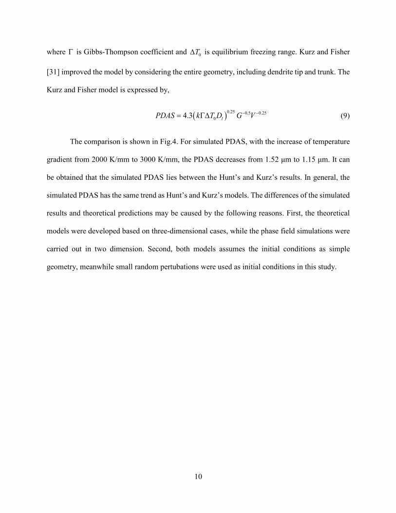
where Γ is Gibbs-Thompson coefficient and 0T∆ is equilibrium freezing range. Kurz and Fisher
[31] improved the model by considering the entire geometry, including dendrite tip and trunk. The
Kurz and Fisher model is expressed by,
( )0.25 0.5 0.2504.3 lPDAS k T D G V− −= Γ∆ (9)
The comparison is shown in Fig.4. For simulated PDAS, with the increase of temperature
gradient from 2000 K/mm to 3000 K/mm, the PDAS decreases from 1.52 μm to 1.15 μm. It can
be obtained that the simulated PDAS lies between the Hunt’s and Kurz’s results. In general, the
simulated PDAS has the same trend as Hunt’s and Kurz’s models. The differences of the simulated
results and theoretical predictions may be caused by the following reasons. First, the theoretical
models were developed based on three-dimensional cases, while the phase field simulations were
carried out in two dimension. Second, both models assumes the initial conditions as simple
geometry, meanwhile small random pertubations were used as initial conditions in this study.

11
Fig. 4: Comparison of simulated PDAS with theoretical predictions. The scan speed is
fixed at 400 mm/s.
The effect of scan speed on Ti-6Al-4V was studied by fixing the temperature gradient at
2000 K/mm and varing the scan speed from 200 mm/s to 800 mm/s. The comparision of the
simulated PDAS with the theoretial predictions is plotted in Fig.5. For simulation results, as the
increase of scan speed from 200 mm/s to 800 mm/s, the PDAS decreases from 1.58 μm to 1.36
μm, which is comparable with analytical model results.
0
0.5
1
1.5
2
2.5
3
2000 2200 2400 2600 2800 3000
PDA
S (µ
m)
G (K/mm)
Kurz and Fisher [34]Hunt [33]Simulation

12
Fig. 5: Comparison of simulated PDAS with theoretical predictions. The temperature
gradient is fixed at 2000 K/mm.
3. Conclusions
The microstructure evolution of additively manufactured Ti-6Al-4V during rapid
solidification has been investigated using phase field method. The conclusions are summarized as
follows:
(1) The phase field method can simulate the phase transformation from liquid phase to
solid phase of Ti-6Al-4V during solidification. The morphology shows a columnar
structure.
0
0.5
1
1.5
2
2.5
3
3.5
200 300 400 500 600 700 800
PDA
S (µ
m)
V (mm/s)
Kurz and Fisher [34]Hunt [33]Simulation

13
(2) The growth of the dendritic arms is along the direction of the heat flow. Droplets are
found formed inside dendrites. Solute enriches in the liquid near the dendritic tips and
between dendritic arms.
(3) Temperature gradient, scan speed and cooling rate have significant influence on
dendrites morphology and growth velocity. Higher temperature gradient, scan speed
and cooling rate will result in small dendritic arm spacing and higher growth velocity.
The simulated results are in good agreement with experimental data and theoretical
predictions.
Acknowledgment
We acknowledge the financial support provided by the Walmart Foundation (project title:
Optimal Plastic Injection Molding Tooling Design and Production through Advanced Additive
Manufacturing), and Praxair's TruForm AMbition Grant awarded to JZ.

14
References
[1] L. E. Murr, S. M. Gaytan, D. A. Ramirez, E. Martinez, J. Hernandez, K. N. Amato, et al., "Metal Fabrication by Additive Manufacturing Using Laser and Electron Beam Melting Technologies," Journal of Materials Science & Technology, vol. 28, pp. 1-14, 2012/01/01 2012.
[2] H. L. Wei, J. Mazumder, and T. DebRoy, "Evolution of Solidification Texture during Additive Manufacturing," Scientific Reports, vol. 5, p. 16446, 2015.
[3] S. Zherebtsov, G. Salishchev, R. Galeyev, and K. Maekawa, "Mechanical Properties of Ti-6Al-4V Titanium Alloy with Submicrocrystalline Structure Produced by Severe Plastic Deformation," Materials Transactions, vol. 46, pp. 2020-2025, 2005.
[4] C. Qiu, N. J. E. Adkins, and M. M. Attallah, "Microstructure and tensile properties of selectively laser-melted and of HIPed laser-melted Ti–6Al–4V," Materials Science and Engineering: A, vol. 578, pp. 230-239, 8/20/ 2013.
[5] T. Vilaro, C. Colin, and J. D. Bartout, "As-Fabricated and Heat-Treated Microstructures of the Ti-6Al-4V Alloy Processed by Selective Laser Melting," Metallurgical and Materials Transactions A, vol. 42, pp. 3190-3199, 2011// 2011.
[6] H. Galarraga, D. A. Lados, R. R. Dehoff, M. M. Kirka, and P. Nandwana, "Effects of The Microstructure and Porosity on Properties of Ti-6Al-4V Alloy Fabricated by Electron Beam Melting (EBM)," Additive Manufacturing, vol. 10, pp. 47-57, 2016.
[7] N. Hrabe and T. Quinn, "Effects of Processing on Microstructure and Mechanical Properties of A Titanium Alloy (Ti–6Al–4V) Fabricated Using Electron Beam Melting (EBM), Part 1: Distance from Build Plate and Part Size," Materials Science and Engineering: A, vol. 573, pp. 264-270, 2013.
[8] N. Hrabe and T. Quinn, "Effects of processing on microstructure and mechanical properties of a titanium alloy (Ti–6Al–4V) fabricated using electron beam melting (EBM), Part 2: Energy input, orientation, and location," Materials Science and Engineering: A, vol. 573, pp. 271-277, 6/20/ 2013.
[9] F. Luca, M. Emanuele, R. Pierfrancesco, and M. Alberto, "Microstructure and Mechanical Properties of Ti‐6Al‐4V Produced by Electron Beam Melting of Pre‐Alloyed Powders," Rapid Prototyping Journal, vol. 15, pp. 171-178, 2009.
[10] Q. Liu, Y. Wang, H. Zheng, K. Tang, L. Ding, H. Li, et al., "Microstructure and mechanical properties of LMD–SLM hybrid forming Ti6Al4V alloy," Materials Science and Engineering: A, vol. 660, pp. 24-33, 4/13/ 2016.
[11] N. Moelans, B. Blanpain, and P. Wollants, "Quantitative Analysis of Grain Boundary Properties in A Generalized Phase Field Model For Grain Growth in Anisotropic Systems," Physical Review B, vol. 78, p. 024113, 2008.
[12] L. Wu, Y. Zhang, Y.-G. Jung, and J. Zhang, "Three-dimensional phase field based finite element study on Li intercalation-induced stress in polycrystalline LiCoO2," Journal of Power Sources, vol. 299, pp. 57-65, 12/20/ 2015.
[13] N. Moelans, B. Blanpain, and P. Wollants, "An Introduction to Phase-Field Modeling of Microstructure Evolution," Calphad, vol. 32, pp. 268-294, 2008.
[14] A. F. Ferreira, A. J. d. Silva, and J. A. d. Castro, "Simulation of The Solidification of Pure Nickel via The Phase-Field Method," Materials Research, vol. 9, pp. 349-356, 2006.
[15] R. Kobayashi, "Phase Field Simulations of Dendritic Solidification," in Advanced Materials ed, 1994, pp. 529-532.

15
[16] S. G. Kim, W. T. Kim, and T. Suzuki, "Phase-Field Model for Binary Alloys," Physical Review E, vol. 60, pp. 7186-7197, 1999.
[17] Z. Bi and R. F. Sekerka, "Phase-Field Model of Solidification of A Binary Alloy," Physica A: Statistical Mechanics and its Applications, vol. 261, pp. 95-106, 1998.
[18] B. Echebarria, R. Folch, A. Karma, and M. Plapp, "Quantitative Phase-Field Model of Alloy Solidification," Physical Review E, vol. 70, p. 061604, 2004.
[19] J. C. Ramirez, C. Beckermann, A. Karma, and H. J. Diepers, "Phase-Field Modeling of Binary Alloy Solidification with Coupled Heat and Solute Diffusion," Physical Review E, vol. 69, p. 051607, 2004.
[20] X. Gong and K. Chou, "Phase-Field Modeling of Microstructure Evolution in Electron Beam Additive Manufacturing," JOM, vol. 67, pp. 1176-1182, 2015.
[21] S. Sahoo and K. Chou, "Phase-Field Simulation of Microstructure Evolution of Ti–6Al–4V in Electron Beam Additive Manufacturing Process," Additive Manufacturing, vol. 9, pp. 14-24, 2016.
[22] G. Supriyo, M. Li, O.-O. Nana, and E. G. Jonathan, "On the primary spacing and microsegregation of cellular dendrites in laser deposited Ni–Nb alloys," Modelling and Simulation in Materials Science and Engineering, vol. 25, p. 065002, 2017.
[23] L. Wu and J. Zhang, "Phase Field Simulation of Dendritic Solidification of Ti-6Al-4V During Additive Manufacturing Process," JOM, vol. 70, pp. 2392-2399, October 01 2018.
[24] L. Nastac, J. Valencia, J.Xu, and H. Dong, "Assessment of Solidification-Kinetics Parameters for Titanium-Base Alloys," Proceedings of the International Symposium on Liquid Metals Processing and Casting, pp. 207-233, 1999.
[25] L. Nastac, "Solute Redistribution, Liquid/Solid Interface Instability, and Initial Transient Regions during the Unidirectional Solidification of Ti-6-4 and Ti-17 Alloys," in CFD Modeling and Simulation in Materials Processing, ed: John Wiley & Sons, Inc., 2012, pp. 123-130.
[26] G. Supriyo, M. Li, O.-O. Nana, and E. G. Jonathan, "On The Primary Spacing and Microsegregation ff Cellular Dendrites in Laser Deposited Ni–Nb Alloys," Modelling and Simulation in Materials Science and Engineering, vol. 25, p. 065002, 2017.
[27] V. Fallah, M. Amoorezaei, N. Provatas, S. F. Corbin, and A. Khajepour, "Phase-Field Simulation of Solidification Morphology in Laser Powder Deposition of Ti–Nb Alloys," Acta Materialia, vol. 60, pp. 1633-1646, 2012.
[28] H. K. Rafi, N. V. Karthik, H. Gong, T. L. Starr, and B. E. Stucker, "Microstructures and Mechanical Properties of Ti6Al4V Parts Fabricated by Selective Laser Melting and Electron Beam Melting," Journal of Materials Engineering and Performance, vol. 22, pp. 3872-3883, 2013.
[29] T. F. Broderick, A. G. Jackson, H. Jones, and F. H. Froes, "The Effect of Cooling Conditions on The Microstructure of Rapidly Solidified Ti-6Al-4V," Metallurgical Transactions A, vol. 16, pp. 1951-1959, 1985.
[30] M. H. Burden and J. D. Hunt, "Cellular and Dendritic Growth. I," Journal of Crystal Growth, vol. 22, pp. 99-108, 1974.
[31] W. Kurz and D. J. Fisher, "Dendrite Growth at The Limit of Stability: Tip Radius and Spacing," Acta Metallurgica, vol. 29, pp. 11-20, 1981.


![TheInfluenceofPriorNaturalAgingon ... · 2019. 7. 31. · using gravity die casting (GDC), resulting in a dendritic microstructure (see [12, 17] for more details of the casting parameters,](https://static.fdocuments.net/doc/165x107/60c9b47836d427709c1d3921/theiniuenceofpriornaturalagingon-2019-7-31-using-gravity-die-casting.jpg)