Phakomatoses(Ophthalmology)
30
Phacomatoses Dr Shylesh B Dabke Resident Dept of ophthalmology KMC Mangalore
-
Upload
shylesh-dabke -
Category
Health & Medicine
-
view
194 -
download
0
Transcript of Phakomatoses(Ophthalmology)

Phacomatoses
Dr Shylesh B DabkeResidentDept of ophthalmologyKMC Mangalore

• Syndromes characterized by hamartomas of the skin, eye, central nervous system (CNS), and other viscera are collectively called phacomatoses.
• These disorders produce significant visual and neurologic disturbances.
• Although most of these syndromes arise from mutations in single genes inherited in a Mendelian pattern, some have no apparent hereditary basis.
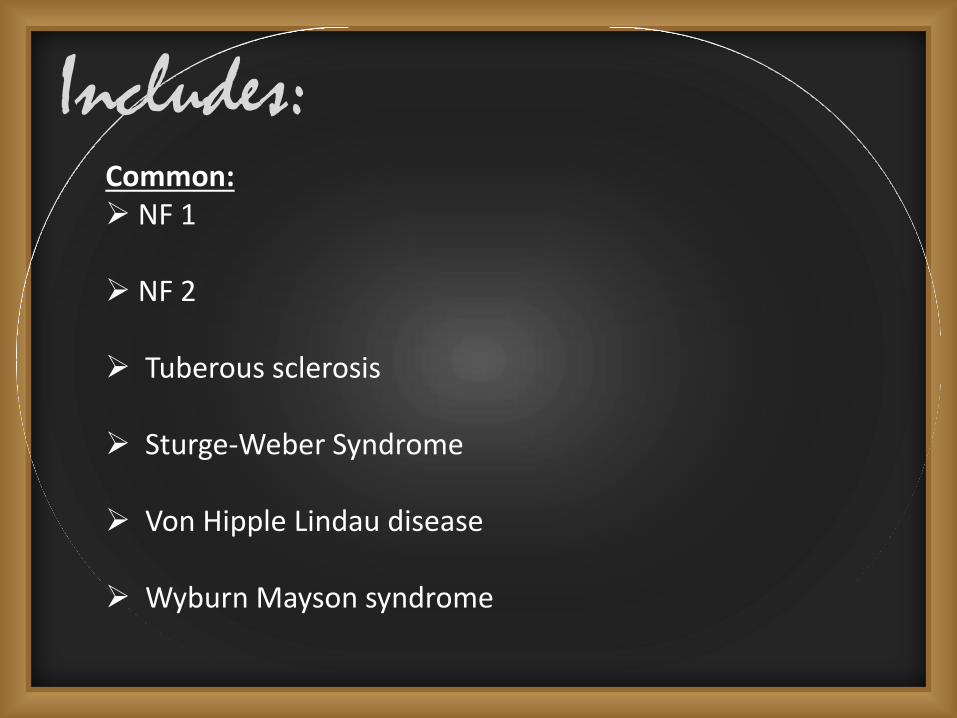
Includes:Common: NF 1
NF 2
Tuberous sclerosis
Sturge-Weber Syndrome
Von Hipple Lindau disease
Wyburn Mayson syndrome

UncommonKlipple trenaunay Weber syndrome
Louis bar syndrome
Diffuse congenital hemangiomatosis
Oculodermal melanocytosis
Basal cell nevus syndrome

Neurofibromatoses Neuroectodermal tumors arising within multiple organs.
Autosomal dominant inheritance.
Two types :-1.NF1 (Von Recklinghausen’s neurofibromatosis or Peripheral
Neurofibromatoses)
2.NF2 (central or bilateral acoustic neurofibromatosis)

Neurofibromatoses 1
The most common type of the two and the most common autosomal dominant disorder.
Characterized by:* Cafe´-au-lait spots. * Peripheral neurofibromas.* Lisch nodules.
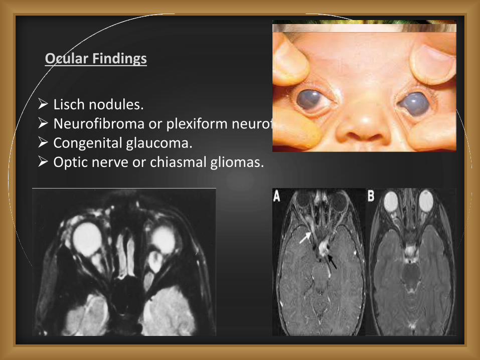
Ocular Findings
Lisch nodules. Neurofibroma or plexiform neurofibroma of eyelid. Congenital glaucoma. Optic nerve or chiasmal gliomas.

Orbital involvement. Strabismus. Retinal abnormalities.

Uveal tract. Lignes grises – Hyperplastic intrastromalnerves. Conjunctival hamartomas.

Neurofibromatoses 2 CNS tumors are the major feature in NF2.
Meningiomas, gliomas, and schwannomas are common, but bilateral vestibular schwannomas are nearly always present.
Ocular findings may predate the onset of symptoms. * Characteristic: PSCO or Wedge cortical cataract* less common findings are retinal hamartoma and combined
hamartomas of the retina and retinal pigment epithelium

Tuberous Scleroses Also known as Bourneville disease.
Vogt triad: Mental retardationSeizuresFacial angiofibromas
Primary features of this disorder:* Facial angiofibroma* Ungual fibromas (multiple)* Cortical tuber* Subependymal nodule (giant cell astrocytoma)* Multiple retinal astrocytomas

Hypopigmented lesions analogous to white spots of the skin are occasionally seen in the iris or choroid.
Characteristic ocular manifestation of TS is the retinal astrocytic hamartoma or Retinal Phakoma.
* Phakomas can develop anywhere in the fundus.* Arise from astrocytes of sensory retina.* vary in size from about half to twice the DD.* Vision is rarely affected Significantly.
Three types
* Type1 :-Flat, soft and semitransparent lesionsUsually at posterior poleBoundaries are grey or faint yellow
* Type 2:-Elevated,nodular and solid apparing massesUsually near disc margin and also at midperiphery
* Type 3:-Border is flat, soft and transparentCentrally elevated and nodular

Von Hippel Lindau Disease Autosomal dominantly inherited disorder of incomplete penetrance manifesting both benign and malignant tumors of many organ systems.
Most common abnormalities are vascular tumors(hemangioblastomas) of the retina and CNS, most often the cerebellum.

Retinal lesions usually become visible ophthalmoscopicallybetween ages 10 and 35 years, about a decade before the peak clinical incidence of cerebellar disease.
Tumors are multiple in the same eye in about one-third of cases and bilateral in as many as one-half of cases.
Tumors typically occur in the peripheral fundus, but lesions adjacent to the optic disc have also been described.

The incipient retinal lesion appears as a minor, nonspecific vascular anomaly or a small reddish dot in the fundus.
The lesion ultimately acquires the fully developed appearance of a pink globular mass 1 to 3 or more disc diameters in size.
The hallmark of the mature tumor is a pair of markedly dilated vessels (artery and vein) running between the lesion and the optic disc, indicating Significant arteriovenousshunting.
Histopathologically, retinal angiomas - relatively well formed capillaries - fluorescein angiography shows these vessels to be leaky.

Transudation of fluid into the subretinal space - lipid accumulation, retinal detachment - loss of vision.
Secondary degenerative changes including cataract and glaucoma.
Management and prognosis* cryotherapy or laser photocoagulation(Small lesions)* Multiple treatment sessions may be necessary * Early diagnosis increases the likelihood of successful
treatment.
Ocular lesions of VHL are asymptomatic prior to retinal detachment - children known to be at risk for the disease should undergo periodic ophthalmologic evaluation.

Cutaneous LesionsAdenoma sebaceum
Ash leaf spots
Shagreen patch
Confetti lesions
Periangual fibroma

Sturge-Weber Syndrome
Encephalotrigeminal angiomatosis.
Characterstic lesions:-* Cutaneous facial nevus flammeus ( in distribution of
branches of trigeminal nerve )* Ipsilateral diffuse cavernous hemangioma choroid* Ipsilateral meningeal hemangiomatosis
These lesions are always present at birth in affected patients
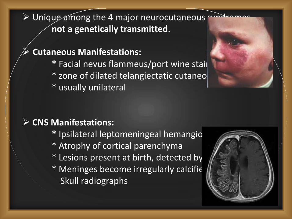
Unique among the 4 major neurocutaneous syndromes -not a genetically transmitted.
Cutaneous Manifestations:* Facial nevus flammeus/port wine stain* zone of dilated telangiectatic cutaneous capillaries* usually unilateral
CNS Manifestations:* Ipsilateral leptomeningeal hemangiomatosis* Atrophy of cortical parenchyma* Lesions present at birth, detected by MRI or CT* Meninges become irregularly calcified, detected by
Skull radiographs

Ocular Manifestations* Increased conjunctival vascularity commonly
produces a pinkish discoloration.* An abnormal plexus of episcleral vessels.
choroid is the site of the most significance - increased numbers of well-formed choroidal vessels - bright red or redorange color.
Glaucoma is the most common and serious ocular complication(70% Cases).
Cause of elevated lOP is uncertain but is likely secondary to:* Elevated episcleral venous pressure. * Hyperemia of the Ciliary body with hypersecretion of
aqueous * Or developmental anomaly of the anterior chamber
angle.

Management
SWS glaucoma is difficult to treat, and there is no universallyaccepted treatment scheme.
Initial therapy with topical drops can be effective
Surgery is indicated when medical treatment is inadequate.
Adequate long-term pressure control can frequently be achieved, although multiple operations are typically necessary.
A particular risk of glaucoma surgery - massive intraoperativeor postoperative exudation or hemorrhage - anomalous choroidal vessels - rapid ocular decompression.

Wyburn-Mason Syndrome
racemose angioma.
noninhereditary arteriovenous malformation of the eye and brain.
Typically involving the optic disc or retina and the midbrain.
Ocular manifest - unilateral and congenital.

Lesions are not distinct tumors - Not a true phakomatoses.
Most patients have unilateral lesions.
Vision ranges from normal to markedly reduced in the involved eye.
Intraocular hemorrhage and secondary neovascularglaucoma are possible complications.
No treatment is indicated for primary ocular lesions.

Klippel – Trenaunay-Weber syndrome
Triad of:* Cutaneous hemangiomas extending over the limbs.* Varicosities in the affected limb* Hypertrophy of bone and soft tissue.
Ocular findings:* Enophthalmos* Conjunctival telangiectasia.* Heterochromia iridis* Iris coloboma* Retinal varicosities* Choroidal angiomas

Louis Bar Syndrome/Ataxia Telangiectasia
Recessive inherited multisystem disease
Ocular lesions – * Bulbar conjunctival telangiectasia* Oculomotor apraxia with Supranuclearpalsies, Nystagmus & Strabismus
CNS – progressive ataxia in childhood
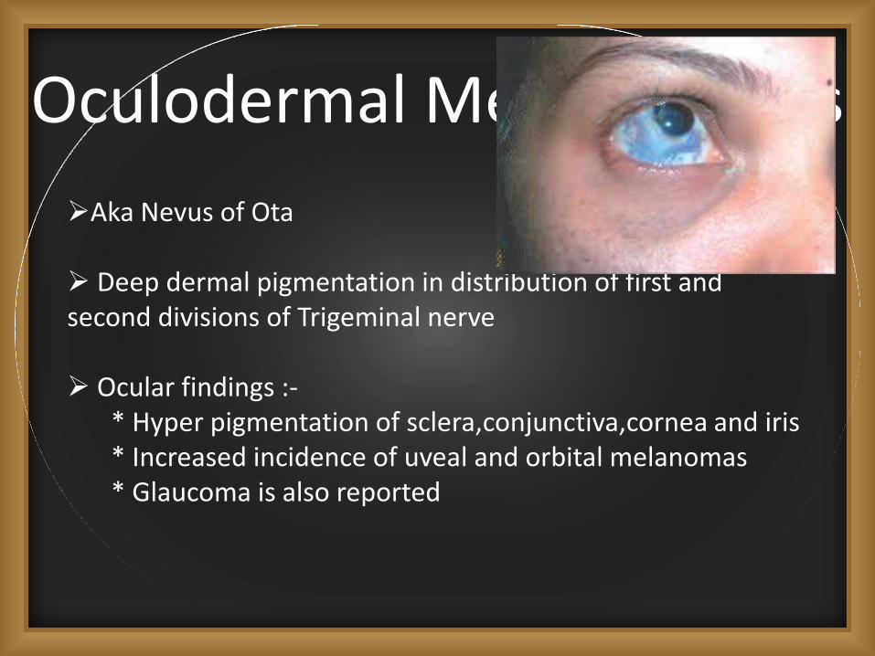
Oculodermal Melanocytosis
Aka Nevus of Ota
Deep dermal pigmentation in distribution of first and second divisions of Trigeminal nerve
Ocular findings :-* Hyper pigmentation of sclera,conjunctiva,cornea and iris* Increased incidence of uveal and orbital melanomas* Glaucoma is also reported

Diffuse congenital hemangiomatosis
Multiple small cutaneous hemangiomas.
Ocular findings * Hemangiomas of iris,conjunctiva & lid.* Abnormal chorioretinal vasculature* Microphthalmos* Galucoma* Central system hemangiomas - cortical blindness.

Basal cell nevus syndromeMultiple skin tumors
Autosomal dominant inheritance
Most lesions are benign
Ocular findings:-* Congenital cataract* Coloboma of choroid and optic disc* Corneal leucomata