Phaeohyphomycosis infection in the knee - SciELO · r ev bras ortop. 2016;51(2):231–234 Case...
4
r e v b r a s o r t o p . 2 0 1 6; 5 1(2) :231–234 www.rbo.org.br Case Report Phaeohyphomycosis infection in the knee David Sadigursky ∗ , Luisa Nogueira e Ferreira, Liz Moreno de Oliveira Corrêa Hospital COT, Salvador, BA, Brazil a r t i c l e i n f o Article history: Received 22 March 2015 Accepted 28 April 2015 Available online 23 February 2016 Keywords: Infection Fungi Knee Dermatomycoses a b s t r a c t Phaeohyphomycosis is caused by cutaneous fungi and rarely affects large joints. This is a case report on phaeohyphomycosis in the left knee of an elderly individual without immuno- suppression. It was accompanied by pain and swelling the anterior knee. The case was first suspected to be suprapatellar bursitis, and was treated with nonsteroidal anti-inflammatory drugs, without remission of symptoms. Surgical treatment was performed, with resection of the suprapatellar bursa and anterior region of the quadriceps tendon. The material was sent for anatomopathological examination and culturing. The pathological examination showed phaeohyphomycosis. The treatment instituted consisted of itraconazole, 200 mg/day for six weeks, and complete remission of symptoms was achieved. The physical examination remained normal after one year of follow-up. This is the first published case of phaeohy- phomycosis infection in the suprapatellar region of the knee. Although almost all the cases reported have been associated with immunosuppressed patients, this was an exception. It is important to suspect phaeohyphomycosis in cases of knee infection, in the area of the suprapatellar bursa, when the symptoms do not resolve after clinical treatment. © 2016 Sociedade Brasileira de Ortopedia e Traumatologia. Published by Elsevier Editora Ltda. All rights reserved. Infecc ¸ão por feohifomicose em joelho Palavras-chave: Infecc ¸ão Fungos Joelho Dermatomicoses r e s u m o A feohifomicose, causada por fungos demáceos, raramente acomete grandes articulac ¸ ões. Este é um relato de caso de feohifomicose, em joelho esquerdo de idoso não imunos- suprimido, acompanhado de dor e aumento de volume em região anterior do joelho. Suspeitou-se de bursite suprapatelar, sendo medicado com anti-inflamatório não esteroidal, sem apresentar remissão dos sintomas. Fez-se tratamento cirúrgico, foram ressecadas a bursa suprapatelar e a região anterior do tendão do quadríceps sendo a pec ¸a encaminhada Work performed in the COT Hospital, Salvador, BA, Brazil. ∗ Corresponding author. E-mail: [email protected] (D. Sadigursky). http://dx.doi.org/10.1016/j.rboe.2016.02.004 2255-4971/© 2016 Sociedade Brasileira de Ortopedia e Traumatologia. Published by Elsevier Editora Ltda. All rights reserved.
-
Upload
truonglien -
Category
Documents
-
view
215 -
download
0
Transcript of Phaeohyphomycosis infection in the knee - SciELO · r ev bras ortop. 2016;51(2):231–234 Case...
r e v b r a s o r t o p . 2 0 1 6;5 1(2):231–234
C
P
D
H
a
A
R
A
A
K
I
F
K
D
P
I
F
J
D
h2
www.rbo.org .br
ase Report
haeohyphomycosis infection in the knee�
avid Sadigursky ∗, Luisa Nogueira e Ferreira, Liz Moreno de Oliveira Corrêa
ospital COT, Salvador, BA, Brazil
r t i c l e i n f o
rticle history:
eceived 22 March 2015
ccepted 28 April 2015
vailable online 23 February 2016
eywords:
nfection
ungi
nee
ermatomycoses
a b s t r a c t
Phaeohyphomycosis is caused by cutaneous fungi and rarely affects large joints. This is a
case report on phaeohyphomycosis in the left knee of an elderly individual without immuno-
suppression. It was accompanied by pain and swelling the anterior knee. The case was first
suspected to be suprapatellar bursitis, and was treated with nonsteroidal anti-inflammatory
drugs, without remission of symptoms. Surgical treatment was performed, with resection of
the suprapatellar bursa and anterior region of the quadriceps tendon. The material was sent
for anatomopathological examination and culturing. The pathological examination showed
phaeohyphomycosis. The treatment instituted consisted of itraconazole, 200 mg/day for
six weeks, and complete remission of symptoms was achieved. The physical examination
remained normal after one year of follow-up. This is the first published case of phaeohy-
phomycosis infection in the suprapatellar region of the knee. Although almost all the cases
reported have been associated with immunosuppressed patients, this was an exception. It
is important to suspect phaeohyphomycosis in cases of knee infection, in the area of the
suprapatellar bursa, when the symptoms do not resolve after clinical treatment.
© 2016 Sociedade Brasileira de Ortopedia e Traumatologia. Published by Elsevier Editora
Ltda. All rights reserved.
Infeccão por feohifomicose em joelho
alavras-chave:
r e s u m o
A feohifomicose, causada por fungos demáceos, raramente acomete grandes articulacões.
nfeccãoungos
oelho
Este é um relato de caso de feohifomicose, em joelho esquerdo de idoso não imunos-
suprimido, acompanhado de dor e aumento de volume em região anterior do joelho.
Suspeitou-se de bursite suprapatelar, sendo medicado com anti-inflamatório não esteroidal,
issão dos sintomas. Fez-se tratamento cirúrgico, foram ressecadas a
ermatomicoses sem apresentar rembursa suprapatelar e a região anterior do tendão do quadríceps sendo a peca encaminhada
� Work performed in the COT Hospital, Salvador, BA, Brazil.∗ Corresponding author.
E-mail: [email protected] (D. Sadigursky).ttp://dx.doi.org/10.1016/j.rboe.2016.02.004255-4971/© 2016 Sociedade Brasileira de Ortopedia e Traumatologia. Published by Elsevier Editora Ltda. All rights reserved.
232 r e v b r a s o r t o p . 2 0 1 6;5 1(2):231–234
para exame anatomopatológico e cultura. No exame anatomopatológico foi possível eviden-
ciar o diagnóstico de feohifomicose. O tratamento instituído foi itraconazol, 200 mg/dia por
seis semanas, apresentando remissão completa do quadro. O exame físico se manteve nor-
mal após um ano de seguimento. Este é o primeiro caso publicado a respeito da infeccão por
feohifomicose em região suprapatelar. Apesar de quase todos os casos registrados estarem
associados a pacientes imunossuprimidos, este foi uma excecão. É importante que se sus-
peite de feohifomicose nas infeccões de joelho, na área da bursa suprapatelar, quando os
sintomas não resolverem após o tratamento clínico medicamentoso.
© 2016 Sociedade Brasileira de Ortopedia e Traumatologia. Publicado por Elsevier
Editora Ltda. Todos os direitos reservados.
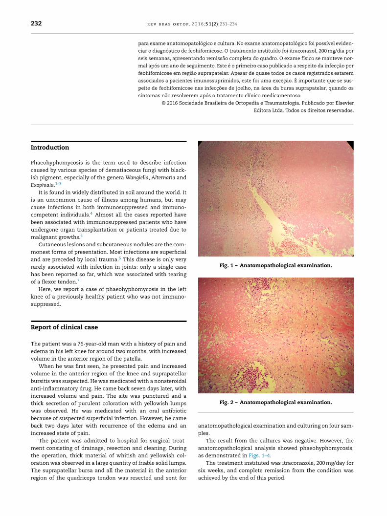
Fig. 1 – Anatomopathological examination.
Introduction
Phaeohyphomycosis is the term used to describe infectioncaused by various species of dematiaceous fungi with black-ish pigment, especially of the genera Wangiella, Alternaria andExophiala.1–3
It is found in widely distributed in soil around the world. Itis an uncommon cause of illness among humans, but maycause infections in both immunosuppressed and immuno-competent individuals.4 Almost all the cases reported havebeen associated with immunosuppressed patients who haveundergone organ transplantation or patients treated due tomalignant growths.5
Cutaneous lesions and subcutaneous nodules are the com-monest forms of presentation. Most infections are superficialand are preceded by local trauma.6 This disease is only veryrarely associated with infection in joints: only a single casehas been reported so far, which was associated with tearingof a flexor tendon.7
Here, we report a case of phaeohyphomycosis in the leftknee of a previously healthy patient who was not immuno-suppressed.
Report of clinical case
The patient was a 76-year-old man with a history of pain andedema in his left knee for around two months, with increasedvolume in the anterior region of the patella.
When he was first seen, he presented pain and increasedvolume in the anterior region of the knee and suprapatellarbursitis was suspected. He was medicated with a nonsteroidalanti-inflammatory drug. He came back seven days later, withincreased volume and pain. The site was punctured and athick secretion of purulent coloration with yellowish lumpswas observed. He was medicated with an oral antibioticbecause of suspected superficial infection. However, he cameback two days later with recurrence of the edema and anincreased state of pain.
The patient was admitted to hospital for surgical treat-ment consisting of drainage, resection and cleaning. During
the operation, thick material of whitish and yellowish col-oration was observed in a large quantity of friable solid lumps.The suprapatellar bursa and all the material in the anteriorregion of the quadriceps tendon was resected and sent forFig. 2 – Anatomopathological examination.
anatomopathological examination and culturing on four sam-ples.
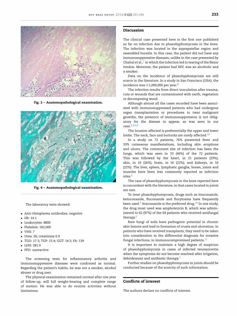
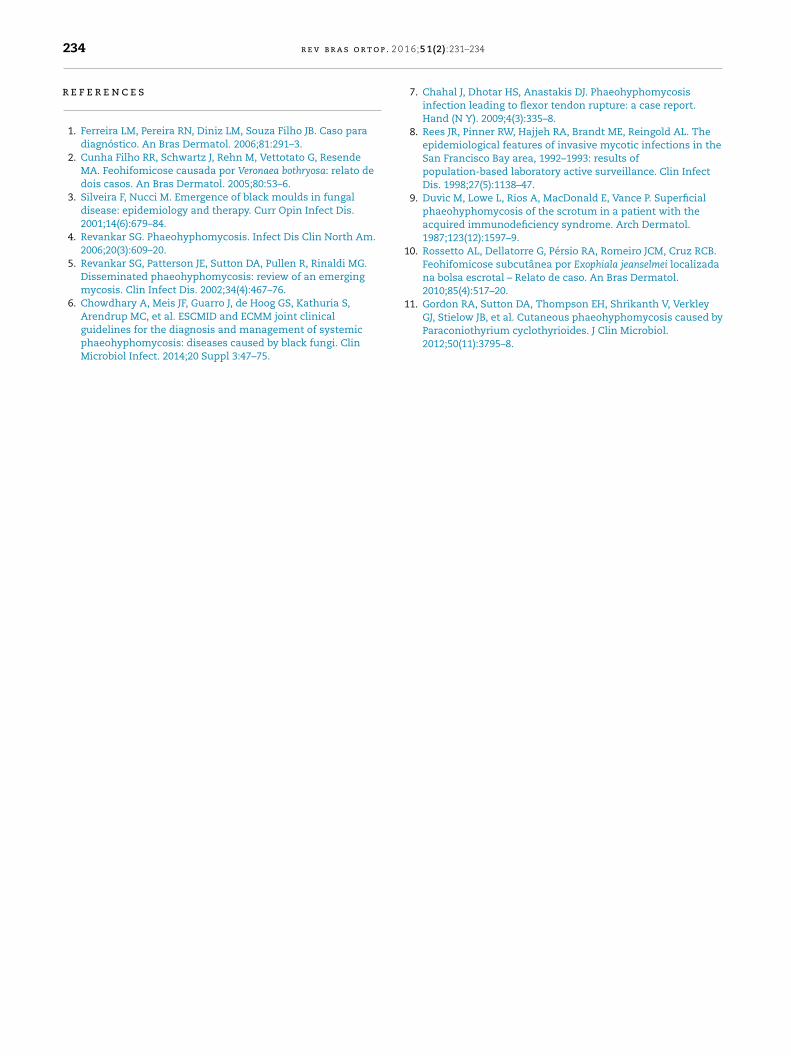
The result from the cultures was negative. However, theanatomopathological analysis showed phaeohyphomycosis,as demonstrated in Figs. 1–4.
The treatment instituted was itraconazole, 200 mg/day forsix weeks, and complete remission from the condition wasachieved by the end of this period.
r e v b r a s o r t o p . 2 0 1 6
Fig. 3 – Anatomopathological examination.
•••••••••
iRa
ool
Conflicts of interest
Fig. 4 – Anatomopathological examination.
The laboratory tests showed:
Anti-Histoplasma antibodies: negative Hb: 14.1 Leukocytes: 8800 Platelets: 182,000 VHS: 7 Urea: 26; creatinine 0.9 TGO: 17.3; TGP: 15.4; GGT: 14.5; FA: 139 LDH: 281.9 PPD: unreactive
The screening tests for inflammatory arthritis andmmunosuppressive diseases were confirmed as normal.egarding the patient’s habits, he was not a smoker, alcoholbuser or drug user.
The physical examination remained normal after one yearf follow-up, will full weight-bearing and complete rangef motion. He was able to do routine activities without
imitations.
;5 1(2):231–234 233
Discussion
The clinical case presented here is the first one publishedso far on infection due to phaeohyphomycosis in the knee.The infection was located in the suprapatellar region andresembled bursitis. In this case, the patient did not have anyimmunosuppressive diseases, unlike in the case presented byChahal et al.,7 in which the infection led to tearing of the flexortendon. Moreover, the patient had HIV, was an alcoholic anda smoker.
Data on the incidence of phaeohyphomycosis are stillscarce in the literature. In a study in San Francisco (USA), theincidence was 1:1,000,000 per year.8
The infection results from direct inoculation after trauma,cuts or wounds that are contaminated with earth, vegetationor decomposing wood.
Although almost all the cases recorded have been associ-ated with immunosuppressed patients who had undergoneorgan transplantation or procedures to treat malignantgrowths, the presence of immunosuppression is not oblig-atory for the disease to appear, as was seen in ourcase.2,3,5,9
The location affected is preferentially the upper and lowerlimbs. The neck, face and buttocks are rarely affected.10
In a study on 72 patients, 76% presented fever and33% cutaneous manifestations, including skin eruptionsand ulcers. The commonest site of infection has been thelungs, which was seen in 33 (46%) of the 72 patients.This was followed by the heart, in 21 patients (29%);skin, in 19 (26%); brain, in 16 (22%); and kidneys, in 16(22%). The liver, spleen, lymphatic ganglia, bones, joints andmuscles have been less commonly reported as infectionsites.5
The case of phaeohyphomycosis in the knee reported hereis concordant with the literature, in that cases located in jointsare rare.
To treat phaeohyphomycosis, drugs such as itraconazole,ketoconazole, fluconazole and flucytosine have frequentlybeen used.5 Itraconazole is the preferred drug.10 In one study,the drug most used was amphotericin B, which was admin-istered to 62 (97%) of the 64 patients who received antifungaltherapy.5
Rare fungi of soils have pathogenic potential in chronicskin lesions and lead to formation of crusts and ulceration. Inpatients who have received transplants, they need to be takeninto consideration in the differential diagnosis for invasivefungal infections, in immunocompromised patients.11
It is important to maintain a high degree of suspicionof phaeohyphomycosis in cases of infected tenosynovitiswhen the symptoms do not become resolved after irrigation,debridement and antibiotic therapy.7
Further studies on phaeohyphomycosis in joints should beconducted because of the scarcity of such information.
The authors declare no conflicts of interest.
p . 2 0
r
1
11. Gordon RA, Sutton DA, Thompson EH, Shrikanth V, Verkley
234 r e v b r a s o r t o
e f e r e n c e s
1. Ferreira LM, Pereira RN, Diniz LM, Souza Filho JB. Caso paradiagnóstico. An Bras Dermatol. 2006;81:291–3.
2. Cunha Filho RR, Schwartz J, Rehn M, Vettotato G, ResendeMA. Feohifomicose causada por Veronaea bothryosa: relato dedois casos. An Bras Dermatol. 2005;80:53–6.
3. Silveira F, Nucci M. Emergence of black moulds in fungaldisease: epidemiology and therapy. Curr Opin Infect Dis.2001;14(6):679–84.
4. Revankar SG. Phaeohyphomycosis. Infect Dis Clin North Am.2006;20(3):609–20.
5. Revankar SG, Patterson JE, Sutton DA, Pullen R, Rinaldi MG.Disseminated phaeohyphomycosis: review of an emergingmycosis. Clin Infect Dis. 2002;34(4):467–76.
6. Chowdhary A, Meis JF, Guarro J, de Hoog GS, Kathuria S,
Arendrup MC, et al. ESCMID and ECMM joint clinicalguidelines for the diagnosis and management of systemicphaeohyphomycosis: diseases caused by black fungi. ClinMicrobiol Infect. 2014;20 Suppl 3:47–75.1 6;5 1(2):231–234
7. Chahal J, Dhotar HS, Anastakis DJ. Phaeohyphomycosisinfection leading to flexor tendon rupture: a case report.Hand (N Y). 2009;4(3):335–8.
8. Rees JR, Pinner RW, Hajjeh RA, Brandt ME, Reingold AL. Theepidemiological features of invasive mycotic infections in theSan Francisco Bay area, 1992–1993: results ofpopulation-based laboratory active surveillance. Clin InfectDis. 1998;27(5):1138–47.
9. Duvic M, Lowe L, Rios A, MacDonald E, Vance P. Superficialphaeohyphomycosis of the scrotum in a patient with theacquired immunodeficiency syndrome. Arch Dermatol.1987;123(12):1597–9.
0. Rossetto AL, Dellatorre G, Pérsio RA, Romeiro JCM, Cruz RCB.Feohifomicose subcutânea por Exophiala jeanselmei localizadana bolsa escrotal – Relato de caso. An Bras Dermatol.2010;85(4):517–20.
GJ, Stielow JB, et al. Cutaneous phaeohyphomycosis caused byParaconiothyrium cyclothyrioides. J Clin Microbiol.2012;50(11):3795–8.