Peroxisome Proliferator-activated Receptor Co-activator 1 (PGC-1 ...
11
Peroxisome Proliferator-activated Receptor Co-activator 1 (PGC-1) and Sirtuin 1 (SIRT1) Reside in Mitochondria POSSIBLE DIRECT FUNCTION IN MITOCHONDRIAL BIOGENESIS * □ S Received for publication, September 25, 2009, and in revised form, April 14, 2010 Published, JBC Papers in Press, May 6, 2010, DOI 10.1074/jbc.M109.070169 Katia Aquilano ‡ , Paola Vigilanza ‡ , Sara Baldelli ‡ , Beatrice Pagliei ‡ , Giuseppe Rotilio ‡§ , and Maria Rosa Ciriolo ‡§1 From the ‡ Department of Biology, University of Rome “Tor Vergata,” 00133 Rome and the § Isituto di Ricovero e Cura a Carattere Scientifico, San Raffaele “La Pisana,” Via dei Bonacolsi, 00165 Rome, Italy The transcriptional co-activator PGC-1 and the NAD -de- pendent deacetylase SIRT1 are considered important inducers of mitochondrial biogenesis because in the nucleus they regu- late transcription of nucleus-encoded mitochondrial genes. We demonstrate that PGC-1 and SIRT1 are also present inside mitochondria and are in close proximity to mtDNA. They inter- act with mitochondrial transcription factor A (TFAM) as assessed by confocal microscopy analysis and by blue native- PAGE. Nucleoid purification allowed us to identify SIRT1 and PGC-1 as proteins associated with native and cross-linked nucleoids, respectively. After mtDNA immunoprecipitation analysis, carried out on mitochondrial extracts, we found that PGC-1 is present on the same D-loop region recognized by TFAM. Finally, by oligonucleotide pulldown assay, we found PGC-1 and SIRT1 associated with the TFAM consensus sequence (human mitochondrial transcription factor-binding site H). The results obtained suggest that in mitochondria PGC-1 and SIRT1 may function as their nuclear counterparts and represent the genuine factors mediating the cross-talk between nuclear and mitochondrial genome. Finally, this work adds new knowledge on the function of SIRT1 and PGC-1 and highlights the direct involvement of such proteins in regulation of mitochondrial biogenesis. The mitochondrial proteome includes 1500 proteins among which only 13 are expressed by the mitochondrial genome (1). Accordingly, abundance, morphology, and func- tional properties of mitochondria are finely controlled at the nuclear genome, where an interconnected network of tran- scription factors regulates the expression of mitochondrial pro- teins, including those that control replication and transcription of the mitochondrial genome. The same transcriptional net- work senses alterations of energetic homeostasis of the cells and is able to adapt mitochondrial function and biogenesis in response to nutrient availability and energy demand (2, 3). In the last few years, the NAD -dependent protein deacety- lase sirtuin 1 (SIRT1) is emerging as a crucial regulator of mito- chondrial biogenesis (4). Mammalian SIRT1 is homologous to the yeast silent information regulator 2 (Sir2) and belongs to class III histone deacetylates (HDACIII) that include other sir- tuins (SIRT2–7) with specific subcellular localization and pro- tein substrates. The dependence of SIRT1 on NAD links its enzymatic activity directly to the energy status of the cell, thus being activated in conditions of nutrient deprivation such as fasting and caloric restriction (5). Besides its role in modifica- tion of chromatin and silencing of transcription, by heterochro- matin formation through histones modification, SIRT1 targets a wide range of transcriptional factors, including protein 53 (p53) and forkhead box O (FoxO) (6, 7). However, by regulating peroxisome proliferator-activated receptor co-activator 1 (PGC-1), through a functional protein-protein interaction, SIRT1 influences the activity of one of the most versatile met- abolic transcriptional co-activators of genes involved in energy metabolism (5, 8, 9). In particular, PGC-1 represents an upstream inducer of genes of mitochondrial metabolism by positively affecting the activity of some hormone nuclear recep- tors (peroxisome proliferator-activated receptor and estro- gen-related receptor ) and nuclear transcription factors (NRF- 1,2) (10). NRF-1 is a downstream effector of SIRT1/PGC-1 and activates the expression of OxPhos components, mito- chondrial transporters, and ribosomal proteins. Additionally, NRF-1 regulates the activation of the Tfam, Tfb1m, and Tfb2m promoters and indirectly affects the expression of Cox genes, Glut4 and PGC-1 itself (1). Importantly, the coordination of the two genomes seems to be exclusively achieved by the nucle- us-encoded proteins TFAM, 2 TFB1M, and TFB2M, among which the mitochondrial transcription factor A seems to play a central role being essential for transcription, replication, and maintenance of mtDNA (11, 12). mtDNA is packaged into the matrix in DNA-protein com- plexes, termed nucleoids, containing several copies of mtDNA and several different proteins. The molecular structure of nucleoids is largely unknown, but a recent model presented by Bogenhagen et al. (13) proposes that nucleoids are a “layered” structure formed by a central core and a peripheral zone. The first region contains proteins involved in mtDNA transcription and replication including TWINKLE, mitochondrial single- * This work was supported in part by grants from Ministero della Salute and MIUR and by Fondazione Roma Research Grant 2008 (to P. V.). □ S The on-line version of this article (available at http://www.jbc.org) contains supplemental Figs. 1 and 2. 1 To whom correspondence should be addressed: Dept. of Biology, Univer- sity of Rome “Tor Vergata,” Via della Ricerca Scientifica, 1, I-00133 Rome, Italy. Tel.: 39-06-7259-4369; Fax: 39-06-7259-4311; E-mail: ciriolo@bio. uniroma2.it. 2 The abbreviations used are: TFAM, mitochondrial transcription factor A; BrdUrd, 5-bromo-2-deoxyuridine; BN-PAGE, blue native-PAGE; GAPDH, glyceraldehyde-3-phosphate dehydrogenase; pAb, polyclonal antibody; mAb, monoclonal antibody; siRNA, small interfering RNA; siscr, scramble small interference RNA. THE JOURNAL OF BIOLOGICAL CHEMISTRY VOL. 285, NO. 28, pp. 21590 –21599, July 9, 2010 © 2010 by The American Society for Biochemistry and Molecular Biology, Inc. Printed in the U.S.A. 21590 JOURNAL OF BIOLOGICAL CHEMISTRY VOLUME 285 • NUMBER 28 • JULY 9, 2010 by guest on April 5, 2018 http://www.jbc.org/ Downloaded from
Transcript of Peroxisome Proliferator-activated Receptor Co-activator 1 (PGC-1 ...
Peroxisome Proliferator-activated Receptor � Co-activator 1�(PGC-1�) and Sirtuin 1 (SIRT1) Reside in MitochondriaPOSSIBLE DIRECT FUNCTION IN MITOCHONDRIAL BIOGENESIS*□S
Received for publication, September 25, 2009, and in revised form, April 14, 2010 Published, JBC Papers in Press, May 6, 2010, DOI 10.1074/jbc.M109.070169
Katia Aquilano‡, Paola Vigilanza‡, Sara Baldelli‡, Beatrice Pagliei‡, Giuseppe Rotilio‡§, and Maria Rosa Ciriolo‡§1
From the ‡Department of Biology, University of Rome “Tor Vergata,” 00133 Rome and the §Isituto di Ricovero e Cura a CarattereScientifico, San Raffaele “La Pisana,” Via dei Bonacolsi, 00165 Rome, Italy
The transcriptional co-activator PGC-1� and the NAD�-de-pendent deacetylase SIRT1 are considered important inducersof mitochondrial biogenesis because in the nucleus they regu-late transcription of nucleus-encodedmitochondrial genes. Wedemonstrate that PGC-1� and SIRT1 are also present insidemitochondria and are in close proximity tomtDNA. They inter-act with mitochondrial transcription factor A (TFAM) asassessed by confocal microscopy analysis and by blue native-PAGE. Nucleoid purification allowed us to identify SIRT1 andPGC-1� as proteins associated with native and cross-linkednucleoids, respectively. After mtDNA immunoprecipitationanalysis, carried out on mitochondrial extracts, we found thatPGC-1� is present on the same D-loop region recognized byTFAM. Finally, by oligonucleotide pulldown assay, we foundPGC-1� and SIRT1 associated with the TFAM consensussequence (human mitochondrial transcription factor-bindingsite H). The results obtained suggest that in mitochondriaPGC-1� and SIRT1 may function as their nuclear counterpartsand represent the genuine factors mediating the cross-talkbetween nuclear and mitochondrial genome. Finally, this workadds new knowledge on the function of SIRT1 and PGC-1� andhighlights the direct involvement of such proteins in regulationof mitochondrial biogenesis.
The mitochondrial proteome includes �1500 proteinsamong which only 13 are expressed by the mitochondrialgenome (1). Accordingly, abundance, morphology, and func-tional properties of mitochondria are finely controlled at thenuclear genome, where an interconnected network of tran-scription factors regulates the expression ofmitochondrial pro-teins, including those that control replication and transcriptionof the mitochondrial genome. The same transcriptional net-work senses alterations of energetic homeostasis of the cells andis able to adapt mitochondrial function and biogenesis inresponse to nutrient availability and energy demand (2, 3).In the last few years, the NAD�-dependent protein deacety-
lase sirtuin 1 (SIRT1) is emerging as a crucial regulator of mito-
chondrial biogenesis (4). Mammalian SIRT1 is homologous tothe yeast silent information regulator 2 (Sir2) and belongs toclass III histone deacetylates (HDACIII) that include other sir-tuins (SIRT2–7) with specific subcellular localization and pro-tein substrates. The dependence of SIRT1 on NAD� links itsenzymatic activity directly to the energy status of the cell, thusbeing activated in conditions of nutrient deprivation such asfasting and caloric restriction (5). Besides its role in modifica-tion of chromatin and silencing of transcription, by heterochro-matin formation through histones modification, SIRT1 targetsa wide range of transcriptional factors, including protein 53(p53) and forkhead boxO (FoxO) (6, 7). However, by regulatingperoxisome proliferator-activated receptor � co-activator 1�(PGC-1�), through a functional protein-protein interaction,SIRT1 influences the activity of one of the most versatile met-abolic transcriptional co-activators of genes involved in energymetabolism (5, 8, 9). In particular, PGC-1� represents anupstream inducer of genes of mitochondrial metabolism bypositively affecting the activity of somehormone nuclear recep-tors (peroxisome proliferator-activated receptor � and estro-gen-related receptor�) andnuclear transcription factors (NRF-1,2) (10). NRF-1 is a downstream effector of SIRT1/PGC-1�and activates the expression of OxPhos components, mito-chondrial transporters, and ribosomal proteins. Additionally,NRF-1 regulates the activation of the Tfam, Tfb1m, and Tfb2mpromoters and indirectly affects the expression of Cox genes,Glut4 and PGC-1� itself (1). Importantly, the coordination ofthe two genomes seems to be exclusively achieved by the nucle-us-encoded proteins TFAM,2 TFB1M, and TFB2M, amongwhich the mitochondrial transcription factor A seems to play acentral role being essential for transcription, replication, andmaintenance of mtDNA (11, 12).mtDNA is packaged into the matrix in DNA-protein com-
plexes, termed nucleoids, containing several copies of mtDNAand several different proteins. The molecular structure ofnucleoids is largely unknown, but a recent model presented byBogenhagen et al. (13) proposes that nucleoids are a “layered”structure formed by a central core and a peripheral zone. Thefirst region contains proteins involved inmtDNA transcriptionand replication including TWINKLE, mitochondrial single-* This work was supported in part by grants from Ministero della Salute and
MIUR and by Fondazione Roma Research Grant 2008 (to P. V.).□S The on-line version of this article (available at http://www.jbc.org) contains
supplemental Figs. 1 and 2.1 To whom correspondence should be addressed: Dept. of Biology, Univer-
sity of Rome “Tor Vergata,” Via della Ricerca Scientifica, 1, I-00133 Rome,Italy. Tel.: 39-06-7259-4369; Fax: 39-06-7259-4311; E-mail: [email protected].
2 The abbreviations used are: TFAM, mitochondrial transcription factor A;BrdUrd, 5-bromo-2-deoxyuridine; BN-PAGE, blue native-PAGE; GAPDH,glyceraldehyde-3-phosphate dehydrogenase; pAb, polyclonal antibody;mAb, monoclonal antibody; siRNA, small interfering RNA; siscr, scramblesmall interference RNA.
THE JOURNAL OF BIOLOGICAL CHEMISTRY VOL. 285, NO. 28, pp. 21590 –21599, July 9, 2010© 2010 by The American Society for Biochemistry and Molecular Biology, Inc. Printed in the U.S.A.
21590 JOURNAL OF BIOLOGICAL CHEMISTRY VOLUME 285 • NUMBER 28 • JULY 9, 2010
by guest on April 5, 2018
http://ww
w.jbc.org/
Dow
nloaded from

stranded DNA-binding protein, mitochondrial DNA polymer-ase �, andTFAM,whereas the second region is fundamental forcomplex assembly and translation. Numerous sets of proteinshave been identified in nucleoids, including several metabolicproteins, chaperons, and antioxidant enzymes (11, 13–16).SIRT1 and PGC-1� have been recognized to have a primary
role in mitochondrial biogenesis/metabolism, and they werefound mainly located inside the nuclear compartment. How-ever, their possible direct involvement in the regulation ofexpression of mitochondrion-encoded genes has not yet beeninvestigated. In this study, we found that SIRT1 and PGC-1�are localized inside mitochondria, are associated with nucle-oids, and form a multiprotein complex with TFAM, suggestingtheir possible involvement in regulation of mitochondrial bio-genesis and metabolism.
EXPERIMENTAL PROCEDURES
Cell Cultures and Transfection—Human SH-SY5Y neuro-blastoma, HeLa cervix carcinoma cells, and human embryonickidney cells (HEK293) were purchased from the European Col-lection of Cell Cultures (Salisbury, UK) and cultured accordingto the manufacturer’s instructions. Cells were maintained inculture medium supplemented with 10% fetal calf serum, 2 mM
glutamine, 100 units/ml penicillin/streptomycin (Lonza Sales,Switzerland) and maintained at 37 °C in an atmosphere of 5%CO2 in air.SH-SY5Y neuroblastoma cells were transiently transfected
with the Addgene plasmid pSV-PGC1 (Addgene, Cambridge,MA) (17) by electroporation using a Gene Pulser Xcell system(Bio-Rad), according to the manufacturer’s instructions, andwere immediately seeded into freshmedium. Transfection effi-ciency was estimated by co-transfecting the cells withpMAX-FP-GreenC vector (Lonza, Basel, Switzerland). Onlyexperiments that gave �80% transfection efficiency were con-sidered. SH-SY5Y cells were transfected with a 21-nucleotidesiRNA duplex (siPGC-1�) directed against the followinghuman PGC-1� target sequence (18): 5�-AAGACCAGCCU-CUUUGCCCAG-3�. Transfection with a scramble siRNAduplex (siscr), with no homology to other human mRNAs, wasused as control. All the experiments were performed onuntransfected cells as additional control. Because no differ-ences were found between siscr and untransfected cells, onlysiscr were reported. Cells were transfected by electroporationas described previously (19), and transfection efficiency ofsiRNA was evaluated by co-transfecting siRNAs with nonspe-cific rhodamine-conjugated oligonucleotides. Only experi-ments that gave transfection efficiency of �80% wereconsidered.Isolation of Mitochondria from HeLa Cells and Mice Organs—
Crude mitochondria from HeLa cells were obtained accordingto Sun et al. (20). Isolation of mitochondria from mouse brain,muscle, and liverwas performed according to the protocol fromFrezza et al. (21). Enriched fractions of mitochondria fromHeLa cells and mice organs were purified on Percoll� (Sigma)gradient according to the protocol from Pellon-Maison et al.(22). To eliminate the possible presence of nuclear DNA con-taminants, Percoll�-purified mitochondria were incubated for1 h at 4 °C in the presence of DNase I (20 units/mg), and reac-
tion was then stopped by addition of 5 mM EDTA, pH 8.0. Ascheme of mitochondria purification is shown in Fig. 2A. Allexperiments were made to minimize animal suffering and toreduce the number of mice used, in accordance with the Euro-pean Community Council Directive of November 24, 1986(86/609/EEC).Isolation of Native and Cross-linked Nucleoids—Native
nucleoids were purified on a sucrose gradient according toKaufman et al. (15)with the followingmodification: before cen-trifugation on a sucrose gradient, isolated mitochondria werehomogenized with a glass Dounce potter. Cross-linked nucle-oidswere isolated on aCsCl gradient according toGarrido et al.(23) with some modifications. In brief, isolated mitochondriawere cross-linked with 1% formaldehyde and homogenized in aglass Dounce potter, and nucleoid-enriched pellets were recov-ered by 20% sucrose centrifugation. The pellet was treated withRNase I for 1 h and charged on CsCl (RI � 1.365) in TE bufferwith 1% Sarkosyl. Gradients were formed by centrifugationat 260,000 � g overnight in a Beckman SW41Ti rotor. Thegradient was divided into 14 fractions that were collectedfrom the bottom of the tube. A scheme of the procedure isshown in Fig. 4.Platelet Isolation—Peripheral human blood from four healthy
donors was layered on top of Histopaque 1077 (Sigma), and cellswere separated by centrifugation according to the manufactur-er’s instructions. The ring containing leukocytes and plateletswas collected and centrifuged at 800 rpm for 10min. The super-natant containing platelets was centrifuged three times at 800rpm for 10 min to eliminate leukocytes (pellet). Platelets werethen pelleted by centrifugation at 1500 rpm for 10 min. Thepresence of leukocyte contamination was excluded by visual-ization through an optical microscope.Western Blotting and Dot Blot—Isolated mitochondria or
platelets were lysed in RIPA buffer (50 mM Tris-HCl, pH 8.0,150 mM NaCl, 12 mM deoxycholic acid, 0.5% Nonidet P-40)containing protease inhibitor mixture (Sigma). Fractions fromsucrose gradient were first dialyzed against TE buffer. Samplesfrom CsCl gradient were incubated at 65 °C overnight in thepresence of 1%SDS for cross-linking reversion. Protein sampleswere used for SDS-PAGE followed byWestern blotting. Nitro-cellulose membranes were stained with the following: mousecytochrome c oxidase sub-IV mAb (1:500) (Molecular Probes);rabbit SOD2 pAb (1:2000) (Upstate Biotechnology, Inc.);mouse PGC-1� mAb (1:1000) (Calbiochem); mouse SIRT1mAb (1:1000) (Cell Signaling); rabbit HSP60 pAb (1:2000); rab-bit H2B pAb (1:1000); rabbit PGC-1� pAb (clone H-300)(1:1000); rabbit SIRT1 pAb (clone H-300) (1:1000); rabbitLamin B pAb (1:500) (Santa Cruz Biotechnology); and rabbitTFAM pAb (1:1000) (kind gift from Prof. Dan Bogenhagen,Stony Brook University, Stony Brook NY). Afterward, themembranes were incubated with the appropriate horseradishperoxidase-conjugate secondary antibody (Bio-Rad), andimmunoreactive bands were detected by a Fluorchem Imagingsystem (Alpha Innotech Corp.).PGC-1�was detected byWestern blotwith the following two
alternative antibodies: mouse mAb from Calbiochem and rab-bit pAb from Santa Cruz Biotechnology. The majority of theexperiments was performedwith the antibody from Santa Cruz
Mitochondrial SIRT1 and PGC-1�
JULY 9, 2010 • VOLUME 285 • NUMBER 28 JOURNAL OF BIOLOGICAL CHEMISTRY 21591
by guest on April 5, 2018
http://ww
w.jbc.org/
Dow
nloaded from

Biotechnology as it displayed a more efficient cross-reactivitytoward mouse PGC-1� (see below).For dot blot, 25�l of fractions derived from sucrose gradients
were spotted on nitrocellulose membrane using a Bio-Rad DotBlot Apparatus (Bio-Rad) according to the manufacturer’sinstruction. Nitrocellulose membrane was stained with goatTFAM pAb (clone E-16) (1:500) (Santa Cruz Biotechnology).DNA Extraction and PCR—The analysis of mtDNA after
CsCl and sucrose gradient was performed as follows. 50 �l ofeach fraction obtained by the CsCl gradient was diluted oncewith TE buffer, extracted two times with phenol/chloroform/isoamyl alcohol, 25:24:1 (Sigma), and precipitated overnightwith 0.1 volume of 3 M sodium acetate and 2 volumes of 2-pro-panol. Precipitates were washed with 75% ethanol and resus-pended in 10�l of TEbuffer. Alternatively, after dialysis 50�l ofthe fractions obtained from sucrose gradient were used forDNA extraction. Primers (Sigma) (forward, 5�-TCAGACATCTGGTTCTTACTTCAG-3�, and reverse, 5�-CATGAATAATTA GCC TTA GGT GAT-3�) gave an amplicon of 263 bpfrom the D-loop region of mouse mtDNA (�15,783/�16,045).PCR cycle was performed as follows: 95 °C for 2 min, 25 cycles(95 °C for 30 s; 59 °C for 30 s; 72 °C for 1 min), with final exten-sion at 72 °C for 5min. PCRproductwas loaded on1.2% agarosegel electrophoresis.Reverse Transcription-PCR Analysis—Total RNAs were pre-
pared using TRIzol� reagent (Invitrogen) according to themanufacturer’s instructions and used for the detection ofPGC-1� mRNAs in semi-quantitative reverse transcription-PCR. GAPDHwas used as internal control. Briefly, 3�g of totalRNA was retrotranscripted in the presence of random primersandMoloneymurine leukemia virus according to themanufac-turer’s instructions. PCRs of PGC-1� and GAPDH cDNA (30cycles of 15 s melting at 95 °C, 30 s annealing at 56 °C, and 30 sof extension at 72 °C) were performed with Platinum TaqDNApolymerase (Invitrogen) using the primers listed below:PGC-1� forward, 5�-AATTCACAATCACAGGATCAGA-ACA-3�, and PGC-1� reverse, 5�-ACTTAAGGTGCGTTCA-ATAGTCTT-3�; GAPDH forward, 5�-CTCCTCCACCTTTG-ACGCTG-3�, and GAPDH reverse, 5�-CCACCCTGTTGCT-GTAGCCA-3�. The products obtained were of 465 bp forPGC-1� and 100 bp for GAPDH.mtDNA Immunoprecipitation—mtDNA immunoprecipita-
tion was performed according to Kucej et al. (24) with somemodifications. Briefly, after Percoll� gradient purification andDNase I treatment, mitochondria from mouse liver werewashed and collected.Mitochondria were cross-linked with 1%formaldehyde at 4 °C and shared on ice using a Branson Sonifier250 (Branson Ultrasonix Corp.) (output 20%, 10 times for 25 s,with a 20-s pause each time). The fragmentation of mtDNA(about 500 bp) was verified by agarose gel electrophoresis afterrapid cross-linking reversion (95 °C for 30 min). Samples werethen pre-cleared with protein A/G-agarose (Santa Cruz Bio-technology) for 1 h at 4 °C, and 500�g/ml proteins were immu-noprecipitated overnight at 4 °C using 4 �g of goat anti-PGC-1� or goat anti-TFAM (Santa Cruz Biotechnology) aspositive control. As negative control, immunoprecipitationwasperformedwith control goat IgG. Immunocomplexeswere pre-cipitated for 2 h at 4 °C using 20 �l of protein A/G-agarose in
the presence of 5 �g of shared salmon sperm DNA (Sigma) toreduce unspecific DNA-bead interactions. Immunoprecipi-tateswerewashed under stringent conditions (50mMTris-HCl,pH 7.4, 500mMNaCl, 2 mM EDTA) and incubated overnight at65 °C in the presence of 1% SDS for cross-linking reversion.DNA was extracted from supernatants, and 50 ng of theobtained mtDNA was used for PCR experiments. Primers(Sigma) (forward, 5�-GGA CAT ATC TGT GTT ATC TGACAT-3�, and reverse, 5�-CCGATACCATCGAGATGTCTTA-3�) (�15,600/15,868) gave an Amplicon of 269 bp from themouse mtDNA D-loop region. PCR was performed asdescribed above.Oligonucleotide Pulldown Assay—The assay was performed
essentially as described previously by Westerheide et al. (25).Mitochondria were purified from HEK293 cells and lysed incold lysis buffer (25 mM HEPES, pH 7.4, 100 mM NaCl, 5 mM
EDTA, 20mM �-glycerophosphate, 20mM p-nitrophenyl phos-phate, 0.5% Triton X-100, 20 mM 100 �M sodium orthovana-date) containing protease inhibitors. Protein extracts wereincubated with 1 �g of human TFAM consensus sequencehuman mitochondrial transcription factor-binding site H(MT-TFH) biotinylated at 5� (5�-CGCTGCTAACCCCATAC-CCCGA-3� and 5�-TCGGGGTATGGGGTTAGCAGCG-3�),and proteins were allowed to bind the oligonucleotide for 30min at room temperature. The oligonucleotides were precipi-tated with UltraLink streptavidin beads (Pierce) for 1 h at 4 °C.Bound fractions were washed three times with wash buffer (20mM Tris-HCl, pH 7.5, 1 mM EDTA, 10% glycerol, 0.1% TritonX-100), eluted with denaturing buffer, and analyzed by West-ern blotting.Blue Native-PAGE (BN-PAGE)—6–20% BN-PAGE and sec-
ond dimension 10% SDS-PAGE were carried out on proteinsfrom purified liver mitochondria according to Camacho-Car-vajal et al. (26). This technique allows finely separating veryhigh molecular weight multiprotein complexes.Microscopy Analyses—Cells were seeded directly on glass
coverslips and after 24 h were fixed with 4% paraformaldehydeand permeabilized by incubation with 0.2% Triton X-100. Foranalysis of mitochondrial localization of SIRT1 and PGC-1�and SIRT1-PGC-1� interaction, SH-SY5Y cells were incubatedwith mouse cytochrome c mAb (1:200) and rabbit SIRT1 pAb(1:100) or rabbit PGC-1� pAb (1:100) or goat PGC-1� pAb(clone K-15) (Santa Cruz Biotechnology). After staining withthe appropriate AlexaFluor�-conjugated secondary antibodies(1:1000) (Invitrogen), fluorescence images were acquired byOlympus IX 70 equipped with Nanomover� and SoftworxDeltaVision (Applied Precision Inc.) with aU-PLAN-APO60�objective. Incubation withHoechst 33342 (Invitrogen) was car-ried out to visualize nuclei. The images presented were cap-tured under constant exposure time, gain, and offset. Toimprove the contrast and resolution of digital images capturedin the microscope, they were elaborated by deconvolutionsoftware.For co-localization of PGC-1� and SIRT1 with TFAM, HeLa
cells were incubated with goat TFAM pAb (1:100) and rabbitPGC-1� pAb (1:100) or rabbit SIRT1 pAb (1:100) (Santa CruzBiotechnology, CA). Images were visualized with an OlympusFluoview 1000 confocal laser scanning system. In addition,
Mitochondrial SIRT1 and PGC-1�
21592 JOURNAL OF BIOLOGICAL CHEMISTRY VOLUME 285 • NUMBER 28 • JULY 9, 2010
by guest on April 5, 2018
http://ww
w.jbc.org/
Dow
nloaded from

Pearson’s correlation coefficient, R(r), was calculated for fluo-rescence intensities of both the confocal channels. This coeffi-cient describes the correlation between the intensity distribu-tion, or pattern overlap, in two channels in terms of a leastsquares fit. This value can be between �1 and 1, and R � 1indicates complete correlation between the two channels. R(r)values higher than 0.5 (50% of co-localization) were consideredsignificant.For identification of the association of PGC-1� and SIRT1
with mtDNA, HeLa cells were metabolically labeled withBrdUrd for 6 h and stained with anti-BrdUrd antibody (Pro-mega, UK) according to Garrido et al. (23). Protein concentra-tion was determined by the method of Lowry et al. (27).Statistical Analysis—The results are presented as means �
S.D. Statistical evaluation was conducted by Student’s t test.Differences were considered to be significant at p 0.05.
RESULTS
Validation of the Antibodies Used for Detection of PGC-1�—In this study, we utilized different antibodies against PGC-1�for accuracy in the detection of the protein. In particular, wechecked the efficacy of the anti-PGC-1� mouse monoclonalantibody (fromCalbiochem) and the rabbit polyclonal antibody(from Santa Cruz Biotechnology) raised against amino acids1–120 and 1–300, respectively. The results reported insupplemental Fig. 1A show that in total extracts of SH-SY5Yneuroblastoma cells, a band at 113 kDa was detected by theCalbiochem antibody, and on the same sample, a prominentband at 90 kDa was detected by the Santa Cruz Biotechnologyantibody. These different molecular weights for PGC-1� are inline with those reported by the manufacturers and most prob-ably are due to diverse forms of the protein, which could beprofoundly modified by post-transcriptional processes, includ-ing the splicing and/or post-translational processes (e.g. acety-lated, phosphorylated, etc.) (5, 28–30). However, to be moreconvinced that the two bands correspond to PGC-1�, we usedtwo different approaches. On the one hand, we overexpressedthemouse PGC-1� using a pSV-PGC1 vector in SH-SY5Y cells.Western blot analysis revealed that the band both at 90 and 113kDa was efficiently increased indicating that the antibodies
used really recognized different forms of PGC-1� (supple-mental Fig. 1B).On the other hand, we down-regulated PGC-1�by transiently transfecting SH-SY5Y cells with siRNA againstPGC-1� (siPGC-1�). As negative control, we used a scrambledsequence matching no intracellular mRNA (siscr). Thesupplemental. Fig. 1C shows that PGC-1� mRNA content wasefficiently down-regulated, andWestern blot analyses revealedthat both the 90- and the 113-kDa bands were down-regulated,confirming that they correspond to PGC-1�.PGC-1� and SIRT1 Are Localized within Mitochondria—
SIRT1 and PGC-1� are considered nuclear proteins, and todate only their effect on transcription of nucleus-encodedmitochondrial target genes has been documented (31). Weexplored whether SIRT1 and PGC-1� could be localized withinmitochondria by analyzing their distribution in paraformalde-hyde-fixed SH-SY5Y cells thorough an “Olympus Delta Vision”epifluorescence microscope equipped with a Nanomover�.These cells were selected on the basis of their well definedmito-chondrial network. As observed in the images elaborated bydeconvolution software and reported in the Fig. 1A, PGC-1�and SIRT1 fluorescence matches with that of Hoechst 33342according to their well known nuclear localizations. Surpris-ingly, these proteins also exhibit a heterogeneous distributionin the cytoplasmic milieu that nicely coincides with mitochon-drial lattice as evidenced by cytochrome c counterstaining. Wealso verified whether the well known PGC-1�-SIRT1 interac-tion was operative inside the mitochondria. The images illus-trated in Fig. 1B show that, in SH-SY5Y cells, fluorescence sig-nals derived from PGC-1� and SIRT1 significantly overlappedboth at the cytoplasmic and nuclear levels. It is important topoint out that an alternative goat anti-PGC-1� was used in Fig.1B. Such antibody equally evidenced a distribution of PGC-1�outside the nucleus, suggesting that the cytoplasmic fluores-cent signal reported in Fig. 1A was not due to a nonspecificstaining.To confirm the mitochondrial localization of PGC-1� and
SIRT1, we carried out Western blot analysis of both PGC-1�and SIRT1 on purified mitochondria from HeLa cells andmouse muscle, brain, and liver. Fig. 2A shows the procedure of
FIGURE 1. Detection of mitochondrial SIRT1 and PGC-1� by immunofluorescence microscopy in SH-SY5Y cells. A, mitochondrial network and nuclei wereevidenced by incubation with mouse anti-cytochrome c and Hoechst 33342, respectively. PGC-1� or SIRT1 was stained by using rabbit antibodies (Santa CruzBiotechnology). B, PGC-1� and SIRT1 were stained by using goat anti-PGC-1� (Santa Cruz Biotechnology) and rabbit anti-SIRT1. Cells were analyzed by anOlympus Delta vision deconvolution fluorescent microscope. The yellow-orange color obtained by image merge indicates the regions where the green and redsignals superimpose. White arrows indicate some regions of co-localization signals. Scale bar, 10 �m.
Mitochondrial SIRT1 and PGC-1�
JULY 9, 2010 • VOLUME 285 • NUMBER 28 JOURNAL OF BIOLOGICAL CHEMISTRY 21593
by guest on April 5, 2018
http://ww
w.jbc.org/
Dow
nloaded from
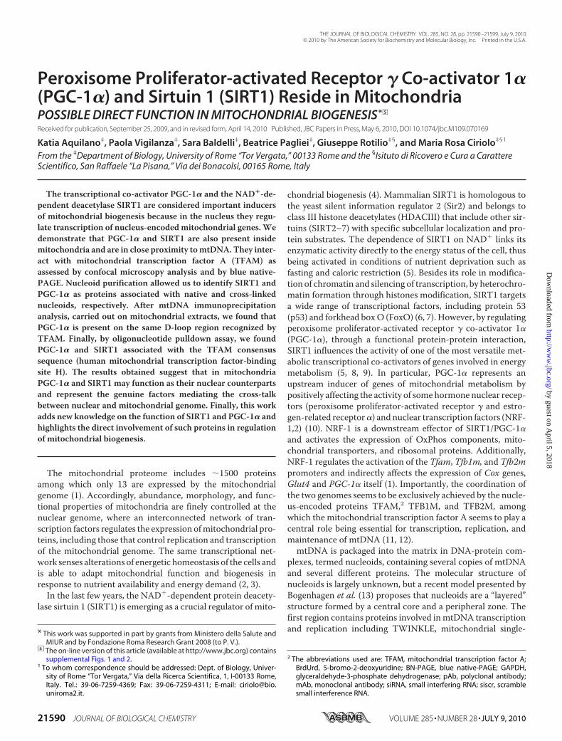
mitochondria purification. In brief, HeLa cells or mice organswere homogenized in a Teflon potter and centrifuged to sepa-rate cytoplasm (S1) from nuclei and unbroken cells (P1). S1 wasthen centrifuged to collect mitochondria-enriched fraction(P2). Finally, P2 was layered on top of 30% Percoll� solution,and mitochondria were separated from associated micro-vesicles through a Percoll� self-forming gradient according tothe method of Pellon-Maison et al. (22). Although isolatedmitochondria were treated with DNase I, the possible contam-ination of mitochondria with nuclei components was excludedby carrying out Western blot analysis of two of the most abun-dant nuclear proteins, i.e.H2B or lamin B for HeLa and mouseorgans, respectively (Fig. 2, B and C). High amounts of mito-chondrial proteins, such as subunit IV of cytochrome c oxidase(COX IV), SOD2, and TFAM, were observed in mitochondriafrom HeLa cells and the screened mice organs, suggesting thatthese organelles were efficiently isolated. Immunoblot analysiswas then carried out to confirm the presence of PGC-1� andSIRT1 inside the mitochondria using rabbit anti-SIRT1 andanti-PGC-1� from Santa Cruz Biotechnology. Protein extractsof purifiedmitochondria fromHeLa cells, mouse brain, mus-cle, and liver displayed the presence of these proteins (Fig. 2,B and C). SIRT1 was also detected by using an alternativeantibody from Calbiochem that recognized the same proteinband (data not shown). Moreover, the results reported insupplemental Fig. 1D show that in mitochondria from mouse
liver both the bands of 113 and 90 kDa were detected by usingthe two different anti-PGC-1� antibodies (Santa Cruz Biotech-nology and Calbiochem, as described previously). Moreover,the Calbiochem antibody cross-reacts with additional proteinsin the mitochondria of mouse liver, one of which could beascribed to the 90-kDa PGC-1� (supplemental Fig. 1D). The113- and the 90 kDa-bandswere also obtained on isolatedmito-chondria fromHeLa cells supplemental Fig. 1E, confirming thatthe same electrophoretic pattern of PGC-1� was present intotal extracts and mitochondria.We then looked at the possible presence of PGC-1� and
SIRT1 in purified human platelets that are known to be cellscompletely void of nuclei, and thus the presence of the nuclearPGC-1� and SIRT1 could be excluded. The immunoblotsreported in Fig. 2D demonstrate that purification of humanplatelets was properly achieved without any presence of leuko-cyte contamination. In fact, platelet protein extracts do notcontain any nuclear components, as demonstrated by theabsence ofH2B.On the contrary,mitochondrial protein such asTFAM and SOD2 are highly expressed. Fig. 2D also shows thatSIRT1 and PGC-1� are expressed in platelets derived from dif-ferent health subjects, indicating that such proteins are nor-mally localized also inside mitochondria.SIRT1 and PGC-1� Are Present in Mitochondrial Nucleoids—
Given the results obtained in the in vitro and in vivomitochon-drial localization of PGC-1� and SIRT1, and the knowledge of
FIGURE 2. Detection of mitochondrial SIRT1 and PGC-1� in mitochondria purified from HeLa cells and mouse organs and in human platelets. A, schemefor purification of mitochondria from HeLa and mouse organs. B, 10 �g of proteins obtained from HeLa purified mitochondria (Mito) and nuclei-enriched pellets(P1) were subjected to SDS-PAGE followed by Western blot analysis. The possible presence of nuclear protein contaminants was measured by incubatingnitrocellulose membrane with rabbit anti-H2B. The effective mitochondria isolation was assessed by staining with rabbit anti-TFAM and mouse anti-cyto-chrome c oxidase sub IV (COX IV). PGC-1� and SIRT1 were detected by using rabbit Santa Cruz Biotechnology antibodies. C, 10 �g of proteins obtained fromnuclei-enriched pellets (P1) and mitochondria (Mito) isolated from mice brain (B), skeletal muscle (M), and liver (L) were subjected to SDS-PAGE followed byWestern blot analysis. The possible presence of nuclear protein contaminants was measured by incubating nitrocellulose membrane with rabbit anti-lamin B.The effective mitochondrial isolation was assessed by staining with rabbit anti-SOD2. PGC-1� and SIRT1 were detected by using Santa Cruz Biotechnologyantibodies. D, human platelets were purified from peripheral blood, and 20 �g of protein extracts were subjected to SDS-PAGE followed by Western blotanalysis. The possible presence of nuclear proteins was measured by incubating the nitrocellulose membrane with rabbit anti-H2B. PGC-1� and SIRT1 weredetected by using Santa Cruz Biotechnology antibodies. Immunoblots reported are from one experiment representative of at least three that gave similarresults.
Mitochondrial SIRT1 and PGC-1�
21594 JOURNAL OF BIOLOGICAL CHEMISTRY VOLUME 285 • NUMBER 28 • JULY 9, 2010
by guest on April 5, 2018
http://ww
w.jbc.org/
Dow
nloaded from

the function of these proteins as regulators of gene expression,we speculated on a possible association of these proteins withnucleoids. Actually, nucleoids are functional structures local-ized in the mitochondrial matrix containing mtDNA packagedwith several proteins belonging to mitochondrial transcrip-tional machinery, including the mitochondrion-specific tran-scription factor TFAM, and are considered the mitochondrialunits of inheritance (13, 23). Isolated liver mitochondria werelysed, and after centrifugation, the nucleoid-enriched pelletwaslayered on a sucrose gradient according to Garrido et al. (23).After sucrose gradient centrifugation, nucleoid-containingfractions were identified by assessing the content of bothTFAMandmtDNA, and the puritywas verified by detecting thepresence of non-nucleoid mitochondrial protein such as cyto-chrome c and COX IV byWestern blot analysis (Fig. 3). More-over, other proteins, such as HSP60 and SOD2, with whichassociation with nucleoids is still debated, were also analyzed.Nucleoids were contained in fractions 6–9 because such frac-tions concomitantly stained positively with TFAM antibodyand showed the presence of mtDNA. However, TFAM wasmainly localized in fraction 1 according to its presence inmito-chondria also as mtDNA-unbound protein. The completeabsence of cytochrome c andCOX IV in fractions 6–9 excludedthe presence of protein contaminants in fractions containingnucleoids. These non-nucleoids proteins were instead particu-larly abundant in fraction 1 and were present to a minor extent
in fractions 2–5 and 10. The sametrend was evidenced for SOD2 andHSP60 indicating that they are notlikely associated with liver nativenucleoids. Being both SOD2 andHSP60 matrix proteins, a possiblematrix-protein contamination inthe nucleoid containing fractionswas finally excluded. Surprisingly,SIRT1 but not PGC-1� was foundnatively associated with nucleoids.In fact, SIRT1was found in fractions6–9.The absence of PGC-1� in native
nucleoids did not exclude that itcould be weakly or indirectly associ-ated with mtDNA. For this reason,liver mitochondria were cross-linked with 1% formaldehyde andpelleted through 20% sucrose fol-lowed by CsCl gradient (Fig. 4A).The putative nucleoid-containingfraction(s) was identified by assay-ing the presence of mtDNA andTFAM through PCR analysis ofD-loop and dot blot analysis ofTFAM protein, respectively (Fig.4B). The presence of genomic DNAwas excluded by performing PCRanalysis of actin. Western blot anal-ysis of cytochrome c and COX IVwas also performed on each col-
lected fraction to exclude the presence of protein contaminantsin nucleoid-containing fractions (Fig. 4B). Cytochrome c andCOX IV were not present in any fraction, strongly indicatingthat the majority of mitochondrial protein contaminants wasalready eliminated after centrifugation on 20% sucrose (Fig.4A). As shown in Fig. 4B, among the collected fractions, frac-tion 9 contains the higher level of both TFAM and mtDNAstrongly indicating that in such fractions purified nucleoidswere included. Fraction 9 also positively stained for HSP60,which is recognized as protein packaged into cross-linkednucleoids (Fig. 4B) (15). As expected, SIRT1 was present infraction 9 containing cross-linked nucleoids (Fig. 4B). More-over, Western blot analysis carried out with rabbit antibodyfrom Santa Cruz Biotechnology indicated that PGC-1� waspresent in fraction 9, suggesting that also PGC-1� is a com-ponent of the nucleoid structure. The same result wasobtained by performing Western blot analysis of PGC-1�with the mouse monoclonal antibody from Calbiochem.Also in this case, the two antibodies recognized PGC-1� atdifferent molecular masses (90 and 113 kDa, respectively).The fraction containing purified nucleoids was also loaded inparallel with a mitochondrial lysate to corroborate both thespecificity of the antibodies used and the goodness of nucleoidpurification (supplemental Fig. 2).In searching for the role of PGC-1� within nucleoids, we
asked whether PGC-1� could interact at the level of the
FIGURE 3. Assay of the presence of SIRT1 and PGC-1� in mitochondrial native nucleoids. Native nucleoidswere obtained from isolated liver mitochondria as described under “Experimental Procedures.” After sucrosegradient, nucleoid-containing fractions were identified by detecting the presence of TFAM by dot blot (usinggoat anti-TFAM) and mtDNA by PCR analysis of the D-loop region. Western blot analyses with mouse anti-cytochrome c oxidase subunit IV (COX IV), mouse anti-cytochrome c (Cyt c), rabbit anti-SOD2, and rabbit anti-HSP60 were carried out to determine the possible presence of protein contaminants. Fractions 6 –9 wereconsidered to contain native nucleoids due to the absence of protein contaminants and to the presence ofTFAM and mtDNA. PGC-1� and SIRT1 were detected by using Santa Cruz Biotechnology antibodies. Immuno-blots reported are from one experiment representative of at least three that gave similar results.
Mitochondrial SIRT1 and PGC-1�
JULY 9, 2010 • VOLUME 285 • NUMBER 28 JOURNAL OF BIOLOGICAL CHEMISTRY 21595
by guest on April 5, 2018
http://ww
w.jbc.org/
Dow
nloaded from
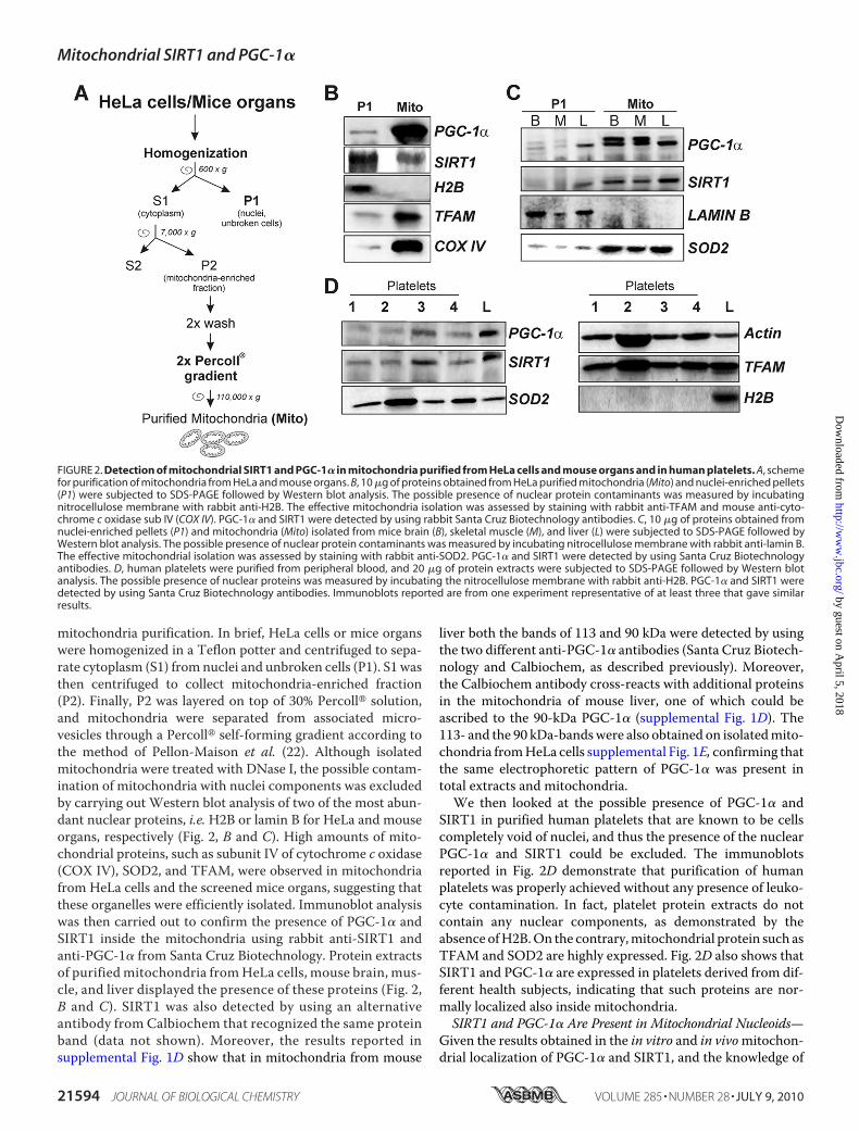
mtDNAD-loop region. To this end, isolated cross-linkedmito-chondria were immunoprecipitated with anti-PGC-1�, anti-TFAM (positive control), or an IgG isotype (negative control).
As expected, PCR analysis of �15,600/�15,868 mtDNAshowed that TFAM is highly associated with the D-loop region.No PCR product was detectable in samples immunoprecipi-
tated with the IgG isotype. Thepresence of D-loop region was iden-tified in samples immunoprecipi-tated with anti-PGC-1�, suggestingthe association of PGC-1� withmtDNA (Fig. 4C). These data wereconfirmed by performing an oligo-nucleotide-based pulldown assayin which mitochondrial proteinextracts were incubated with aTFAM biotinylated consensus oli-gonucleotide. After oligonucleotidepulldown, samples were subjectedto Western blot analysis. Fig. 4Dshows that TFAM protein was effi-ciently precipitated together withPGC-1� and SIRT1, suggesting thatthese proteins in some way have theability to interact with TFAM at thelevel of DNA.To further demonstrate that
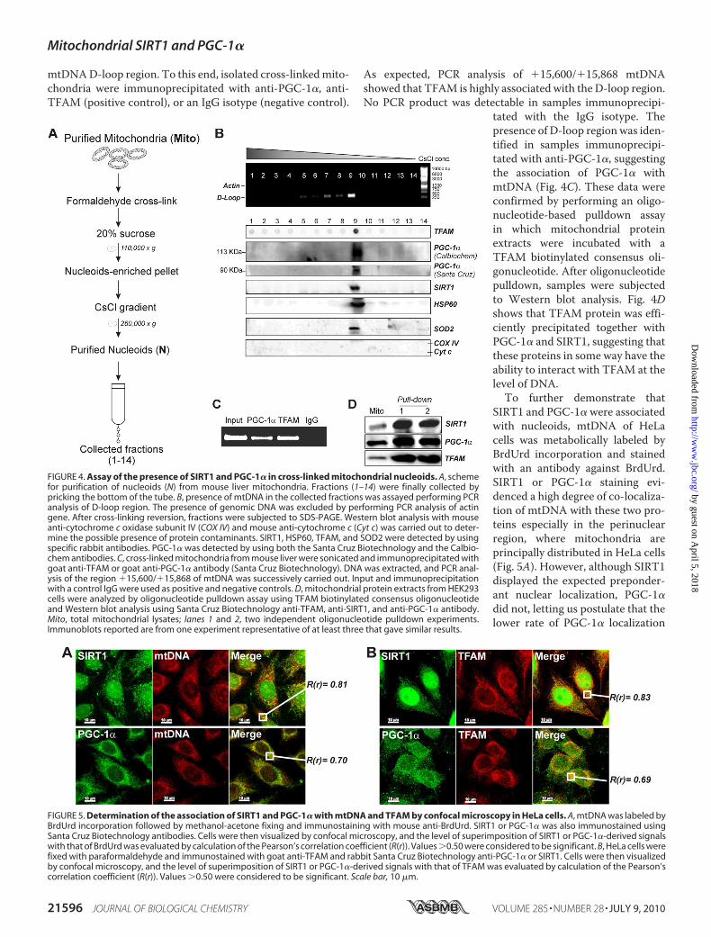
SIRT1 and PGC-1� were associatedwith nucleoids, mtDNA of HeLacells was metabolically labeled byBrdUrd incorporation and stainedwith an antibody against BrdUrd.SIRT1 or PGC-1� staining evi-denced a high degree of co-localiza-tion of mtDNA with these two pro-teins especially in the perinuclearregion, where mitochondria areprincipally distributed in HeLa cells(Fig. 5A). However, although SIRT1displayed the expected preponder-ant nuclear localization, PGC-1�did not, letting us postulate that thelower rate of PGC-1� localization
FIGURE 4. Assay of the presence of SIRT1 and PGC-1� in cross-linked mitochondrial nucleoids. A, schemefor purification of nucleoids (N) from mouse liver mitochondria. Fractions (1–14) were finally collected bypricking the bottom of the tube. B, presence of mtDNA in the collected fractions was assayed performing PCRanalysis of D-loop region. The presence of genomic DNA was excluded by performing PCR analysis of actingene. After cross-linking reversion, fractions were subjected to SDS-PAGE. Western blot analysis with mouseanti-cytochrome c oxidase subunit IV (COX IV) and mouse anti-cytochrome c (Cyt c) was carried out to deter-mine the possible presence of protein contaminants. SIRT1, HSP60, TFAM, and SOD2 were detected by usingspecific rabbit antibodies. PGC-1� was detected by using both the Santa Cruz Biotechnology and the Calbio-chem antibodies. C, cross-linked mitochondria from mouse liver were sonicated and immunoprecipitated withgoat anti-TFAM or goat anti-PGC-1� antibody (Santa Cruz Biotechnology). DNA was extracted, and PCR anal-ysis of the region �15,600/�15,868 of mtDNA was successively carried out. Input and immunoprecipitationwith a control IgG were used as positive and negative controls. D, mitochondrial protein extracts from HEK293cells were analyzed by oligonucleotide pulldown assay using TFAM biotinylated consensus oligonucleotideand Western blot analysis using Santa Cruz Biotechnology anti-TFAM, anti-SIRT1, and anti-PGC-1� antibody.Mito, total mitochondrial lysates; lanes 1 and 2, two independent oligonucleotide pulldown experiments.Immunoblots reported are from one experiment representative of at least three that gave similar results.
FIGURE 5. Determination of the association of SIRT1 and PGC-1� with mtDNA and TFAM by confocal microscopy in HeLa cells. A, mtDNA was labeled byBrdUrd incorporation followed by methanol-acetone fixing and immunostaining with mouse anti-BrdUrd. SIRT1 or PGC-1� was also immunostained usingSanta Cruz Biotechnology antibodies. Cells were then visualized by confocal microscopy, and the level of superimposition of SIRT1 or PGC-1�-derived signalswith that of BrdUrd was evaluated by calculation of the Pearson’s correlation coefficient (R(r)). Values �0.50 were considered to be significant. B, HeLa cells werefixed with paraformaldehyde and immunostained with goat anti-TFAM and rabbit Santa Cruz Biotechnology anti-PGC-1� or SIRT1. Cells were then visualizedby confocal microscopy, and the level of superimposition of SIRT1 or PGC-1�-derived signals with that of TFAM was evaluated by calculation of the Pearson’scorrelation coefficient (R(r)). Values �0.50 were considered to be significant. Scale bar, 10 �m.
Mitochondrial SIRT1 and PGC-1�
21596 JOURNAL OF BIOLOGICAL CHEMISTRY VOLUME 285 • NUMBER 28 • JULY 9, 2010
by guest on April 5, 2018
http://ww
w.jbc.org/
Dow
nloaded from
into the nucleus may be a characteristic of this tumor cell line.The extent of co-localization was estimated by calculation ofPearson’s correlation coefficient. This analysis gave scores of0.81 and 0.70 for SIRT1/mtDNAand PGC-1�/mtDNA, respec-tively. TFAM is a protein that was found to be acetylated on asingle lysine residue in rat liver (32). Because TFAM is a tran-scription factor, it was reasonable to postulate not only a possi-blemodulation by SIRT1 but also by PGC-1� in a similar way tothat operating at nuclear levels. Thus, we first evaluated thepossible interaction of SIRT1 and/or PGC-1� with TFAM byconfocal microscopy. As reported in Fig. 5B, TFAM stainingwas mainly visualized in the perinuclear region and as an intri-cate cytoplasmic network according to its mitochondrial local-ization. We found that fluorescence of SIRT1 and PGC-1�overlapped well with that of TFAM at the mitochondrial level,and calculation of Pearson’s correlation coefficient finally dem-onstrated that they are co-localized with TFAM (0.83 and 0.69,respectively).PGC-1� and SIRT1 Interact with TFAM to Form Multipro-
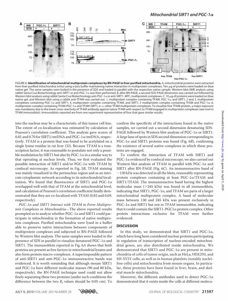
tein Complexes in Mitochondria—The above reported resultsprompted us to analyze whether PGC-1� and SIRT1 could par-ticipate in mitochondria in the formation of native multipro-tein complexes. Purified mitochondria were lysed in a bufferable to preserve native interactions between components ofmultiprotein complexes and subjected to BN-PAGE followedbyWestern blot analysis. The same samples were loaded in thepresence of SDS in parallel to visualize denatured PGC-1� andSIRT1. The immunoblots reported in Fig. 6A shows that bothproteins are present as free factors inmitochondrial lysates andalso form proteinmacro-complexes. A superimposable patternof anti-SIRT1 and anti-PGC-1� immunoreactive bands wasevidenced. It is worth reminding that although mouse SIRT1and PGC-1� have different molecular masses (90 and 80 kDa,respectively), the BN-PAGE technique used could not allowfinely separating these two proteins (under such conditions thedifference between the two Rf values should be 0.05 cm). To
confirm the specificity of the interactions found in the nativesamples, we carried out a second dimension denaturing SDS-PAGE followed by Western blot analysis of PGC-1� or SIRT1.A large lane of spots in SDS seconddimension corresponding toPGC-1� and SIRT1 proteins was found (Fig. 6B), confirmingthe existence of several native complexes in which these pro-teins are engaged.To confirm the interaction of TFAM with SIRT1 and
PGC-1� evidenced by confocal microscopy, we also carried outWestern blot analysis of TFAM in parallel with PGC-1� andSIRT1 after BN-PAGE (Fig. 6C). An immunoreactive band of�130 kDawas detected in all the blots, reasonably representingprotein complexes containing at least PGC-1�/TFAM andSIRT1/TFAM. The immunoreactive band having the highestmolecular mass (�245 kDa) was found in all immunoblots,indicating that SIRT1, PGC-1�, and TFAM are parts of a largermitochondrial multiprotein complex. A band of molecularmass between 130 and 245 kDa was present exclusively inPGC-1� and SIRT1 but not in TFAM immunoblot, indicatingthat it could contain the SIRT1-PGC1� protein complex.Otherprotein interactions exclusive for TFAM were furtherevidenced.
DISCUSSION
In this study, we demonstrated that SIRT1 and PGC-1�,which have long been considered nuclear proteins participatingin regulation of transcription of nucleus-encoded mitochon-drial genes, are also distributed inside mitochondria. Wedemonstrated that SIRT1 and PGC-1� are present in mito-chondria of cells of tumor origin, such as HeLa, HEK293, andSH-SY5Y cells, as well as in human platelets (notably nuclei-free cells) and mitochondria from mouse organs. In particu-lar, these proteins have been found in liver, brain, and skel-etal muscle mitochondria.Moreover, the different antibodies used to detect PGC-1�
demonstrated that it exists inside the cells at different molecu-
FIGURE 6. Identification of mitochondrial multiprotein complexes by BN-PAGE in liver purified mitochondria. A, mitochondrial proteins were extractedfrom liver purified mitochondria (mito) using a lysis buffer maintaining native interaction in multiprotein complexes. Ten �g of proteins were loaded on bluenative gel. The same samples were boiled in the presence of SDS and loaded in parallel with the respective native sample. Western blot (WB) analysis usingrabbit Santa Cruz Biotechnology anti-SIRT1 or anti-PGC-1� was then performed. B, after BN-PAGE, a second SDS-PAGE dimension was carried out followed byWestern blot analysis using rabbit Santa Cruz Biotechnology anti-PGC-1� or anti-SIRT1. MPC, multiprotein complexes. C, 10 �g of proteins were loaded on bluenative gel, and Western blot using a rabbit anti-TFAM was carried out. 1, multiprotein complex containing TFAM, PGC-1�, and SIRT1; 2 and 3, multiproteincomplexes containing PGC-1� and SIRT1; 4, multiprotein complex containing TFAM, and SIRT1; 5 multiprotein complex containing TFAM and PGC-1�; 6,multiprotein complex containing TFAM-PGC-1� and TFAM-SIRT1; a– c, other TFAM multiprotein complexes. To visualize free TFAM protein, a major exposurewas mandatory due to the lower cross-reactivity of TFAM antibody against native TFAM with respect to TFAM engaged in multiprotein complexes (see inset inTFAM immunoblot). Immunoblots reported are from one experiment representative of four that gave similar results.
Mitochondrial SIRT1 and PGC-1�
JULY 9, 2010 • VOLUME 285 • NUMBER 28 JOURNAL OF BIOLOGICAL CHEMISTRY 21597
by guest on April 5, 2018
http://ww
w.jbc.org/
Dow
nloaded from

larmasses, and in particular, wemainly evidenced two differentbands at 90 and 113 kDa, which were both modulated by eitheroverexpression or down-regulation. The diverse molecularmass forms of PGC-1� were in line with those reported by themanufacturers and most probably due to post-transcriptionaland/or post-translational processes (28, 29). Work is in pro-gress in our laboratory to identify themodifications responsiblefor such molecular weights difference.During mitochondrial biogenesis, SIRT1 participates inside
the nucleus in the induction of PGC-1� that is pivotal toorchestrate the activation of a broad set of transcription factorsand nuclear hormone receptors enhancing the expression ofthe nucleus-encoded mitochondrial genes. The presence ofSIRT1 and PGC-1� within the mitochondria suggests thatthese proteins could exert similar functions in this compart-ment. Among the putative targets of their actions, we focusedon TFAM as, being a transcription factor, it can be likely mod-ulated by co-activator(s) and by an acetylation/deacetylationmechanism. Data from Dinardo et al. (32) in part support thisassumption as they demonstrated that TFAM is acetylated at asingle lysine residue, a modification that is supposed to have arole in regulating the expression of mitochondrial genes or ingeneral the mtDNA maintenance. Also, unpublished resultsfrom our laboratory indicate that in liver mitochondria TFAMis present both in acetylated and de-acetylated status.3 To date,neither a deacetylase nor a co-activator has been identified tomodulate TFAM activity. The presence of SIRT1 in samplesprecipitated after incubationwith oligonucleotide representingTFAM consensus sequence strongly indicates a possible actionof SIRT1 onTFAM. Purification of liver nucleoids allowed us todiscover that SIRT1 as well as TFAM are natively associatedwith nucleoids, whereas PGC-1� is not. These observations ledus to speculate that, at least in its nucleoid-associated state,SIRT1 could be the factor responsible formodulation of TFAMacetylation level. We are confident that the presence of SIRT1in native nucleoids is not due to contamination, because mito-chondrial HSP60, which is considered a protein contaminant innative purified nucleoids (13), and other non-nucleoid associ-ated proteins were not detected. Moreover, the cross-reactivityof the SIRT1 antibody (recognizing an �90-kDa band) withmitochondrial SIRT3–5 has to be excluded because these fam-ily members have very lower molecular weights with respect toSIRT1 as reported in several protein databases (e.g. Swiss Protand TrEMBL). The evidence that SIRT1 has been recently rec-ognized to be located in cytoplasm of cardiomyocytes (33)strongly supports our findings about the presence of SIRT1inside mitochondria. Interestingly, our data also indicate thatSOD2 is not associated with liver native nucleoids as wasinstead recently demonstrated for Jurkat cells by Kienhofer etal. (14). However, in line with our results, the same authorsdemonstrated that SOD2 could be absent in native nucleoids ofother cell types, such as bovine endothelial cells. The resultsobtained from mitochondria cross-linking confirm that SOD2is in some way associated with nucleoids and further support
the role of this antioxidant enzyme in ensuring mtDNAintegrity.The absence of PGC-1� in natively purified nucleoids is in
agreement with its inability to directly bind DNA. In contrast,its presence in cross-linked nucleoids nicely correlates with itsfunction as a transcriptional co-activator also in mitochondria.Results obtained by confocal images confirmed the associationof both PGC-1� and SIRT1 with nucleoids, as a very significantPearson’s correlation coefficient of such proteins with mtDNAwas observed.We demonstrate that PGC-1� and SIRT1 are present intra-
mitochondrially as free proteins and also interact with TFAM.Inmitochondria, SIRT1 andPGC-1� form severalmultiproteincomplexes among which are included PGC-1�/SIRT1, TFAM/PGC-1�, and TFAM/SIRT1. Moreover, both PGC-1� andSIRT1 reasonably participate with TFAM in the formation of ahigher molecular weight multiprotein complex, which couldcorrespond to macro-complexes involved in mtDNA transac-tions. We also have strong evidence that PGC-1� could posi-tively influence the activity of human TFAM at least on theD-loop region.Not only the oligonucleotide pulldown assay butalso data from mtDNA immunoprecipitation analysis demon-strated that TFAM is bound in association with SIRT1 and/orPGC-1� to mtDNA.
The way in which SIRT1 and PGC-1� are included in mito-chondria remains obscure, having no known mitochondrialimport sequences, and it deserves further investigation. How-ever, an import mechanism similar to that of other mitochon-drial proteins, mediated by the translocases of outer and innermitochondrial membrane, may be operative (3).The presence of SIRT1 in themitochondrial compartment is
not in contrast with that of mitochondria resident sirtuins(SIRT3–5), which have been uniquely assigned to regulatingmetabolic mitochondrial enzyme activity (34), and at the sametime it reinforces the role of SIRT1 inmodulating DNA remod-eling and transcription.All together, our results suggest that SIRT1 and PGC-1�
have to be considered not only nuclear but also mitochondrialimported proteins that are associated with nucleoids. The dataobtained also indicate that in this submitochondrial region,SIRT1 and PGC-1� could function as regulators of TFAM pos-sibly mediating its deacetylation and mtDNA binding activity.On the basis of such findings, other avenues are now open, Forinstance, is the acetylation/deacetylation status of PGC-1� andTFAM responsible for their association with nucleoids and formodulating mtDNA transcription? Are mitochondrial SIRT1and PGC-1� the effective mediators of the cross-talk betweencellular metabolism and mitochondrial biogenesis?Finally, this work strengthens the role of SIRT1/PGC-1�
pathway in regulation of mitochondrial biogenesis and metab-olism and gives new inputs in the comprehension of theregulatory mechanisms governing mitochondrial genomeexpression, replication, and maintenance. This unexpectedlocalization also highlights the intriguing perspective that alter-ations in mitochondrial residents PGC-1� and SIRT1 could becausative of mitochondrial homeostasis impairment underly-ing pathophysiological processes, including aging, metabolicsyndromes, myopathies, and neurodegenerative diseases.
3 K. Aquilano, P. Vigilanza, S. Baldelli, B. Pagliei, G. Rotilio, and M. R. Ciriolo,unpublished results.
Mitochondrial SIRT1 and PGC-1�
21598 JOURNAL OF BIOLOGICAL CHEMISTRY VOLUME 285 • NUMBER 28 • JULY 9, 2010
by guest on April 5, 2018
http://ww
w.jbc.org/
Dow
nloaded from

Acknowledgments—We gratefully acknowledge Palma Mattioli andElena Romano (Dept. of Biology, University of Rome Tor Vergata,Rome, Italy) for their assistance in fluorescent and confocal micros-copy and images analysis. We thank Dr. Dan Bogenhagen (StonyBrook University, Stony Brook, NY) for the kind gift of polyclonal rab-bit TFAM antibody. We also thank Dr. Bruce Spiegelman (Dept. ofMedicine, University of Chicago) for providing pSV-PGC-1� vectorinsert in Addgene deposit plasmid. We thank Dr. Teresa C. Leone forhelpful discussions about PGC-1� antibody utilization. We are alsograteful to the animal house staff of the University of Rome “TorVergata.”
REFERENCES1. Hock, M. B., and Kralli, A. (2009) Annu. Rev. Physiol. 71, 177–2032. Chow, L. S., Greenlund, L. J., Asmann, Y.W., Short, K. R., McCrady, S. K.,
Levine, J. A., and Nair, K. S. (2007) J. Appl. Physiol. 102, 1078–10893. Scarpulla, R. C. (2008) Physiol. Rev. 88, 611–6384. Guarente, L. (2007) Cold Spring Harbor Symp. Quant. Biol. 72, 483–4885. Rodgers, J. T., Lerin, C., Gerhart-Hines, Z., and Puigserver, P. (2008) FEBS
Lett. 582, 46–536. Cheng, H. L., Mostoslavsky, R., Saito, S., Manis, J. P., Gu, Y., Patel, P.,
Bronson, R., Appella, E., Alt, F.W., andChua, K. F. (2003)Proc. Natl. Acad.Sci. U.S.A. 100, 10794–10799
7. Brunet, A., Sweeney, L. B., Sturgill, J. F., Chua, K. F., Greer, P. L., Lin, Y.,Tran,H., Ross, S. E.,Mostoslavsky, R., Cohen,H. Y., Hu, L. S., Cheng,H. L.,Jedrychowski, M. P., Gygi, S. P., Sinclair, D. A., Alt, F. W., and Greenberg,M. E. (2004) Science 303, 2011–2015
8. Nemoto, S., Fergusson, M. M., and Finkel, T. (2005) J. Biol. Chem. 280,16456–16460
9. Knutti, D., and Kralli, A. (2001) Trends Endocrinol. Metab. 12, 360–36510. Finkel, T. (2006) Nature 444, 151–15211. Wang, Y., and Bogenhagen, D. F. (2006) J. Biol. Chem. 281, 25791–2580212. Kang, D., Kim, S. H., and Hamasaki, N. (2007)Mitochondrion 7, 39–4413. Bogenhagen, D. F., Rousseau, D., and Burke, S. (2008) J. Biol. Chem. 283,
3665–367514. Kienhofer, J., Haussler, D. J., Ruckelshausen, F., Muessig, E., Weber, K.,
Pimentel, D., Ullrich, V., Burkle, A., and Bachschmid,M.M. (2009) FASEBJ. 23, 2034–2044
15. Kaufman, B. A., Newman, S. M., Hallberg, R. L., Slaughter, C. A., Perlman,P. S., and Butow, R. A. (2000) Proc. Natl. Acad. Sci. U.S.A. 97, 7772–7777
16. Bogenhagen, D. F. (1985) J. Biol. Chem. 260, 6466–647117. Puigserver, P., Wu, Z., Park, C. W., Graves, R., Wright, M., and
Spiegelman, B. M. (1998) Cell 92, 829–83918. Kawakami, Y., Tsuda, M., Takahashi, S., Taniguchi, N., Esteban, C. R.,
Zemmyo, M., Furumatsu, T., Lotz, M., Belmonte, J. C., and Asahara, H.(2005) Proc. Natl. Acad. Sci. U.S.A. 102, 2414–2419
19. Aquilano, K., Vigilanza, P., Rotilio, G., and Ciriolo, M. R. (2006) FASEB J.20, 1683–1685
20. Sun, F. C.,Wei, S., Li, C.W., Chang, Y. S., Chao, C. C., and Lai, Y. K. (2006)Biochem. J. 396, 31–39
21. Frezza, C., Cipolat, S., and Scorrano, L. (2007) Nat. Protoc. 2, 287–29522. Pellon-Maison, M., Montanaro, M. A., Coleman, R. A., and Gonzalez-
Baro, M. R. (2007) Biochim. Biophys. Acta 1771, 830–83823. Garrido, N., Griparic, L., Jokitalo, E., Wartiovaara, J., van der Bliek, A. M.,
and Spelbrink, J. N. (2003)Mol. Biol. Cell 14, 1583–159624. Kucej, M., Kucejova, B., Subramanian, R., Chen, X. J., and Butow, R. A.
(2008) J. Cell Sci. 121, 1861–186825. Westerheide, S. D., Anckar, J., Stevens, S. M., Jr., Sistonen, L., and Mori-
moto, R. I. (2009) Science 323, 1063–106626. Camacho-Carvajal, M. M., Wollscheid, B., Aebersold, R., Steimle, V., and
Schamel, W. W. (2004)Mol. Cell. Proteomics 3, 176–18227. Lowry, O. H., Rosebrough, N. J., Farr, A. L., and Randall, R. J. (1951) J. Biol.
Chem. 193, 265–27528. Chang, J. S., Huypens, P., Zhang, Y., Black, C., Kralli, A., and Gettys, T.W.
(2010) J. Biol. Chem. 285, 18039–1805029. Handschin, C., and Spiegelman, B. M. (2006) Endocr. Rev. 27, 728–73530. Zhang, Y., Huypens, P., Adamson, A. W., Chang, J. S., Henagan, T. M.,
Boudreau, A., Lenard, N. R., Burk, D., Klein, J., Perwitz, N., Shin, J.,Fasshauer, M., Kralli, A., and Gettys, T. W. (2009) J. Biol. Chem. 284,32813–32826
31. Lin, J. D. (2009)Mol. Endocrinol. 23, 2–1032. Dinardo, M. M., Musicco, C., Fracasso, F., Milella, F., Gadaleta, M. N.,
Gadaleta, G., and Cantatore, P. (2003) Biochem. Biophys. Res. Commun.301, 187–191
33. Tanno, M., Kuno, A., Yano, T., Miura, T., Hisahara, S., Ishikawa, S., Shi-mamoto, K., and Horio, Y. (2010) J. Biol. Chem. 285, 8375–8382
34. Schlicker, C., Gertz, M., Papatheodorou, P., Kachholz, B., Becker, C. F.,and Steegborn, C. (2008) J. Mol. Biol. 382, 790–801
Mitochondrial SIRT1 and PGC-1�
JULY 9, 2010 • VOLUME 285 • NUMBER 28 JOURNAL OF BIOLOGICAL CHEMISTRY 21599
by guest on April 5, 2018
http://ww
w.jbc.org/
Dow
nloaded from
Maria Rosa CirioloKatia Aquilano, Paola Vigilanza, Sara Baldelli, Beatrice Pagliei, Giuseppe Rotilio and
MITOCHONDRIAL BIOGENESISSirtuin 1 (SIRT1) Reside in Mitochondria: POSSIBLE DIRECT FUNCTION IN
) andα (PGC-1α Co-activator 1γPeroxisome Proliferator-activated Receptor
doi: 10.1074/jbc.M109.070169 originally published online May 6, 20102010, 285:21590-21599.J. Biol. Chem.
10.1074/jbc.M109.070169Access the most updated version of this article at doi:
Alerts:
When a correction for this article is posted•
When this article is cited•
to choose from all of JBC's e-mail alertsClick here
Supplemental material:
http://www.jbc.org/content/suppl/2010/05/06/M109.070169.DC1
http://www.jbc.org/content/285/28/21590.full.html#ref-list-1
This article cites 34 references, 20 of which can be accessed free at
by guest on April 5, 2018
http://ww
w.jbc.org/
Dow
nloaded from