Periprosthetic Femoral Fractures in Total Knee...
16
Chapter 18 Periprosthetic Femoral Fractures in Total Knee Arthroplasty Vladan Stevanović, Zoran Vukašinović, Zoran Baščarević, Branislav Starčević, Dragana Matanović and Duško Spasovski Additional information is available at the end of the chapter http://dx.doi.org/10.5772/55226 1. Introduction Total joint arthroplasty has greatly improved the treatment of knee arthrosis, but still is not without complications. Supracondylar fractures above total knee replacements are an uncom‐ mon complication (incidence 0,3% to 2.5%), occuring more frequently in patients older than 60 years with osteoporotic bone. The rate of these fractures is expected to increase in the future because of the growing number of total knee replacements and greater level of acitivity among elderly patients. The timing of such fractures has been reported to range from early in the postoperative period to more than a decade after surgery, with a mean of 2 to 4 years. During the past two decades authors were not agreed in the definition of periprosthetic supracondylar region: the lower 3 inches (7cm) of the femur [1]; 9 cm proximal to the knee joint line [2]; all fractures within 15 cm proximal to the knee joint line [3]. Generally, based on the older literature, supracondylar periprosthetic fractures were those within 15 cm of the joint line, or in the case of stemmed component, within 5 cm of the proximal end of the implant. Never‐ theless, the most important is understanding that these fractures occur in regions of stress concentration adjacent to a prosthetic component, and that the presence of the prosthesis has a significant effect on fracture treatment. So, we suggest that fractures above total knee replacement should be considered supracondylar fractures if they extend within 7 cm of the prosthetic joint line or if they are within 2 cm of the femoral prosthetic flange. The most commonly suggested predisposing factors for a periprosthetic femoral fracture after total knee arthroplasty are osteopenia, revision arthroplasty, rheumatoid arthritis, use of steroids, existing neurological disorders, misalignment of the components, and notching of © 2013 Stevanović et al.; licensee InTech. This is an open access article distributed under the terms of the Creative Commons Attribution License (http://creativecommons.org/licenses/by/3.0), which permits unrestricted use, distribution, and reproduction in any medium, provided the original work is properly cited.
Transcript of Periprosthetic Femoral Fractures in Total Knee...

Chapter 18
Periprosthetic Femoral Fractures inTotal Knee Arthroplasty
Vladan Stevanović, Zoran Vukašinović,Zoran Baščarević, Branislav Starčević,Dragana Matanović and Duško Spasovski
Additional information is available at the end of the chapter
http://dx.doi.org/10.5772/55226
1. Introduction
Total joint arthroplasty has greatly improved the treatment of knee arthrosis, but still is notwithout complications. Supracondylar fractures above total knee replacements are an uncom‐mon complication (incidence 0,3% to 2.5%), occuring more frequently in patients older than60 years with osteoporotic bone. The rate of these fractures is expected to increase in the futurebecause of the growing number of total knee replacements and greater level of acitivity amongelderly patients. The timing of such fractures has been reported to range from early in thepostoperative period to more than a decade after surgery, with a mean of 2 to 4 years. Duringthe past two decades authors were not agreed in the definition of periprosthetic supracondylarregion: the lower 3 inches (7cm) of the femur [1]; 9 cm proximal to the knee joint line [2]; allfractures within 15 cm proximal to the knee joint line [3]. Generally, based on the olderliterature, supracondylar periprosthetic fractures were those within 15 cm of the joint line, orin the case of stemmed component, within 5 cm of the proximal end of the implant. Never‐theless, the most important is understanding that these fractures occur in regions of stressconcentration adjacent to a prosthetic component, and that the presence of the prosthesis hasa significant effect on fracture treatment. So, we suggest that fractures above total kneereplacement should be considered supracondylar fractures if they extend within 7 cm of theprosthetic joint line or if they are within 2 cm of the femoral prosthetic flange.
The most commonly suggested predisposing factors for a periprosthetic femoral fracture aftertotal knee arthroplasty are osteopenia, revision arthroplasty, rheumatoid arthritis, use ofsteroids, existing neurological disorders, misalignment of the components, and notching of
© 2013 Stevanović et al.; licensee InTech. This is an open access article distributed under the terms of theCreative Commons Attribution License (http://creativecommons.org/licenses/by/3.0), which permitsunrestricted use, distribution, and reproduction in any medium, provided the original work is properly cited.

the anterior femoral cortex. Different factors were found in the pathogenesis of the fracture:stress-shielding from the anterior flange of the femoral component, inadequate osseousremodeling due to postoperative hypovascularity, relative difference in elastic modulusbetween the implant-covered distal part of the femur and femoral cortex, endosteal ischemiafrom metal or bone cement, and osteolysis of the distal part of the femur secondary topolyethylene wear debris. The majority of these fractures results from a combination of axialand torsion loads. Most of them occur following minimal falls, while the rest of them aresecondary to motor-vehicle accidents, seizers or closed manipulation of a stiff knee after totalknee arthroplasty
2. Prevalence and pathogenesis
The prevalence of supracondylar femoral fracture in patients with total knee replacementranges from 0.3 to 4.2%. Most of the patients who sustain fractures about a total knee arthro‐plasty are women, usually in their seventh decade of life. As with other supracondylarfractures in the elderly, periprosthetic fractures usually occurs after low energy trauma.Osteoporosis is often present as well, due to a number of factors including stress shieldingbecause of a rigid implant, pharmacologic causes, hormonal influences and senility. Anassociation with rheumatoid arthritis, especially when the patient is receiving oral corticoste‐roid treatment, has been noted. Neurologic disorders have also been involved in the occurrenceof these fractures, due to either medication induced osteoporosis or gait disturbance. Inaddition, revision arthroplasty has been associated with an increased incidence of peripros‐thetic fractures, more commonly when constrained implants are used, as they transfer appliedtorque more directly to bone that is potentially already deficient. Notching of the anteriorfemoral cortex during total knee arthroplasty has been indicated as one factor contributing tothese periprosthetic femoral fractures. The prevalence of inadvertent cortical notching of thefemur during total knee arthroplasty has been reported to be as high as 27% and there areseveral studies performed to quantify the reduction in bending and torsion strength resultingfrom femoral notching in attempt to provide the clinician with useful information related tothe postoperative management [5, 6]. Clearly, notching of the anterior femoral cortex is neitherthe only risk factor nor the principal risk factor for supracondylar femoral fracture after kneereplacement. Of a total of 6470 total knee arthroplasties included in reports on this subject,only seventeen (0.26%) were complicated by a supracondylar femoral fracture associated withanterior notching compared with nearly three times as many fractures that occurred in theabsence of notching [5]; biomechanical effects of femoral notching following total kneearthroplasty showed mean decrease in bending strength of 18% (8-31%) and mean reductionin torsion strength of 39.2% (19-73%) in cadaveric specimens [6]. Based on Wolff’s law, distalpart of the femur would strengthen after the operation as result of remodeling, thus reductionin femoral bone strength should primarily be expected in the immediate postoperative period.Therefore a clear recommendation should be given to the patients who sustain inadvertentnotching that they should have additional protection in the early postoperative period, and toconsider the use a femoral component with stem as a means to bypass the stress riser of the
Arthroplasty - Update422

anterior cortical notch. Most important, authors believe that an anterior cortical notch shouldbe considered as a contraindication for manipulation of the knee prosthesis in the earlypostoperative period [7, 8].
Anterior defects may be present without notching, such as in cases of cystic lesions of degen‐erative or rheumatoid origin near the proximal aspect of the anterior femoral flange. Adequateremodeling may not be possible after those cysts are filled with cement at the time of arthro‐plasty. These defects remain as permanent stress risers, which may predispose to fracture.Large anterior effects might be better managed during primary knee arthroplasty with bonegrafting and protection of the distal femur with an intramedullary stem [9].
Another recently recognized factor leading to late supracondylar femoral fracture is thepresence of a massive debris-related osteolytic defect in the distal femur; such defects havebeen reported in association with asymptomatic well-fixed cementless femoral component.Ankylosis of a total knee arthroplasty may also predispose a fracture by producing increasedstress in the distal femoral metaphysis [10, 11].
3. Risk factors / etiology
Literature data show that patients with osteopenia are at greater risk to acquire supracondylarfemoral fracture after total knee arthroplasty, followed by rheumatoid arthritis, corticosteroidtreatment, female gender and older age [12,13,14]. Additional risk factors are: neurologicaldisorders, a revision total knee replacement (TKR) and rotationally constrained implants thatcreate increased torsion load transfer to bone [15] (Table1).
OsteopeniaRheumatoid arthritisSteroid useNeurologic disordersRevision TKRFemale genderSeventh decade of lifeDistal femoral osteolysisAnterior femoral notching +/-
Table 1. Risk factors for supracondylar femoral fractures, in decreasing order
Clinical and biomechanical data on anterior notching of the distal femoral cortex confirm theincrease of fracture risk, and theoretical mathematical analysis calculated that a three-millime‐ter notch results in a 30% reduction in torsion bone strength [9]. On the other hand, a series of 670total knee prosthesis with 20% femurs with anterior notching of at 3 mm at least, and found onlytwo supracondylar fractures [11]. Different fracture patterns are associated with notched and nonotched femurs: notched femurs tend to have short oblique fractures originating from the notchedcortex, whereas no notched femurs tend to have diaphyseal fractures.
Periprosthetic Femoral Fractures in Total Knee Arthroplastyhttp://dx.doi.org/10.5772/55226
423

Furthermore, there is a general feeling that the most significant risk factor causing supracon‐dylar fracture is the increase in activity that elderly patients achieve after knee replacement,exposing them to a greater risk of slipping and falling.
4. Diagnostic algorithm
Patients with this type of injury usually provide a history of minor trauma, such as fall duringambulation. They usually present with pain and inability to bear weight. Since these aretypically low energy injuries, major tissue swelling is uncommon. Unless marked displace‐ment is present, deformity may not be apparent on examination.
A thorough evaluation includes careful physical examination, a review of the patient’s medicalhistory and adequate radiographic studies. The injured limb should be assessed for soft tissueintegrity and neurovascular status. The location of previous skin incisions must also be noted.
A complete radiographic examination of a fracture about a total knee arthroplasty includesstandard anteroposterior and lateral radiographs as well as long leg views of the involvedlimb; oblique images and tomography are also often useful (Table 2). The diagnostic evaluationmust include a direct lateral view of the distal femur in order to guide subsequent treatment:the direct lateral view facilitates assessment of fracture displacement, while also revealing thebone available for fixation devices, the location of femoral lugs of posterior cruciate retainingcomponents and the proximal extent of the central femoral recess in cases with posteriorstabilized components. Radiographically, nondisplaced or minimally displaced fractures maybe obscured by the femoral flange; it is important to identify nondisplaced fractures sincedisplacement may occur later.
Fracture displacement and comminution
Axial limb alignment
Quality of bone stock
Location of the fracture relative to the prosthesis
Stability of the prosthesis
Table 2. Characteristics of radiography assessment
Review of prefracture radiographs can provide important data regarding baseline limbalignment, implant fixation and the presence of regions of osteolysis or polyethylene wear.The type and technical specifications of the implant and templates in place will influence theselection of fixation device if open reduction is necessary [16, 17].
The first step is to establish whether the implant is loose; if so even if the fracture is well alignedand heals, treatment that does not include revision will lead to poor result. Prefracturemisalignment, osteolysis and polyethylene wear are important factors in the decision makingprocess.
Arthroplasty - Update424

The second step in the treatment is to identify fracture displacement and to decide whetherreduction is needed. Any alteration in limb axis resulting from fracture can result in altered loadingof the prosthesis, which may in turn lead to enhanced wear and/or accelerated implant loosening.
The third step is to determine the appropriate treatment for displaced fracture (Figure 1).
Figure 1. Diagnostic algorithm for periprosthetic supracondylar femoral fracture above total knee arthroplasty
5. Classification
Numerous systems of classification of supracondylar femoral fractures after total kneearthroplasty have been developed. Most of the classifications were based on supracondylarfractures without knee arthroplasty (Neer et al, DiGioia and Rubash, Chen et al.) (Table 3).
Neer et al. Type I Undisplaced (<5mm displacement or <50
angulation)*
Type II Displaced >1cmType IIa With lateral femoral shaft displacementType IIb With medial femoral shaft displacementType III Displaced and comminuted
DiGioia and Rubash Group IGroup IIGroup III
Extraarticular, undisplaced*Extraarticular, displaced*
Severely displaced (loss of cortical contact) orangulated
Chen et al Type IType II
Nondisplaced(Neer I)Displaced or comminuted (Neer I or II)
Table 3. Classification of supracondylar femoral fractures above total knee arthroplasty reprinted from Su ET, De WalH, Di Cesare P. Periprosthetic Femoral Fractures Above Total Knee Replacements J Am Acad Orthop Surg, 2004; 12:12– 20. - with permission (personal communication)
For identifying fracture displacement and deciding whether reduction is needed Rorabeck etal. [18] created classification that takes into account both the status of the prosthesis (intact orfailing) and the displacement of the fracture:
Periprosthetic Femoral Fractures in Total Knee Arthroplastyhttp://dx.doi.org/10.5772/55226
425

Type I: fracture is undisplaced and the prosthesis is intact; Type II: fracture is displaced andthe prosthesis is intact; Type III: fracture is displaced or undisplaced and the prosthesis is looseor failing
Summarizing above mentioned classifications, we strongly support suggested and explainedin article by Su et al.[4] which is transcripted (Figure 2)
(a)
Type I: Fracture proximal to femoral knee component
Type III: Fracture line is distal to the upper edge of the anterior flange of the femoral knee component
Type II: Fracture originating at the proximal aspect of the femoral knee component and extending proximally
Figure 2. Reprinted from Su ET, De Wal H, Di Cesare P. Periprosthetic Femoral Fractures Above Total Knee Replace‐ments J Am Acad Orthop Surg, 2004; 12:12 – 20. - with permission (personal communication)
Arthroplasty - Update426
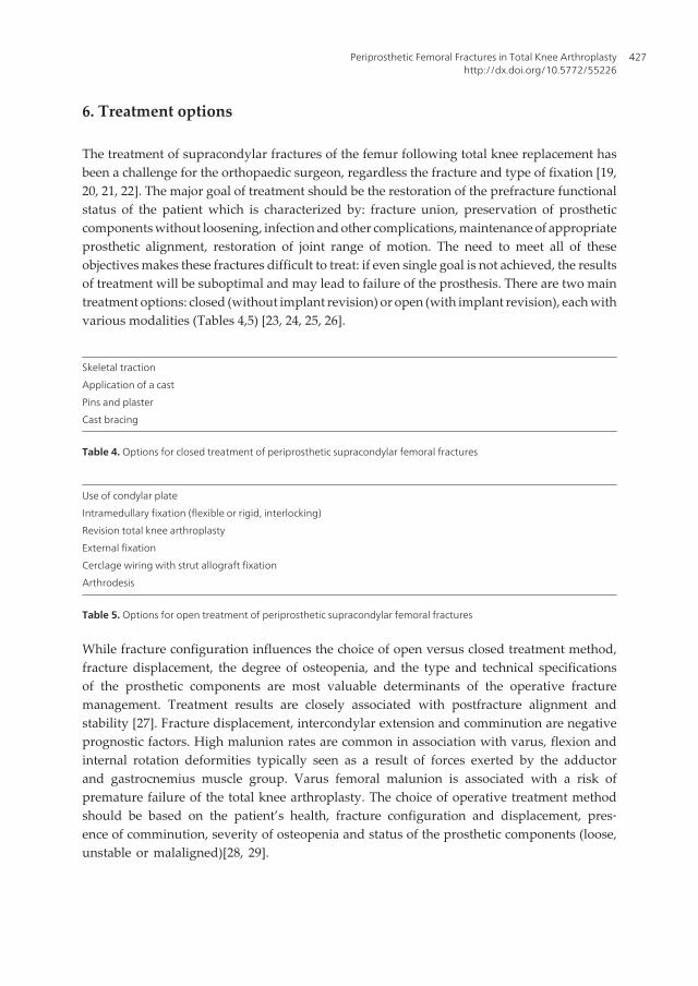
6. Treatment options
The treatment of supracondylar fractures of the femur following total knee replacement hasbeen a challenge for the orthopaedic surgeon, regardless the fracture and type of fixation [19,20, 21, 22]. The major goal of treatment should be the restoration of the prefracture functionalstatus of the patient which is characterized by: fracture union, preservation of prostheticcomponents without loosening, infection and other complications, maintenance of appropriateprosthetic alignment, restoration of joint range of motion. The need to meet all of theseobjectives makes these fractures difficult to treat: if even single goal is not achieved, the resultsof treatment will be suboptimal and may lead to failure of the prosthesis. There are two maintreatment options: closed (without implant revision) or open (with implant revision), each withvarious modalities (Tables 4,5) [23, 24, 25, 26].
Skeletal traction
Application of a cast
Pins and plaster
Cast bracing
Table 4. Options for closed treatment of periprosthetic supracondylar femoral fractures
Use of condylar plate
Intramedullary fixation (flexible or rigid, interlocking)
Revision total knee arthroplasty
External fixation
Cerclage wiring with strut allograft fixation
Arthrodesis
Table 5. Options for open treatment of periprosthetic supracondylar femoral fractures
While fracture configuration influences the choice of open versus closed treatment method,fracture displacement, the degree of osteopenia, and the type and technical specificationsof the prosthetic components are most valuable determinants of the operative fracturemanagement. Treatment results are closely associated with postfracture alignment andstability [27]. Fracture displacement, intercondylar extension and comminution are negativeprognostic factors. High malunion rates are common in association with varus, flexion andinternal rotation deformities typically seen as a result of forces exerted by the adductorand gastrocnemius muscle group. Varus femoral malunion is associated with a risk ofpremature failure of the total knee arthroplasty. The choice of operative treatment methodshould be based on the patient’s health, fracture configuration and displacement, pres‐ence of comminution, severity of osteopenia and status of the prosthetic components (loose,unstable or malaligned)[28, 29].
Periprosthetic Femoral Fractures in Total Knee Arthroplastyhttp://dx.doi.org/10.5772/55226
427

7. Nonoperative treatment
The advantages of nonoperative treatment are: noninvasiveness and negligible infection rate.Since fracture union is likely in nondisplaced fractures, nonoperative treatment is uniformlyrecommended as the initial management in these cases. Disadvantages include: a relativelyhigh malunion rate and functional loss, particularly in patients with displaced fracture throughosteopenic bone in whom maintance of reduction is difficult. Nonoperative treatment is bestreserved for nondisplaced fractures that do not demonstrate intercondylar extension. Non‐surgical management does eliminate surgical risks such as bleeding, infection, loss of fixationand anesthetic complications. On the other hands, prolonged recumbency in elderly patientscarries the significant risk of decubitus ulcers, pneumonia, pulmonary embolia, deep venousthrombosis and diffuse muscle atrophy [30].
8. Operative treatment
Management of periprosthetic fractures of the femur above total knee arthroplasty dependson displacement at the fracture site, bone quality, size of distal fragment and condition ofimplants (Table 6) [31, 32, 33, 34].
Fracture type Description of fracture Treatment recommendation
I Undisplaced fracture and well fixed prosthesis Bracing, nonweightbearing
II Displaced fracture and well fixed prosthesis
Good quality bone
Internal fixation using conventional
plate, intramedullary nail or locking
plate
Poor quality bone with osteopenia and
comminution
Intramedullary nail or locking plate
Decent size distal fragment
Extremely distal fracture
Locking plate or buttress plate with
strut allograft
III Displaced fracture, loose prosthesis
No metahyseal bone loss
Revision knee arthroplasty using a
long stemmed femoral implant
Metaphyseal bone loss or nonunion following
previous surgery
Structural allograft prosthesis
composite or distal femoral
replacement prosthesis
Table 6. Operative guidelines for the treatment of periprosthetic supracondylar fractures above total kneearthroplasty
Open reduction and fixation with a condylar plate provides the potential advantages of anatomi‐cal reconstruction, rigid fixation and an early range of motion exercise. Maintenance of reduc‐tion can be a problem, particularly when a patient has a comminuted fracture through osteopenicbone, and malunion is commonly observed. Use of condylar plate is best reserved for lesscomminuted, displaced fractures with satisfactory bone stock. Using the buttress condylar plate
Arthroplasty - Update428

include the ability to place the multiple screws distally in many directions and excellent visuali‐zation of the fracture to obtain an anatomic reduction. Disadvantages include extensive soft tissuestripping and less rigid fixation than with a nail or fixed angle condylar plate.
Use of flexible intramedullary rods is an efficient and less invasive treatment option, althoughshortening and rotational malunion occasionally occur as a result of the reduced axial androtational stability. This technique should be considered for mildly displaced fractures patientswith unstable general condition. It is minimally invasive procedure with limited morbidity.
New locking plates offer advantages over conventional plates for the treatment of periprostheticfracture associated with total knee arthroplasty. These devices provide stable fixation inosteopenic bone, they are adaptable to different types of fracture and prosthesis and can beinserted using a minimally invasive approach. These plates are particularly useful in presenceof an implant in proximal femur as it allows unicortical screw fixation if there is overlappingthe distal part of the proximal implant, thus avoiding a stress riser between the two implants.
Rigid supracondylar interlocking rod fixation offers the advantage of being minimally invasive whileproviding good axial, angular and rotational stability. It can be performed with use of minimalpatellar tendon splitting approach with percutaneous placement of interlocking screws in caseswith lesser comminution with maintenance of the fracture hematoma and osseous blood supply.Contraindications include loose total knee components, severe comminution, extremely distalfracture and a presence of long total hip intramedullary stem,. This technique has severaladvantages over traditional open reduction with plate fixation: intramedullary implants arebiomechanically superior to subperiostally placed fixation devices, who have significantly largerbending moments; there is no need for periosteal stripping, which can compromise blood supplyto the fracture site and increase the risk of nonunion; plate fixation can be technically demand‐ing and often requires the use of supplemental bone grafting.
Revision total knee arthroplasty provides the advantage of stable fixation with a dyaphisisengaging intramedullary femoral stem, allowing early range of motion and weight bearing.This technique is selected for extremely distal or comminuted fractures when stable fixationis difficult to secure with other methods, or for any fracture associated with loose, unstable, orsubstantially malaligned total knee components. Revision total knee arthroplasty is frequentlyrequired in cases where other methods, nonoperative or operative, have failed.
The most difficult cases involve a loose prosthesis coupled with deficient metaphyseal bonestock, rendering a basic revision procedure impossible. Such cases require excision of distalfracture fragment and replacement with either a distal femoral replacement prosthesis or astructural allograft. These treatment methods may also be required for nonunion resulting fromfailed osteosynthesis. Distal femoral replacement implants should be considered as a limbsalvage option when other surgical options are not feasible. The use of stemmed constrainedrevision component with structural distal femoral allograft composite has been described asthe effective means of providing both implant and fracture stability.
Periprosthetic fractures have a higher rate of nonunion than other supracondylar femoralfractures in the elderly. This has been attributed to alterations in vascularity at the fracture site
Periprosthetic Femoral Fractures in Total Knee Arthroplastyhttp://dx.doi.org/10.5772/55226
429

due to primary surgery, the presence of metal implant and intramedullary polymethylmethacrylate (PMMA), or long term oral corticosteroid administration.
The goals of treatment, whether surgical or nonsurgical, are fracture healing, restoration andmaintenance of knee range of motion, and pain free ambulation. A good result is a minimum of90 degrees of knee motion, with femoral shortening less then 2 cm, varus/valgus malalignmentless than 5 degrees, and flexion/extension malalignment less than 10 degrees. Fulfillment of thesecriteria enables satisfactory knee function, which is of paramout importance to the patient.
9. Complications
Major early complications include nonunion and malunion, which often lead to prostheticloosening, pain and revision. The treatment of delayed unions with bone grafting is possibleand is advocated if appropriate limb alignment and fracture fixation are maintained. In casesof deformity, early signs of prosthetic failure or inability to secure rigid fixation, revision maybe the most appropriate. The most devastating complication of operative care of these fracturesis infection. Incidence of periprosthetic fracture following total knee arthroplasty is graduallyincreasing, and management of these fractures can be challenging with complications thatseverely influence both the patient and surgeon. Furthermore, treatment complication raterange from 20 to 75 percent according to literature data [35]: in a review of 415 cases, therewere reported a nonunion rate of 9%, fixation failure in 4%, an infection rate of 3% and revisionsurgery rate of 13%. Following case will demonstrate some of these problems in treatingsupracondylar periprosthetic femoral fractures.
Figure 3. Anteroposterior and lateral view of type II supracondylar femoral fracture
Arthroplasty - Update430

Figure 4. Operative treatment with DCP (anatomic reduction with rigid fixation)
Figure 5. Loss of reduction and fixation two months following surgery
Periprosthetic Femoral Fractures in Total Knee Arthroplastyhttp://dx.doi.org/10.5772/55226
431

Figure 6. Revision total knee arthroplasty for loose femoral component and fracture treatment
Figure 7. Devastating complication, infection, and limb salvage procedure with antibiotic cement spacer
Arthroplasty - Update432

10. Aftertreatment, rehabilitation
Rehabilitation process is generally guided by the characteristics of the fracture and chosentreatmen methods. As previously said, non-operative treatment is reserved for nondisplacedsupracondylar fractures with stable implant and includes longer rehabilitation period toachieve patient’s preambulatory status, if it is possible at all. Since surgery and more stableimplants including intramedullary nails and angular locking plates allow for faster after-treatment program, rehabilitation protocol is similar to post fracture treatment in cases withoutknee arthroplasty. Main goals are fracture healing, implant stability and prefracture functionalstatus.
11. Prevention
Since supracondylar femoral fractures above total knee arthroplasty are mostly seen inosteoporotic patients, prevention of osteopenia and osteoporosis including treatment with bis-phosphonate and regular exercise will be good for the well-being of the patient and implant.Surgical factors, such as anterior femoral notching, bone cement hypovascularisation andtermal necrosis, and uncontrolled soft tissue manipulation should be kept in mind on regularbasis in order to minimize surgeon’s impact on development of potential complicationsincluding supracondylar fracture.
12. Conclusion
Periprosthetic femoral fractures above total knee prosthesis are increasing complication withconstantly growing incidence since the number of total knee replacements and populationagings are convergating factors. Risk factors analysis and prevention should be in surgeon andpatient focus on this topic. Treatment options include first step to establish whether the implantis loose and, if so even if the fracture is well aligned and heals, treatment that does not includerevision will lead to poor result. Prefracture misalignment, osteolysis and polyethylene wearare important factors in the decision making process. The second step in the treatment is toidentify fracture displacement and to decide whether reduction is needed. The most appro‐priate criteria of acceptable fracture alignment are for supracondylar fractures without kneeprosthesis: less than 5 mm of translation; less than 5 to10 degrees of angulation; less than 10mm of shortening and less than 10 degrees of rotational displacement. Any alteration in limbaxis resulting from fracture can result in altered loading of the prosthesis, which may in turnlead to enhanced wear and/or accelerated implant loosening. The goals of treatment, whethersurgical or nonsurgical, are fracture healing, restoration and maintenance of knee range ofmotion, and pain free ambulation. A good result is a minimum of 90 degrees of knee motion,with femoral shortening less then 2 cm, varus/valgus malalignment less than 5 degrees, andflexion/extension malalignment less than 10 degrees. Fulfillment of these criteria enablessatisfactory knee function, which is of paramout importance to the patient.
Periprosthetic Femoral Fractures in Total Knee Arthroplastyhttp://dx.doi.org/10.5772/55226
433

Acknowledgements
This work is supported by grant number III 41004, Ministry of Education and Science Republicof Serbia.
Author details
Vladan Stevanović1*, Zoran Vukašinović1,2, Zoran Baščarević1,2, Branislav Starčević2,3,Dragana Matanović2,4 and Duško Spasovski1,2
*Address all correspondence to: [email protected]
1 Institute for Orthopaedic Surgery „Banjica“, Belgrade, Serbia
2 Faculty of Medicine, University of Belgrade, Belgrade, Serbia
3 Clinic for Orthopaedic Surgery and Traumatology, Clinical Center of Serbia, Belgrade, Serbia
4 Clinic for Physical Therapy and Rehabilitation, Clinical Center of Serbia, Belgrade, Serbia
References
[1] Neer, C. S I. I, Grantham, S. A, & Shelton, M. L. Supracondylar fracture of the adultfemur: A study of one hundred and ten cases. J Bone Joint Surge Am (1967). , 49,591-613.
[2] Culp, R. W, Schmidt, R. G, Hanks, G, & Mak, A. Esterhai JL Jr, Heppenstall RB. Su‐pracondylar fracture of the femur following prosthetic knee arthroplasty. Clin Or‐thop (1987). , 222, 212-22.
[3] Sisto, D. J, Lachiewicz, P. F, & Insall, J. N. Treatment of supracondylar fractures fol‐lowing prosthetic arthroplasty of the knee. Clin Orthop (1985). , 196, 265-72.
[4] Su, E. T, & De Wal, H. Di Cesare P. Periprosthetic Femoral Fractures Above TotalKnee Replacements. J Am Acad Orthop Surg (2004). , 12, 12-20.
[5] Hirsch, D. M, Bhalla, S, & Roffman, M. Supracondylar fracture of the femur follow‐ing total knee replacement. Report of four cases. J Bone Joint Surg Am (1981). , 63,162-3.
[6] Lesh, M. L, Schneider, D. J, Deol, G, Davis, B, Jacobs, C. R, & Pellegrini, V. D. Theconsequences of anterior femoral notching in total knee arthroplasty: a biomechani‐cal study. J Bone Joint Surg Am (2000). , 82, 1096-101.
Arthroplasty - Update434

[7] Dennis, D. A. Periprosthetic fractures following total knee arthroplasty: the good,bad and ugly. Orthopedics (1998). , 21, 1048-50.
[8] Scott, R. D. Anterior femoral notching and ipsilateral supracondylar femur fracturein total knee arthroplasty. J Arthroplasty (1988).
[9] Shawen, S. B. Belmont PJ Jr, Klemme WR, Topoleski LDT, Xenos JS, Orchowski JR.Osteoporosis and anterior femoral notching in periprosthetic supracondylar frac‐tures. A biomechanical study. J Bone Joint Surg Am (2003). , 85, 115-21.
[10] Dennis, A. D. Periprosthetic fractures following total knee arthroplasty. J Bone JointSurg Am (2001). , 83, 120-4.
[11] Ritter, M. A, Faris, P. M, & Keating, E. M. Anterior femoral notching and ipsilateralsupracondylar femur fracture in total knee arthroplasty. J Arthroplasty (1988). , 3,185-7.
[12] Henry, S. L. Booth RE Jr. Management of supracondylar fractures above total kneeprosthesis. Tech Orthop (1995). , 9, 243-52.
[13] Merkel, K. D. Johnson EW Jr. Supracondylar fracture of the femur after total knee ar‐throplasty. J Bone Joint Surg Am (1986). , 68, 29-43.
[14] Aaron, R. K, & Scott, R. Supracondylar fracture of the femur after total knee arthro‐plasty. Clin Orthop (1987). , 219, 136-9.
[15] Dennis, D. A. Periprosthetis fractures following total knee arthroplasty. Tech Orthop(1999). , 14, 138-43.
[16] DiGioia AM 3d, Rubash HE. Periprosthetic fractures of the femur after total knee ar‐throplasty. A literature review and treatment algorithm. Clin Orthop (1991). , 271,135-42.
[17] Rorabeck, C. H, Angliss, R. D, & Lewis, P. L. Fractures of the femur, tibia and patellaafter total knee arthroplasty: decision making and principles of management. InstrCourse Lect (1998). , 47, 449-60.
[18] Rorabeck, C. H, & Taylor, J. W. Classification of periprosthetic fractures complicatingtotal knee arthroplasty. Orthop Clin North Am (1999). , 30, 209-14.
[19] Shatzker, J, & Lambert, D. C. Supracondylar fractures of the femur. Clin Orthop(1979). , 138, 77-83.
[20] Insall, J. M. Fractures in the distal femur. In: Insall JM, editor. Surgery of the knee.New York: Churchill Livingstone, (1984). , 413-48.
[21] Hohl, M. Fractures about the knee. In: Rockwood CA, Green DP, editors. Fractures inadults.. Philadelphia: JB Lippincott,(1984). , 1478-9.
[22] Rorabeck, C. H, & Taylor, J. W. Periprosthetic fractures of the femur complicating to‐tal knee arthroplasty. Orthop Clin North Am (1999). , 30, 265-77.
Periprosthetic Femoral Fractures in Total Knee Arthroplastyhttp://dx.doi.org/10.5772/55226
435

[23] Harloww, M. L, & Hofmann, A. A. Periprosthetic fractures. In: Scot WN, editor. Theknee. St Louis: CV Mosby, (1994). , 1405-17.
[24] Sochart, D. H, & Hardinge, K. Nonsurgical management of supracondylar fractureabove total knee arthroplasty. Still the nineties option. J Arthroplasty (1997). , 12,830-4.
[25] Ayers, D. C. Supracondylar fracture of the distal femur proximal to a total knee re‐placement. Instr Course Lect (1997). , 46, 197-203.
[26] Chmell, M. J, Moran, M. C, & Scott, R. D. Periarticular fractures after total knee ar‐throplasty: principles of management. J Am Acad Orthop Surg (1996). , 4, 109-16.
[27] Figgie, M. P, & Goldberg, V. M. Figgie HE III. The results of treatment of supracon‐dylar fracture above total knee arthroplasty. J Arthroplasty (1990). , 5, 267-76.
[28] Kang Il KimEgol KA, Hozack WJ, Parvizi J. Periprosthetic fractures after total kneearthroplasties. Clin Orthop (2006). , 446, 167-75.
[29] Chen, F, Mont, M. A, & Bachner, R. S. Management of ipsilateral supracondylar fe‐muir fractures following total knee arthroplasty. J Arthroplasty (1994). , 9, 521-6.
[30] Sochart, D. H, & Hardinge, K. Nonsurgical management of supracondylar fractureabove total knee arthroplasty. Still the nineties option. J Arthroplasty (1997). , 12,830-4.
[31] Engh, G. A, & Ammeen, D. J. Periprosthetic fractures adjacent to total knee implants.Treatment and clinical results. J Bone Joint Surg Am (1997). , 79, 1100-13.
[32] Cusick, R. P, Lucas, G. L, Mcqueen, D. A, & Graber, C. D. Construct stiffness of dif‐ferent fixation methods for supracondylar femoral fractures above total knee pros‐thesis. Am J Orthop (2000). , 29, 695-9.
[33] Lewis, P. L, & Rorabeck, C. H. Periprosthetic fractures. In: Engh GA, Rorabeck CH,editors. Revision total knee arthroplasty. Baltimore MD: Williams & Willkins;(1997). , 275-295.
[34] Kolb, K, Grützner, P. A, Marx, F, & Kolb, W. Fixation of Periprosthetic Supracondy‐lar Femur Fractures Above Total Knee Arthroplasty- The Indirect Reduction Techni‐que with the Condylar Blade Plate and the Minimally Invasive Technique with theLISS. In: Fokter S, editor. Recent Advances in Hip and Knee Arthroplasty. Rijeka: In‐Tech, (2012). , 315-42.
[35] Herrera, D. A, Kregor, P. J, Cole, P. A, Levy, B. A, Jonssos, A, & Zlowodzki, M. Treat‐ment of acute distal femur fractures above a total knee arthroplasty: systematic re‐vew of 415 cases (1981-2006). Acta Orthop (2008). , 79, 22-7.
Arthroplasty - Update436