
Peripheral Nervous System
52
Peripheral Nervous System 31 spinal nerves – We’ve already discussed their structure 12 cranial nerves – How do they differ from spinal nerves? – We need to learn their: Names Locations Functions
-
Upload
elliott-boyle -
Category
Documents
-
view
32 -
download
0
description
Peripheral Nervous System. 31 spinal nerves We’ve already discussed their structure 12 cranial nerves How do they differ from spinal nerves? We need to learn their: Names Locations Functions. 12 Cranial Nerves. How do you remember which nerve is which number? - PowerPoint PPT Presentation
Transcript of Peripheral Nervous System

Peripheral Nervous System
31 spinal nerves – We’ve already
discussed their structure
12 cranial nerves– How do they differ from
spinal nerves?– We need to learn their:
Names Locations Functions
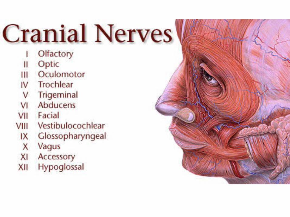

12 Cranial Nerves
How do you remember which nerve is which number?– Here is a G-rated mnemonic
devices: Old Opie occasionally tries
trigonometry and feels very gloomy, vague, and hypoactive.
– There are also several R-rated ones
Some cranial nerves are sensory, some motor, and some are both (mixed)?– Some say marry money but my
brother says big butts matter more.

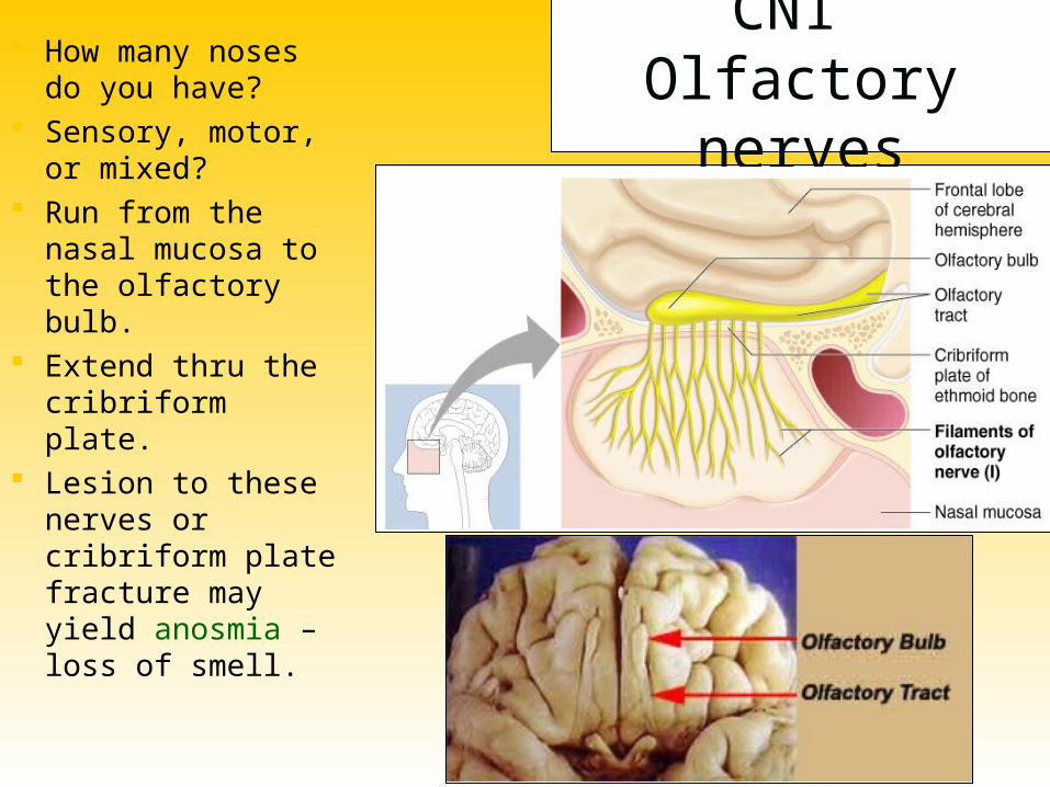
CN1 Olfactory nerves
How many noses do you have?
Sensory, motor, or mixed?
Run from the nasal mucosa to the olfactory bulb.
Extend thru the cribriform plate.
Lesion to these nerves or cribriform plate fracture may yield anosmia – loss of smell.

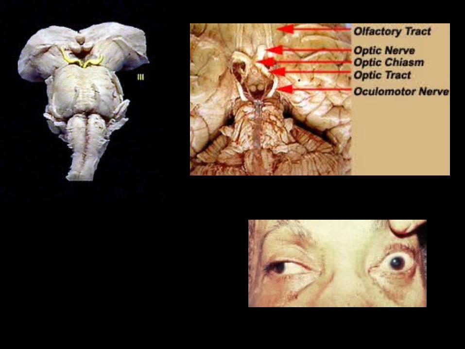
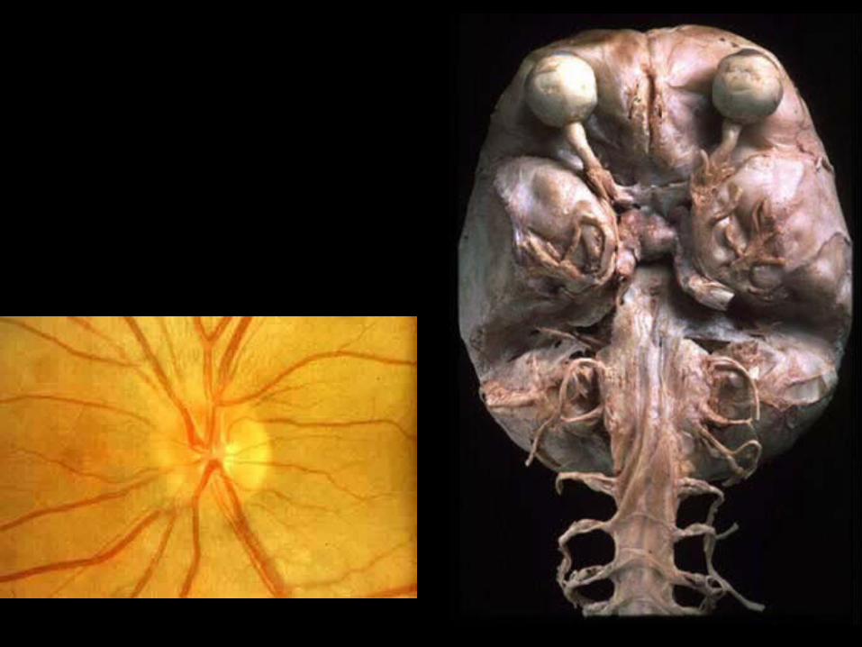
CN2 Optic Nerves
How many eyes do you have?
Sensory, motor, or mixed?
Begin at the retina, run to the optic chiasm, cross over, continue as the optic tract and synapse in the thalamus.
Optic nerve damage yields blindness in the eye served by the nerve. Optic tract damage yields partial visual loss.
Visual defects = anopsias

CN3 Oculomotor Nerves “Eye mover”
Sensory, motor, or mixed?
Originate at the ventral midbrain.
Synapse on:– Extraocular muscles
Inferior oblique; Inferior, medial, and superior rectus
– Iris constrictor muscle– Ciliary muscle
Disorders can result in eye paralysis, diplopia or ptosis.

CN4 Trochlear Nerves
Controls the superior oblique muscle which depresses the eye via pulling on the superior oblique tendon which loops over a ligamentous pulley known as the trochlea.
Originates on the dorsal midbrain and synapses on the superior oblique
Sensory, motor, or mixed? Trauma can result in
double vision. Why?


Sensory, motor, or mixed? Biggest cranial nerve Originates in the pons and
eventually splits into 3 divisions:– Ophthalmic (V1), Maxillary (V2),
&
Mandibular (V3).
Sensory info (touch, temp., and pain) from face.
Motor info to muscles of mastication
Damage?
CN5 Trigeminal Nerves


Sensory, motor, or mixed?
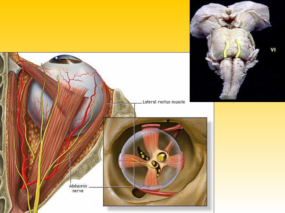
Runs between inferior pons and lateral rectus.
CN5 Abducens Nerves
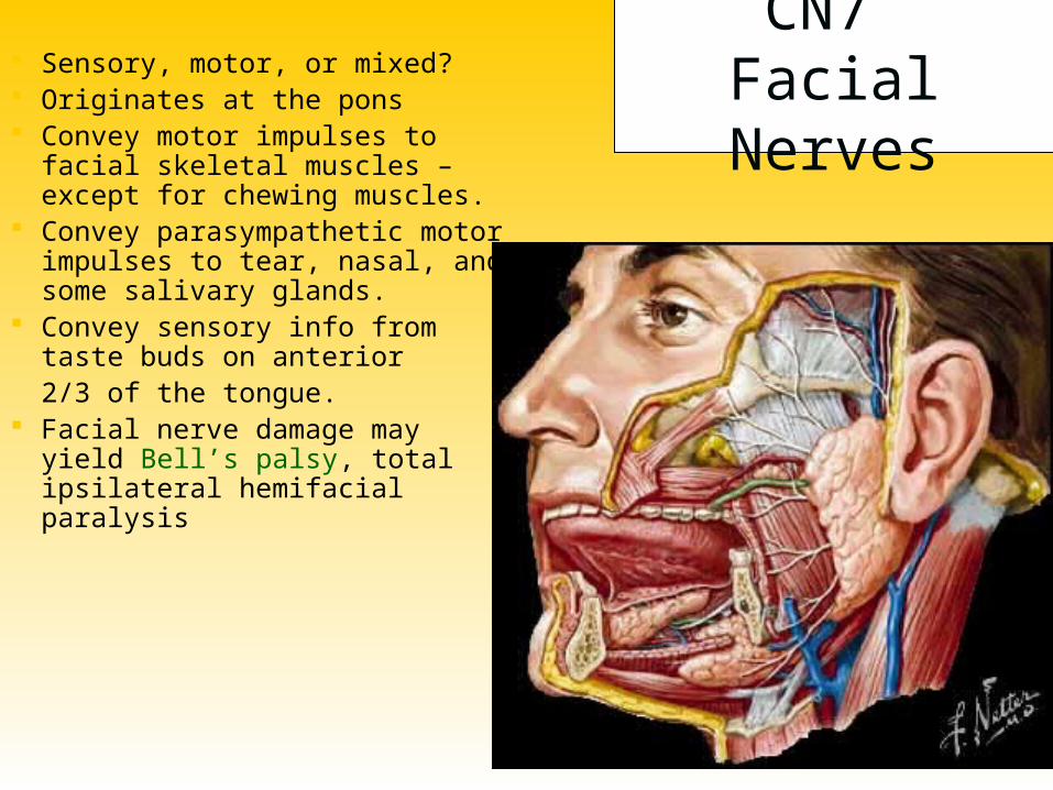
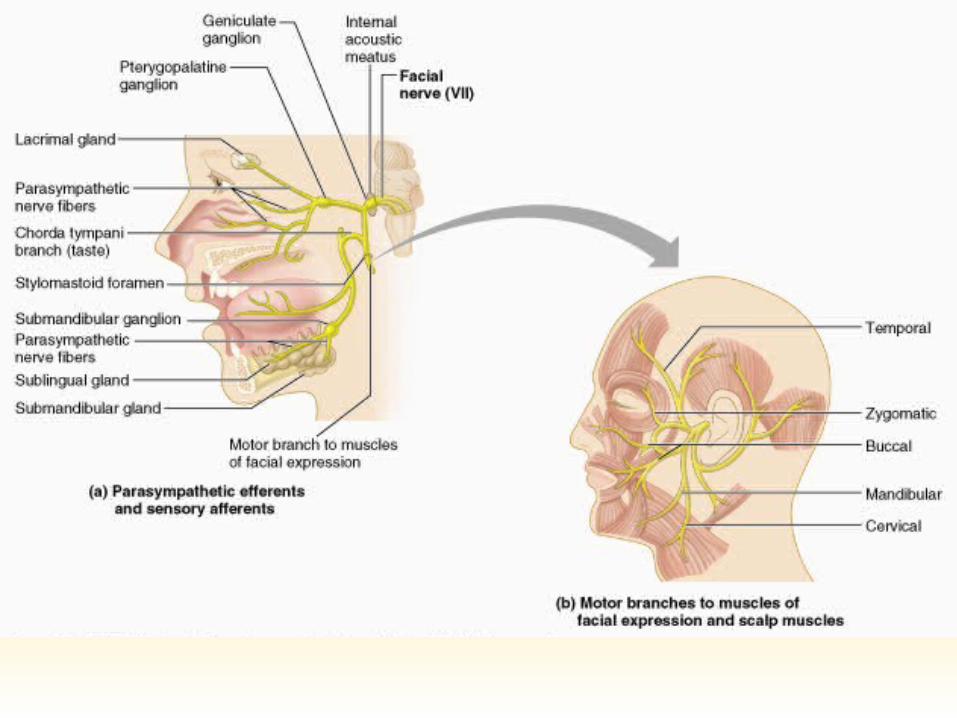
CN7 Facial Nerves
Sensory, motor, or mixed? Originates at the pons Convey motor impulses to facial
skeletal muscles – except for chewing muscles.
Convey parasympathetic motor impulses to tear, nasal, and some salivary glands.
Convey sensory info from taste buds on anterior 2/3 of the tongue.
Facial nerve damage may yield Bell’s palsy, total ipsilateral hemifacial paralysis

CN8 Auditory/Vestibulocochlear
Nerves
Sensory, motor, or mixed? Originates at the pons 2 divisions:
– Cochlear Afferent fibers from
cochlea in the inner ear HEARING
– Vestibular Afferent fibers from
equilibrium receptors in inner ear
BALANCE
Functional impairment?

CN9 Glossopharyngeal Nerves
Sensory, motor, or mixed? Fibers run emerge from
medulla and run to the throat. Motor Functions:
– Motor fibers to some swallowing muscles
– Parasympathetic fibers to some salivary glands
Sensory Functions:– Taste, touch, heat from pharynx
and posterior tongue.– Info from chemoreceptors on the
level of O2 and CO2 in the blood. Info from baroreceptors on BP.
Chemoreceptors and baroreceptors are located in the carotid sinus – a dilation in the internal carotid artery.

CN10 Vagus Nerves Sensory, motor, or mixed? Only cranial nerves to extend
beyond head and neck.– Fibers emerge from medulla, leave
the skull, and course downwards into the thorax and abdomen.
Motor Functions:– Parasympathetic efferents to the
heart, lungs, and abdominal organs.
Sensory Functions:– Input from thoracic and abdominal
viscera; from baro- and chemoreceptors in the carotid sinus; from taste buds in posterior tongue and pharynx


CN11 Accessory Nerves Sensory, motor, or mixed?
Formed by the union of a cranial root and a spinal root.– CR arises from medulla while
SR arises from superior spinal cord. SR passes thru the FM and joins with CR to form the accessory nerve. They then leave the skull via the jugular foramen.
– Cranial division then joins vagus and innervates larynx, pharynx, and soft palate.
– Spinal division innervates sternocleidomastoids and trapezius.

CN12 Hypoglossal Nerves Sensory, motor, or mixed? Arise from the medulla and
exit the skull via the hypoglossal canal and innervate the tongue.
Innervate the intrinsic & extrinsic muscles of the tongue.– Swallowing, speech, food
manipulation.
Damage?
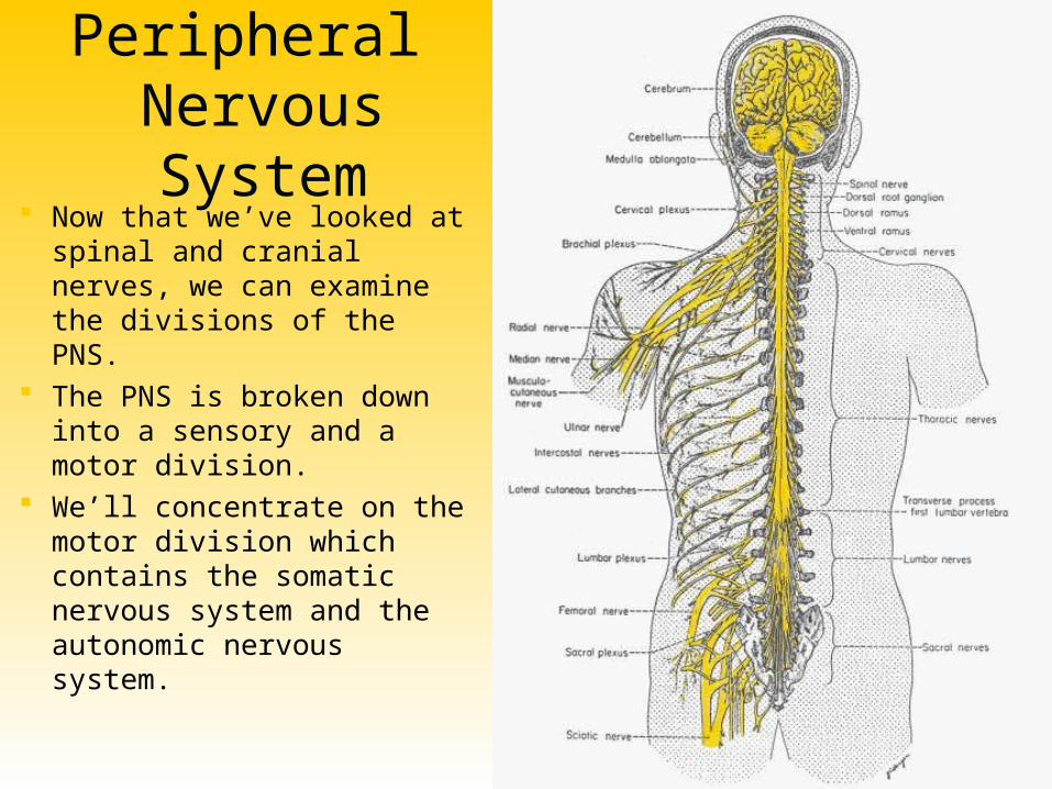
Peripheral Nervous System
Now that we’ve looked at spinal and cranial nerves, we can examine the divisions of the PNS.
The PNS is broken down into a sensory and a motor division.
We’ll concentrate on the motor division which contains the somatic nervous system and the autonomic nervous system.

Somatic vs. Autonomic
Voluntary Skeletal muscle Single efferent neuron Axon terminals release
acetylcholine Always excitatory Controlled by the
cerebrum
Involuntary Smooth, cardiac muscle;
glands Multiple efferent neurons Axon terminals release
acetylcholine or norepinephrine
Can be excitatory or inhibitory
Controlled by the homeostatic centers in the brain – pons, hypothalamus, medulla oblongata
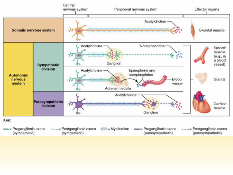

Autonomic Nervous System
2 divisions:– Sympathetic
“Fight or flight” “E” division
– Exercise, excitement, emergency, and embarrassment
– Parasympathetic “Rest and digest” “D” division
– Digestion, defecation, and diuresis

Antagonistic Control
Most internal organs are innervated by both branches of the ANS which exhibit antagonistic control
A great example is heart rate. An increase in sympathetic stimulation causes HR to increase whereas an increase in parasympathetic stimulation causes HR to decrease
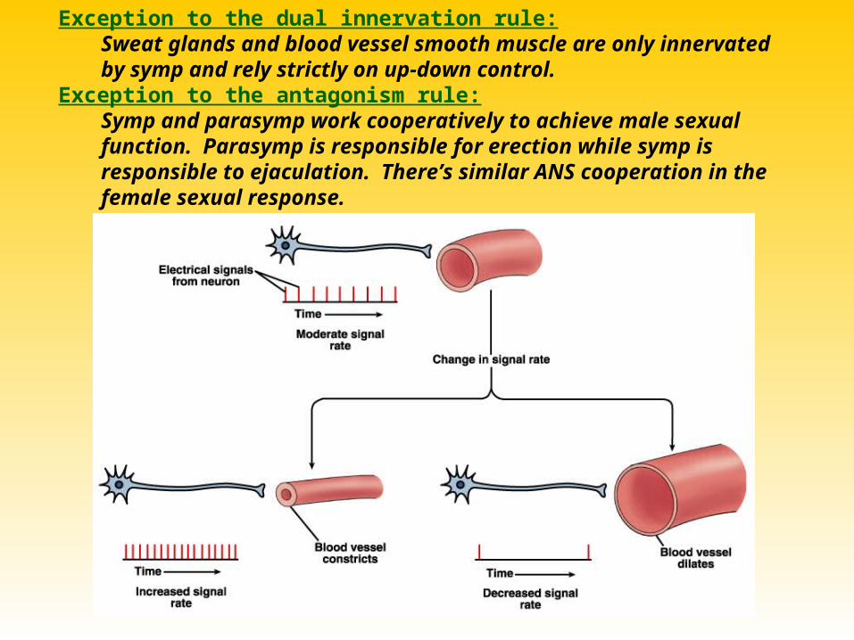
Exception to the dual innervation rule:Sweat glands and blood vessel smooth muscle are only innervated by symp and rely strictly on up-down control.
Exception to the antagonism rule:Symp and parasymp work cooperatively to achieve male sexual function. Parasymp is responsible for erection while symp is responsible to ejaculation. There’s similar ANS cooperation in the female sexual response.
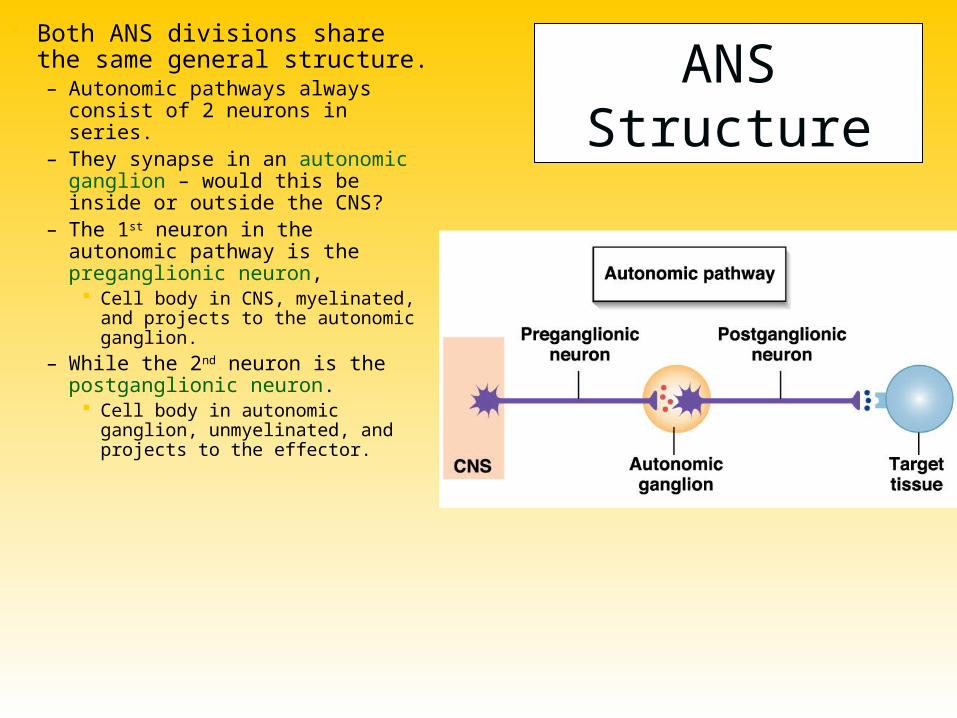
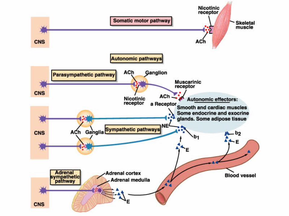
ANS Structure
Both ANS divisions share the same general structure. – Autonomic pathways always
consist of 2 neurons in series.– They synapse in an autonomic
ganglion – would this be inside or outside the CNS?
– The 1st neuron in the autonomic pathway is the preganglionic neuron,
Cell body in CNS, myelinated, and projects to the autonomic ganglion.
– While the 2nd neuron is the postganglionic neuron.
Cell body in autonomic ganglion, unmyelinated, and projects to the effector.

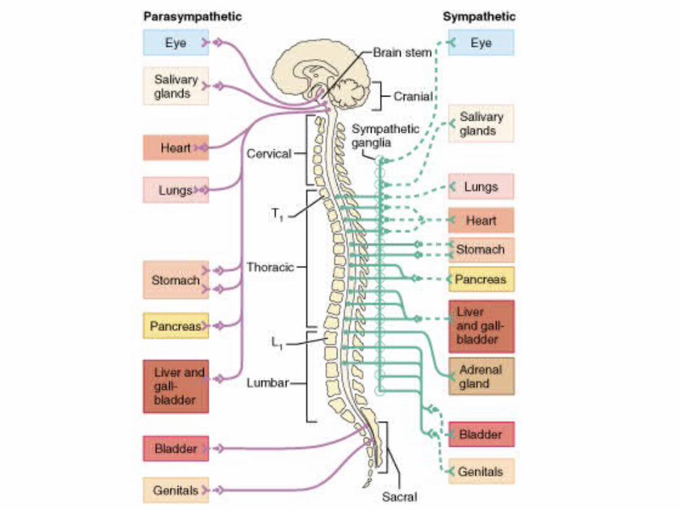
Sympathetic vs. Parasympathetic Structural Differences:
Symp. Parasymp.
Point of CNS OriginPoint of CNS Origin T1 L2
(thoracolumbar)
Brainstem,
S2 S4
(craniosacral)
Site of Peripheral Site of Peripheral GangliaGanglia
Paravertebral – in sympathetic chain
On or near target tissue
Length of Length of preganglionic fiberpreganglionic fiber
Short Long
Length of Length of postganglionic fiberpostganglionic fiber
Long Short

Sympathetic vs. Parasympathetic Receptor/NT Differences:
Symp. Parasymp.
NT at Target NT at Target SynapseSynapse
Norepinephrine
(adrenergic neurons)
Acetylcholine
(cholinergic neurons)
Type of NT Type of NT Receptors at Receptors at Target SynapseTarget Synapse
Alpha and Beta
( and )
Muscarinic
NT at GanglionNT at Ganglion Acetylcholine Acetylcholine
Receptor at Receptor at GanglionGanglion
Nicotinic Nicotinic

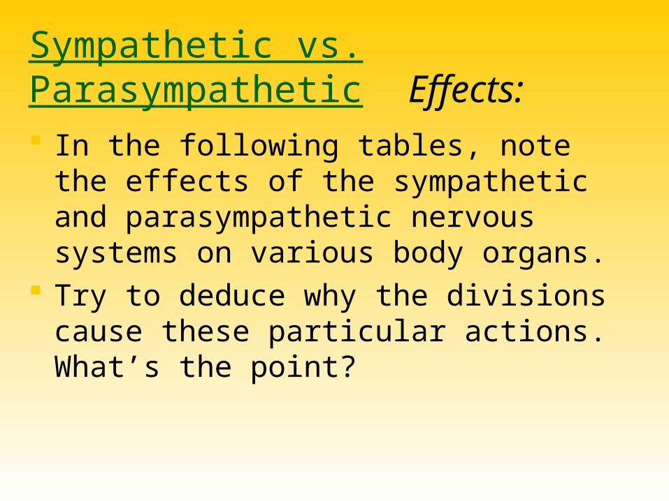
Sympathetic vs. Parasympathetic Effects: In the following tables, note the effects of
the sympathetic and parasympathetic nervous systems on various body organs.
Try to deduce why the divisions cause these particular actions. What’s the point?

Target OrganTarget Organ Parasympathetic Parasympathetic EffectsEffects
Sympathetic Sympathetic EffectsEffects
Eye (Iris)Eye (Iris) Stimulates constrictor muscles. Pupil constriction.
Stimulates dilator muscles. Pupil dilates.
Eye (Ciliary Eye (Ciliary muscle)muscle)
Stimulates. Lens accommodates – allows for close vision.
No innervation.
Salivary GlandsSalivary Glands Watery secretion. Mucous secretion.
Sweat GlandsSweat Glands No innervation. Stimulates sweating in large amounts. (Cholinergic)
GallbladderGallbladder Stimulates smooth muscle to contract and expel bile.
Inhibits gallbladder smooth muscle.
Arrector PiliArrector Pili No innervation Stimulates contraction. Piloerection (Goosebumps)

Target OrganTarget Organ Parasympathetic Parasympathetic EffectsEffects
Sympathetic Sympathetic EffectsEffects
Cardiac MuscleCardiac Muscle Decreases HR. Increases HR and force of contraction.
Coronary Blood Coronary Blood VesselsVessels
Constricts. Dilates
Urinary Bladder; Urinary Bladder; UrethraUrethra
Contracts bladder smooth muscle; relaxes urethral sphincter.
Relaxes bladder smooth muscle; contracts urethral sphincter.
LungsLungs Contracts bronchiole (small air passage) smooth muscle.
Dilates bronchioles.
Digestive OrgansDigestive Organs Increases peristalsis and enzyme/mucus secretion.
Decreases glandular and muscular activity.
Liver Liver No innervation No innervation (indirect effect).

Target OrganTarget Organ Parasympathetic Parasympathetic EffectsEffects
Sympathetic Sympathetic EffectsEffects
KidneyKidney No innervation. Releases the enzyme renin which acts to increase BP.
PenisPenis Vasodilates penile arteries. Erection.
Smooth muscle contraction. Ejaculation.
Vagina; ClitorisVagina; Clitoris Vasodilation. Erection. Vaginal reverse peristalsis.
Blood CoagulationBlood Coagulation No effect. Increases coagulation rate.
Cellular Cellular MetabolismMetabolism
No effect. Increases metabolic rate.
Adipose TissueAdipose Tissue No effect. Stimulates fat breakdown.

Target OrganTarget Organ Parasympathetic Parasympathetic EffectsEffects
Sympathetic Sympathetic EffectsEffects
Mental ActivityMental Activity No innervation. Increases alertness.
Blood VesselsBlood Vessels Little effect. Constricts most blood vessels and increases BP. Exception – dilates blood vessels serving skeletal muscle fibers (cholinergic).
UterusUterus Depends on stage of the cycle.
Depends on stage of the cycle.
Endocrine Endocrine PancreasPancreas
Stimulates insulin secretion.
Inhibits insulin secretion.

Duration/Location of Parasympathetic Effects
Parasympathetic preganglionic neurons synapse on only a few postganglionic neurons.
Would you expect parasympathetic activity to be widespread or local?
All parasympathetic fibers release ACh.– ACh is quickly broken down by what enzyme?
What can you say about the duration of parasympathetic effects?
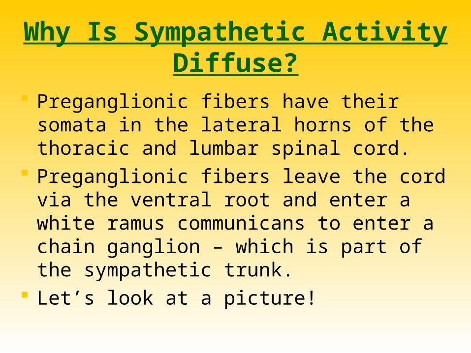
Why Is Sympathetic Activity Diffuse?
Preganglionic fibers have their somata in the lateral horns of the thoracic and lumbar spinal cord.
Preganglionic fibers leave the cord via the ventral root and enter a white ramus communicans to enter a chain ganglion – which is part of the sympathetic trunk.
Let’s look at a picture!


Once a preganglionic axon reaches the chain ganglion, it may:
…synapse with a
ganglionic neuron w/i
the same chain ganglion.
…ascend or
descend in the trunk
to synapse within
another chain
ganglion.
…pass thru the chain ganglion and emerge
from the chain w/o synapsing.


If the preganglionic axon synapses in a chain ganglion (routes 1 and 2)…
It will enter the ventral or dorsal ramus of the adjoining spinal nerve via a gray ramus communicans.
From here it may give branches to sweat glands, arrector pili, and vascular smooth muscle – while it continues to its final destination which could be the iris muscles, the heart, or something else.
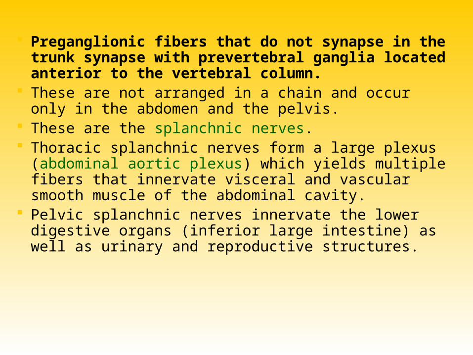
Preganglionic fibers that do not synapse in the trunk synapse with prevertebral ganglia located anterior to the vertebral column.
These are not arranged in a chain and occur only in the abdomen and the pelvis.
These are the splanchnic nerves. Thoracic splanchnic nerves form a large plexus (abdominal
aortic plexus) which yields multiple fibers that innervate visceral and vascular smooth muscle of the abdominal cavity.
Pelvic splanchnic nerves innervate the lower digestive organs (inferior large intestine) as well as urinary and reproductive structures.
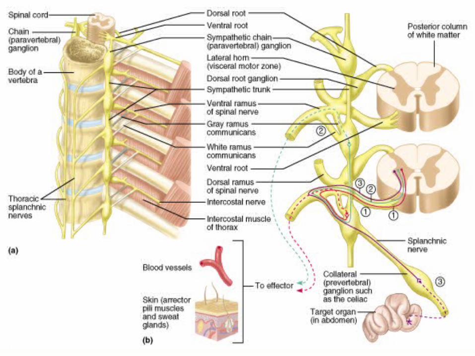

Certain splanchnic nerves synapse on hormone-producing cells of the adrenal medulla – the interior of the adrenal glands which sit upon the kidneys.
How does this contribute to the “diffuseness” of sympathetic activity?
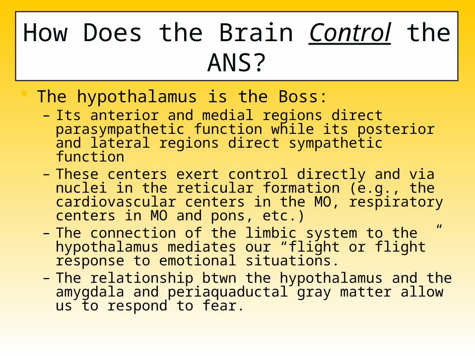
How Does the Brain Control the ANS?
The hypothalamus is the Boss:– Its anterior and medial regions direct parasympathetic
function while its posterior and lateral regions direct sympathetic function
– These centers exert control directly and via nuclei in the reticular formation (e.g., the cardiovascular centers in the MO, respiratory centers in MO and pons, etc.)
– The connection of the limbic system to the hypothalamus mediates our “flight or flight” response to emotional situations.
– The relationship btwn the hypothalamus and the amygdala and periaquaductal gray matter allow us to respond to fear.