PERIODONTOLOGY,2000, Acute,gingival,and…eprints.ucm.es/30392/1/Acute periodontal lesions....
55
1 PERIODONTOLOGY 2000 Acute gingival and periodontal lesions David Herrera, Bettina Alonso, Lorenzo de Arriba, Isabel Santa Cruz, Cristina Serrano, Mariano Sanz ETEP (Etiology and Therapy of Periodontal Diseases) Research Group, University Complutense, Madrid, Spain. Corresponding author: David Herrera ETEP Research Group Faculty of Odontology, Universidad Complutense de Madrid Plaza Ramón y Cajal s/n (Ciudad Universitaria) 28040 Madrid (Spain) Telephone number: (+34) 913942021 Fax number: (+34) 913941910 email: [email protected] Running title: Acute lesions Key words: periodontal abscess, necrotizing gingivitis, necrotizing periodontitis, necrotizing stomatitis, necrotizing periodontal diseases. Title series: Aggressive and Acute Periodontal Diseases Editor: Albandar JM
Transcript of PERIODONTOLOGY,2000, Acute,gingival,and…eprints.ucm.es/30392/1/Acute periodontal lesions....

1
PERIODONTOLOGY 2000
Acute gingival and periodontal lesions
David Herrera, Bettina Alonso, Lorenzo de Arriba, Isabel Santa Cruz, Cristina Serrano,
Mariano Sanz
ETEP (Etiology and Therapy of Periodontal Diseases) Research Group, University
Complutense, Madrid, Spain.
Corresponding author:
David Herrera
ETEP Research Group
Faculty of Odontology, Universidad Complutense de Madrid
Plaza Ramón y Cajal s/n (Ciudad Universitaria)
28040 Madrid (Spain)
Telephone number: (+34) 913942021
Fax number: (+34) 913941910
e-‐mail: [email protected]
Running title: Acute lesions
Key words: periodontal abscess, necrotizing gingivitis, necrotizing periodontitis,
necrotizing stomatitis, necrotizing periodontal diseases.
Title series: Aggressive and Acute Periodontal Diseases
Editor: Albandar JM

2
ABSTRACT
This is a review and update on acute conditions affecting the gingival tissues, including
abscesses in the periodontium, necrotizing periodontal diseases, and other acute
conditions that cause gingival lesions with acute presentation, such as infectious
processes not associated with oral bacterial biofilms, muco-‐cutaneous disorders, and
traumatic and allergic lesions. A periodontal abscess is clinically important since it is a
relatively frequent dental emergency, it can compromise the periodontal prognosis of
the affected tooth, and because bacteria within the abscess can spread and cause
infections in other body sites. Different types of abscesses have been identified, mainly
by the type of etiology, and there are clear differences between those affecting a
previously existing periodontal pocket and those affecting healthy sites. Therapy for
this acute condition consists of drainage and tissue debridement, with individual
evaluation of the need for systemic antimicrobial therapy. The definitive treatment of
the pre-‐existing condition should be accomplished after the acute phase is controlled.
Necrotizing periodontal diseases (NPD) present three typical clinical features: papilla
necrosis, gingival bleeding, and pain. Although the prevalence of these diseases is not
high, their importance is clear, since they represent the most severe conditions
associated with the dental biofilm, with very rapid tissue destruction. In addition to
bacteria, the etiology of NPD includes numerous factors that alter the host response
and predispose to these diseases, including HIV infection, malnutrition, stress, and
tobacco smoking. The treatment consists of superficial debridement, careful
mechanical oral hygiene, rinsing with chlorhexidine, and daily re-‐evaluation. Systemic
antimicrobials may be used adjunctively in severe cases or in non-‐responding
conditions, and the best option is metronidazole. Once the acute disease is under
control, definitive treatment should be provided, including the adequate therapy for
the pre-‐existing gingivitis or periodontitis. Among other acute conditions affecting the
periodontal tissues, but not caused by the microorganisms present in oral biofilms, are
infectious diseases, muco-‐cutaneous diseases and traumatic or allergic lesions. In most
cases, the gingival involvement is not severe, though they are common and may
prompt a dental emergency visit. These conditions may have the appearance of an
erythematous lesion, sometime erosive. Erosive lesions may the direct result of a
trauma or the consequence of the breaking of vesicles and bullae. A proper differential

3
diagnosis is important for an adequate management of the case.

4
INTRODUCTION
Acute lesions in the periodontium, such as abscesses and necrotizing periodontal
diseases, are among the few clinical situations in periodontics where patients may
seek urgent care, mostly due to the associated pain. In addition, and in contrast to
most other periodontal conditions, a rapid destruction of periodontal tissues may
occur during the course of these lesions, thus stressing the importance of prompt
diagnosis and treatment. In spite of this, the available scientific knowledge on these
conditions is limited and based on somewhat outdated literature. This lack of
contemporary data makes it rather challenging to devise evidence-‐based therapeutic
guidelines. Hence, an update is imperative, though it must be confined to an
evaluation of narrative reviews and expert opinions.
Other gingival and periodontal lesions may also show an acute presentation, including
different infectious processes not related to oral bacterial biofilms, muco-‐cutaneous
disorders or traumatic and allergic lesions.
This chapter provides an overview and update of existing information on acute
conditions affecting the gingival tissues, including abscesses in the periodontium,
necrotizing periodontal diseases, and other acute conditions.

5
ABSCESSES IN THE PERIODONTIUM
Definition of periodontal abscess
Abscesses in the periodontium are odontogenic infections, which may be caused by
pulp necrosis, periodontal infections, pericoronitis, trauma, or surgery (109).
Odontogenic or dental abscesses are classified, according to the infection source, into
periapical (dentoalveolar) abscess, periodontal abscess, and pericoronal abscess (327).
A periodontal abscess has been defined as a localized purulent infection in the
periodontal tissues (210). A more comprehensive definition has also been proposed: a
lesion with an expressed periodontal breakdown occurring during a limited period of
time, and with easily detectable clinical symptoms, including a localised accumulation
of pus located within the gingival wall of the periodontal pocket (130). See Figure 1.
Classification of periodontal abscesses
Different criteria have been used to classify periodontal abscesses:
-‐ By the location of the abscess: gingival and periodontal abscesses (110). A gingival
abscess is a localised painful swelling, affecting only the marginal and interdental
gingiva, normally associated with subgingivally impacted foreign objects. These
conditions may occur in a previously healthy gingiva (7). A periodontal abscess is a
localised painful swelling, affecting deeper periodontal structures, including deep
pockets, furcations and vertical osseous defects, and they are usually located beyond
the mucogingival line. Histologically, both lesions are identical, but the gingival abscess
affects only the marginal soft tissues of previously healthy sites, while the periodontal
abscess occurs in a periodontal pocket related to a periodontitis lesion (76).
-‐ By the course of the lesion: acute and chronic periodontal abscesses. The acute
periodontal abscess usually manifests symptoms like pain, tenderness, sensitivity to
palpation, and suppuration upon gentle pressure. The chronic abscess is normally
associated with a sinus tract, and it is usually asymptomatic, although the patient can
refer mild symptoms (250). A localised acute abscess may become a chronic abscess

6
when drainage is established through a sinus or through the sulcus. Similarly, a chronic
abscess may have an acute exacerbation.
-‐ By the number of abscesses: single and multiple periodontal abscesses (321). A single
periodontal abscess is usually associated with local factors, which contribute to the
closure of the drainage of a periodontal pocket. Multiple periodontal abscesses have
been reported in uncontrolled diabetes mellitus, medically compromised patients, and
in patients with untreated periodontitis after systemic antibiotic therapy for non-‐oral
reasons (125, 126, 321). Multiple abscesses have also been described in a patient with
multiple external root resorptions (339).
-‐ By the type of etiological factors: a periodontal abscesses is usually associated a
previously existing periodontal pocket, though it also can develop without presence of
a pocket (130). This classification is detailed in the section on etiology.
-‐ According to the International Workshop for a Classification of Periodontal Diseases
and Conditions (1999), abscesses in the periodontium include gingival, periodontal,
pericoronal and periapical abscesses (210). A gingival abscess is defined as “a localized,
painful, rapidly expanding lesion involving the marginal gingiva or interdental papilla
sometimes in a previously disease-‐free area”. A periodontal abscess (that can be acute
or chronic) is defined as “a localized accumulation of pus within the gingival wall of a
periodontal pocket resulting in the destruction of the collagen fibre attachment and
the loss of nearby alveolar bone. A pericoronal abscess is “a localized accumulation of
pus within the overlying gingival flap surrounding the crown of an incompletely
erupted tooth” (Figure 2).
Clinical significance of a periodontal abscess
-‐ A common periodontal emergency:
Periodontal abscesses represented approximately 14% of all dental emergencies in a
study in the USA (7). In general practices in the United Kingdom, 6–7‰ of the patients
treated in one month suffered from a periodontal abscess, being the third most
prevalent infection demanding emergency treatment, after dentoalveolar abscesses

7
(14– 25‰) and pericoronitis (10–11‰) (177). In an army dental clinic, 3.7% of the
patients had periodontitis, and among these 27.5% had a periodontal abscess, with
clear differences between patients undergoing active periodontal treatment (13.5%)
and untreated patients (59.7%) (117). Among patients in supportive periodontal
therapy (SPT), a periodontal abscess was detected in 37% of the patients and 3.7 of the
teeth, followed for 5–29 years before (mean 12.5 years) (209); in the Nebraska
prospective longitudinal study, 53% of the patients, followed for 7 years, showed a
periodontal abscess, and 85% of these abscesses were associated to teeth only treated
with coronal scaling. Sixteen out of 27 abscess sites had initial probing pocket depths
(PPD) deeper than 6 mm, and in eight sites PPD was 5–6 mm (156). Abscesses occur
more often in molar sites, representing more than 50% of all sites affected by abscess
formation (128, 209, 300), probably due to the presence of furcation, and complex
anatomy and root morphology. However, one case series suggested that the lower
anterior incisors were the most frequently affected teeth (149).
-‐ Periodontal abscesses may lead to tooth loss, especially if they affect teeth with
previous moderate to severe attachment loss, as it occurs during SPT in patients with
severe chronic periodontitis. In fact, it has been considered the main cause for tooth
extraction during SPT (55, 209, 294, 300). Similarly, teeth with repeated abscess
formation were considered to have a hopeless prognosis (36), and 45% of teeth with a
periodontal abscess during SPT were extracted (209). The main reason for tooth
extraction in teeth with a questionable prognosis, followed for 8.8 years, was a
periodontal abscess (55). Taken into account these reports, it has been suggested that
when facing a periodontal abscess in SPT patients, early diagnosis and adequate
therapy are crucial to preserve the prognosis of the affected tooth (294).
-‐ Periodontal abscesses may be associated with systemic dissemination of a localized
infection. Numerous case reports have described the occurrence of systemic infections
from a suspected source in a periodontal abscess, either through dissemination
occurring during therapy or related to an untreated abscess. During the treatment of
an abscess, the concomitant bacteraemia may lead to colonization of pathogenic
microorganisms in other body sites and the developing of different infections, such as:

8
pulmonary actinomycosis (315), a brain abscess containing P. melaninogenica and
other Prevotella spp. (103), or a total knee arthroplasty infection (330). It has been
suggested that the risk of bacteraemia during abscess drainage may be reduced if a
needle aspiration of the content of the abscess is performed before the procedure (99,
265).
There are also case reports of bacteraemia originating from untreated abscesses, such
as in cellulitis in breast cancer patients (198), a cervical necrotizing fascitis (57), a
necrotising cavernositis containing Peptostreptococcus spp. and Fusobacterium spp.
(245), or a sickle cell crisis in a patient with sickle cell anaemia (255).
Etiology, pathogenesis and histopathology of a periodontal abscess
Periodontal abscesses may develop in periodontitis-‐affected sites (with a pre-‐existing
periodontal pocket) or in healthy sites (without a previous pocket):
-‐ In periodontitis, a periodontal abscess may represent a period of disease
exacerbation, favoured by the existence of tortuous pockets, the presence of furcation
involvement (Figure 3) or a vertical defect, in which the marginal closure of the pocket
may lead to an extension of the infection into the surrounding periodontal tissues (76,
158, 224). Also changes in the composition of the subgingival microbiota, with an
increase in bacterial virulence, or a decrease in the host defences, could also result in
an inefficient capacity to drain the increased suppuration. Among periodontal
abscesses in periodontitis patients, different subgroups can be distinguished:
• Acute exacerbation of an untreated periodontitis (74). See Figure 4.
• After non-‐surgical periodontal therapy: after scaling or professional prophylaxis,
dislodged calculus fragments can be pushed into the tissues (74), or inadequate
scaling may allow calculus to remain in deep pocket areas, while the coronal
part will occlude the normal drainage (156).
• After surgical periodontal therapy: associated with the presence of foreign
bodies such as membranes for regeneration or sutures (106).
• Acute exacerbation in refractory periodontitis (97).
• Acute exacerbation in supportive periodontal therapy, as described above (55,
209, 294).

9
• Systemic antimicrobial intake without subgingival debridement, in patients
with advanced periodontitis may also cause abscess formation (125, 126, 321),
probably related to an overgrowth of opportunistic bacteria (125).
-‐ Non-‐periodontitis periodontal abscesses. Periodontal abscesses can occur in
previously healthy sites, due to the impaction of foreign bodies or to alteration of the
root surfaces:
• Different foreign bodies have been described associated with the development
of a periodontal abscess: an orthodontic elastic (250), a piece of dental floss (5),
a dislodged cemental tear (124), a piece of a toothpick (100), pieces of nails in
subjects with nail-‐biting habits (304), etc. The term ‘‘oral hygiene abscesses’’
has been proposed for abscesses caused by the impaction of foreign bodies
that are oral hygiene aids (110).
• The root surface may be altered due to different factors: perforation by an
endodontic instrument (4); cervical cemental tears (124, 146); external root
resorption (339); invaginated tooth (60); cracked tooth (116).
In the development of a periodontal abscess, the first step may be the bacterial
invasion of the soft tissues surrounding the periodontal pocket, which will develop an
inflammatory process through the chemotactic factors released by bacteria that
attract inflammatory cells and lead to the destruction of the connective tissues and the
encapsulation of the bacterial infection and the production of pus. Once the abscess is
formed, the rate of destruction within the abscess will depend on the growth of
bacteria inside the foci, its virulence and the local pH (an acidic environment will
favour the activity of lysosomal enzymes) (76).
The histopathology of periodontal abscess lesions was described by the evaluation of
biopsies retrieved from 12 abscesses (76), describing the following areas from the
outside to the inside of the lesion:
• A normal oral epithelium and lamina propria.
• An acute inflammatory infiltrate.

10
• An intense focus of inflammation, with presence of neutrophils and
lymphocytes in an area of destroyed and necrotic connective tissue.
• A destroyed and ulcerated pocket epithelium.
In seven biopsies observed through electronic microscopy, gram-‐negative bacteria
were identified invading the pocket epithelium and the affected connective tissue
forming a mass of granular, acidophilic and amorphous debris.
Microbiological findings
Purulent oral infections are usually poly-‐microbial and caused by commensal bacteria
(317). In microbiological reports on periodontal abscesses, gram-‐negative bacteria
predominated over gram-‐positives, and rods over cocci (224), with a predominant
composition of large proportions of strict anaerobes (128, 224, 321).
The most prevalent bacterial species identified in periodontal abscess using cultural or
molecular-‐based diagnostic techniques are Porphyromonas gingivalis ranging from 50-‐
100% (82, 123, 128, 149, 224, 321, 327)(Figure 5). Other strict anaerobes frequently
detected included: P. intermedia, P. melaninogenica, F. nucleatum, Tannerella
forsythia, Treponema spp. Parvimonas micra, Actinomyces spp. and Bifidobacterium
spp. Among the facultative anaerobic gram-‐negative bacteria, Campylobacter spp.,
Capnocytophaga spp. and Aggregatibacter actinomycetemcomitans have been
reported (123), as well as gram-‐negative enteric rods (149).
In summary, previous studies have shown that the microbiota of periodontal abscess is
not different from the microbiota of chronic periodontitis lesions. It is poly-‐microbial
and dominated by non-‐motile, gram-‐negative, strict anaerobic, rod-‐shaped species.
Among these bacteria, P. gingivalis is probably the most virulent and relevant
microorganism. Although it is not clearly mentioned, these studies described the
microbiology of abscesses in patients with periodontitis. Conversely, there is limited
information on the microbiota of other type of abscesses, except those associated with
systemic antibiotic intake in periodontitis patients for non-‐oral reasons (125, 126,
321): in these abscesses, the occurrence of periodontal pathogens is similar to other
abscesses in periodontitis, although opportunistic bacteria were also detected,
11
especially Staphylococcus aureus, leading the authors to suggest that this type of
abscess could be considered as a super-‐infection.
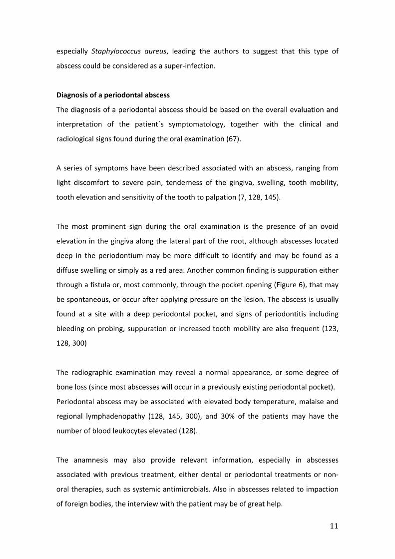
Diagnosis of a periodontal abscess
The diagnosis of a periodontal abscess should be based on the overall evaluation and
interpretation of the patient´s symptomatology, together with the clinical and
radiological signs found during the oral examination (67).
A series of symptoms have been described associated with an abscess, ranging from
light discomfort to severe pain, tenderness of the gingiva, swelling, tooth mobility,
tooth elevation and sensitivity of the tooth to palpation (7, 128, 145).
The most prominent sign during the oral examination is the presence of an ovoid
elevation in the gingiva along the lateral part of the root, although abscesses located
deep in the periodontium may be more difficult to identify and may be found as a
diffuse swelling or simply as a red area. Another common finding is suppuration either
through a fistula or, most commonly, through the pocket opening (Figure 6), that may
be spontaneous, or occur after applying pressure on the lesion. The abscess is usually
found at a site with a deep periodontal pocket, and signs of periodontitis including
bleeding on probing, suppuration or increased tooth mobility are also frequent (123,
128, 300)
The radiographic examination may reveal a normal appearance, or some degree of
bone loss (since most abscesses will occur in a previously existing periodontal pocket).
Periodontal abscess may be associated with elevated body temperature, malaise and
regional lymphadenopathy (128, 145, 300), and 30% of the patients may have the
number of blood leukocytes elevated (128).
The anamnesis may also provide relevant information, especially in abscesses
associated with previous treatment, either dental or periodontal treatments or non-‐
oral therapies, such as systemic antimicrobials. Also in abscesses related to impaction
of foreign bodies, the interview with the patient may be of great help.

12
Differential diagnosis is critical, since periodontal abscesses may be similar to other
oral conditions:
• Other abscesses in the mouth: periapical or dento-‐alveolar or endodontic
abscesses, lateral periapical cyst, vertical root fractures, endo-‐periodontal
abscess, postoperative infection (7). A combination of different factors, such as
pulp vitality, presence of dental caries, presence of periodontal pockets, the
location of the abscess, and a careful radiographic examination should be
carefully assessed to reach an accurate diagnosis.
• Other serious oral diseases may also have a similar appearance: osteomyelitis
in periodontitis patients (241), squamous cell carcinomas (162, 163, 322), a
metastatic carcinoma from pancreatic origin (289), a metastatic head and neck
cancer (85), an eosinophilic granuloma (111) or a pyogenic granuloma (237).
Therefore, in cases not responding to conventional therapy, a biopsy and histo-‐
pathologic diagnosis should be recommended.
• Self-‐inflicted gingival injuries: including trauma of the gingiva with a pencil
(267) or with a safety pin (37), or nail biting habit (304). A careful anamnesis is
the key factor in the diagnosis of these lesions.
Treatment of a periodontal abscess
The treatment of a periodontal abscess should include two distinct phases: the control
of the acute condition to arrest tissue destruction and control the symptoms, and the
management of the pre-‐existing and/or residual lesion, especially in periodontitis
patients.
Control of the acute condition
Four therapeutic alternatives have been proposed: tooth extraction, drainage and
debridement, systemic or local antimicrobials and surgery.
If the tooth is severely damaged, and its prognosis is hopeless after the destruction
caused by the abscess, the preferred treatment should be tooth extraction (300).

13
The most logical treatment of a periodontal abscess, as in other abscesses in medicine,
should include drainage (through the pocket or through an external incision),
compression and debridement of the soft tissue wall, and the application of topical
antiseptics after the drainage. If the abscess is associated with a foreign body
impaction, the object has to be eliminated through careful debridement (5), although
it may not be present anymore.
Systemic antimicrobials may be used as sole treatment, as initial treatment or as an
adjunctive treatment to drainage. As sole or as initial treatment may be only
recommended if there is a need of pre-‐medication, or when the infection is not well
localized or when adequate drainage cannot be ascertained (176). As an adjunctive
treatment, it should always be considered if a clear systemic involvement is present (7,
176). The duration and the type of the antibiotic therapy is also a matter of discussion,
including the recommendation of shorter courses of therapy (176, 202). The available
scientific evidence on the efficacy of these therapies, however, is very limited, with
only two prospective case series and two randomized clinical trials. Smith & Davies
(1986) evaluated the incision and drainage of the abscess, together with adjunctive
systemic metronidazole (200 mg, tid, 5 days), followed by a delayed periodontal
therapy, in 22 abscesses for up to 3 years (300). Hafström et al. (1994) proposed
drainage through the periodontal pocket, irrigation with sterile saline, supragingival
scaling and tetracycline for 2 weeks (1 g/day), and tested this therapy in 20 abscesses,
with 13 followed for 180 days, highlighting the importance of drainage and the
potential of regeneration (123). Herrera et al. (2000) compared azithromycin (500 mg,
once per day, 3 days) versus amoxicillin plus clavulanate (500+125 mg, tid, 8 days),
with delayed scaling (after 12 days), in 29 patients with abscess followed for 1 month,
concluding that both protocols were similarly effective (129). Eguchi et al. (2008)
compared irrigation with sterile physiological saline and 2% minocycline hydrochloride
ointment, versus irrigation with sterile physiological saline without the local antibiotic
in 91 patients for 7 days (82).
Surgical procedures have also been proposed, mainly for abscesses associated with
deep vertical defects (158), or in cases occurring after periodontal debridement in

14
which calculus is left subgingivally after the treatment (74). A case series evaluating a
combination of an access flap with deep scaling and irrigation with doxycycline is also
available, reporting “good results”, but scientific data was not provided (316).
After drainage and debridement the patient should be recalled in 24-‐48 hours to
evaluate the resolution of the abscess (Figures 7a and 7b) and the duration of the
antimicrobial intake. Once the acute phase has resolved, the patient should be
scheduled for a follow-‐up therapeutic phase.
In summary, numerous treatment protocols have been proposed, but sufficient
scientific evidence is not available to recommend a definitive approach. It is, however,
clear that drainage and debridement should be established when the systemic
condition and the access to the abscess is adequate. When the immediate drainage is
not possible or a systemic affect is evident, a therapy with systemic antimicrobials
should be considered. The drug with the most adequate profile is metronidazole
(normally prescribed, for acute conditions, at 250 mg, tid). Azithromycin (500 mg, once
per day) and amoxicillin plus clavulanate (500+125 mg, tid) have also shown good
clinical results. The duration of the therapy should be restricted to the duration of the
acute lesion, which is normally 2-‐3 days.
Management of a pre-‐existing and/or residual lesion
Since most periodontal abscesses occur in a previously existing periodontal pocket,
periodontal therapy should be evaluated after resolution of the acute phase. In cases
where the patient has not been treated previously, the appropriate periodontal
treatment should be provided. If the patient is within the active phase of therapy, once
the acute lesion is treated the periodontal therapy should be completed. In patients
during SPT, a careful evaluation of the recurrence of the abscesses should be made as
well as an adequate evaluation of the tissue damage and how this affects tooth
prognosis.
Summary
15
Abscesses in the periodontium are important since they are a relatively frequent
dental emergency, they can compromise the periodontal prognosis of the affected
tooth and because the bacteria within the abscess can spread and cause infections in
other body sites.
Although histologically all abscess lesions are similar, different type of abscesses have
been identified, mainly by their different etiology, since there are clear differences
between those affecting a previously existing periodontal pocket and those affecting
healthy sites.
For the management of this condition, a rapid and accurate diagnosis, mainly based on
clinical features, and provision of early therapy is mandatory. Therapy for the acute
condition should be based on drainage and debridement, with individual evaluation of
the need of systemic antimicrobial therapy. When the supporting tissues have been
destroyed to that extend of compromising the tooth prognosis, tooth extraction may
be the only valid alternative. The definitive treatment of the pre-‐existing condition
should be accomplished after the acute phase is controlled, since most abscesses are
found in untreated periodontitis patients.
NECROTIZING PERIODONTAL DISEASES (NPD)
Definition
NPD is a group of infectious diseases that includes: necrotizing ulcerative gingivitis
(NUG), necrotizing ulcerative periodontitis (NUP) and necrotizing stomatitis (NS).
However, it has also been suggested that these conditions may be different stages of
the same disease, since they have similar etiology, clinical characteristics, and
treatment, although they vary in disease severity (138, 140). These diseases share
common clinical features consisting of an acute inflammatory process and the
presence of periodontal destruction (Figure 8).

16
NUG has been diagnosed for centuries though termed various names, such as:
Vincent’s disease, trench-‐mouth disease, necrotizing gingivo-‐estomatitis, fuso-‐
spirochaetal stomatitis, ulcerative membranous gingivitis, acute ulcerative gingivitis,
necrotizing ulcerative gingivitis or acute necrotizing ulcerative gingivitis (138, 152, 269).
NUP was defined both in the 1989 World Workshop (54) and in the 1993 European
Workshop (26). At the International Workshop for a Classification of Periodontal
Diseases and Conditions in 1999 (22), the new category of “Necrotizing Periodontal
Diseases” was introduced, which includes NUG and NUP. More recently, the terms
necrotizing gingivitis (NG) and necrotizing periodontitis (NP) have been used instead of
the terms NUG and NUP (137). The simpler terms NG and NP adequately describe
these diseases and these term will be used in the rest of the text.
Classification of necrotizing periodontal diseases
According to the location of the tissue affected by the acute disease process, NPD can
be classified as (138, 140):
• Necrotizing gingivitis: when only the gingival tissues are affected.
• Necrotizing periodontitis: when the necrosis progresses into the periodontal
ligament and the alveolar bone, leading to attachment loss.
• Necrotizing stomatitis: the necrosis progresses to deeper tissues beyond the
mucogingival line, including the lip or cheek mucosa, the tongue, etc.
Clinical significance of necrotizing periodontal diseases
NPD are considered among the most severe inflammatory conditions associated with
oral biofilm bacteria (138). It is, therefore important to control predisposing factors,
and once the disease developed, to act quickly in order to limit its progression and
exacerbation.
The prevalence of NPD in systemically healthy populations has not been adequately
established, since most studies have focused on specific group of patients with clear
predisposing factors, such as military personal (141, 248), students (108, 188, 307),
patients positive for human immunodeficiency virus (HIV) (136), or subjects with
severe malnutrition (88, 234). In addition, data from these conditions originate from

17
hospitals or dental clinic settings, which may overestimate the true epidemiological
information. A high prevalence of NG (14%) was observed in civil and military
populations during the 2nd World War (12, 138), and a moderately high rate (2.2%) in
North America in the 1950s (122). After the war, the prevalence clearly decreased in
developed countries and nowadays it has a low prevalence.
NPD have been described in young people, both in developed and in developing
countries (12). In North America and Europe these diseases have been studied mostly
in groups aged late teens to mid-‐twenties. In 326 North-‐American students in the
1960s (108) the prevalence ranged from 2.5-‐6.7%. The current estimates in developed
countries are 0.5% or less (33, 141), with 0.001% reported in young Danish army
recruits (138)
In developing countries, the reported prevalence is higher, especially in children (12).
In Chile, among 9.203 students, 6.7% presented, at least a papilla with necrosis. In
India, 54-‐68% of the cases were observed in children younger than 10 years (249). In
South Africa, 3.3% of subjects aged 3-‐48 years presented NG, with 73% aged 5-‐12
years and mostly from a low socioeconomic class (20). In Nigeria, the prevalence of NG
ranged between 1.7-‐15% in 2-‐6 years old children, and was 27.2% in children with
severe malnutrition (12, 88, 292). In Kenya, 0.15% of patients attended the Nairobi
Hospital during one year were diagnosed with NG, and 58.5% of them were younger
than 11 years (155). In spite of these figures, subjects of any age may be affected.
NP is less frequent and it has been most frequently reported in HIV-‐positive patients,
with a prevalence 0-‐11% among these patients (40, 136, 261, 338). This prevalence is
lower when studies were performed outside hospitals or dental clinics. In HIV-‐positive
patients under anti-‐retroviral therapy (204, 272, 318) the prevalence of NP may not
differ from that in the general population.
Necrotizing periodontal diseases can progress rapidly and cause severe tissue
destruction. It is therefore, important that these conditions are managed promptly,
since there is evidence proving that NPD can be controlled by means of an adequate

18
periodontal treatment, combined with effective oral hygiene measures and control of
predisposing factors (152). NG patients, however, are frequently susceptible to future
disease recurrence, mostly due to the difficulties in controlling predisposing factors as
well as the difficulty in achieving proper supragingival biofilm control, in part due to
the sequelae of these diseases, mainly the presence of gingival craters (196).
NG can heal without clinical sequelae (38), but often the necrotizing lesion extends
laterally from the papilla to the gingival margin, affecting both the buccal and lingual
sites and progresses to other sites in the mouth, evolving from a localized into a
generalized disease (Figure 9). It may also progress apically evolving into NP (Figure 10).
In fact NP can be the result of one or various episodes of NG, or it can be the result of a
NPD affecting a site previously affected with periodontitis (227). NPD can also become
chronic, with a slow reduction in its symptomatology and progression, with ensuing
destruction, although at a slower rate (138, 248). Some authors believe that these
conditions remain acute and may be “recurrent” (152).
In cases of severe systemic involvement, such as in AIDS or severe malnutrition NG and
NP can progress further with rapid involvement of the oral mucosae. The severity of
these lesions are normally related to the severity of the systemic condition and the
compromised immune host-‐response, leading to extensive bone destruction and
presence of large osteitis lesions and oral-‐antral fistulae (335). NE has common
features with Cancrun Oris or NOMA. Some investigators suggested that NOMA is a
progression of NE affecting the skin, whereas others believe that NE and NOMA are
two distinct clinical entities. NOMA is a destructive gangrenous disease affecting the
facial tissues. It is associated with high mortality and morbidity rates (32, 89, 91), and it
is almost exclusively observed in developing countries, especially in children suffering
from systemic diseases, including severe malnutrition. NOMA is normally preceded by
measles, malaria, severe diarrhea and NG; which highlights the importance of
prevention, early detection and treatment during the first stages of the disease (269).
Etiology, pathogenesis and histopathology of necrotizing periodontal diseases

19
NPD are caused by infectious agents, although the predisposing factors, such as the
compromised host immune response are the main factors facilitating the bacterial
pathogenicity. This bacterial etiology was already demonstrated by Plaut in 1894 and
by Vicent in 1896 (269), since the microscopic exams of the plaque sample retrieved
from affected subjects clearly showed the presence of spirochetes and fusiform
bacteria, even within the tissues. Moreover, clinical resolution was observed after
mechanical debridement and antimicrobial treatments (302). However, the knowledge
on the pathogenesis of these diseases is limited, since it is not clear whether these
bacteria observed in the lesions are the cause or the consequence, as secondary
bacterial colonization may ensue after periodontal destruction, since the necrotic
tissues are the perfect environment for bacterial colonization and tissue invasion (138,
269).
The spirochetes and fusiform bacteria described in the necrotic lesions have the
capacity to invade the epithelium (131) and connective tissue (181), as well as to
release endotoxins that may cause periodontal tissue destruction through the
activation or modification of the host response.
In NG lesions observed through light microscopy have shown a distinct pathology (181),
with presence of an ulcer within the stratified squamous epithelium and the superficial
layer of the gingival connective tissue surrounded with a non-‐specific acute
inflammatory reaction. Four regions have been described within these lesions:
• The bacterial area with a superficial fibrous mesh composed of degenerated
epithelial cells, leukocytes, cellular rests, and a wide variety of bacterial cells,
including rods, fusiforms and spirochetes.
• The neutrophil-‐rich zone, composed of a great number of leukocytes, especially
neutrophils, and numerous spirochetes of different sizes and other bacterial
morphotypes located between the host cells.
• The necrotic zone, with presence of disintegrated cells, together with medium
and large size spirochetes and fusiform bacteria.

20
• The spirochetal infiltration zone, where the tissue components are adequately
preserved, but infiltrated by large and medium size spirochetes. Other bacterial
morphotypes are not found.
Microbiology of necrotizing periodontal diseases
The microbiology of NPD has been described in different studies with a similar
composition of the associated microbiota, including Treponema spp., Selenomonas
spp., Fusobacterium spp. and P. intermedia. Other microorganisms have also been
described although defined as “variable” flora and not present in all cases (187). Since
this typical microbiological description can also be detected in healthy, gingivitis or
periodontitis sites, the use of the microbiological testing does not provide relevant
diagnostic information (67, 152).
In HIV-‐positive patients with NPD the microbiological findings are also non-‐specific
(204, 266, 338), except in regards to the counts of yeast (namely, Candida albicans)
and the presence herpes viruses (62, 272, 338, 340), or the detection of superinfecting
bacterial species, such as enterics including Enterococcus avium, E. faecalis,
Clostridium clostridiiforme, C. difficile, Mycoplasma spp. and Klebsiella pneumoniae
(258, 340). Most recently, with the use of molecular technologies (polymerase chain
reaction), some bacterial species have been frequently associated with NPD lesions,
including Eubacterium saphenum, E. sabburum, Filifactor alocis, Dialister spp. and P.
endodontalis, while the typical periodontitis-‐associated pathogens P. gingivalis and T.
forsythia were less frequently found (243).
Some researchers have pointed out the possible etiological role of viruses in NPD,
including human cytomegalovirus (HCMV) (78, 273). In children with NG in Nigeria, in
addition to the presence of HCMV in the lesions, other viruses were detected including
Epstein Barr virus type 1 and herpes simplex virus (64).
Predisposing factors for necrotizing periodontal diseases
The most common predisposing factors are those altering the host immune response,
although usually more than one factor is necessary for initiating the disease (138). In a

21
study in USA, the most important factor was HIV infection. In non-‐HIV patients, the
most important factors were previous history of NPD, poor oral hygiene, inadequate
sleep, unusual psychological stress, poor diet, recent systemic diseases, alcohol abuse,
tobacco smoking, Caucasian ethnicity and age below 21 (140).
Systemic conditions
Those conditions lowering the host immune response favor NPD. Patients infected
with HIV or diseases affecting leukocytes (e.g. leukemia) are among the most
important predisposing factors. Other systemic conditions that have shown a positive
association include: malnutrition, measles, chickenpox, tuberculosis, herpetic gingiva-‐
stomatitis, malaria, or even diabetes, which was identified in a study with Chilean
adolescents (188).
Among predisposing systemic conditions, HIV infection and malnutrition have been
studied more in depth:
• In HIV-‐positive patients, NPD are more frequent and show a faster progression,
although no differences have been detected between the characteristics of the
disease in HIV-‐negative and positive patients. It has been suggested that HIV-‐
positive patients have a higher tendency for recurrence and diminished
response to both mechanical and/or pharmacological periodontal treatment
(227). In HIV-‐positive patients, the reduction in the counts of peripheral CD4
lymphocytes has been correlated with NG and NP (113, 303) and therefore, the
diagnosis of NPD should prompt the likelihood that the patient may have an
HIV infection, and therefore the affected subjects should be screened for HIV
(138, 139).
• Malnutrition has also been reported as predisposing factor (269) especially in
developing countries (88, 90, 234). The basis for this interaction has been
termed “protein-‐energy malnutrition”, implying a marked reduction in key
antioxidant nutrients and an altered acute phase response against infection.
Other consequences are an inversed proportion in the ratio of
helper/suppressor T-‐lymphocytes, histaminemia, increased free cortisol in
blood and saliva, and defects in mucosal integrity (90).

22
Psychological stress and insufficient sleep
Acute psychological stress and acute stressing situations have been associated with NG
(140, 152, 291). Certain situations may predispose individuals to NPD, such as military
personnel in wartime, new recruits for military services, drug-‐abusers during
abstinence syndrome, students during exam periods, and patients with depression or
other psychological conditions (108, 122, 248). During these stress periods, not only
the immune response is altered, but also the subject’s behavior, leading to inadequate
oral hygiene, poor diet or increased tobacco consumption. The proposed mechanisms
to explain this association are based on a reduction in the gingival microcirculation and
salivary flow and an increase in serum and urine levels of 17-‐hydroxycorticosteroid
(17-‐OHCS), which are associated with an alteration in the function of
polymorphonuclear leukocytes (PMN) and lymphocytes, or even an increase in the
levels of periodontal pathogens, such as P. intermedia (187). In patients with NG,
higher urine levels of 17-‐OHCS have been reported, when compared with healthy or
treated patients; in addition, those patients demonstrated statistically significant
higher levels of anxiety, depression or emotional alteration (138). Patients with NG
also contained PMNs with altered functions, since their bactericidal, phagocytic and
chemotactic capacities were depressed (63).
Inadequate oral hygiene, pre-‐existing gingivitis and previous history of NPD
Plaque accumulation has been considered a predisposing factor (140, 152), although it
may also be the consequence of the presence of ulcer and crater lesions that may limit
tooth brushing due to pain. NPD usually occurs over a previously existing periodontal
disease, usually chronic gingivitis (248) (Figure 11). In one study, 28% of the patients
with NPD reported a history of painful gingival inflammation and 21% showed lesions
compatible with previous NPD (140).
Alcohol and tobacco consumption
Smoking is a risk factor for NPD (152, 153, 264) and in fact, most HIV-‐negative patients
diagnosed of NPD were smokers (108, 248, 307). The mechanisms explaining this
association are probably related to the effect of smoking on inflammation and tissue

23
response, since smoking interferes with both PMN and lymphocyte function and
nicotine induces vasoconstriction in gingival blood vessels.
Alcohol consumption has also been associated with the physiological and psychological
factors favoring NPD (139).
Young age and ethnicity
In developed countries, young people are more prone to suffer NPD, mostly between
21-‐24 years, usually combined with other predisposing factors, such as smoking and
stress (139, 140, 307). In developing countries, NPD affects even younger people,
being malnutrition and occurrence of infections the most frequent predisposing
factors, (89, 90, 269). Studies in North America have reported that up to 95% of cases
of NPD occurred in Caucasian patients (33, 140, 307), although more studies are
needed to confirm this finding.
Diagnosis of necrotizing periodontal diseases
The diagnosis of NPD is mainly based on the clinical findings (67, 269). Although the
reported histology and the typical microbiota associated with these lesions have a
distinctive character, neither biopsy nor microbiological sampling are usually essential
diagnostic tools in these diseases (67). The different stages of NPD share clinical
features, but also distinct findings depending on the extent and severity of the lesions
(1, 67, 138, 152, 269):
Necrotizing gingivitis
The diagnosis is based on the presence of necrosis and ulcers in the free gingiva. These
lesions usually start at the interdental papilla with the typical “punched-‐out”
appearance. In addition, a marginal erythema, named “lineal erythema”, may be
present, separating the healthy and the diseased gingiva. These necrotic lesions can
progress to the marginal gingiva. The most typical location is the anterior teeth,
especially in the mandible (Figure 12). In NG gingival bleeding is a frequent finding,
usually spontaneous or after minimal contact (Figure 13). Pain normally occurs soon
and with different degrees of severity, depending on the severity and extension of the

24
lesions. The bouts of pain increase with eating and with the oral hygiene practices and
it is normally the reason for patient’s consultation.
Other less common findings include the presence of:
• pseudo-‐membranous formation over the necrotic area. It consists on a
meshwork of whitish-‐yellow color, composed of necrotic tissue, fibrin,
erythrocytes, leukocytes and bacterial cells. When this “membrane” is removed,
the underlying connective tissue becomes exposed and bleeds;
• halitosis, although it is not an exclusive sign of NG;
• adenopathies, which are usually found in the most severe cases. If present,
submandibular lymph nodes are more affected than those in the cervical area
(150, 151).
• Fever and a general feeling of discomfort.
Necrotizing periodontitis
The same clinical picture described in NG occurs in NP, but in addition in NP the
following characteristics may be present:
• The necrosis affects the periodontal ligament and alveolar bone, leading to loss
of attachment. Since there is concomitant necrosis of the soft tissue, presence
of pockets is not a usual finding (Figure 14).
• As the disease progresses (Figure 15), the interdental papilla is divided in a
buccal and a lingual/palatal part, with a necrotic area in the middle, known as
interproximal crater. If the craters are deep, the interdental crestal bone
becomes exposed and denudated. In addition, crater formation favors disease
progression by allowing the accumulation of more bacteria. Interproximal
necrotic areas spread laterally and merge with the neighboring areas, creating
an extensive zone of destruction.
• In severe cases, especially in immune-‐compromised patients, bone sequestrum
(necrotic bone fragments within the tissues but separated from the healthy
bone) may occur, mainly interdentally, but also in buccal or lingual/palatal
alveolar bone.

25
Necrotizing stomatitis
When bone denudation extends through the alveolar mucosa, larger bone sequestra
may occur, with large areas of osteitis and oral-‐antral fistulae. The severity of these
lesions is associated with severely compromised systemic patients, including AIDS
patients and severe malnutrition (335).
Management of necrotizing periodontal diseases
Due to the previously listed specific features of NPD (tissue destruction, acute course,
pain), diagnosis and treatment have to be performed as soon as possible, and
conventional periodontal therapies may need adjunctive therapeutic measures (2,
152).
The treatment should be organized in successive stages, including the treatment of the
acute phase and a subsequent treatment phase that should include the treatment of
the pre-‐existing condition, the corrective treatment of the disease sequelae, and the
supportive or maintenance phase.
Treatment of the Acute Phase (138)
Two main objectives should be achieved with the therapy: to arrest the disease
process and tissue destruction, and to control the patient´s general feeling of
discomfort and pain, which is interfering with nutrition and oral hygiene practices. The
first task should be a careful superficial debridement, to remove the soft and
mineralized deposits. Power-‐driven debridement devices (e.g. ultrasonics) are usually
recommended exerting minimum pressure over the ulcerated soft tissues. The
debridement should be performed daily, getting deeper as the tolerance of the patient
improves, and lasting for as long as the acute phase lasts (normally, 2-‐4 days).
Mechanical oral hygiene measures should be limited, in order to avoid pain, since
brushing directly in the wounds may impair healing. During this period the patient is
advised to use chemical plaque control agents, such as chlorhexidine-‐based mouth
rinses (at 0.12-‐0.2%, twice daily). Other products have also been suggested, such as 3%
hydrogen peroxide diluted 1:1 in warm water, and other oxygen-‐releasing agents,
which not only contribute to the mechanical cleaning of the lesions, but also provide

26
the antibacterial effect of oxygen against anaerobes (333). Other oxygen therapies
have also been evaluated, such as a local oxygen therapy, that may help in a reduction
or even eradication of microorganisms, resulting in a faster clinical healing with less
periodontal destruction (101).
In cases that show unsatisfactory response to debridement or systemic effects (fever,
malaise), the use of systemic antimicrobials may be considered. Metronidazole, at 250
mg, every 8 hours, may represent the first alternative, due to its action over strict
anaerobes (187). Other systemic drugs have also been proposed, with acceptable
results, including penicillin, tetracyclines, clindamycin, amoxicillin or amoxicillin plus
clavulanate. Conversely, locally delivered antimicrobials are not recommended, due to
the large amount of bacteria present within the tissues, where the local drug will not
be able to achieve adequate concentrations.
These patients have to be followed-‐up very closely, daily if possible, and as the
symptoms and signs improve, strict mechanical hygiene measures should be enforced,
as well as complete debridement of the lesions can be scheduled.
Treatment of the pre-‐existing condition
NPD normally occurs over a chronic gingivitis or periodontitis. Once the acute phase
has been controlled, the treatment of the pre-‐existing chronic condition should be
implemented, including professional prophylaxis and/or scaling and root planing. Oral
hygiene instructions and motivation should be enforced. Existing predisposing local
factors, such as overhanging restorations, interdental open spaces and tooth
malposition should be carefully evaluated and treated (140). At this stage, and also
during the acute phase, attention should be paid to the control of the systemic
predisposing factors, including smoking, adequate sleep, reduction of stress or
treatment of the systemic conditions.
Corrective treatment of disease sequelae
The correction of the altered gingival topography caused by the disease should be
considered (Figure 16), since gingival craters may favor plaque accumulation and

27
disease recurrence. Gingivectomy and/or gingivoplasty procedures may be helpful to
treat superficial craters; for deep craters, periodontal flap surgery, or even
regenerative surgery represent more suitable options (138).
Supportive or maintenance phase
During this phase, the main goal becomes the compliance with the oral hygiene
practices (Figure 17) and the control of the predisposing factors.
Specific considerations for HIV-‐positive patients
HIV-‐positive patients may not be aware of their serologic status. Occurrence of NPD in
systemically healthy individuals is suggestive of HIV infection, and therefore the
affected individuals should be screened for HIV (132, 138, 140). The specific
management of NPD in HIV-‐positive patients includes debridement of bacterial
deposits combined with the irrigation of the site with iodine povidone, based on its
hypothetic anesthetic and bleeding control effects (338), although no sound scientific
evidence is available to support this protocol (271, 336, 338). Careful consideration
should be made regarding the use of systemic antimicrobials, due to the risk of over-‐
infections with Candida spp. Metronidazole has been recommended due to its narrow
spectrum, with limited effects on Gram-‐positive bacteria, which prevent Candida spp.
overgrowth (272, 336, 338), although HIV-‐positive patients may not need antibiotic
prophylaxis for the treatment of NPD (194). In non-‐responding cases, the use of
antifungals may be beneficial, including clotrimazole lozenges, nystatin vaginal tablets,
systemic fluoconazole or itraconazole, mainly in cases of severe immune-‐suppression
(272, 338). In HIV-‐positive patients the systemic status should be closely monitored,
including the viral load and the hematologic and immune condition, leading to a
customized periodontal treatment plan (266, 272, 338).
Summary
NPD includes NG, NP and NS, and these may be considered different stages of the
same pathologic process. This group of diseases always presents three typical clinical
features: papilla necrosis, bleeding and pain, which make them different from other
periodontal diseases. Although their prevalence is not high, their importance is clear,
28
since they represent the most severe biofilm-‐related periodontal conditions, leading to
rapid tissue destruction. In their etiology, together with the bacteria, numerous factors
that alter the host response may predispose to these diseases, including HIV infection,
malnutrition, stress or tobacco smoking.
Due to their acute presentation, together with the associated pain and tissue
destruction, the treatment should be provided immediately once diagnosed, including
superficial debridement, careful mechanical oral hygiene, rinsing with chlorhexidine
and daily revisions. Systemic antimicrobials may be used adjunctively in severe cases
or in non-‐responding conditions, and the best option is metronidazole. Once the acute
disease is under control, definitive treatment should be provided, including the
adequate therapy for the pre-‐existing gingivitis or periodontitis as well as adequate
oral hygiene practices and supportive therapy. Surgical treatment of the sequelae
should be individually considered.
OTHER ACUTE CONDITIONS IN THE PERIODONTIUM
This group of acute gingival lesions includes lesions manifesting initially as acute
conditions or as acute episodes of a chronic condition. They can appear as isolated
lesions or as part of complex clinical pictures and they are frequently the cause for
emergency consultations.
The presenting clinical lesion is usually an ulcer or erosion, which may be the primary
lesion, or secondary to a vesicle-‐bullous lesion. The most frequent symptom is localized
pain, which initiates with the lesion or may precede it, although it may also occur in
conjunction with pain in the pharynx or dysphagia. To properly establish the diagnosis, a
clinical history, anamnesis and careful examination are mandatory, since most of the
times there is a direct and recent relation with the cause (68). In the following
description, the lesions are classified according to their etiology, since their clinical
appearance is similar and a careful differential diagnosis is key to their therapy (22, 134,
326).

29
Gingival diseases of infectious origin
Gingival lesions of specific bacterial origin
Specific bacterial infections localized in the oral mucosa are not frequent. They may be
caused by bacteria normally present in the oral cavity, that eventually become
pathogenic, and also by bacteria exogenous to the oral cavity, such as by gonococci,
tularemia or anthrax. In addition, the lesions present in the oral cavity may be a
secondary location of generalized infectious disease, as it may happen in scarlatina,
diphtheria, syphilis or tuberculosis.
Both staphylococci and streptococci may cause oral infections with gingival
involvement, leading to a lesion with a non-‐specific appearance (usually erythematous
or erosive) (102, 201). Group B streptococcal infections frequently result in pharynx-‐
amigdalitis that affects all oral mucosae and may be associated with fever and malaise.
Its therapy includes re-‐hydration, resting, and the prescription of systemic
antimicrobials (154, 157, 160, 183). Stomatitis associated with Staphylococcus aureus is
characterized by bullous generalized dermatitis, with vesicles and desquamation,
affecting lips, oral mucosa and other mucosae. Its appearance is similar to multiform
erythema or impetigo, and it is usually treated with systemic antimicrobials (102, 299).
Despite the presence of the oral epithelium as a protective barrier against infection of
Neisseria gonorrhoeae, gonococcic lesions may appear as a result of a direct inoculation.
In the newborn, it may be the result of a contact when passing through the birth canal.
In adults, transmission may be bucco-‐genital and superficial lesions are found, as white-‐
yellowish plaques or pseudo-‐membranes that, when removed, result in a bleeding ulcer.
Salivary flow may be reduced and saliva can be denser. The clinical history is crucial and
treatment should consist of the administration of systemic antimicrobials (61, 92, 293,
332).
Among the generalized infectious diseases, syphilis may often affect the gingival tissues.
Syphilis is caused by Treponema pallidum and may be difficult to diagnose due to the
similarities with other systemic conditions (96, 172, 256). Syphilis can be congenital and

30
acquired. In the acquired syphilis, the incubation period may vary from 12-‐40 days, and
the lesions may follow different stages. In primary syphilis, the lesion is located in the
point of transmission, normally as a chancre that may be located on the lips, tongue or
tonsils (11). The gingiva may be affected by the secondary syphilis, after 6-‐8 weeks or
even 6 months, with the presence of a plaque (elevated papule with central erosion),
that may last for weeks or even a year (18, 168, 193, 197, 257, 268). Tertiary syphilis is
not frequent and, within the oral cavity, it may affect the palate and the tongue (171).
Differential diagnosis is critical and sometimes difficult. The treatment consists of the
administration of specific systemic antimicrobials (96, 197).
Viral infections
Different viruses may cause lesions in the oral cavity, with or without concomitant skin
involvement (215). The most frequently associated viruses causing gingival and
periodontal lesions are from the Herpesviridae family (herpes simplex virus type 1 (HSV-‐
1), causal agent of oral and labial herpes lesions; herpes simplex virus type 2 (HSV-‐2),
associated with genital herpes; and varicela zoster virus (HSV-‐3), responsible for varicela
and herpes zoster) (64-‐66, 119, 137, 211, 240, 298). Herpes virus adapt easily to the host,
and after the primary infection they remain inside the infected cells in a latent or silent
state; they show tropism for epithelial and neural cells, and the preferred site for
latency is the nervous ganglia (66, 83, 84, 324, 337).
Varicela is the result of an infection by Herpesvirus varicellae (HSV-‐3), being the result of
the first contact with the virus. It results in a generalized condition, especially in children,
with vesicle eruptions in kin after an incubation period of 1-‐3 weeks. Before or during
the skin lesions, vesicles can be evident in the oral cavity, including the gingiva, and they
break easily, forming ulcers surrounded by an erythematous halo. The result of the re-‐
activation of the mentioned virus (HSV-‐3), residing in regional sensitive ganglia, is
herpes zoster, especially in the elderly or associated with immune-‐depression. It may
affect the trigeminal ganglion with clinical manifestation preceded by pain or itching
feeling. Clinical lesions are vesicles surrounded by erythematous halo, with unilateral
distribution; vesicles easily break, forming erosive areas (214, 262, 312). Primary
herpetic infection caused by HSV-‐1 is normally suffered without notice, but it is

31
sometimes very evident in the form of generalized gingivo-‐stomatitis, with dysphagia,
fever, malaise and submandibular adenopathy. It is more frequently observed in
children 2-‐5 years, with oral lesions in the form of ulcers or erosions, after the vesicles
are broken (Figure 1). Treatment may include antiviral agents and adequate nutritional
support, mostly when pain compromises eating (66, 84, 216, 287, 324).
Recurrent herpetic infection by HSV-‐1 may be either intraoral or labial. Initial symptoms
include local discomfort in the form of itching or stinging feeling. The lesions develop as
an erythema, and then a variable number of grouped vesicles break forming an erosion
in the oral mucosae gingiva or the lip (Figure 2). Lesions normally last 7-‐10 days and heal
without scaring. Differential diagnosis with other conditions showing ulcers, such as
recurrent aphthous stomatitis, is important; although the latter lesions will not affect
keratinized tissues (178, 252, 254, 284, 341). Since the condition is self-‐limiting, no
treatment is usually required, although if present in immune-‐compromised patients,
antiviral agents should be prescribed (216, 287, 311, 337).
Other viruses, such as Epstein Barr virus (herpes simplex virus type 4, HSV-‐4),
Cytomegalovirus (CMV, or herpes simplex virus type 5, HSV-‐5) or Coxsackie virus, are
frequently transmitted via saliva and may result in specific oral manifestations:
infectious mononucleosis (HSV-‐4) or hand, foot and mouth disease (Coxsackie virus).
Sometime, they may also cause unspecific conditions, including acute stomatitis with
oral ulcers and malaise and adenopathy. These conditions can be important in immune-‐
compromised patients (25, 127, 189, 218, 231).
In addition to the previous acute conditions, viruses are related to chronic conditions
that mat require emergency attention due to complications during their development,
trauma or unexpected findings, including viral wart or condyloma acuminata.
Fungal infections
Many fungal species are part of the resident flora of the mouth, but they may cause
pathology when local or systemic factors trigger their overgrowth (114, 207, 222).
Among these opportunistic fungal infections, candidiasis is the most frequent, normally
32
affecting immune-‐compromised subjects, especially HIV-‐positive patients or during
infancy and elderly (49, 118, 133, 229, 278, 328). Although its localized forms are usually
not severe, it may spread and lead to more severe infections, such as esophagic or
systemic candidiasis. Among Candida spp., C. albicans is the most relevant for oral
infections (263).
Multiple factors are associated with the pathogenesis of oral candidiasis, and these may
be divided into host-‐related and environmental (49, 134, 275, 278, 314). Among the
host-‐related factors are the presence of concomitant systemic diseases (such as
diabetes) or debilitating conditions leading to a reduced host response. The most
frequent local associated factors include removable prosthesis, systemic antimicrobials,
corticosteroids, and tobacco smoking (9, 49, 104, 114, 118, 134, 207, 275, 278, 314).
Acute candidiasis can present as pseudomembranous or as erythematous candidiasis,
with itching or stinging feeling, being the diagnosis mostly clinical (28, 135). Treatment
will include antifungal agents, such as nystatin, amphotericin B or miconazole in
solutions or gels. In severe cases or immune-‐compromised patients the treatment of
choice is with systemic fluconazole. It is also mandatory to evaluate and control the
predisposing/adjunctive local and/or systemic factors associated (9, 43, 104, 319).
Gingival manifestations of systemic conditions
Muco-‐cutaneous disorders
These represent a group of chronic autoimmune diseases, characterized by the
presence of vesicle-‐bullous lesions, with liquid content (either serum or hemorrhagic).
Although the evolution of these diseases is chronic, acute bouts may occur and affect an
intra-‐oral location, as well as lesions in the skin or in other mucosae (50, 310, 331).
Vesicle-‐bullous diseases usually present in the gingival tissues as desquamative
gingivitis. This manifestation reflects the presence of epithelial desquamation and
erythematous, vesicle-‐bullous or erosive lesions in the gingiva. Although it is normally
part of a chronic process, painful acute phases can also occur, with intense and diffuse
33
redness in the attached and free gingiva. The epithelium is fragile and easily detached,
leaving a reddish surface, which bleeds after minimum stimulus (313). When the lesions
are limited to the gingival tissues, they are more frequently located in dentate areas at
buccal sites (Figure 4). Women in the 4th-‐5th decades of life are more prone to present
these lesions (172, 184, 185, 199, 310, 325). Therapy is usually based on application of
topical or systemic corticoids (51, 52, 95, 115, 191, 226).
Lichen planus is a chronic inflammatory disease affecting skin and mucosae, being the
most frequent non-‐infectious disease of the oral cavity. The oral manifestation is very
frequent, mostly in adult women (30-‐50 years) of Caucasian ethnicity (186, 213, 280).
These oral lesions usually precede the skin lesions or are just the only lesion (Figure 5).
Clinical manifestations range from its typical reticular form affecting the buccal and
cheek mucosae, to erosive forms usually located in the tongue and gingiva that cause
pain and tenderness, as well as bleeding. When present in the gingiva they usually
present as a desquamative gingivitis affecting the attached gingiva (Figure 5). The
diagnosis is both clinical (bilateral and symmetric lesions, whitish reticular lesions or red
lesions, not disappearing after scraping) and histological (53, 186, 213, 281). If some of
the diagnosis criteria are not fulfilled, the lesions will be defined as lichenoid reactions,
normally associated with restorative materials, pharmaceutical drugs, graft versus host
disease or some systemic conditions (10, 75, 80, 147, 169, 233, 246).
Pemphigus is a severe autoimmune muco-‐cutaneous disease, with a chronic and
aggressive progression, characterized by the destruction of the intercellular adhesion
systems between keratinocytes, leading to intra-‐epithelial bulla formation (39, 170, 221,
288). Among the different clinical forms, only the vulgaris and the vegetans can affect
the oral mucosae, although the latter is very infrequent. Pemphigus vulgaris is more
frequent in women, 40-‐60 years of age, with a Mediterranean or Jewish background.
Skin lesions are more frequent, but in 50% of the cases oral lesions may precede the
skin lesions (295). Intraoral lesions if affecting the gingiva appear as desquamative
gingivitis (86). Diagnosis may be difficult and will be based on histology and immune-‐
florescence (48, 87, 212, 230, 282, 286, 288, 296).

34
Pemphigoid includes a group of autoimmune muco-‐cutaneous diseases, either affecting
the skin (bullous pemphigoid and gestational herpes) or the mucosae (benign mucous
membrane pemphigoid or cicatricial pemphigoid). In the latter, the autoimmune
reaction affects the basal membrane (sub-‐epithelial bulla), frequently occurring in
women older than 50 years, presenting oral lesions in more than 90% of the cases.
These lesions are usually located in the gingiva, later spreading to neighboring tissues or
other sites. Gingival tissues are affected in 40-‐100% of the cases, as desquamative
gingivitis including periods of exacerbation and remission (19, 31, 170, 239, 274, 285).
Other muco-‐cutaneous conditions, characterized as vesicle-‐bullous conditions, include:
linear IgA disease, bullous epidermolisis, erythema multiforme and lupus
erythematosus. When affecting the gingival, they manifest as desquamative gingivitis
and the differential diagnosis should be based on histological or laboratory grounds, as
well as the involvement of other body sites or organs (13, 50, 94, 200, 225, 310, 323,
331).
Allergic reactions
Allergy is an abnormal reaction of the human body, responding exaggeratedly after a
contact with a foreign substance or product (allergen), which will not induce a similar
reaction in other individuals (3, 27, 44, 72, 195, 228). Food products, including fruits,
seafood, nuts or some vegetables, induce most allergic reactions; however, some
medicines, including antibiotics (e.g. penicillin) or non-‐steroidal anti-‐inflammatory drugs
(e.g. acetyl-‐salicylic acid) can be very relevant (15, 17, 23, 42, 46, 70, 72, 112, 179, 228,
235). Other potentially allergenic products are haptens, which needs to be linked to
proteins in order to become allergens, and are very relevant in dentistry since they
include some metals and dental materials (3, 41, 72, 73, 164, 169, 173, 203, 217), topical
anesthesia, oral hygiene products ingredients (161), as well as components of rubber
dam, latex examination gloves and cosmetics (16, 45, 79, 169, 180, 208, 277, 297).
Allergies in the mouth may have different clinical manifestations ranging from the
typical urticarial reaction to angioedema, although they do not normally affect the
gingival tissues (27, 159, 205).

35
Exudative erythema multiforme is a disseminated hypersensitivity reaction, which may
affect the majority of the human systems and even compromise the patient’s life (3, 24,
29, 34, 47, 58, 81, 93, 98, 107, 167, 260, 270, 279, 306). An inductor or precipitating
factor is normally present, including herpes simplex virus infections, drugs such as
sulphamide, penicillin or salicylate, or gastro-‐intestinal conditions, including Crohn
disease and ulcerative colitis (29, 190, 270). Clinically, it may affect skin and mucosae,
with minor and major (also known as Steven Johnson syndrome) forms, and it is
characterized by bullae formation (24, 34, 47, 93, 143, 190, 223, 260). The diagnosis is
mainly clinical and, when affecting gingival tissues, differential diagnosis with other
conditions that manifest as desquamative gingivitis should be considered (24, 47, 144).
In mild cases, topical corticoids, analgesics and a soft diet may be sufficient. In severe
cases, systemic corticoids are the first option, and in refractory cases the treatment is
with azathioprine or dapsone (144, 192, 223, 232).
Contact allergy of intraoral location is an ill-‐defined entity (3, 27, 72, 73), normally
associated with drugs (antimalarial, non-‐steroidal anti-‐inflammatory, antihypertensive
or anti-‐diabetic medications) or metals (especially amalgam (41, 169, 208), gold (334),
nickel (73), or other dental materials, including acrylic resins or dental composites (6,
164, 173, 203, 217, 277) (Figure 6). Contact allergies associated with toothpaste, mouth
rinse or chewing gum are rare (16, 79, 161, 180, 297). The lesions are usually not
clinically distinguishable from a tooth-‐related irritation or trauma (73, 305, 329).
Symptoms include burning, itching or stinging feeling and the lesion is often defined as a
liquenoid reaction (165, 206). The clinical aspect of intraoral contact allergies includes
erythematous and edematous gingival tissues, sometimes with ulcers and whitish areas.
The same lesions can be observed on the lip, buccal or lingual mucosae. Diagnosis,
therefore, is often difficult, with a need to find a direct association between the clinical
lesions and the exposure to allergen. The most important therapeutic measure is to
remove the allergen, although this is not always easy, and sometimes it will not solve
the case, including cases of wrong diagnosis (16, 79, 161, 180, 297).
Traumatic lesions

36
The clinical presentation of lesions associated with physical and chemical agents will
depend of the aggression, being either direct or indirect. The effect of a direct action
depends on the type of agent, the time of action and the extension of mucosa affected.
Indirect effects appear as color alteration, erythema, erosion, ulcer, gingival
enlargement, hemorrhage or dysgeusia (59, 236, 259).
Among traumatic lesions, iatrogenic lesions are relevant in dentistry, produced during a
therapeutic intervention or as the result of the therapy (Figure 7). They are normally
considered as treatment complications, since dental instruments and chemical products
can cause injuries or burns, including lesions in the gingival tissues (8, 59, 71, 105, 112,
220, 236, 247, 251, 259, 276, 277, 290, 305).
Physical (mechanical and thermal) injury
Physical mechanical injuries may appear as erosions or ulcers, associated with gingival
recession. But also can be observed as hyper-‐queratosis, vesicles or bullae, sometimes
in combination with other oral lesions on lips, tongue or teeth; they may be
asymptomatic, but they can also induce intense, localized pain at the area of the lesion.
The correct diagnosis is based not only on the clinical aspect, but also on the
identification of the noxious agent, and for that, patient collaboration is crucial (148,
182, 236, 259, 305).
Physical mechanical injuries are most frequently caused by traumatic accidents, but
they can also be related to incorrect oral hygiene habits and para-‐functions (182, 238,
242, 301). If the physical trauma is limited, but continuous over time, the gingival lesion
will be frictional hyper-‐queratosis that may lead to a leukoplasic lesion. If the physical
trauma is more aggressive, superficial laceration or a more severe tissue loss can be
produced, eventually resulting in gingival recession (Figure 8). As an example, the use of
an abrasive dentifrice and vigorous horizontal brushing will result in an ulcer or an
erosion in the gingiva, frequently leading to tooth abrasion (182, 301). The use of dental
floss can also result in gingival ulceration or inflammation, especially at the most coronal
part of the interdental papilla (142).

37
Some physical traumatic lesions are self-‐induced and they are named as self-‐induced
gingitivis, patomimias, factitious lesions or artifact (14, 30, 37, 77, 120, 121, 244, 308,
309). Lesions present as ulcers in the gingival margin, associated with gingival recession;
patients are frequently children or teenagers, sometimes associated with psychological
conditions. There is a clear tendency for recurrence, and the traumatic agent can be the
fingers, nails or different instruments (pencil, pen…) (69, 166, 253, 259).
Another group of physical traumatic lesions are related to dental fractures or root rests,
orthodontic or prosthetic appliances and oral piercing (56, 148, 174, 175). Normally,
they are observed as ulcers or erosions, but dental or periodontal abscesses can be
triggered, as well as chronic infections such as sub-‐prosthesis palatitis or lesions as
fissured epulis. Frictional hyper-‐queratosis can also occur. In addition, the risk of pre-‐
cancer or carcinoma lesions is present, especially if other factors (infection, irritation,
chemicals…) act also over the traumatized area.
Ionizing radiations may also cause physical injuries in the gingival tissues (320). This type
of lesion is observed after irradiation therapy as part of the head-‐and-‐neck cancer
treatment. The gingival tissues show mucositis, with erythema, followed by epithelial
necrosis with whitish plaques, which leaves bleeding surfaces after dislodging (283).
Lesions related to hot, cold or electrical trauma are not frequent, although burns caused
by very hot food or liquids may need emergency consultation (35, 219, 220, 236). The
patient will feel pain in the affected area, which appears with erythema or
desquamated, sometimes with vesicles, erosions or ulcers. Normally, diagnosis is
straightforward (Figure 9).
Chemical and pharmacological injury
Numerous agents can induce chemical injury, including acids and bases, medicines,
alcohol, tobacco, and also lesions associated with the use of mouth rinses and
dentifrices. Lesions may appear after the direct contact with the agent over the mucosa,
leading to a maculae, vesicles, erosions or ulcers, with different extension and

38
relevance, depending on the causal agent, the extension of the lesion and the time of
contact (8, 71, 105, 112, 276, 290).
Summary
This is a group of periodontal lesions, with acute onset, which are not etiologically
associated with oral biofilm microorganisms. They however, result in pain and difficult
oral hygiene practices. Among these conditions, infectious diseases, muco-‐cutaneous
diseases and traumatic or allergic lesions can be listed.
In most cases, the gingival involvement is not severe, but it is important to be familiar
with these lesions, since they may be frequent and may represent a reason for
emergency dental consultations. In addition, some of them may be the first lesions of a
severe systemic disease, as in muco-‐cutaneous diseases and they usually manifest with
the characteristic gingival lesion of desquamative gingivitis.
Many of these conditions have the appearance of an erythematous lesion, sometime
erosive, with pain induced by tooth brushing or chewing. The differential diagnosis is
fundamental with special focus on the anamnesis and clinical history and progression
of the lesions. Erosive lesions may be the result of direct aggressions, such as trauma
or indirect reactive lesions related to dental or iatrogenic interventions. Similarly the
proper differential diagnosis will lead to the appropriate therapeutic intervention.

39
List of figures
Figure 1. A periodontal abscess, manifesting as a localised purulent lesion within the
gingival wall of the periodontal pocket.
Figure 2. Pericoronal abscess associated to the right lower third molar.
Figure 3. Periodontal abscess with the presence of furcation involvement.
Figure 4. Periodontal abscess in untreated periodontitis.
Figure 5. Microbiological sampling in a periodontal abscess.
Figure 6. Periodontal abscess demonstrating suppuration through the periodontal
pocket.
Figure 7a. Periodontal abscess before treatment.
Figure 7b. Periodontal abscess, 12 days later, after treatment with azithromycin.
Figure 8. Multiple gingival crater formation. The clinical picture includes rapid
progression and severe periodontal destruction.
Figure 9. Necrotic area in a localized papilla: necrotizing gingivitis starts at the
interdental papilla and different degrees of destruction can be observed in different
areas of the same mouth.
Figure 10. Necrotizing periodontitis: it can occur after numerous episodes of
necrotizing gingivitis, or as a necrotizing disease over a pre-‐existing periodontitis.
Figure 11. Plaque and calculus accumulation in a chronic gingivitis patient: necrotizing
periodontal diseases affect tissues already affected by chronic periodontal conditions.
Figure 12. Necrotizing gingivitis affects more frequently the lower anterior sextant.
Figure 13. Bleeding observed in necrotizing gingivitis: it can occur spontaneously or
after a minimum stimulus.
Figure 14. Papilla destruction in necrotizing gingivitis: no pocket formation is observed
due to the loss of soft tissues.
Figure 15. Crater formation and bleeding in a posterior area.
Figure 16. Gingival craters are important sequelae since they can limit mechanical
plaque control.
Figure 17. Aesthetic consequences of necrotizing periodontal diseases: the best
treatment is prevention by means of the control of the predisposing factors.

40
Figure 18. Herpetic gingivo-‐stomatitis: painful gingivitis with generalized vesicles in
primary herpetic infection.
Figure 19. Recurrent herpetic infection: vesicles break, leading to erosive lesions.
Gingival location is not infrequent.
Figure 20. Oral candidiasis oral: one of the predisposing factors is removable prosthesis
and erythematous lesions are among the most frequent lesions.
Figure 21. Muco-‐cutaneous disorders: most of them may have oral and gingival,
normally with a chronic evolution but also with acute periods.
Figure 22. Lichen planus: gingival involvement includes erosive lesions and reticular
whitish lesions.
Figure 23. Liquenoid lesions: identical to lichen planus lesions, but clearly associated to
a causative fact (such amalgam fillings), and disappearing after the elimination of the
factor.
Figure 24. Iatrogenic lesions: gingival erosions.
Figure 25. Traumatic lesions: often self-‐inflicted, or associated to traumatic tooth
brushing or incorrect oral hygiene habits.
Figure 26. Burning lesion: associated to very hot food intake, leading to lesions with
vesicles that lead to erosive areas.

41
References
1. AAP. Parameter on acute periodontal diseases. American Academy of Periodontology. J Periodontol 2000;71:863-‐6.
2. AAP. Treatment of plaque-‐induced gingivitis, chronic periodontitis, and other clinical conditions. J Periodontol 2001;72:1790-‐800.
3. Abdollahi M, Radfar M. A review of drug-‐induced oral reactions. J Contemp Dent Pract 2003;4:10-‐31.
4. Abrams H, Cunningham CJ, Lee SB. Periodontal changes following coronal/root perforation and formocresol pulpotomy. J Endo 1992;18:399-‐402.
5. Abrams H, Kopczyk RA. Gingival sequela from a retained piece of dental floss. J Am Dent Assoc 1983;106:57-‐8.
6. Agner T, Menne T. Sensitization to acrylates in a dental patient. Contact Dermatitis 1994;30:249-‐50.
7. Ahl DR, Hilgeman JL, Snyder JD. Periodontal emergencies. Dent Clin North Am 1986;30:459-‐72.
8. Akman AC, Demiralp B, Guncu GN, et al. Necrosis of gingiva and alveolar bone caused by acid etching and its treatment with subepithelial connective tissue graft. J Can Dent Assoc 2005;71:477-‐9.
9. Akpan A, Morgan R. Oral candidiasis. Postgrad Med J 2002;78:455-‐9.
10. Al-‐Hashimi I, Schifter M, Lockhart PB, et al. Oral lichen planus and oral lichenoid lesions: diagnostic and therapeutic considerations. Oral Surg Oral Med Oral Pathol Oral Radiol Endod 2007;103 Suppl:S25 e1-‐12.
11. Alam F, Argiriadou AS, Hodgson TA, et al. Primary syphilis remains a cause of oral ulceration. Br Dent J 2000;189:352-‐4.
12. Albandar JM, Tinoco EM. Global epidemiology of periodontal diseases in children and young persons. Periodontol 2000 2002;29:153-‐76.
13. Aliko A, Alushi A, Tafaj A, et al. Oral mucosa involvement in rheumatoid arthritis, systemic lupus erythematosus and systemic sclerosis. Int Dent J 2010;60:353-‐8.
14. Alonso Chevitarese AB, Della Valle D, Primo L. Self-‐inflicted gingival injury in a pediatric patient: a case report. J Dent Child (Chic) 2004;71:215-‐7.
15. Amlot PL, Urbanek R, Youlten LJ, et al. Type I allergy to egg and milk proteins: comparison of skin prick tests with nasal, buccal and gastric provocation tests. Int Arch Allergy Appl Immunol 1985;77:171-‐3.
16. Anil S. Plasma cell gingivitis among herbal toothpaste users: a report of three cases. J Contemp Dent Pract 2007;8:60-‐6.
17. Antico A. Oral allergy syndrome induced by chestnut (Castanea sativa). Ann Allergy Asthma Immunol 1996;76:37-‐40.
18. Aquilina C, Viraben R, Denis P. Secondary syphilis simulating oral hairy leukoplakia. J Am Acad Dermatol 2003;49:749-‐51.
19. Arduino PG, Lopetuso E, Carcieri P, et al. Professional oral hygiene treatment and detailed oral hygiene instructions in patients affected by mucous membrane pemphigoid with specific gingival localization: a pilot study in 12 patients. Int J Dent Hyg 2012;10:138-‐41.
20. Arendorf TM, Bredekamp B, Cloete CA, et al. Seasonal variation of acute necrotising ulcerative gingivitis in South Africans. Oral Dis 2001;7:150-‐4.
21. Armitage GC. Classifying periodontal diseases-‐-‐a long-‐standing dilemma. Periodontol 2000 2002;30:9-‐23.

42
22. Armitage GC. Development of a classification system for periodontal diseases and conditions. Ann Periodontol 1999;4:1-‐6.
23. Asero R, Mistrello G, Roncarolo D, et al. Detection of clinical markers of sensitization to profilin in patients allergic to plant-‐derived foods. J Allergy Clin Immunol 2003;112:427-‐32.
24. Assier H, Bastuji-‐Garin S, Revuz J, et al. Erythema multiforme with mucous membrane involvement and Stevens-‐Johnson syndrome are clinically different disorders with distinct causes. Arch Dermatol 1995;131:539-‐43.
25. Attard AA, Praveen P, Dunn PJ, et al. Epstein-‐Barr virus-‐positive mucocutaneous ulcer of the oral cavity: the importance of having a detailed clinical history to reach a correct diagnosis. Oral Surg Oral Med Oral Pathol Oral Radiol 2012;114:e37-‐9.
26. Attström R, van der Velden U. Consensus report of session I. In Lang NP, Karring T eds. Proceedings of the 1st European Workshop on Periodontology. London: Quintessence Books, 1993:120-‐6.
27. Axell T. Hypersensitivity of the oral mucosa: clinics and pathology. Acta Odontol Scand 2001;59:315-‐9.
28. Axell T, Samaranayake LP, Reichart PA, et al. A proposal for reclassification of oral candidosis. Oral Surg Oral Med Oral Pathol Oral Radiol Endod 1997;84:111-‐2.
29. Ayangco L, Sheridan PJ, Rogers RS. Erythema multiforme secondary to herpes simplex infection: a case report. J Periodontol 2001;72:953-‐7.
30. Baer PN, Archard HO. Factitial disease of the gingiva and buccal mucosa. Report of a case. N Y State Dent J 1974;40:33-‐6.
31. Bagan J, Lo Muzio L, Scully C. Mucosal disease series. Number III. Mucous membrane pemphigoid. Oral Dis 2005;11:197-‐218.
32. Baratti-‐Mayer D, Pittet B, Montandon D, et al. Noma: an "infectious" disease of unknown aetiology. The Lancet infectious diseases 2003;3:419-‐31.
33. Barnes GP, Bowles WF, 3rd, Carter HG. Acute necrotizing ulcerative gingivitis: a survey of 218 cases. J Periodontol 1973;44:35-‐42.
34. Barrett AW, Scully CM, Eveson JW. Erythema multiforme involving gingiva. J Periodontol 1993;64:910-‐3.
35. Baruchin AM, Lustig JP, Nahlieli O, et al. Burns of the oral mucosa. Report of 6 cases. J Craniomaxillofac Surg 1991;19:94-‐6.
36. Becker W, Berg L, Becker BE. The long term evaluation of periodontal treatment and maintenance in 95 patients. Int J Perio Res Dent 1984;4:54-‐71.
37. Beckett H, Buxey-‐Softley G, Gilmour AG. Self-‐inflicted gingival injury. Br Dent J 1995;178:246.
38. Bermejo-‐Fenoll A, Sanchez-‐Perez A. Necrotising periodontal diseases. Med Oral Pat Oral Cir Oral 2004;9 Suppl:114-‐9; 08-‐14.
39. Black M, Mignogna MD, Scully C. Number II. Pemphigus vulgaris. Oral Dis 2005;11:119-‐30.
40. Bodhade AS, Ganvir SM, Hazarey VK. Oral manifestations of HIV infection and their correlation with CD4 count. J Oral Sci 2011;53:203-‐11.
41. Bolewska J, Hansen HJ, Holmstrup P, et al. Oral mucosal lesions related to silver amalgam restorations. Oral Surg Oral Med Oral Pathol 1990;70:55-‐8.
42. Bucher X, Pichler WJ, Dahinden CA, et al. Effect of tree pollen specific, subcutaneous immunotherapy on the oral allergy syndrome to apple and hazelnut. Allergy 2004;59:1272-‐6.
43. Budtz-‐Jorgensen E, Holmstrup P, Krogh P. Fluconazole in the treatment of Candida-‐associated denture stomatitis. Antimicrob Agents Chemother 1988;32:1859-‐63.
44. Burke FJ. Allergic to dentistry? Dental update 2000;27:421.

43
45. Burke FJ, Wilson MA, McCord JF. Allergy to latex gloves in clinical practice: case reports. Quintessence Int 1995;26:859-‐63.
46. Burks W, Ballmer-‐Weber BK. Food allergy. Mol Nutr Food Res 2006;50:595-‐603.
47. Bystryn JC. Erythema multiforme with mucous membrane involvement and Stevens-‐Johnson syndrome are clinically different disorders. Arch Dermatol 1996;132:711-‐2.
48. Bystryn JC, Rudolph JL. Pemphigus. Lancet 2005;366:61-‐73.
49. Cannon RD, Holmes AR, Mason AB, et al. Oral Candida: clearance, colonization, or candidiasis? J Dent Res 1995;74:1152-‐61.
50. Carbone M, Broccoletti R, Gambino A, et al. Clinical and histological features of gingival lesions: A 17-‐year retrospective analysis in a northern Italian population. Med Oral Patol Oral Cir Bucal 2012;17:e555-‐61.
51. Carbone M, Conrotto D, Carrozzo M, et al. Topical corticosteroids in association with miconazole and chlorhexidine in the long-‐term management of atrophic-‐erosive oral lichen planus: a placebo-‐controlled and comparative study between clobetasol and fluocinonide. Oral Dis 1999;5:44-‐9.
52. Carbone M, Goss E, Carrozzo M, et al. Systemic and topical corticosteroid treatment of oral lichen planus: a comparative study with long-‐term follow-‐up. J Oral Pathol Med 2003;32:323-‐9.
53. Carrozzo M, Gandolfo S. The management of oral lichen planus. Oral Dis 1999;5:196-‐205.
54. Caton J. Periodontal diagnosis and diagnostic aids. In AAP ed. Proceedings of the World Workshop in Clinical Periodontics. Princenton, New Jersey, 1989.
55. Chace R, Sr., Low SB. Survival characteristics of periodontally-‐involved teeth: a 40-‐year study. J Periodontol 1993;64:701-‐5.
56. Chambrone L, Chambrone LA. Gingival recessions caused by lip piercing: case report. Dent Assist 2004;73:14, 6-‐7, 9.
57. Chan CH, McGurk M. Cervical necrotising fasciitis-‐-‐a rare complication of periodontal disease. Br Dent J 1997;183:293-‐6.
58. Chan HL, Stern RS, Arndt KA, et al. The incidence of erythema multiforme, Stevens-‐Johnson syndrome, and toxic epidermal necrolysis. A population-‐based study with particular reference to reactions caused by drugs among outpatients. Arch Dermatol 1990;126:43-‐7.
59. Chen JY, Wang WC, Chen YK, et al. A retrospective study of trauma-‐associated oral and maxillofacial lesions in a population from southern Taiwan. J Appl Oral Sci 2010;18:5-‐9.
60. Chen RJ, Yang JF, Chao TC. Invaginated tooth associated with periodontal abscess. Oral Surg Oral Med Oral Pathol 1990;69:659.
61. Chue PW. Gonorrhea-‐-‐its natural history, oral manifestations, diagnosis, treatment, and prevention. J Am Dent Assoc 1975;90:1297-‐301.
62. Cobb CM, Ferguson BL, Keselyak NT, et al. A TEM/SEM study of the microbial plaque overlying the necrotic gingival papillae of HIV-‐seropositive, necrotizing ulcerative periodontitis. J Perio Res 2003;38:147-‐55.
63. Cogen RB, Stevens AW, Jr., Cohen-‐Cole S, et al. Leukocyte function in the etiology of acute necrotizing ulcerative gingivitis. J Periodontol 1983;54:402-‐7.
64. Contreras A, Falkler WA, Jr., Enwonwu CO, et al. Human Herpesviridae in acute necrotizing ulcerative gingivitis in children in Nigeria. Oral Microbiol Immunol 1997;12:259-‐65.
65. Contreras A, Nowzari H, Slots J. Herpesviruses in periodontal pocket and gingival tissue specimens. Oral Microbiol Immunol 2000;15:15-‐8.
66. Contreras A, Slots J. Herpesviruses in human periodontal disease. J Periodontal Res 2000;35:3-‐16.
67. Corbet EF. Diagnosis of acute periodontal lesions. Periodontol 2000 2004;34:204-‐16.

44
68. Coventry J, Griffiths G, Scully C, et al. ABC of oral health: periodontal disease. BMJ 2000;321:36-‐9.
69. Creath CJ, Steinmetz S, Roebuck R. A case report. Gingival swelling due to a fingernail-‐biting habit. J Am Dent Assoc 1995;126:1019-‐21.
70. da Fonseca MA. Adverse reaction to amoxicillin: a case report. Pediatr Dent 2000;22:401-‐4.
71. De Bruyne MA, De Moor RJ, Raes FM. Necrosis of the gingiva caused by calcium hydroxide: a case report. Int Endod J 2000;33:67-‐71.
72. De Rossi SS, Greenberg MS. Intraoral contact allergy: a literature review and case reports. J Am Dent Assoc 1998;129:1435-‐41.
73. de Silva BD, Doherty VR. Nickel allergy from orthodontic appliances. Contact Dermatitis 2000;42:102-‐3.
74. Dello Russo NM. The post-‐prophylaxis periodontal abscess: etiology and treatment. Int J Perio Res Dent 1985;5:28-‐37.
75. DeRossi SS, Ciarrocca KN. Lichen planus, lichenoid drug reactions, and lichenoid mucositis. Dent Clin North Am 2005;49:77-‐89, viii.
76. DeWitt GV, Cobb CM, Killoy WJ. The acute periodontal abscess: microbial penetration of the soft tissue wall. Int J Perio Res Dent 1985;5:38-‐51.
77. Dilsiz A, Aydin T. Self-‐inflicted gingival injury due to habitual fingernail scratching: a case report with a 1-‐year follow up. Eur J Dent 2009;3:150-‐4.
78. Dodd CL, Winkler JR, Heinic GS, et al. Cytomegalovirus infection presenting as acute periodontal infection in a patient infected with the human immunodeficiency virus. J Clin Periodontol 1993;20:282-‐5.
79. Duffin P, Cowan GC. An allergic reaction to toothpaste. J Ir Dent Assoc 1985;31:11-‐2.
80. Dunsche A, Kastel I, Terheyden H, et al. Oral lichenoid reactions associated with amalgam: improvement after amalgam removal. Br J Dermatol 2003;148:70-‐6.
81. Edmond BJ, Huff JC, Weston WL. Erythema multiforme. Pediatr Clin North Am 1983;30:631-‐40.
82. Eguchi T, Koshy G, Umeda M, et al. Microbial changes in patients with acute periodontal abscess after treatment detected by PadoTest. Oral Dis 2008;14:180-‐4.
83. Ehrlich J, Cohen GH, Hochman N. Specific herpes simplex virus antigen in human gingiva. J Periodontol 1983;54:357-‐60.
84. Eisen D. The clinical characteristics of intraoral herpes simplex virus infection in 52 immunocompetent patients. Oral Surg Oral Med Oral Pathol Oral Radiol Endod 1998;86:432-‐7.
85. Elkhoury J, Cacchillo DA, Tatakis DN, et al. Undifferentiated malignant neoplasm involving the interdental gingiva: a case report. J Periodontol 2004;75:1295-‐9.
86. Endo H, Rees TD, Hallmon WW, et al. Disease progression from mucosal to mucocutaneous involvement in a patient with desquamative gingivitis associated with pemphigus vulgaris. J Periodontol 2008;79:369-‐75.
87. Endo H, Rees TD, Matsue M, et al. Early detection and successful management of oral pemphigus vulgaris: a case report. J Periodontol 2005;76:154-‐60.
88. Enwonwu CO. Cellular and molecular effects of malnutrition and their relevance to periodontal diseases. J Clin Periodontol 1994;21:643-‐57.
89. Enwonwu CO. Epidemiological and biochemical studies of necrotizing ulcerative gingivitis and noma (cancrum oris) in Nigerian children. Arch Oral Biol 1972;17:1357-‐71.
90. Enwonwu CO. Infectious oral necrosis (cancrum oris) in Nigerian children: a review. Com Dent Oral Epidemiol 1985;13:190-‐4.
91. Enwonwu CO, Falkler WA, Jr., Phillips RS. Noma (cancrum oris). Lancet 2006;368:147-‐56.

45
92. Escobar V, Farman AG, Arm RN. Oral gonococcal infection. Int J Oral Surg 1984;13:549-‐54.
93. Fabbri P, Panconesi E. Erythema multiforme ("minus" and "maius") and drug intake. Clin Dermatol 1993;11:479-‐89.
94. Farthing P, Bagan JV, Scully C. Mucosal disease series. Number IV. Erythema multiforme. Oral Dis 2005;11:261-‐7.
95. Femiano F, Gombos F, Scully C. Pemphigus vulgaris with oral involvement: evaluation of two different systemic corticosteroid therapeutic protocols. J Eur Acad Dermatol Venereol 2002;16:353-‐6.
96. Ficarra G, Carlos R. Syphilis: the renaissance of an old disease with oral implications. Head Neck Pathol 2009;3:195-‐206.
97. Fine DH. Microbial identification and antibiotic sensitivity testing, an aid for patients refractory to periodontal therapy. A report of 3 cases. J Clin Periodontol 1994;21:98-‐106.
98. Fischer PR, Shigeoka AO. Familial occurrence of Stevens-‐Johnson syndrome. Am J Dis Child 1983;137:914-‐6.
99. Flood TR, Samaranayake LP, MacFarlane TW, et al. Bacteraemia following incision and drainage of dento-‐alveolar abscesses. Br Dent J 1990;169:51-‐3.
100. Fuss Z, Bender IB, Rickoff BD. An unusual periodontal abscess. J Endo 1986;12:116-‐8.
101. Gaggl AJ, Rainer H, Grund E, et al. Local oxygen therapy for treating acute necrotizing periodontal disease in smokers. J Periodontol 2006;77:31-‐8.
102. Galeone M, Colucci R, D'Erme AM, et al. Potential Infectious Etiology of Behcet's Disease. Patholog Res Int 2012;2012:595380.
103. Gallagher DM, Erickson K, Hollin SA. Fatal brain abscess following periodontal therapy: a case report. Mt Sinai J Med 1981;48:158-‐60.
104. Galle F, Sanguinetti M, Colella G, et al. Oral candidosis: characterization of a sample of recurrent infections and study of resistance determinants. New Microbiol 2011;34:379-‐89.
105. Garip H, Salih IM, Sener BC, et al. Management of arsenic trioxide necrosis in the maxilla. J Endod 2004;30:732-‐6.
106. Garrett S, Polson AM, Stoller NH, et al. Comparison of a bioabsorbable GTR barrier to a non-‐absorbable barrier in treating human class II furcation defects. A multi-‐center parallel design randomized single-‐blind trial. J Periodontol 1997;68:667-‐75.
107. Gebel K, Hornstein OP. Drug-‐induced oral erythema multiforme. Results of a long-‐term retrospective study. Dermatologica 1984;168:35-‐40.
108. Giddon DB, Zackin SJ, Goldhaber P. Acute Necrotizing Ulcerative Gingivitis in College Students. J Am Dent Assoc 1964;68:380-‐6.
109. Gill Y, Scully C. Orofacial odontogenic infections: review of microbiology and current treatment. Oral Surg Oral Med Oral Pathol 1990;70:155-‐8.
110. Gillette WB, Van House RL. Ill effects of improper oral hygeine procedure. J Am Dent Assoc 1980;101:476-‐80.
111. Girdler NM. Eosinophilic granuloma presenting as a chronic lateral periodontal abscess: a lesson in diagnosis? Br Dent J 1991;170:250.
112. Glick GL, Chaffee RB, Jr., Salkin LM, et al. Oral mucosal chemical lesions associated with acetyl salicylic acid. Two case reports. N Y State Dent J 1974;40:475-‐8.
113. Glick M, Muzyka BC, Salkin LM, et al. Necrotizing ulcerative periodontitis: a marker for immune deterioration and a predictor for the diagnosis of AIDS. J Periodontol 1994;65:393-‐7.
114. Godara N, Godara R, Khullar M. Impact of inhalation therapy on oral health. Lung India 2011;28:272-‐5.

46
115. Gonzalez-‐Moles MA, Ruiz-‐Avila I, Rodriguez-‐Archilla A, et al. Treatment of severe erosive gingival lesions by topical application of clobetasol propionate in custom trays. Oral Surg Oral Med Oral Pathol Oral Radiol Endod 2003;95:688-‐92.
116. Goose DH. Cracked tooth syndrome. Br Dent J 1981;150:224-‐5.
117. Gray JL, Flanary DB, Newell DH. The prevalence of periodontal abscess. J Indiana Dent Assoc 1994;73:18-‐20, 2-‐3; quiz 4.
118. Grbic JT, Mitchell-‐Lewis DA, Fine JB, et al. The relationship of candidiasis to linear gingival erythema in HIV-‐infected homosexual men and parenteral drug users. J Periodontol 1995;66:30-‐7.
119. Greenberg MS. Herpesvirus infections. Dent Clin North Am 1996;40:359-‐68.
120. Greene PR. An unusual self-‐inflicted gingival injury. Br Dent J 1994;177:23-‐4.
121. Groves BJ. Self-‐inflicted periodontal injury. Br Dent J 1979;147:244-‐6.
122. Grupe HE. Acute necrotizing gingivitis. Med Bull U.S. Army 1956;13:187-‐9.
123. Hafstrom CA, Wikstrom MB, Renvert SN, et al. Effect of treatment on some periodontopathogens and their antibody levels in periodontal abscesses. J Periodontol 1994;65:1022-‐8.
124. Haney JM, Leknes KN, Lie T, et al. Cemental tear related to rapid periodontal breakdown: a case report. J Periodontol 1992;63:220-‐4.
125. Helovuo H, Hakkarainen K, Paunio K. Changes in the prevalence of subgingival enteric rods, staphylococci and yeasts after treatment with penicillin and erythromycin. Oral Microbiol Immunol 1993;8:75-‐9.
126. Helovuo H, Paunio K. Effects of penicillin and erythromycin on the clinical parameters of the periodontium. J Periodontol 1989;60:467-‐72.
127. Hernandez G, Jimenez C, Arriba L, et al. Resolution of oral ulcerations after decreasing the dosage of tacrolimus in a liver transplantation recipient. Oral Surg Oral Med Oral Pathol Oral Radiol Endod 2001;92:526-‐31.
128. Herrera D, Roldan S, Gonzalez I, et al. The periodontal abscess (I). Clinical and microbiological findings. J Clin Periodontol 2000;27:387-‐94.
129. Herrera D, Roldan S, O'Connor A, et al. The periodontal abscess (II). Short-‐term clinical and microbiological efficacy of 2 systemic antibiotic regimes. J Clin Periodontol 2000;27:395-‐404.
130. Herrera D, Roldan S, Sanz M. The periodontal abscess: a review. J Clin Periodontol 2000;27:377-‐86.
131. Heylings RT. Electron microscopy of acute ulcerative gingivitis (Vincent's type). Demonstration of the fusospirochaetal complex of bacteria within pre-‐necrotic gingival epithelium. Br Dent J 1967;122:51-‐6.
132. Hodge WG, Discepola MJ, Deschenes J. Adenoviral keratoconjunctivitis precipitating Stevens-‐Johnson syndrome. Can J Ophthalmol 1994;29:198-‐200.
133. Holmes AR, Cannon RD, Jenkinson HF. Interactions of Candida albicans with bacteria and salivary molecules in oral biofilms. J Ind Microbiol 1995;15:208-‐13.
134. Holmstrup P. Non-‐plaque-‐induced gingival lesions. Ann Periodontol 1999;4:20-‐31.
135. Holmstrup P, Axell T. Classification and clinical manifestations of oral yeast infections. Acta Odontol Scand 1990;48:57-‐9.
136. Holmstrup P, Westergaard J. HIV infection and periodontal diseases. Periodontol 2000 1998;18:37-‐46.
137. Holmstrup P, Westergaard J. Necrotizing Periodontal Disease. In Lindhe J, Lang NP, Karring T eds. Clinical Periodontology and Implant Dentistry. 5th ed. Oxford: Wiley-‐Blackwell, 2008:459-‐74.
138. Holmstrup P, Westergaard J. Periodontal diseases in HIV-‐infected patients. J Clin Periodontol 1994;21:270-‐80.

47
139. Horning GM. Necotizing gingivostomatitis: NUG to noma. Comp Cont Educ Dent 1996;17:951-‐4, 6, 7-‐8 passim; quiz 64.
140. Horning GM, Cohen ME. Necrotizing ulcerative gingivitis, periodontitis, and stomatitis: clinical staging and predisposing factors. J Periodontol 1995;66:990-‐8.
141. Horning GM, Hatch CL, Lutskus J. The prevalence of periodontitis in a military treatment population. J Am Dent Assoc 1990;121:616-‐22.
142. Huang CT, Ho CC, Tsai YJ, et al. The first case report of dental floss pick-‐related injury presenting with massive hemoptysis: a case report. J Med Case Rep 2008;2:78.
143. Huff JC, Weston WL, Tonnesen MG. Erythema multiforme: a critical review of characteristics, diagnostic criteria, and causes. J Am Acad Dermatol 1983;8:763-‐75.
144. Hurwitz S. Erythema multiforme: a review of its characteristics, diagnostic criteria, and management. Pediatr Rev 1990;11:217-‐22.
145. Ibbott CG, Kovach RJ, Carlson-‐Mann LD. Acute periodontal abscess associated with an immediate implant site in the maintenance phase: a case report. Int J Oral Maxillofac Imp 1993;8:699-‐702.
146. Ishikawa I, Oda S, Hayashi J, et al. Cervical cemental tears in older patients with adult periodontitis. Case reports. J Periodontol 1996;67:15-‐20.
147. Issa Y, Duxbury AJ, Macfarlane TV, et al. Oral lichenoid lesions related to dental restorative materials. Br Dent J 2005;198:361-‐6; disussion 549; quiz 372.
148. Jacobsen N, Hensten-‐Pettersen A. Changes in occupational health problems and adverse patient reactions in orthodontics from 1987 to 2000. Eur J Orthod 2003;25:591-‐8.
149. Jaramillo A, Arce RM, Herrera D, et al. Clinical and microbiological characterization of periodontal abscesses. J Clin Periodontol 2005;32:1213-‐8.
150. Jimenez LM, Duque FL, Baer PN, et al. Necrotizing ulcerative periodontal diseases in children and young adults in Medellin, Colombia, 1965-‐-‐2000. J Int Acad Periodontol 2005;7:55-‐63.
151. Jimenez M, Baer PN. Necrotizing ulcerative gingivitis in children: a 9 year clinical study. J Periodontol 1975;46:715-‐20.
152. Johnson BD, Engel D. Acute necrotizing ulcerative gingivitis. A review of diagnosis, etiology and treatment. J Periodontol 1986;57:141-‐50.
153. Johnson GK, Guthmiller JM. The impact of cigarette smoking on periodontal disease and treatment. Periodontol 2000 2007;44:178-‐94.
154. Jorgensen MG, Slots J. Practical antimicrobial periodontal therapy. Compend Contin Educ Dent 2000;21:111-‐4, 6, 8-‐20 passim; quiz 24.
155. Kaimenyi JT. Demography and seasonal variation of acute necrotising gingivitis in Nairobi, Kenya. Int Dent J 1999;49:347-‐51.
156. Kaldahl WB, Kalkwarf KL, Patil KD, et al. Long-‐term evaluation of periodontal therapy: I. Response to 4 therapeutic modalities. J Periodontol 1996;67:93-‐102.
157. Kara C, Demir T, Tezel A, et al. Aggressive periodontitis with streptococcal gingivitis: a case report. Eur J Dent 2007;1:251-‐5.
158. Kareha MJ, Rosenberg ES, DeHaven H. Therapeutic considerations in the management of a periodontal abscess with an intrabony defect. J Clin Periodontol 1981;8:375-‐86.
159. Karlis V, Glickman RS, Stern R, et al. Hereditary angioedema: case report and review of management. Oral Surg Oral Med Oral Pathol Oral Radiol Endod 1997;83:462-‐4.
160. Katz J, Guelmann M, Rudolph M, et al. Acute streptococcal infection of the gingiva, lower lip, and pharynx-‐-‐a case report. J Periodontol 2002;73:1392-‐5.
161. Kerr DA, McClatchey KD, Regezi JA. Allergic gingivostomatitis (due to gum chewing). J Periodontol 1971;42:709-‐12.

48
162. Kim OS, Uhm SW, Kim SC, et al. A Case of Squamous Cell Carcinoma Presenting as Localized Severe Periodontitis in the Upper Gingiva. J Periodontol 2011.
163. Kirkham DB, Hoge HW, Sadeghi EM. Gingival squamous cell carcinoma appearing as a benign lesion: report of case. J Am Dent Assoc 1985;111:767-‐9.
164. Koch G, Magnusson B, Nyquist G. Contact allergy to medicaments and materials used in dentistry. II. Sensitivity to eugenol and colophony. Odontol Revy 1971;22:275-‐89.
165. Korstanje MJ. Drug-‐induced mouth disorders. Clin Exp Dermatol 1995;20:10-‐8.
166. Krejci CB. Self-‐inflicted gingival injury due to habitual fingernail biting. J Periodontol 2000;71:1029-‐31.
167. Krippaehne JA, Montgomery MT. Erythema multiforme: a literature review and case report. Spec Care Dentist 1992;12:125-‐30.
168. Kumar B, Gupta S, Muralidhar S. Mucocutaneous manifestations of secondary syphilis in north Indian patients: a changing scenario? J Dermatol 2001;28:137-‐44.
169. Laeijendecker R, Dekker SK, Burger PM, et al. Oral lichen planus and allergy to dental amalgam restorations. Arch Dermatol 2004;140:1434-‐8.
170. Laskaris G, Sklavounou A, Stratigos J. Bullous pemphigoid, cicatricial pemphigoid, and pemphigus vulgaris. A comparative clinical survey of 278 cases. Oral Surg Oral Med Oral Pathol 1982;54:656-‐62.
171. Leao JC, Gueiros LA, Porter SR. Oral manifestations of syphilis. Clinics (Sao Paulo) 2006;61:161-‐6.
172. Leao JC, Ingafou M, Khan A, et al. Desquamative gingivitis: retrospective analysis of disease associations of a large cohort. Oral Dis 2008;14:556-‐60.
173. Leggat PA, Kedjarune U. Toxicity of methyl methacrylate in dentistry. Int Dent J 2003;53:126-‐31.
174. Levin L. Alveolar bone loss and gingival recession due to lip and tongue piercing. N Y State Dent J 2007;73:48-‐50.
175. Levin L, Zadik Y. Oral piercing: complications and side effects. Am J Dent 2007;20:340-‐4.
176. Lewis MA, McGowan DA, MacFarlane TW. Short-‐course high-‐dosage amoxycillin in the treatment of acute dento-‐alveolar abscess. Br Dent J 1986;161:299-‐302.
177. Lewis MA, Meechan C, MacFarlane TW, et al. Presentation and antimicrobial treatment of acute orofacial infections in general dental practice. Br Dent J 1989;166:41-‐5.
178. Lewkowicz N, Lewkowicz P, Kurnatowska A, et al. Innate immune system is implicated in recurrent aphthous ulcer pathogenesis. J Oral Pathol Med 2003;32:475-‐81.
179. Liccardi G, D'Amato M, D'Amato G. Oral allergy syndrome after ingestion of salami in a subject with monosensitization to mite allergens. J Allergy Clin Immunol 1996;98:850-‐2.
180. Lilleker R. Constituent of toothpastes. Br Dent J 2007;203:4.
181. Listgarten MA. Electron Microscopic Observations on the Bacterial Flora of Acute Necrotizing Ulcerative Gingivitis. The J Periodontol 1965;36:328-‐39.
182. Litonjua LA, Andreana S, Bush PJ, et al. Toothbrushing and gingival recession. Int Dent J 2003;53:67-‐72.
183. Littner MM, Dayan D, Kaffe I, et al. Acute streptococcal gingivostomatitis. Report of five cases and review of the literature. Oral Surg Oral Med Oral Pathol 1982;53:144-‐7.
184. Lo Russo L, Fierro G, Guiglia R, et al. Epidemiology of desquamative gingivitis: evaluation of 125 patients and review of the literature. Int J Dermatol 2009;48:1049-‐52.
185. Lo Russo L, Guiglia R, Pizzo G, et al. Effect of desquamative gingivitis on periodontal status: a pilot study. Oral Dis 2010;16:102-‐7.

49
186. Lodi G, Scully C, Carrozzo M, et al. Current controversies in oral lichen planus: report of an international consensus meeting. Part 1. Viral infections and etiopathogenesis. Oral Surg Oral Med Oral Pathol Oral Radiol Endod 2005;100:40-‐51.
187. Loesche WJ, Syed SA, Laughon BE, et al. The bacteriology of acute necrotizing ulcerative gingivitis. J Periodontol 1982;53:223-‐30.
188. Lopez R, Fernandez O, Jara G, et al. Epidemiology of necrotizing ulcerative gingival lesions in adolescents. J Perio Res 2002;37:439-‐44.
189. Lopez-‐Pintor RM, Hernandez G, de Arriba L, et al. Oral ulcers during the course of cytomegalovirus infection in renal transplant recipients. Transplant Proc 2009;41:2419-‐21.
190. Lozada F, Silverman S, Jr. Erythema multiforme. Clinical characteristics and natural history in fifty patients. Oral Surg Oral Med Oral Pathol 1978;46:628-‐36.
191. Lozada F, Silverman S, Jr. Topically applied fluocinonide in an adhesive base in the treatment of oral vesiculoerosive diseases. Arch Dermatol 1980;116:898-‐901.
192. Lozada-‐Nur F, Gorsky M, Silverman S, Jr. Oral erythema multiforme: clinical observations and treatment of 95 patients. Oral Surg Oral Med Oral Pathol 1989;67:36-‐40.
193. Lu SY, Eng HL. Secondary syphilis-‐related oral ulcers: report of four cases. Chang Gung Med J 2002;25:683-‐8.
194. Lucartorto FM, Franker CK, Maza J. Postscaling bacteremia in HIV-‐associated gingivitis and periodontitis. Oral Surg Oral Med Oral Pathol 1992;73:550-‐4.
195. Luders G. [Exogenously induced diseases of the mouth mucosa]. Z Hautkr 1987;62:603-‐6, 11-‐2.
196. MacCarthy D, Claffey N. Acute necrotizing ulcerative gingivitis is associated with attachment loss. J Clin Periodontol 1991;18:776-‐9.
197. Mani NJ. Secondary syphilis initially diagnosed from oral lesions. Report of three cases. Oral Surg Oral Med Oral Pathol 1984;58:47-‐50.
198. Manian FA. Cellulitis associated with an oral source of infection in breast cancer patients: report of two cases. Scand J Infect Dis 1997;29:421-‐2.
199. Markopoulos AK, Antoniades D, Papanayotou P, et al. Desquamative gingivitis: a clinical, histopathologic, and immunologic study. Quintessence Int 1996;27:763-‐7.
200. Marques ER, Lourenco SV, Lima DM, et al. Oral lesions in lupus erythematosus-‐cytokines profiles of inflammatory infiltrate. J Cutan Pathol 2010;37:439-‐45.
201. Marrelli M, Tatullo M, Dipalma G, et al. Oral infection by Staphylococcus aureus in patients affected by White Sponge Nevus: a description of two cases occurred in the same family. Int J Med Sci 2012;9:47-‐50.
202. Martin MV, Longman LP, Hill JB, et al. Acute dentoalveolar infections: an investigation of the duration of antibiotic therapy. Br Dent J 1997;183:135-‐7.
203. Martin N, Bell HK, Longman LP, et al. Orofacial reaction to methacrylates in dental materials: a clinical report. J Prosthet Dent 2003;90:225-‐7.
204. Mataftsi M, Skoura L, Sakellari D. HIV infection and periodontal diseases: an overview of the post-‐HAART era. Oral Dis 2011;17:13-‐25.
205. Mathews KP. Urticaria and angioedema. J Allergy Clin Immunol 1983;72:1-‐14.
206. McCartan BE, McCreary CE. Oral lichenoid drug eruptions. Oral Dis 1997;3:58-‐63.
207. McDerra EJ, Pollard MA, Curzon ME. The dental status of asthmatic British school children. Pediatr Dent 1998;20:281-‐7.
208. McGivern B, Pemberton M, Theaker ED, et al. Delayed and immediate hypersensitivity reactions associated with the use of amalgam. Br Dent J 2000;188:73-‐6.

50
209. McLeod DE, Lainson PA, Spivey JD. Tooth loss due to periodontal abscess: a retrospective study. J Periodontol 1997;68:963-‐6.
210. Meng HX. Periodontal abscess. Ann Periodontol 1999;4:79-‐83.
211. Michalowicz BS, Ronderos M, Camara-‐Silva R, et al. Human herpesviruses and Porphyromonas gingivalis are associated with juvenile periodontitis. J Periodontol 2000;71:981-‐8.
212. Mignogna MD, Lo Muzio L, Bucci E. Clinical features of gingival pemphigus vulgaris. J Clin Periodontol 2001;28:489-‐93.
213. Mignogna MD, Lo Russo L, Fedele S. Gingival involvement of oral lichen planus in a series of 700 patients. J Clin Periodontol 2005;32:1029-‐33.
214. Millar EP, Troulis MJ. Herpes zoster of the trigeminal nerve: the dentist's role in diagnosis and management. J Can Dent Assoc 1994;60:450-‐3.
215. Miller CS. Viral infections in the immunocompetent patient. Dermatol Clin 1996;14:225-‐41.
216. Miller CS, Redding SW. Diagnosis and management of orofacial herpes simplex virus infections. Dent Clin North Am 1992;36:879-‐95.
217. Moore MM, Burke FJ, Felix DH. Allergy to a common component of resin-‐bonding systems: a case report. Dent Update 2000;27:432-‐4.
218. Muppa R, Bhupatiraju P, Duddu M, et al. Hand, foot and mouth disease. J Indian Soc Pedod Prev Dent 2011;29:165-‐7.
219. Nahlieli O, Eliav E, Shapira Y, et al. Central palatal burns associated with the eating of microwaved pizzas. Burns 1999;25:465-‐6.
220. Nahlieli O, Shapira Y, Yoffe B, et al. An unusual iatrogenic burn from a heated dental instrument. Burns 2000;26:676-‐8.
221. Navarro CM, Sposto MR, Onofre MA, et al. Gingival lesions diagnosed as pemphigus vulgaris in an adolescent. Case report. J Periodontol 1999;70:808-‐12.
222. Negroni de Bonvehi MB, Fraysse S, Paoletti A, et al. [Mycotic gingivitis]. Rev Asoc Odontol Argent 1986;74:118, 20-‐2.
223. Nesbit SP, Gobetti JP. Multiple recurrence of oral erythema multiforme after secondary herpes simplex: report of case and review of literature. J Am Dent Assoc 1986;112:348-‐52.
224. Newman MG, Sims TN. The predominant cultivable microbiota of the periodontal abscess. J Periodontol 1979;50:350-‐4.
225. Nico MM, Vilela MA, Rivitti EA, et al. Oral lesions in lupus erythematosus: correlation with cutaneous lesions. Eur J Dermatol 2008;18:376-‐81.
226. Nisengard RJ, Rogers RS, 3rd. The treatment of desquamative gingival lesions. J Periodontol 1987;58:167-‐72.
227. Novak MJ. Necrotizing ulcerative periodontitis. Ann Periodontol 1999;4:74-‐8.
228. Nowak-‐Wegrzyn A, Sicherer SH. Immunotherapy for food and latex allergy. Clin Allergy Immunol 2008;21:429-‐46.
229. Odden K, Schenck K, Koppang H, et al. Candidal infection of the gingiva in HIV-‐infected persons. J Oral Pathol Med 1994;23:178-‐83.
230. Ohta M, Osawa S, Endo H, et al. Pemphigus vulgaris confined to the gingiva: a case report. Int J Dent 2011;2011:207153.
231. Olczak-‐Kowalczyk D, Pawlowska J, Cukrowska B, et al. Local presence of cytomegalovirus and Candida species vs oral lesions in liver and kidney transplant recipients. Ann Transplant 2008;13:28-‐33.
232. Osterne RL, Matos Brito RG, Pacheco IA, et al. Management of erythema multiforme associated with recurrent herpes infection: a case report. J Can Dent Assoc 2009;75:597-‐601.

51
233. Ostman PO, Anneroth G, Skoglund A. Amalgam-‐associated oral lichenoid reactions. Clinical and histologic changes after removal of amalgam fillings. Oral Surg Oral Med Oral Pathol Oral Radiol Endod 1996;81:459-‐65.
234. Osuji OO. Necrotizing ulcerative gingivitis and cancrum oris (noma) in Ibadan, Nigeria. J Periodontol 1990;61:769-‐72.
235. Ozcelik O, Haytac MC. Oral challenge test for the diagnosis of gingival hypersensitivity to apple: a case report. Oral Surg Oral Med Oral Pathol Oral Radiol Endod 2006;101:317-‐21.
236. Ozcelik O, Haytac MC, Akkaya M. Iatrogenic trauma to oral tissues. J Periodontol 2005;76:1793-‐7.
237. Panseriya BJ, Hungund S. Pyogenic granuloma associated with periodontal abscess and bone loss -‐ A rare case report. Contemp Clin Dent 2011;2:240-‐4.
238. Panzarini SR, Pedrini D, Poi WR, et al. Dental trauma involving root fracture and periodontal ligament injury: a 10-‐year retrospective study. Braz Oral Res 2008;22:229-‐34.
239. Parisi E, Raghavendra S, Werth VP, et al. Modification to the approach of the diagnosis of mucous membrane pemphigoid: A case report and literature review. Oral Surg Oral Med Oral Pathol Oral Radiol Endod 2003;95:182-‐6.
240. Parra B, Slots J. Detection of human viruses in periodontal pockets using polymerase chain reaction. Oral Microbiol Immunol 1996;11:289-‐93.
241. Parrish LC, Kretzschmar DP, Swan RH. Osteomyelitis associated with chronic periodontitis: a report of three cases. J Periodontol 1989;60:716-‐22.
242. Passi S, Sharma N. Unusual Foreign Bodies in the Orofacial Region. Case Rep Dent 2012;2012:191873.
243. Paster BJ, Russell MK, Alpagot T, et al. Bacterial diversity in necrotizing ulcerative periodontitis in HIV-‐positive subjects. Ann Periodontol 2002;7:8-‐16.
244. Pattison GL. Self-‐inflicted gingival injuries: literature review and case report. J Periodontol 1983;54:299-‐304.
245. Pearle MS, Wendel EF. Necrotizing cavernositis secondary to periodontal abscess. J Urol 1993;149:1137-‐8.
246. Pezelj-‐Ribaric S, Prpic J, Miletic I, et al. Association between oral lichenoid reactions and amalgam restorations. J Eur Acad Dermatol Venereol 2008;22:1163-‐7.
247. Phatale S, Marawar PP, Byakod G, et al. Effect of retraction materials on gingival health: A histopathological study. J Indian Soc Periodontol 2010;14:35-‐9.
248. Pindborg JJ. Gingivitis in military personnel with special reference to ulceromembranous gingivitis. Odontologisk tidskrift 1951;59:403-‐99.
249. Pindborg JJ, Bhat M, Devanath KR, et al. Occurrence of acute necrotizing gingivitis in South Indian children. J Periodontol 1966;37:14-‐9.
250. Pini Prato GP, Cortellini P, Clauser C. Fibrin and fibronectin sealing system in a guided tissue regeneration procedure. A case report. J Periodontol 1988;59:679-‐83.
251. Pope JW, Gargiulo AW, Staffileno H, et al. Effects of electrosurgery on wound healing in dogs. Periodontics 1968;6:30-‐7.
252. Porter S, Scully C. Aphthous ulcers (recurrent). Clin Evid 2003:1610-‐6.
253. Pradeep AR, Sharma DC. Gingival recession and pathologic migration due to an unusual habit. J Int Acad Periodontol 2006;8:74-‐7.
254. Preeti L, Magesh K, Rajkumar K, et al. Recurrent aphthous stomatitis. J Oral Maxillofac Pathol 2011;15:252-‐6.
255. Rada RE, Bronny AT, Hasiakos PS. Sickle cell crisis precipitated by periodontal infection: report of two cases. J Am Dent Assoc 1987;114:799-‐801.

52
256. Rajlawat BP, Evans-‐Jones J, Triantafyllou A, et al. A solitary oral ulcer. Clin Exp Dermatol 2011;36:217-‐9.
257. Ramirez-‐Amador V, Madero JG, Pedraza LE, et al. Oral secondary syphilis in a patient with human immunodeficiency virus infection. Oral Surg Oral Med Oral Pathol Oral Radiol Endod 1996;81:652-‐4.
258. Rams TE, Andriolo M, Jr., Feik D, et al. Microbiological study of HIV-‐related periodontitis. J Periodontol 1991;62:74-‐81.
259. Rawal SY, Claman LJ, Kalmar JR, et al. Traumatic lesions of the gingiva: a case series. J Periodontol 2004;75:762-‐9.
260. Reed RJ. Erythema multiforme. A clinical syndrome and a histologic complex. Am J Dermatopathol 1985;7:143-‐52.
261. Reichart PA. Oral manifestations in HIV infection: fungal and bacterial infections, Kaposi's sarcoma. Med Microbiol Immunol 2003;192:165-‐9.
262. Rentier B, Piette J, Baudoux L, et al. Lessons to be learned from varicella-‐zoster virus. Vet Microbiol 1996;53:55-‐66.
263. Rindum JL, Stenderup A, Holmstrup P. Identification of Candida albicans types related to healthy and pathological oral mucosa. J Oral Pathol Med 1994;23:406-‐12.
264. Rivera-‐Hidalgo F. Smoking and periodontal disease. A review of the literature. J Periodontol 1986;57:617-‐24.
265. Roberts GJ, Sherriff M. 'Bacteraemia following incision and drainage of dento-‐alveolar abscesses'. Br Dent J 1990;169:149.
266. Robinson PG. The significance and management of periodontal lesions in HIV infection. Oral Dis 2002;8 Suppl 2:91-‐7.
267. Rodd HD. Self-‐inflicted gingival injury in a young girl. Br Dent J 1995;178:28-‐30.
268. Rodriguez-‐Diaz E, Moran-‐Estefania M, Lopez-‐Avila A, et al. Clinical expression of secondary syphilis in a patient with HIV infection. J Dermatol 1994;21:111-‐6.
269. Rowland RW. Necrotizing ulcerative gingivitis. Ann Periodontol 1999;4:65-‐73; discussion 8.
270. Ruokonen H, Malmstrom M, Stubb S. Factors influencing the recurrence of erythema multiforme. Proc Finn Dent Soc 1988;84:167-‐74.
271. Ryder MI. Periodontal management of HIV-‐infected patients. Periodontol 2000 2000;23:85-‐93.
272. Ryder MI. An update on HIV and periodontal disease. J Periodontol 2002;73:1071-‐8.
273. Sabiston CB, Jr. A review and proposal for the etiology of acute necrotizing gingivitis. J Clin Periodontol 1986;13:727-‐34.
274. Sacher C, Hunzelmann N. Cicatricial pemphigoid (mucous membrane pemphigoid): current and emerging therapeutic approaches. Am J Clin Dermatol 2005;6:93-‐103.
275. Samaranayake LP, Holmstrup P. Oral candidiasis and human immunodeficiency virus infection. J Oral Pathol Med 1989;18:554-‐64.
276. Sapir S, Bimstein E. Cholinsalicylate gel induced oral lesion: report of case. J Clin Pediatr Dent 2000;24:103-‐6.
277. Sarrami N, Pemberton MN, Thornhill MH, et al. Adverse reactions associated with the use of eugenol in dentistry. Br Dent J 2002;193:257-‐9.
278. Schmid J, Hunter PR, White GC, et al. Physiological traits associated with success of Candida albicans strains as commensal colonizers and pathogens. J Clin Microbiol 1995;33:2920-‐6.
279. Scully C, Bagan JV. Adverse drug reactions in the orofacial region. Crit Rev Oral Biol Med 2004;15:221-‐39.

53
280. Scully C, Beyli M, Ferreiro MC, et al. Update on oral lichen planus: etiopathogenesis and management. Crit Rev Oral Biol Med 1998;9:86-‐122.
281. Scully C, Carrozzo M. Oral mucosal disease: Lichen planus. Br J Oral Maxillofac Surg 2008;46:15-‐21.
282. Scully C, Challacombe SJ. Pemphigus vulgaris: update on etiopathogenesis, oral manifestations, and management. Crit Rev Oral Biol Med 2002;13:397-‐408.
283. Scully C, Epstein J, Sonis S. Oral mucositis: a challenging complication of radiotherapy, chemotherapy, and radiochemotherapy. Part 2: diagnosis and management of mucositis. Head Neck 2004;26:77-‐84.
284. Scully C, Gorsky M, Lozada-‐Nur F. The diagnosis and management of recurrent aphthous stomatitis: a consensus approach. J Am Dent Assoc 2003;134:200-‐7.
285. Scully C, Lo Muzio L. Oral mucosal diseases: mucous membrane pemphigoid. Br J Oral Maxillofac Surg 2008;46:358-‐66.
286. Scully C, Mignogna M. Oral mucosal disease: pemphigus. Br J Oral Maxillofac Surg 2008;46:272-‐7.
287. Scully C, Monteil R, Sposto MR. Infectious and tropical diseases affecting the human mouth. Periodontol 2000 1998;18:47-‐70.
288. Scully C, Paes De Almeida O, Porter SR, et al. Pemphigus vulgaris: the manifestations and long-‐term management of 55 patients with oral lesions. Br J Dermatol 1999;140:84-‐9.
289. Selden HS, Manhoff DT, Hatges NA, et al. Metastatic carcinoma to the mandible that mimicked pulpal/periodontal disease. J Endo 1998;24:267-‐70.
290. Serper A, Ozbek M, Calt S. Accidental sodium hypochlorite-‐induced skin injury during endodontic treatment. J Endod 2004;30:180-‐1.
291. Shannon IL, Kilgore WG, O'Leary TJ. Stres as a predisposing factor in necrotizing ulcerative gingivitis. J Periodontol 1969;40:240-‐2.
292. Sheiham A. An epidemiological survey of acute ulcerative gingivitis in Nigerians. Arch Oral Biol 1966;11:937-‐42.
293. Siegel MA. Syphilis and gonorrhea. Dent Clin North Am 1996;40:369-‐83.
294. Silva GL, Soares RV, Zenobio EG. Periodontal abscess during supportive periodontal therapy: a review of the literature. J Contemp Dent Pract 2008;9:82-‐91.
295. Sirois D, Leigh JE, Sollecito TP. Oral pemphigus vulgaris preceding cutaneous lesions: recognition and diagnosis. J Am Dent Assoc 2000;131:1156-‐60.
296. Sirois DA, Fatahzadeh M, Roth R, et al. Diagnostic patterns and delays in pemphigus vulgaris: experience with 99 patients. Arch Dermatol 2000;136:1569-‐70.
297. Skaare A, Kjaerheim V, Barkvoll P, et al. Skin reactions and irritation potential of four commercial toothpastes. Acta Odontol Scand 1997;55:133-‐6.
298. Slots J, Contreras A. Herpesviruses: a unifying causative factor in periodontitis? Oral Microbiol Immunol 2000;15:277-‐80.
299. Smith AJ, Jackson MS, Bagg J. The ecology of Staphylococcus species in the oral cavity. J Med Microbiol 2001;50:940-‐6.
300. Smith RG, Davies RM. Acute lateral periodontal abscesses. Br Dent J 1986;161:176-‐8.
301. Smukler H, Landsberg J. The toothbrush and gingival traumatic injury. J Periodontol 1984;55:713-‐9.
302. Socransky SS, Haffajee AD. Evidence of bacterial etiology: a historical perspective. Periodontol 2000 1994;5:7-‐25.
303. Sontakke SA, Umarji HR, Karjodkar F. Comparison of oral manifestations with CD4 count in HIV-‐infected patients. Indian J Dent Res 2011;22:732.
54
304. Sousa D, Pinto D, Araujo R, et al. Gingival abscess due to an unusual nail-‐biting habit: a case report. J Contemp Dent Pract 2010;11:085-‐91.
305. Spencer RJ, Haria S, Evans RD. Gingivitis artefacta-‐-‐a case report of a patient undergoing orthodontic treatment. Br J Orthod 1999;26:93-‐6.
306. Stampien TM, Schwartz RA. Erythema multiforme. Am Fam Physician 1992;46:1171-‐6.
307. Stevens AW, Jr., Cogen RB, Cohen-‐Cole S, et al. Demographic and clinical data associated with acute necrotizing ulcerative gingivitis in a dental school population (ANUG-‐demographic and clinical data). J Clin Periodontol 1984;11:487-‐93.
308. Stewart DJ. Minor self-‐inflicted injuries to the gingivae: gingivitis artefacta minor. J Clin Periodontol 1976;3:128-‐32.
309. Stewart DJ, Kernohan DC. Self-‐inflicted gingival injuries. Gingivitis artefacta, factitial gingivitis. Dent Pract Dent Rec 1972;22:418-‐26.
310. Stoopler ET, Sollecito TP, DeRossi SS. Desquamative gingivitis: early presenting symptom of mucocutaneous disease. Quintessence Int 2003;34:582-‐6.
311. Straus SE. Treatment of persistent active herpesvirus infections. J Virol Methods 1988;21:305-‐13.
312. Straus SE, Ostrove JM, Inchauspe G, et al. NIH conference. Varicella-‐zoster virus infections. Biology, natural history, treatment, and prevention. Ann Intern Med 1988;108:221-‐37.
313. Suresh L, Neiders ME. Definitive and Differential Diagnosis of Desquamative Gingivitis Through Direct Immunofluorescence Studies. J Periodontol 2012.
314. Suryawanshi H, Ganvir SM, Hazarey VK, et al. Oropharyngeal candidosis relative frequency in radiotherapy patient for head and neck cancer. J Oral Maxillofac Pathol 2012;16:31-‐7.
315. Suzuki JB, Delisle AL. Pulmonary actinomycosis of periodontal origin. J Periodontol 1984;55:581-‐4.
316. Taani DS. An effective treatment for chronic periodontal abscesses. Quintessence international 1996;27:697-‐9.
317. Tabaqchali S. Anaerobic infections in the head and neck region. Scand J Infect Dis. Supplementum 1988;57:24-‐34.
318. Tappuni AR, Fleming GJ. The effect of antiretroviral therapy on the prevalence of oral manifestations in HIV-‐infected patients: a UK study. Oral surgery, oral medicine, oral pathology, oral radiology, and endodontics 2001;92:623-‐8.
319. Thompson GR, 3rd, Patel PK, Kirkpatrick WR, et al. Oropharyngeal candidiasis in the era of antiretroviral therapy. Oral Surg Oral Med Oral Pathol Oral Radiol Endod 2010;109:488-‐95.
320. Toljanic JA, Siddiqui AA, Patterson GL, et al. An evaluation of a dentifrice containing salivary peroxidase elements for the control of gingival disease in patients with irradiated head and neck cancer. J Prosthet Dent 1996;76:292-‐6.
321. Topoll HH, Lange DE, Muller RF. Multiple periodontal abscesses after systemic antibiotic therapy. J Clin Periodontol 1990;17:268-‐72.
322. Torabinejad M, Rick GM. Squamous cell carcinoma of the gingiva. J Am Dent Assoc 1980;100:870-‐2.
323. Torchia D, Caproni M, Fabbri P. Linear IgA disease and desquamative gingivitis: time for inclusion in mucous membrane pemphigoid. Oral Dis 2008;14:768-‐9 author reply 70.
324. Usatine RP, Tinitigan R. Nongenital herpes simplex virus. Am Fam Physician 2010;82:1075-‐82.
325. Vaillant L, Chauchaix-‐Barthes S, Huttenberger B, et al. [Chronic desquamative gingivitis syndrome: retrospective analysis of 33 cases]. Ann Dermatol Venereol 2000;127:381-‐7.
326. van der Waal I. Non-‐plaque related periodontal lesions. An overview of some common and uncommon lesions. J Clin Periodontol 1991;18:436-‐40.
55
327. van Winkelhoff AJ, Carlee AW, de Graaff J. Bacteroides endodontalis and other black-‐pigmented Bacteroides species in odontogenic abscesses. Infect Immun 1985;49:494-‐7.
328. Velegraki A, Nicolatou O, Theodoridou M, et al. Paediatric AIDS-‐-‐related linear gingival erythema: a form of erythematous candidiasis? J Oral Pathol Med 1999;28:178-‐82.
329. Vilaplana J, Romaguera C, Cornellana F. Contact dermatitis and adverse oral mucous membrane reactions related to the use of dental prostheses. Contact Dermatitis 1994;30:80-‐4.
330. Waldman BJ, Mont MA, Hungerford DS. Total knee arthroplasty infections associated with dental procedures. Clin Orthop Rel Res 1997:164-‐72.
331. Weinberg MA, Insler MS, Campen RB. Mucocutaneous features of autoimmune blistering diseases. Oral Surg Oral Med Oral Pathol Oral Radiol Endod 1997;84:517-‐34.
332. Weinstock H, Workowski KA. Pharyngeal gonorrhea: an important reservoir of infection? Clin Infect Dis 2009;49:1798-‐800.
333. Wennstrom J, Lindhe J. Effect of hydrogen peroxide on developing plaque and gingivitis in man. J Clin Periodontol 1979;6:115-‐30.
334. Wiesenfeld D, Ferguson MM, Forsyth A, et al. Allergy to dental gold. Oral Surg Oral Med Oral Pathol 1984;57:158-‐60.
335. Williams CA, Winkler JR, Grassi M, et al. HIV-‐associated periodontitis complicated by necrotizing stomatitis. Oral Surg Oral Med Oral Pathol 1990;69:351-‐5.
336. Winkler JR, Murray PA, Grassi M, et al. Diagnosis and management of HIV-‐associated periodontal lesions. J Am Dent Assoc 1989;Suppl:25S-‐34S.
337. Woo SB, Lee SF. Oral recrudescent herpes simplex virus infection. Oral Surg Oral Med Oral Pathol Oral Radiol Endod 1997;83:239-‐43.
338. Yin MT, Dobkin JF, Grbic JT. Epidemiology, pathogenesis, and management of human immunodeficiency virus infection in patients with periodontal disease. Periodontol 2000 2007;44:55-‐81.
339. Yusof WZ, Ghazali MN. Multiple external root resorption. J Am Dent Assoc 1989;118:453-‐5.
340. Zambon JJ, Reynolds HS, Genco RJ. Studies of the subgingival microflora in patients with acquired immunodeficiency syndrome. J Periodontol 1990;61:699-‐704.
341. Zunt SL. Recurrent aphthous stomatitis. Dermatol Clin 2003;21:33-‐9.