Perinatal Transmission Of Hiv -...
12
Perinatal Transmission Of Hiv PERINATAL TRANSMISSION OF HIV - AN UPDATE AUTHORS: 1. Dr. Deepak A.* 2. Dr. Pushpalatha Mahesh** Department of Oral Pathology, AECS Maaruti College of Dental Sciences & Research Center *Senior Lecturer, **Professor Abstract Acquired Immunodeficiency Syndrome (AIDS) is a pandemic disease caused by the retrovirus Human Immunodeficiency Virus (HIV) which is characterized by profound immunosuppression resulting in increased susceptibility to opportunistic infections, secondary neoplasms and neurologic manifestations. The route of HIV transmission in children is predominantly Vertical Transmission which occurs through transplacental route, during parturum or postnatally during breastfeeding routes. Pediatric HIV infection can present in neonates, children or adolescents. HIV-infected children are the most vulnerable of all patients. In infants who acquire HIV at the time of delivery, the disease progresses rapidly in the first few months of life, often leading to death. The number of cases of AIDS among children is decreasing due to increased awareness and effective medical intervention in preventing perinatal transmission. Prompt diagnosis and adherence to effective treatment protocol have helped in changing HIV infections from a fatal disease to a chronic, manageable infection.1, 2, 3 Keywords: Acquired Immunodeficiency Syndrome (AIDS), Human Immunodeficiency Virus (HIV), Vertical Transmission Introduction The HIV pandemic affects 36.9 million people worldwide, of whom 1•5 million are pregnant women. HIV infection is strongly associated with increased risks of preterm birth, low birth-weight, small for gestational age and still-birth and weakly associated with term and preterm low birth-weight. The primary mode of HIV acquisition in children is through Mother-To-Child Transmission (MTCT) during pregnancy, childbirth or breastfeeding. It was estimated by the India’s Joint Technical Mission in 2006, that 189 000 pregnancies occur in HIV-positive women and 56 700 HIV-positive babies were born annually in the absence of intervention. The most affected states were found to be Andhra Pradesh, Karnataka, Maharashtra, Tamil Nadu, Nagaland and Manipur. This lapse in intervention arises due to delayed infant diagnosis, lack of appropriate pediatric formulations and lack of skilled health personnel. In infants and children, poorly developed immunity allows greater dissemination
Transcript of Perinatal Transmission Of Hiv -...
Perinatal Transmission Of Hiv PERINATAL TRANSMISSION OF HIV - AN UPDATE
AUTHORS: 1. Dr. Deepak A.* 2. Dr. Pushpalatha Mahesh** Department of Oral Pathology, AECS Maaruti College of Dental Sciences & Research Center *Senior Lecturer, **Professor Abstract Acquired Immunodeficiency Syndrome (AIDS) is a pandemic disease caused by the retrovirus Human Immunodeficiency Virus (HIV) which is characterized by profound immunosuppression resulting in increased susceptibility to opportunistic infections, secondary neoplasms and neurologic manifestations. The route of HIV transmission in children is predominantly Vertical Transmission which occurs through transplacental route, during parturum or postnatally during breastfeeding routes. Pediatric HIV infection can present in neonates, children or adolescents. HIV-infected children are the most vulnerable of all patients. In infants who acquire HIV at the time of delivery, the disease progresses rapidly in the first few months of life, often leading to death. The number of cases of AIDS among children is decreasing due to increased awareness and effective medical intervention in preventing perinatal transmission. Prompt diagnosis and adherence to effective treatment protocol have helped in changing HIV infections from a fatal disease to a chronic, manageable infection.1, 2, 3 Keywords: Acquired Immunodeficiency Syndrome (AIDS), Human Immunodeficiency Virus (HIV), Vertical Transmission Introduction The HIV pandemic affects 36.9 million people worldwide, of whom 1•5 million are pregnant women. HIV infection is strongly associated with increased risks of preterm birth, low birth-weight, small for gestational age and still-birth and weakly associated with term and preterm low birth-weight. The primary mode of HIV acquisition in children is through Mother-To-Child Transmission (MTCT) during pregnancy, childbirth or breastfeeding. It was estimated by the India’s Joint Technical Mission in 2006, that 189 000 pregnancies occur in HIV-positive women and 56 700 HIV-positive babies were born annually in the absence of intervention. The most affected states were found to be Andhra Pradesh, Karnataka, Maharashtra, Tamil Nadu, Nagaland and Manipur. This lapse in intervention arises due to delayed infant diagnosis, lack of appropriate pediatric formulations and lack of skilled health personnel. In infants and children, poorly developed immunity allows greater dissemination

throughout various organs. There is also an increased frequency of malnutrition and infections that may be more persistent, severe and less responsive to treatment.2, 4, 5 HIV The Center for Disease Control, USA first recognized Acquired Immunodeficiency Syndrome in June 1981 which since then has rapidly escalated to pandemic proportions and has been reported from al the continents. The causative agent was discovered simultaneously and named independently in the year 1983 by Dr. Luc Montangier as Lymphadenopathy Associated Virus (LAV) and Dr. Robert C. Gallo as Human T Lymphotropic Virus Type III (HTLV III). Human Immunodeficiency Virus (HIV) are RNA viruses belonging to the family Retroviridae and subfamily Lentivirinae. They are further classified into two major subtypes HIV-1 and HIV-2 depending upon their biologic and molecular characteristics, geologic distribution and extent of transmission. HIV-1 has 3 subgroups HIV1 major group (HIV1 –M), outlier (HIV1 –O) and HIV1 –N group while HIV-2 has 5 subtypes (A-E). HIV1 –Major group can be further subdivided into types A to K excluding I.(fig 1) The strains of HIV isolated from USA and Europe are radically different from strains isolated from Africa and Asia. India predominantly has (HIV1 –M) while subtypes A and B are less frequent. HIV subtypes A, C and D are implicated in vertical transmission of HIV. 1, 6
Fig 1: Classification of HIV
HIV is a 120 nm icosahedral enveloped RNA virus with the lipid layer of the outer envelope containing spikes of gp 120/ gp 41(in HIV-1) and gp 140/ gp36 (in HIV-2). Within this is the protein core which encloses two copies of RNA and the enzymes reverse transcriptase, integrase and protease. HIV replicates by binding of its surface gp 120/140 to CD4 receptors on the surface of host cells such as lymphocytes and macrophages, which is aided by interactions with other co-receptors termed as Cell Infectivity Factors (CCR-5, CXCR-4, CCR-2, CCR-3). This is followed by binding of gp 41/36 with the host cell membrane which causes uncoating of the viral capsid, releasing a ribonucleoprotein complex capable of reverse transcription. Reverse transcription occurs in the presence of viral reverse transcriptase to form complementary DNA which is then transported and integrated into the host nucleus by viral integrase to form the provirus which then synthesizes the necessary viral proteins for replication.(Fig 2) 1, 6

Fig 2: HIV replication 1
Transmission Transmission of HIV in infants and children below 18 months is a significant global problem as it can be transmitted from an infected mother to her child. The route of HIV transmission in children is predominantly vertical transmission and blood transfusion. Routes of vertical transmission include: (i) Antenatally (transplacentally), (ii) Intrapartum (as the infant passes through the birth canal during delivery) or (iii) Postpartum (breast-feeding). The relative contribution of the three modes of transmission is still not well defined. In India the chief mode of HIV transmission to children is through the vertical route. Almost half of the infected infants are clinically symptomatic in the first year of life. The immature immune system predisposes a vertically infected child to a rapid and fulminant disease process. Numerous factors influence HIV perinatal transmission and these are often responsible for the observed variability in transmission rates. The strongest predictor of transmission is the maternal viral load. Maternal immune depletion appears to co-relate with vertical transmission with increased risk of vertical transmission when there is a lowered CD4+ T cell counts or maternal AIDS. Factors that increase the risk of a child contracting HIV from its mother can be attributed to the following maternal factors or infant factors: MATERNAL FACTORS
• Advanced HIV infection or AIDS • High viral load or a low CD4 count • Vaginal delivery in presence of high viral load • STD, Hepatitis C, CMV • Use illicit drugs during pregnancy • Breast-feeding
OBSTETRICAL FACTORS

• Prolonged rupture of membrane (>4hrs) • Intrapartum haemorrhage • Obstetrical procedures (episiotomy or forceps delivery ) • Mode of delivery • Invasive fetal monitoring
INFANT FACTORS
• Prematurity • Compromised Cellular immunity • Multiple pregnancy • Oral factors – cuts/ abrasions/ oral thrush • Gastro Intestinal factors - Low gastric acidity/thin mucosa and microvilli /deficiency of IgA
secreting cells.
Intrapartum events are crucial factors governing mother to child transmission (MTCT). Elective caesarean section prior to onset prevents perinatal transmission. But studies have shown that increased transmission rates occur in emergency caesarean section performed for prolonged or difficult labor. Breastfeeding is an important route of HIV transmission from mother to child as breast milk contains the HIV virus.7, 8. Clinical Features The clinical manifestation of HIV infection varies widely among infants, children and adolescents. Clinical manifestations in children differ from those in adults due to immature developing immune system and subsequent effect of on-going viral replication and inflammation on somatic and neuro developmental growth that allows greater dissemination throughout various organs. The pattern of disease progression may be: (a) Rapid: Consisting of infants who manifest within the first few months of life and who have a rapid downhill course. (b) Intermediate: Manifesting between 2 to 5 years, with persistent generalized lymphadenopathy, hepatosplenomegaly and recurrent bacterial infections including tuberculosis. (c) Late: Revealing minor manifestations later in childhood or those who may be asymptomatic and detected incidentally. The age at which infection occurs influences the presentation of the disease. Intrauterine infection coincides with the period of rapid expansion of CD4+ cells in the fetus resulting in infection of
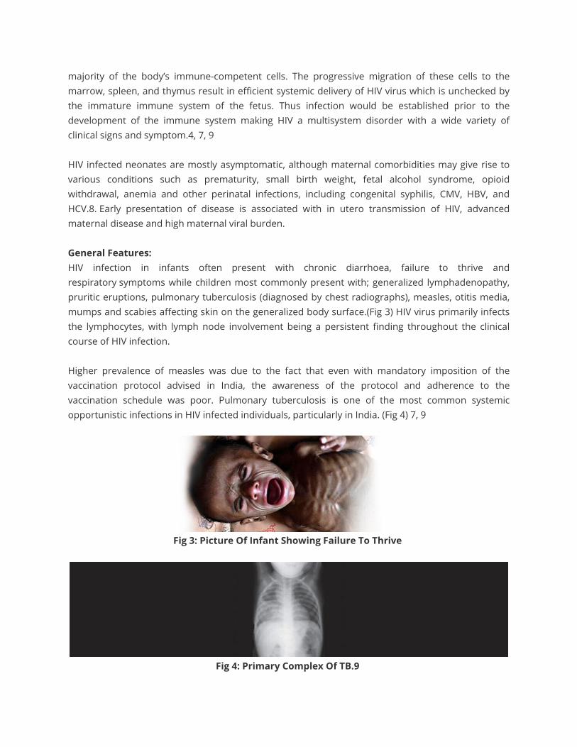
majority of the body’s immune-competent cells. The progressive migration of these cells to the marrow, spleen, and thymus result in efficient systemic delivery of HIV virus which is unchecked by the immature immune system of the fetus. Thus infection would be established prior to the development of the immune system making HIV a multisystem disorder with a wide variety of clinical signs and symptom.4, 7, 9 HIV infected neonates are mostly asymptomatic, although maternal comorbidities may give rise to various conditions such as prematurity, small birth weight, fetal alcohol syndrome, opioid withdrawal, anemia and other perinatal infections, including congenital syphilis, CMV, HBV, and HCV.8. Early presentation of disease is associated with in utero transmission of HIV, advanced maternal disease and high maternal viral burden. General Features: HIV infection in infants often present with chronic diarrhoea, failure to thrive and respiratory symptoms while children most commonly present with; generalized lymphadenopathy, pruritic eruptions, pulmonary tuberculosis (diagnosed by chest radiographs), measles, otitis media, mumps and scabies affecting skin on the generalized body surface.(Fig 3) HIV virus primarily infects the lymphocytes, with lymph node involvement being a persistent finding throughout the clinical course of HIV infection. Higher prevalence of measles was due to the fact that even with mandatory imposition of the vaccination protocol advised in India, the awareness of the protocol and adherence to the vaccination schedule was poor. Pulmonary tuberculosis is one of the most common systemic opportunistic infections in HIV infected individuals, particularly in India. (Fig 4) 7, 9
Fig 3: Picture Of Infant Showing Failure To Thrive
Fig 4: Primary Complex Of TB.9
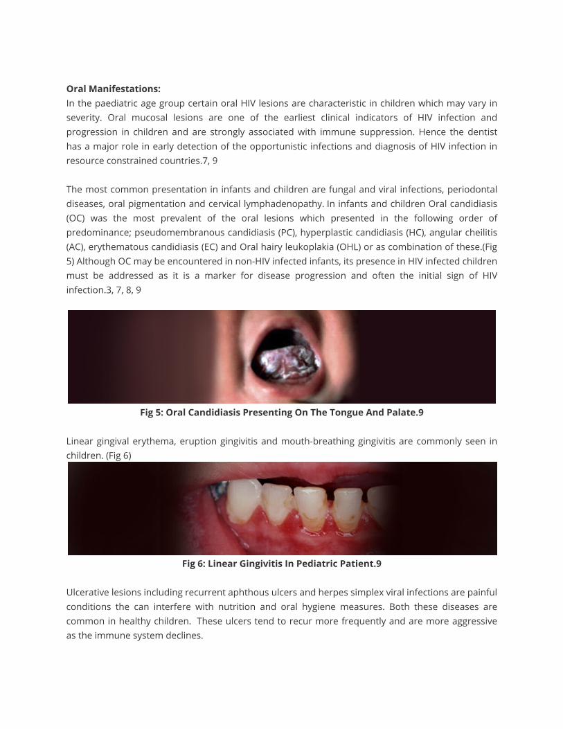
Oral Manifestations: In the paediatric age group certain oral HIV lesions are characteristic in children which may vary in severity. Oral mucosal lesions are one of the earliest clinical indicators of HIV infection and progression in children and are strongly associated with immune suppression. Hence the dentist has a major role in early detection of the opportunistic infections and diagnosis of HIV infection in resource constrained countries.7, 9 The most common presentation in infants and children are fungal and viral infections, periodontal diseases, oral pigmentation and cervical lymphadenopathy. In infants and children Oral candidiasis (OC) was the most prevalent of the oral lesions which presented in the following order of predominance; pseudomembranous candidiasis (PC), hyperplastic candidiasis (HC), angular cheilitis (AC), erythematous candidiasis (EC) and Oral hairy leukoplakia (OHL) or as combination of these.(Fig 5) Although OC may be encountered in non-HIV infected infants, its presence in HIV infected children must be addressed as it is a marker for disease progression and often the initial sign of HIV infection.3, 7, 8, 9
Fig 5: Oral Candidiasis Presenting On The Tongue And Palate.9
Linear gingival erythema, eruption gingivitis and mouth-breathing gingivitis are commonly seen in children. (Fig 6)
Fig 6: Linear Gingivitis In Pediatric Patient.9
Ulcerative lesions including recurrent aphthous ulcers and herpes simplex viral infections are painful conditions the can interfere with nutrition and oral hygiene measures. Both these diseases are common in healthy children. These ulcers tend to recur more frequently and are more aggressive as the immune system declines.

Oral pigmentation was also a common manifestation with pigmentation seen on the dorsal surface of tongue, hard palate and on the buccal mucosa. Anaemia and associated nutritional deficiency causes epithelial atrophy and predisposes to mucositis both of which lead to abnormal oral melanin pigmentation. In addition to anaemia, other causes of pigmentation are the release of a melanocyte-stimulating hormone caused by dysregulation of cytokines in HIV disease, Addison’s disease and drug induced (antiretroviral therapy). Both HIV-associated salivary gland disease and cervical lymphadenopathy, result in diffuse swellings of the face and neck. Concurrent xerostomia and pharyngeal tonsillar enlargement with subsequent mouth-breathing may increase the risk for plaque accumulation, dental caries and oral pseudomembranous candidiasis. Children with lymphproliferative disease have a better prognosis initially but with increased survival are at risk for lymphoma. 3, 7, 8, 9 Most viral infections are commonly associated with childhood infections but their presence in immunocompromised infant/children may result in more severe disease that is challenging to manage or may frequently recur. The occurrence of viral infections with multiple episodes of recurrence within the first year of life of an HIV infected infant are considered to be reflective of moderately symptomatic disease. In general, painful and persistent oral ulcerations may be associated with Herpes simplex, Varicella-zoster and Cytomegalovirus infections. Facial lesions, including the common wart, Molluscum contagiosum are also observed. Non-infective parotid gland enlargement in HIV infection is a common sign and arises due to infiltration of CD8 + cells that are cytotoxic to virally infected cells. (Fig 7)7, 8, 9
Fig 7: Parotid Gland Enlargement In Infant
DIAGNOSIS OF HIV INFECTION In resource-constrained settings where virological testing is not available, a presumptive diagnosis of severe HIV disease in infants < 18 months can be made using clinical criteria set by WHO. The presumptive diagnosis of severe HIV disease should be made if:
• The infant is confirmed HIV antibody positive and

• Diagnosis of any AIDS-indicator condition(s) can be made.
or
• The infant is symptomatic with two or more of the following:
1)Oral thrush 2)Severe pneumonia 3)Severe sepsis Other factors that support the diagnosis of severe HIV disease in an HIV seropositive infant include:
• Recent HIV-related maternal death; or advanced HIV disease in the mother. • CD4+ count < 20%
Confirmation of the diagnosis of HIV infection should be sought as soon as possible.6 Laboratory diagnosis of HIV infections in adults is made on the basis of two positive results of Enzyme Immunoassays which employ different antigens (ELISA, EIA, Rapid or Simple). The positive result of an enzyme assay test is confirmed by a Western Blot for antibodies to the virus. This also differentiates between HIV-1 and HIV-2 infection. The same rapid testing methods cannot be utilized in infants as mothers pass their antibodies to their babies while still in the womb, hence the standard assays cannot accurately diagnose infants until the age of 18 months, after which the mother's antibodies totally clear from the infant's blood and the infected child develops its own antibody to HIV. But this delay in diagnosis results in the deaths of many HIV-infected infants.b, c, d In infants and children the test used to diagnose babies born to HIV-infected mothers is Polymerase Chain Reaction (PCR), which directly detects HIV-1 pro-viral DNA integrated to human genome rather than the HIV antibody in the blood. Using PCR, infants can be tested using PCR as early as six weeks from birth. (Fig 8) 3, 8, 11, 12
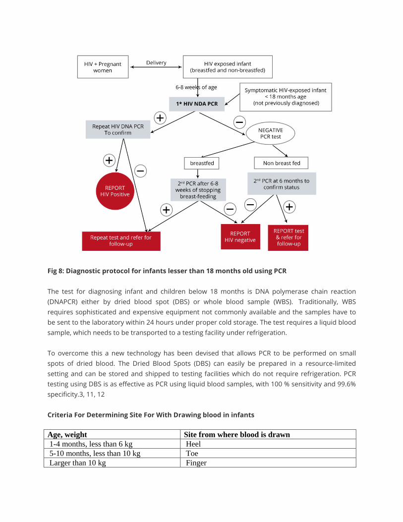
Fig 8: Diagnostic protocol for infants lesser than 18 months old using PCR The test for diagnosing infant and children below 18 months is DNA polymerase chain reaction (DNAPCR) either by dried blood spot (DBS) or whole blood sample (WBS). Traditionally, WBS requires sophisticated and expensive equipment not commonly available and the samples have to be sent to the laboratory within 24 hours under proper cold storage. The test requires a liquid blood sample, which needs to be transported to a testing facility under refrigeration. To overcome this a new technology has been devised that allows PCR to be performed on small spots of dried blood. The Dried Blood Spots (DBS) can easily be prepared in a resource-limited setting and can be stored and shipped to testing facilities which do not require refrigeration. PCR testing using DBS is as effective as PCR using liquid blood samples, with 100 % sensitivity and 99.6% specificity.3, 11, 12 Criteria For Determining Site For With Drawing blood in infants Age, weight Site from where blood is drawn 1-4 months, less than 6 kg Heel 5-10 months, less than 10 kg Toe Larger than 10 kg Finger

The site is first disinfected and blood is drawn using a sterile lancet. The first drop of blood should be wiped away with gauze or cotton wool. Then allow a large drop of blood to collect on the foot before touching it to the circle on the filter paper completely filling the circle. Samples should be stored horizontally out of direct sunlight for at least three hours. Once dry, samples are stored in sealable plastic bags with desiccant packets and a humidity card. Then they are transported to the laboratory. (Fig 9) 3, 11, 12
Fig 9: Drawing blood from heel of infant for preparing dried blood spot
In breast-fed infants, there is a need for repeat DNA-PCR testing at least 6 weeks after cessation of breastfeeding to confirm a HIV negative diagnosis. After 18 months of age a screening test (spot/ ELISA) must be done to confirm the serological status. Infants and children with confirmed HIV infection need a baseline CD4 testing (absolute count and CD4 %) and repeat every 6 months or earlier if clinically indicated. All children on ART should have regular clinical, immunological and virological monitoring for early recognition of treatment failure. The immune classification for monitoring is based on the absolute CD4+ lymphocyte count or the percentage of CD4+ cells. Infants have naturally high CD4 counts compared to adults hence age appropriate normal values must be considered when interpreting. Children under 5 years have less variability in CD4 percentages and therefore can be monitored for following progression (Table 1).The risk of progression is greatest in the first year of life and CD4 counts and viral loads are poorly predictive of progression of disease. At any given CD4 count, infants less than 12 months are more likely to progress to AIDS. 3, 9, 11, 12
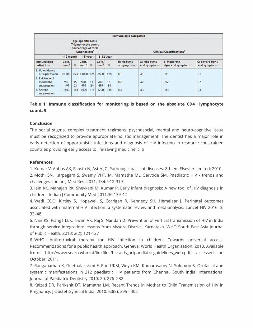
Table 1: Immune classification for monitoring is based on the absolute CD4+ lymphocyte count. 9 Conclusion The social stigma, complex treatment regimens, psychosocial, mental and neuro-cognitive issue must be recognized to provide appropriate holistic management. The dentist has a major role in early detection of opportunistic infections and diagnosis of HIV infection in resource constrained countries providing early access to life-saving medicine. c, b References 1. Kumar V, Abbas AK, Fausto N, Aster JC. Pathologic basis of diseases. 8th ed. Elsevier Limited; 2010. 2. Mothi SN, Karpagam S, Swamy VHT, M. Mamatha ML, Sarvode SM. Paediatric HIV - trends and challenges. Indian J Med Res. 2011; 134: 912-919 3. Jain KK, Mahajan RK, Shevkani M, Kumar P. Early infant diagnosis: A new tool of HIV diagnosis in children. Indian J Community Med 2011;36:139-42 4. Wedi COO, Kirtley S, Hopewell S, Corrigan R, Kennedy SH, Hemelaar J. Perinatal outcomes associated with maternal HIV infection: a systematic review and meta-analysis. Lancet HIV 2016: 3; 33–48 5. Nair KS, Piang1 LLK, Tiwari VK, Raj S, Nandan D. Prevention of vertical transmission of HIV in India through service integration: lessons from Mysore District, Karnataka. WHO South-East Asia Journal of Public Health. 2013: 2(2); 121-127 6. WHO. Antiretroviral therapy for HIV infection in children: Towards universal access. Recommendations for a public health approach. Geneva: World Health Organisation. 2010. Available from: http://www.searo.who.int/linkfiles/hiv-aids_artpaediatricguidelines_web.pdf, accessed on October. 2011. 7. Ranganathan K, Geethalakshmi E, Rao UKM, Vidya KM, Kumarasamy N, Solomon S. Orofacial and systemic manifestations in 212 paediatric HIV patients from Chennai, South India. International Journal of Paediatric Dentistry 2010; 20: 276–282 8. Kaizad DR, Parikshit DT, Mamatha LM. Recent Trends in Mother to Child Transmission of HIV in Pregnancy. J Obstet Gynecol India. 2010: 60(5); 395 - 402

9. Lala MM. Principles of perinatal and pediatric HIV/AIDS. 1st ed. Jaypee publications; 2012. 10. John GC, Kreiss J. Mother-to-Child Transmission of Human Immunodeficiency Virus Type 1. Epidemol rev. 1996: 18(2); 149-157 11. Sherman, Gayle G MD, Dried Blood Spots Improve Access to HIV Diagnosis and Care for Infants in Low-Resource Settings. JAIDS. 2005; 38(5):615-617 12. Li W, Tse FLS. Dried blood spot sampling in combination with LC-MS/MS for quantitative analysis of small molecules. Biomed. Chromatogr. 2010; 24: 49–65