Peri-implant augmentation
23
Scientific Background, Experts’ Treatment Reports and Cases using Alloplastic Biomaterials Peri-implant Bone Regeneration in Immediate and Immediate- Delayed Implantation Leventis M, Scarsdale Dental Aesthetic and Implant Clinic, 67 Earls Court Road- Kensington, London, United Kingdom Caiazzo A, Centro Odontoiatrico Salernitano,Via Enrico Bottiglieri 13, Salerno, Italy Fairbairn P, Scarsdale Dental Aesthetic and Implant Clinic, 67 Earls Court Road- Kensington, London, United Kingdom Flichy Fernández A, Universidad de Valencia, C/Gasco Oliag n1,Valencia, Spain Hollay HC, Zahnarztpraxis Dres. Hollay, Albert-Rosshaupterstr. 94, Munich, Germany Horowitz R A, 2 Overhill Rd., Suite 270, Scarsdale, NY, USA Kakar A, Global Health Research Group, H-8 Masjid Moth, GK-2 New Delhi, India Kirste M, Praxis für Zahn-, Mund- und Kieferheilkunde,Tunnelstr. 18, Frankfurt (Oder), Germany Schug J, Swiss Dental Center, Heinrichstr. 239, Zurich, Switzerland Troedhan A, Zentrum für Gesichtsästhetik, Braeuhausgasse 12–14,Vienna, Austria
Transcript of Peri-implant augmentation
Scientific Background, Experts’ Treatment Reports and Cases using Alloplastic Biomaterials
Peri-implant Bone Regeneration in Immediate and Immediate- Delayed Implantation
Leventis M, Scarsdale Dental Aesthetic and Implant Clinic, 67 Earls Court Road- Kensington, London, United Kingdom
Caiazzo A, Centro Odontoiatrico Salernitano, Via Enrico Bottiglieri 13, Salerno, Italy
Fairbairn P, Scarsdale Dental Aesthetic and Implant Clinic, 67 Earls Court Road- Kensington, London, United Kingdom
Flichy Fernández A, Universidad de Valencia, C/Gasco Oliag n1, Valencia, Spain
Hollay HC, Zahnarztpraxis Dres. Hollay, Albert-Rosshaupterstr. 94, Munich, Germany
Horowitz R A, 2 Overhill Rd., Suite 270, Scarsdale, NY, USA
Kakar A, Global Health Research Group, H-8 Masjid Moth, GK-2 New Delhi, India
Kirste M, Praxis für Zahn-, Mund- und Kieferheilkunde, Tunnelstr. 18, Frankfurt (Oder), Germany
Schug J, Swiss Dental Center, Heinrichstr. 239, Zurich, Switzerland
Troedhan A, Zentrum für Gesichtsästhetik, Braeuhausgasse 12–14, Vienna, Austria
2 3
CONTENT
Introduction ............................................................................................................................................4
Scope and Terminology ...................................................................................................................5
Basic Considerations for Implant Placement .....................................................................6
Aesthetic Risk Profile ........................................................................................................................7
“Experts” Consensus for immediate or immediate-delayed Implantation .....8
Materials ....................................................................................................................................................9
Treatment Pathways ............................................................................................................10 - 11
Immediate Implantation ....................................................................................................14 - 29
Immediate-delayed Implantation .................................................................................30 - 39
Disclaimer .................................................................................................................................................40
Cited References/Links ......................................................................................................41 - 43
ContentPeri-implant bone regeneration
Peri-implant bone regeneration in immediate and immediate-delayed Implantation using alloplastic biomaterials
Bone gaps, smaller bone dehiscence and fenestration defects are frequently encountered when placing implants in an extracted socket (Esposito et al. 2006).
Such defects can be treated before Implantation or at the same time using regenerative therapies to achieve optimal results in terms of both function and aesthetics.
Although the treatment depends on the individual clinical situation, outcomes may be optimized by following general principles and guidelines.
In order to discuss such guidelines, the authors of this Guidebook met in Baden, Switzerland, in January 2015 to develop a guideline for the use of alloplastic materials in the immediate or immediately delayed implant indication. The guideline was based on a review of scientific literature in addition to the authors’ clinical experience.
This Guidebook consolidates scientific evidence on regenerative procedures with the practical considerations after tooth extraction with a specific focus on application of alloplastic biomaterials predominantly in a minimally invasive approach.
The successful application of these treatment guidelines in the clinical practice are further illustrated with original clinical cases by the authors which demonstrate the use of GUIDOR® easy-graft® CLASSIC, GUIDOR easy-graft CRYSTAL and the GUIDOR bioresorbable matrix barrier in cases of immediate and immediate-delayed Implantation.
4 5
Scope and Terminology
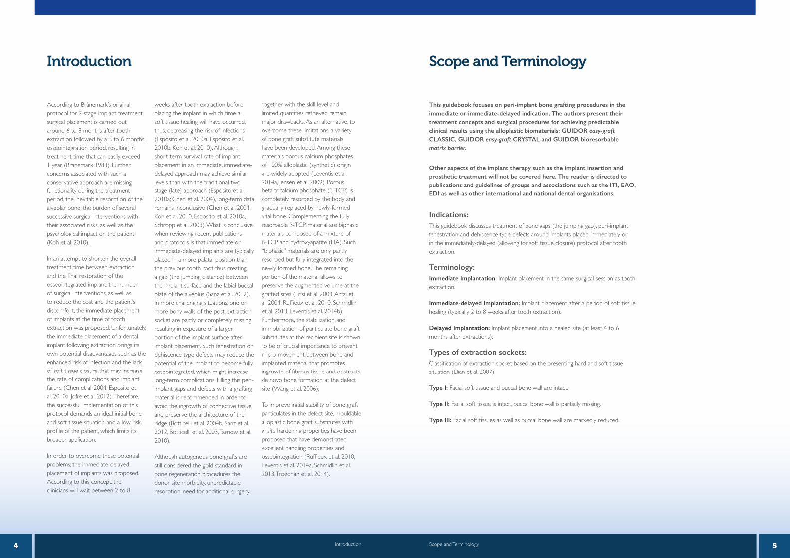
According to Brånemark’s original protocol for 2-stage implant treatment, surgical placement is carried out around 6 to 8 months after tooth extraction followed by a 3 to 6 months osseointegration period, resulting in treatment time that can easily exceed 1 year (Branemark 1983). Further concerns associated with such a conservative approach are missing functionality during the treatment period, the inevitable resorption of the alveolar bone, the burden of several successive surgical interventions with their associated risks, as well as the psychological impact on the patient (Koh et al. 2010).
In an attempt to shorten the overall treatment time between extraction and the final restoration of the osseointegrated implant, the number of surgical interventions, as well as to reduce the cost and the patient’s discomfort, the immediate placement of implants at the time of tooth extraction was proposed. Unfortunately, the immediate placement of a dental implant following extraction brings its own potential disadvantages such as the enhanced risk of infection and the lack of soft tissue closure that may increase the rate of complications and implant failure (Chen et al. 2004, Esposito et al. 2010a, Jofre et al. 2012). Therefore, the successful implementation of this protocol demands an ideal initial bone and soft tissue situation and a low risk profile of the patient, which limits its broader application.
In order to overcome these potential problems, the immediate-delayed placement of implants was proposed. According to this concept, the clinicians will wait between 2 to 8
weeks after tooth extraction before placing the implant in which time a soft tissue healing will have occurred, thus, decreasing the risk of infections (Esposito et al. 2010a; Esposito et al. 2010b, Koh et al. 2010). Although, short-term survival rate of implant placement in an immediate, immediate-delayed approach may achieve similar levels than with the traditional two stage (late) approach (Esposito et al. 2010a; Chen et al. 2004), long-term data remains inconclusive (Chen et al. 2004, Koh et al. 2010, Esposito et al. 2010a, Schropp et al. 2003). What is conclusive when reviewing recent publications and protocols is that immediate or immediate-delayed implants are typically placed in a more palatal position than the previous tooth root thus creating a gap (the jumping distance) between the implant surface and the labial buccal plate of the alveolus (Sanz et al. 2012). In more challenging situations, one or more bony walls of the post-extraction socket are partly or completely missing resulting in exposure of a larger portion of the implant surface after implant placement. Such fenestration or dehiscence type defects may reduce the potential of the implant to become fully osseointegrated, which might increase long-term complications. Filling this peri-implant gaps and defects with a grafting material is recommended in order to avoid the ingrowth of connective tissue and preserve the architecture of the ridge (Botticelli et al. 2004b, Sanz et al. 2012, Botticelli et al. 2003, Tarnow et al. 2010).
Although autogenous bone grafts are still considered the gold standard in bone regeneration procedures the donor site morbidity, unpredictable resorption, need for additional surgery
together with the skill level and limited quantities retrieved remain major drawbacks. As an alternative, to overcome these limitations, a variety of bone graft substitute materials have been developed. Among these materials porous calcium phosphates of 100% alloplastic (synthetic) origin are widely adopted (Leventis et al. 2014a, Jensen et al. 2009). Porous beta tricalcium phosphate (ß-TCP) is completely resorbed by the body and gradually replaced by newly-formed vital bone. Complementing the fully resorbable ß-TCP material are biphasic materials composed of a mixture of ß-TCP and hydroxyapatite (HA). Such “biphasic” materials are only partly resorbed but fully integrated into the newly formed bone. The remaining portion of the material allows to preserve the augmented volume at the grafted sites (Trisi et al. 2003, Artzi et al. 2004, Ruffieux et al. 2010, Schmidlin et al. 2013, Leventis et al. 2014b). Furthermore, the stabilization and immobilization of particulate bone graft substitutes at the recipient site is shown to be of crucial importance to prevent micro-movement between bone and implanted material that promotes ingrowth of fibrous tissue and obstructs de novo bone formation at the defect site (Wang et al. 2006).
To improve initial stability of bone graft particulates in the defect site, mouldable alloplastic bone graft substitutes with in situ hardening properties have been proposed that have demonstrated excellent handling properties and osseointegration (Ruffieux et al. 2010, Leventis et al. 2014a, Schmidlin et al. 2013, Troedhan et al. 2014).
Scope and TerminologyIntroduction
Introduction
This guidebook focuses on peri-implant bone grafting procedures in the immediate or immediate-delayed indication. The authors present their treatment concepts and surgical procedures for achieving predictable clinical results using the alloplastic biomaterials: GUIDOR easy-graft CLASSIC, GUIDOR easy-graft CRYSTAL and GUIDOR bioresorbable matrix barrier.
Other aspects of the implant therapy such as the implant insertion and prosthetic treatment will not be covered here. The reader is directed to publications and guidelines of groups and associations such as the ITI, EAO, EDI as well as other international and national dental organisations.
Indications:This guidebook discusses treatment of bone gaps (the jumping gap), peri-implant fenestration and dehiscence type defects around implants placed immediately or in the immediately-delayed (allowing for soft tissue closure) protocol after tooth extraction.
Terminology:Immediate Implantation: Implant placement in the same surgical session as tooth extraction.
Immediate-delayed Implantation: Implant placement after a period of soft tissue healing (typically 2 to 8 weeks after tooth extraction).
Delayed Implantation: Implant placement into a healed site (at least 4 to 6 months after extractions).
Types of extraction sockets:Classification of extraction socket based on the presenting hard and soft tissue situation (Elian et al. 2007).
Type I: Facial soft tissue and buccal bone wall are intact.
Type II: Facial soft tissue is intact, buccal bone wall is partially missing.
Type III: Facial soft tissues as well as buccal bone wall are markedly reduced.
6 7
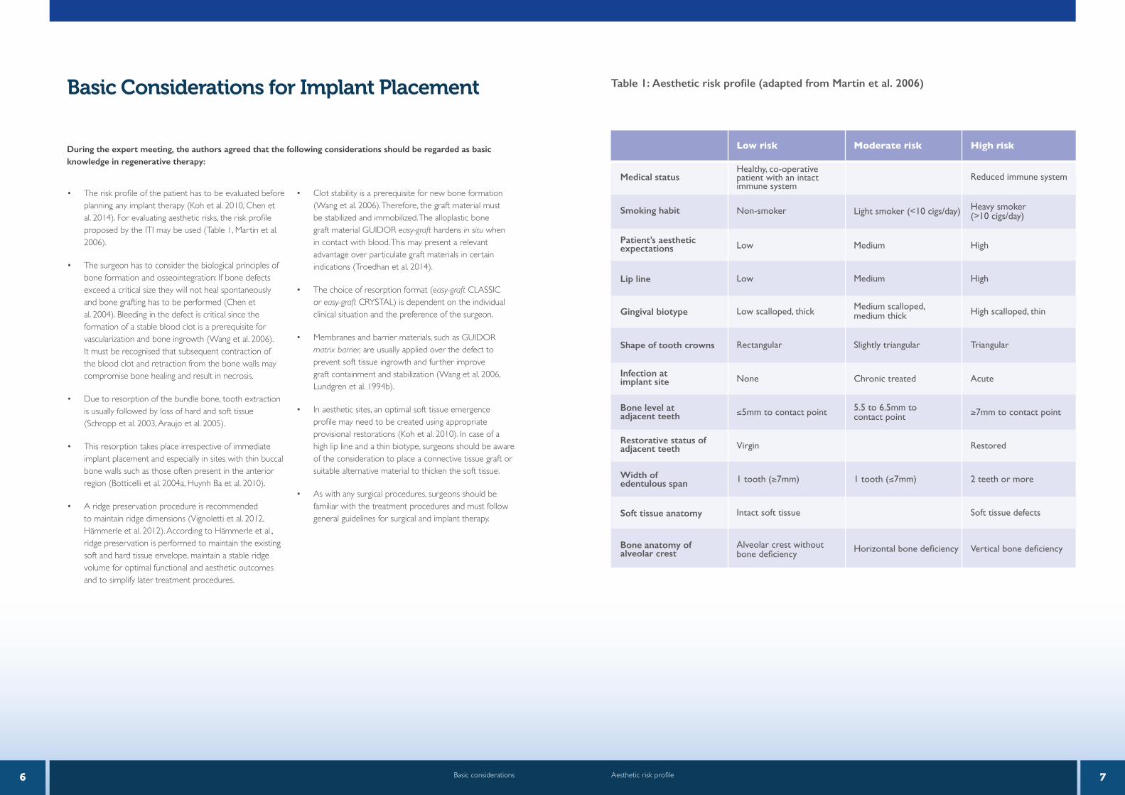
Table 1: Aesthetic risk profile (adapted from Martin et al. 2006)
• The risk profile of the patient has to be evaluated before planning any implant therapy (Koh et al. 2010, Chen et al. 2014). For evaluating aesthetic risks, the risk profile proposed by the ITI may be used (Table 1, Martin et al. 2006).
• The surgeon has to consider the biological principles of bone formation and osseointegration: If bone defects exceed a critical size they will not heal spontaneously and bone grafting has to be performed (Chen et al. 2004). Bleeding in the defect is critical since the formation of a stable blood clot is a prerequisite for vascularization and bone ingrowth (Wang et al. 2006). It must be recognised that subsequent contraction of the blood clot and retraction from the bone walls may compromise bone healing and result in necrosis.
• Due to resorption of the bundle bone, tooth extraction is usually followed by loss of hard and soft tissue (Schropp et al. 2003, Araujo et al. 2005).
• This resorption takes place irrespective of immediate implant placement and especially in sites with thin buccal bone walls such as those often present in the anterior region (Botticelli et al. 2004a, Huynh Ba et al. 2010).
• A ridge preservation procedure is recommended to maintain ridge dimensions (Vignoletti et al. 2012, Hämmerle et al. 2012). According to Hämmerle et al., ridge preservation is performed to maintain the existing soft and hard tissue envelope, maintain a stable ridge volume for optimal functional and aesthetic outcomes and to simplify later treatment procedures.
• Clot stability is a prerequisite for new bone formation (Wang et al. 2006). Therefore, the graft material must be stabilized and immobilized. The alloplastic bone graft material GUIDOR easy-graft hardens in situ when in contact with blood. This may present a relevant advantage over particulate graft materials in certain indications (Troedhan et al. 2014).
• The choice of resorption format (easy-graft CLASSIC or easy-graft CRYSTAL) is dependent on the individual clinical situation and the preference of the surgeon.
• Membranes and barrier materials, such as GUIDOR matrix barrier, are usually applied over the defect to prevent soft tissue ingrowth and further improve graft containment and stabilization (Wang et al. 2006, Lundgren et al. 1994b).
• In aesthetic sites, an optimal soft tissue emergence profile may need to be created using appropriate provisional restorations (Koh et al. 2010). In case of a high lip line and a thin biotype, surgeons should be aware of the consideration to place a connective tissue graft or suitable alternative material to thicken the soft tissue.
• As with any surgical procedures, surgeons should be familiar with the treatment procedures and must follow general guidelines for surgical and implant therapy.
Aesthetic risk profileBasic considerations
Basic Considerations for Implant Placement
During the expert meeting, the authors agreed that the following considerations should be regarded as basic knowledge in regenerative therapy:
Medical status
Moderate risk High risk
Smoking habit
Patient’s aesthetic expectations
Lip line
Gingival biotype
Shape of tooth crowns
Infection at implant site
Bone level at adjacent teeth
Restorative status of adjacent teeth
Width of edentulous span
Soft tissue anatomy
Bone anatomy of alveolar crest
Healthy, co-operative patient with an intact immune system
Low risk
Non-smoker
Low
Low
Low scalloped, thick
Rectangular
None
≤5mm to contact point
Virgin
1 tooth (≥7mm)
Intact soft tissue
Alveolar crest without bone deficiency
Light smoker (<10 cigs/day)
Medium
Medium
Medium scalloped,medium thick
Slightly triangular
Chronic treated
5.5 to 6.5mm to contact point
1 tooth (≤7mm)
Horizontal bone deficiency
Reduced immune system
Heavy smoker (>10 cigs/day)
High
High
High scalloped, thin
Triangular
Acute
≥7mm to contact point
Restored
2 teeth or more
Soft tissue defects
Vertical bone deficiency
8 9
• Tooth extraction has to be performed using an atraumatic method such as that described in the Sunstar GUIDOR “Ridge Preservation Protocol” Guidebook (Schug et al. 2013).
• Risk factors have to be considered for achieving an optimal outcome. These include systemic medical conditions, smoking, alcoholism, oral hygiene and soft tissue profile (biotype).
• The presence of acute infection has to be excluded in case of immediate implant placement.
• Implants can be placed either immediately in an extraction socket, immediate-delayed (after soft tissue closure) or in a healed site.
• Cone Beam Computed Tomography (CBCT) is advised prior to implant placement.
• Implants are typically placed in an optimal 3-dimensional position (Buser et al. 2004) in accordance with the existing consensus such as that from EDI (Nickenig et al. 2014).
• In case of a bony “jumping” gap, fenestration or dehiscence defects around the implant, placement of a bone graft or substitute is advised.
MaterialsExpert agreement and consensus
“Experts” Consensus for immediate or immediate-delayed Implantation During the expert meeting the authors agreed on the following statements:
Materials
GUIDOR easy-graft alloplastic bone graft substituteGUIDOR easy-graft is an alloplastic bone augmentation material that is applied directly from the syringe into the defect. The mouldable adhesive granules can be shaped in the defect. When in contact with blood, the material hardens within minutes to form a porous defect analogue. GUIDOR easy-graft products are 100% alloplastic and do not contain substances of animal or human origin.GUIDOR easy-graft is available in two formats: easy-graft CLASSIC and easy-graft CRYSTAL:
• easy-graft CLASSIC consists of coated, phase pure ß-tricalcium phosphate (ß-TCP). During healing easy-graft CLASSIC is completely resorbed and replaced by newly formed bone within a time period of 5–15 months. No material remains.
• easy-graft CRYSTAL consists of coated biphasic calcium phosphate (compound of 60% hydroxyapatite and 40% ß-TCP). The ß-TCP component is completely resorbed and replaced by newly formed bone whilst the hydroxyapatite portion remains integrated in the newly formed bone providing long-term volume stability.
GUIDOR bioresorbable matrix barrier
GUIDOR matrix barrier is the first and most widely studied alloplastic matrix and barrier technology.
Composed of a resorbable alloplastic polymer GUIDOR matrix barrier is presented in a unique multi-layered design which stabilizes the wound site, aids in early integration of gingival connective tissues and effectively impedes epithelial down-growth. Laser cut pores on the upper and lower layers allow fluid exchange whilst varying pore diameters allow selective tissue integration contributing to a favourable soft-tissue response and outstanding clinical results (Rosen 2010).
GUIDOR matrix barrier is easy to handle, adapts well to varying defect shapes and it provides a barrier effect for a minimum of 6 weeks (Lundgren et al. 1994a) after which the material degrades to water and carbon dioxide.
• GUIDOR matrix barrier is available in a selection of configurations as appropriate to the guided bone (GBR) and guided tissue (GTR) indications. For GTR an additional thick sealing collar and integrated suture ensure “true cell occlusion” (Fugazzotto 2011).
10 11Treatment pathwaysTreatment pathways
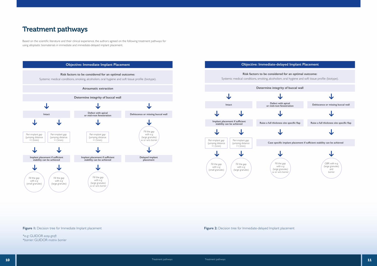
Figure 2: Decision tree for Immediate-delayed Implant placement
Objective: Immediate-delayed Implant Placement
Risk factors to be considered for an optimal outcome: Systemic medical conditions, smoking, alcoholism, oral hygiene and soft tissue profile (biotype).
Determine integrity of buccal wall
Intact
Peri-implant gap (jumping distance
<1.5mm)
Peri-implant gap (jumping distance
>1.5mm)
Fill the gapwith e-g
(small granules)
Fill the gapwith e-g
(large granules)
Defect with apical or mid-root fenestration
Raise a full thickness site specific flap
Fill the gapwith e-g
(large granules)w or w/o barrier
Dehiscence or missing buccal wall
Raise a full thickness site specific flapImplant placement if sufficient
stability can be achieved
Case specific implant placement if sufficient stability can be achieved
GBR with e-g(large granules)
and barrier
Objective: Immediate Implant Placement
Risk factors to be considered for an optimal outcome: Systemic medical conditions, smoking, alcoholism, oral hygiene and soft tissue profile (biotype).
Atraumatic extraction
Determine integrity of buccal wall
Intact
Peri-implant gap (jumping distance
<1.5mm)
Peri-implant gap (jumping distance
>1.5mm)
Implant placement if sufficient stability can be achieved
Fill the gapwith e-g
(small granules)
Fill the gapwith e-g
(large granules)
Defect with apical or mid-root fenestration
Peri-implant gap (jumping distance
>1.5mm)
Implant placement if sufficient stability can be achieved
Fill the gapwith e-g
(large granules)w or w/o barrier
Dehiscence or missing buccal wall
Delayed implantplacement
Fill the gapwith e-g
(large granules)w or w/o barrier
Based on the scientific literature and their clinical experience, the authors agreed on the following treatment pathways for using alloplastic biomaterials in immediate and immediate-delayed implant placement.
Figure 1: Decision tree for Immediate Implant placement
*e-g : GUIDOR easy-graft *barrier : GUIDOR matrix barrier
Treatment pathways
12 13
Case Reports
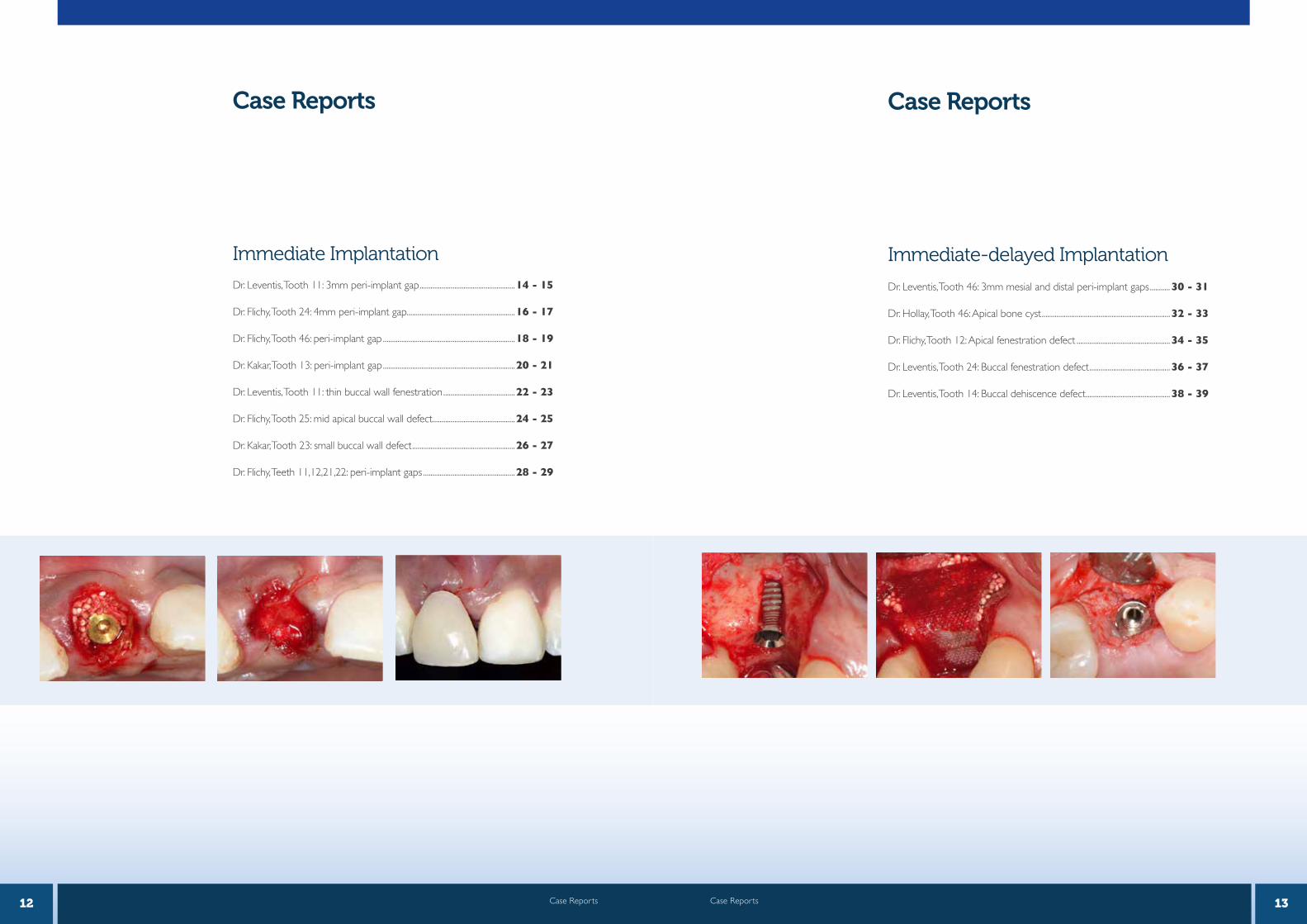
Dr. Leventis, Tooth 46: 3mm mesial and distal peri-implant gaps ...........30 - 31
Dr. Hollay, Tooth 46: Apical bone cyst ......................................................................32 - 33
Dr. Flichy, Tooth 12: Apical fenestration defect ...................................................34 - 35
Dr. Leventis, Tooth 24: Buccal fenestration defect ............................................36 - 37
Dr. Leventis, Tooth 14: Buccal dehiscence defect ..............................................38 - 39
Immediate-delayed Implantation
Case ReportsCase Reports
Case Reports
Dr. Leventis, Tooth 11: 3mm peri-implant gap ....................................................14 - 15
Dr. Flichy, Tooth 24: 4mm peri-implant gap...........................................................16 - 17
Dr. Flichy, Tooth 46: peri-implant gap ........................................................................18 - 19
Dr. Kakar, Tooth 13: peri-implant gap ........................................................................20 - 21
Dr. Leventis, Tooth 11: thin buccal wall fenestration .......................................22 - 23
Dr. Flichy, Tooth 25: mid apical buccal wall defect .............................................24 - 25
Dr. Kakar, Tooth 23: small buccal wall defect ........................................................26 - 27
Dr. Flichy, Teeth 11,12,21,22: peri-implant gaps ..................................................28 - 29
Immediate Implantation
14 15
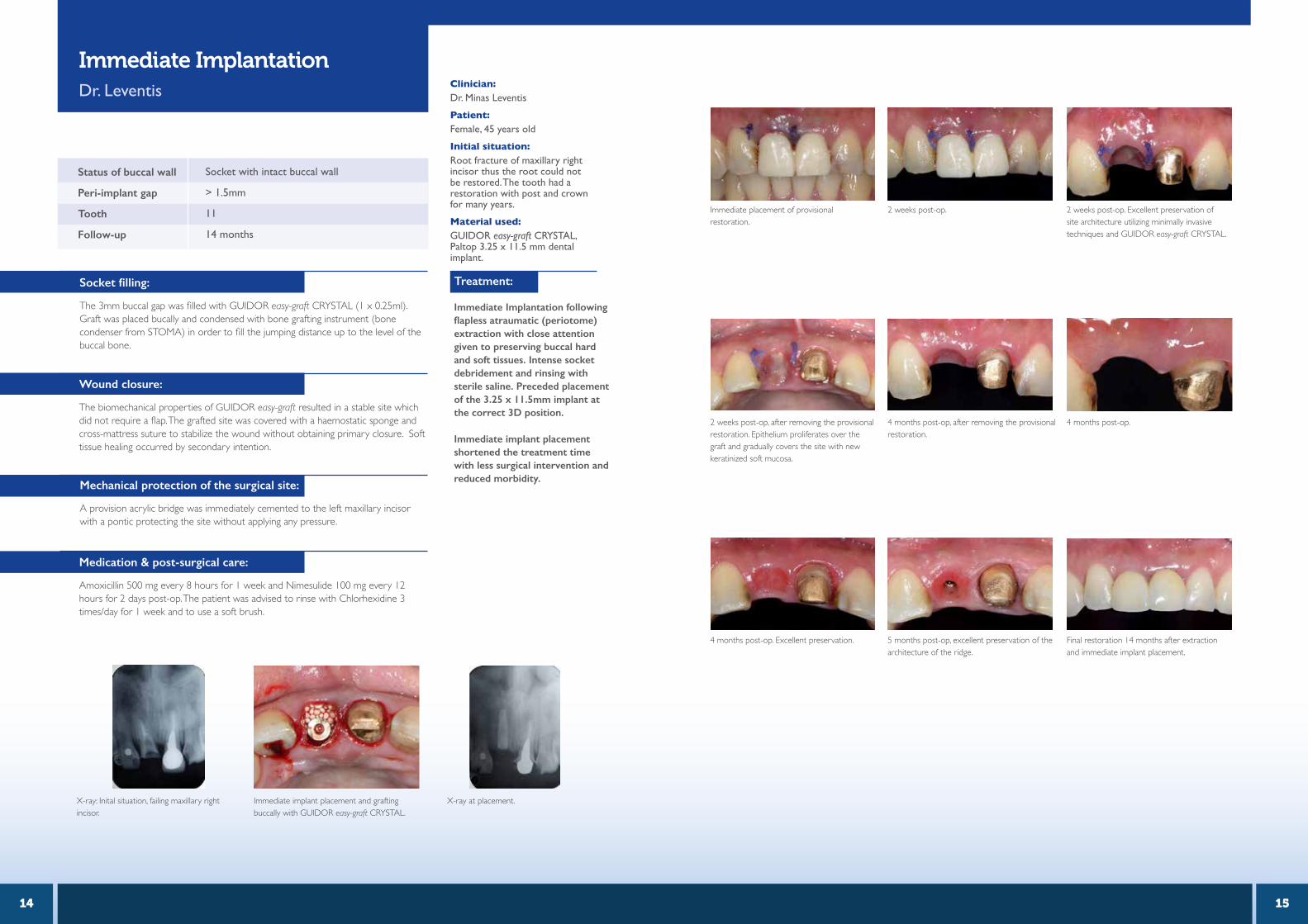
Immediate ImplantationDr. Leventis
Treatment:
Immediate Implantation following flapless atraumatic (periotome) extraction with close attention given to preserving buccal hard and soft tissues. Intense socket debridement and rinsing with sterile saline. Preceded placement of the 3.25 x 11.5mm implant at the correct 3D position. Immediate implant placement shortened the treatment time with less surgical intervention and reduced morbidity.
Clinician:Dr. Minas Leventis
Patient:Female, 45 years old
Initial situation:Root fracture of maxillary right incisor thus the root could not be restored. The tooth had a restoration with post and crown for many years.
Material used:GUIDOR easy-graft CRYSTAL, Paltop 3.25 x 11.5 mm dental implant.
Socket filling:
The 3mm buccal gap was filled with GUIDOR easy-graft CRYSTAL (1 x 0.25ml). Graft was placed bucally and condensed with bone grafting instrument (bone condenser from STOMA) in order to fill the jumping distance up to the level of the buccal bone.
Wound closure:
The biomechanical properties of GUIDOR easy-graft resulted in a stable site which did not require a flap. The grafted site was covered with a haemostatic sponge and cross-mattress suture to stabilize the wound without obtaining primary closure. Soft tissue healing occurred by secondary intention.
Mechanical protection of the surgical site:
A provision acrylic bridge was immediately cemented to the left maxillary incisor with a pontic protecting the site without applying any pressure.
Medication & post-surgical care:
Amoxicillin 500 mg every 8 hours for 1 week and Nimesulide 100 mg every 12 hours for 2 days post-op. The patient was advised to rinse with Chlorhexidine 3 times/day for 1 week and to use a soft brush.
Status of buccal wall
Peri-implant gap
Tooth
Follow-up
Socket with intact buccal wall
> 1.5mm
11
14 months
Immediate placement of provisional restoration.
2 weeks post-op, after removing the provisional restoration. Epithelium proliferates over the graft and gradually covers the site with new keratinized soft mucosa.
4 months post-op. Excellent preservation.
2 weeks post-op.
4 months post-op, after removing the provisional restoration.
5 months post-op, excellent preservation of the architecture of the ridge.
2 weeks post-op. Excellent preservation of site architecture utilizing minimally invasive techniques and GUIDOR easy-graft CRYSTAL.
4 months post-op.
Final restoration 14 months after extraction and immediate implant placement.
X-ray: Inital situation, failing maxillary right incisor.
Immediate implant placement and grafting buccally with GUIDOR easy-graft CRYSTAL.
X-ray at placement.
16 17
Immediate ImplantationDr. Flichy
Treatment:
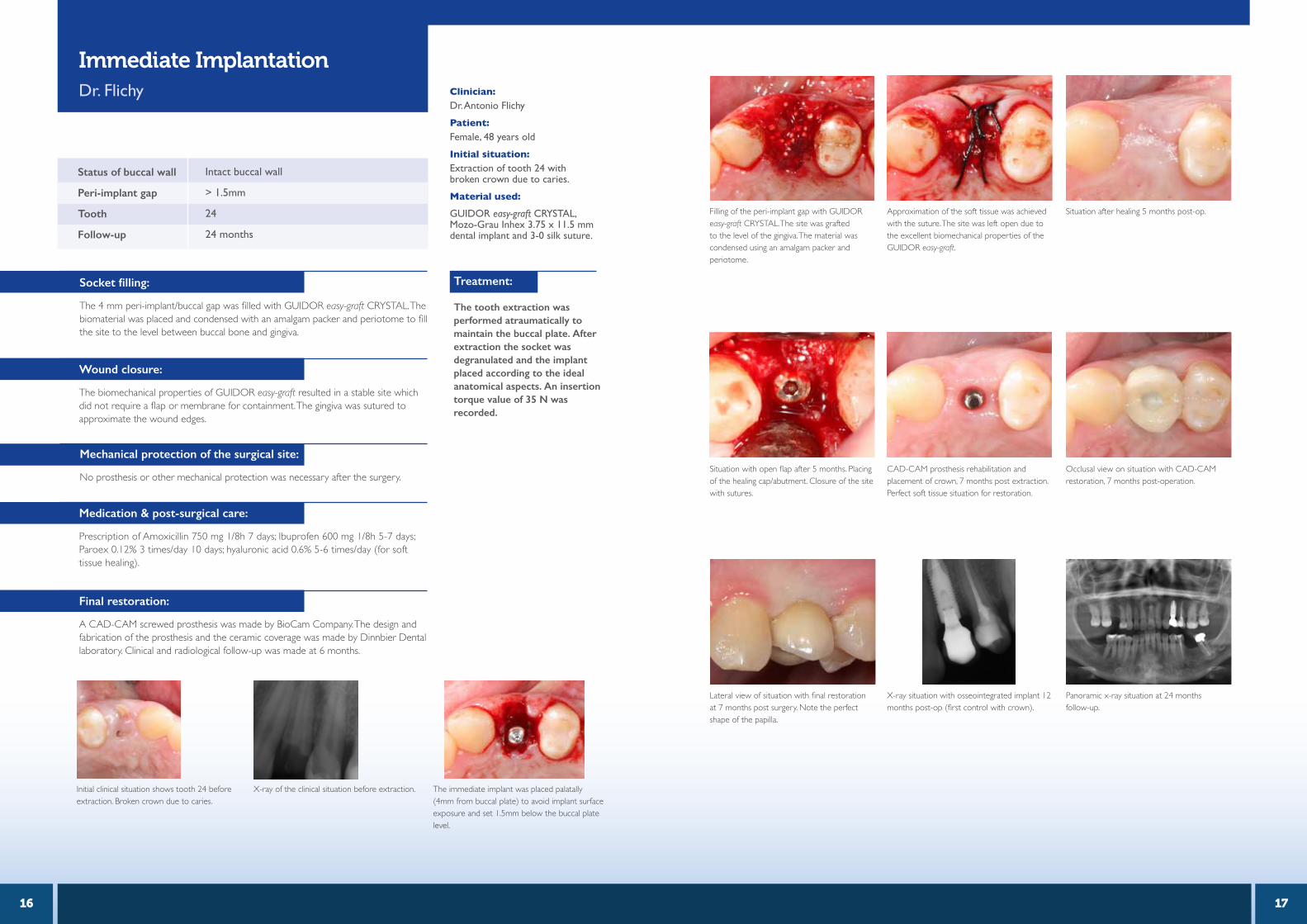
The tooth extraction was performed atraumatically to maintain the buccal plate. After extraction the socket was degranulated and the implant placed according to the ideal anatomical aspects. An insertion torque value of 35 N was recorded.
Clinician:Dr. Antonio Flichy
Patient:Female, 48 years old
Initial situation:Extraction of tooth 24 with broken crown due to caries.
Material used:
GUIDOR easy-graft CRYSTAL, Mozo-Grau Inhex 3.75 x 11.5 mm dental implant and 3-0 silk suture.
Socket filling:
The 4 mm peri-implant/buccal gap was filled with GUIDOR easy-graft CRYSTAL. The biomaterial was placed and condensed with an amalgam packer and periotome to fill the site to the level between buccal bone and gingiva.
Wound closure:
The biomechanical properties of GUIDOR easy-graft resulted in a stable site which did not require a flap or membrane for containment. The gingiva was sutured to approximate the wound edges.
Mechanical protection of the surgical site:
No prosthesis or other mechanical protection was necessary after the surgery.
Medication & post-surgical care:
Prescription of Amoxicillin 750 mg 1/8h 7 days; Ibuprofen 600 mg 1/8h 5-7 days; Paroex 0.12% 3 times/day 10 days; hyaluronic acid 0.6% 5-6 times/day (for soft tissue healing).
Status of buccal wall
Peri-implant gap
Tooth
Follow-up
Intact buccal wall
> 1.5mm
24
24 months
Filling of the peri-implant gap with GUIDOR easy-graft CRYSTAL. The site was grafted to the level of the gingiva. The material was condensed using an amalgam packer and periotome.
Situation with open flap after 5 months. Placing of the healing cap/abutment. Closure of the site with sutures.
Lateral view of situation with final restoration at 7 months post surgery. Note the perfect shape of the papilla.
Approximation of the soft tissue was achieved with the suture. The site was left open due to the excellent biomechanical properties of the GUIDOR easy-graft.
CAD-CAM prosthesis rehabilitation and placement of crown, 7 months post extraction. Perfect soft tissue situation for restoration.
X-ray situation with osseointegrated implant 12 months post-op (first control with crown).
Situation after healing 5 months post-op.
Occlusal view on situation with CAD-CAM restoration, 7 months post-operation.
Panoramic x-ray situation at 24 months follow-up.
Initial clinical situation shows tooth 24 before extraction. Broken crown due to caries.
X-ray of the clinical situation before extraction. The immediate implant was placed palatally (4mm from buccal plate) to avoid implant surface exposure and set 1.5mm below the buccal plate level.
Final restoration:
A CAD-CAM screwed prosthesis was made by BioCam Company. The design and fabrication of the prosthesis and the ceramic coverage was made by Dinnbier Dental laboratory. Clinical and radiological follow-up was made at 6 months.
18 19
Immediate ImplantationDr. Flichy
Treatment:
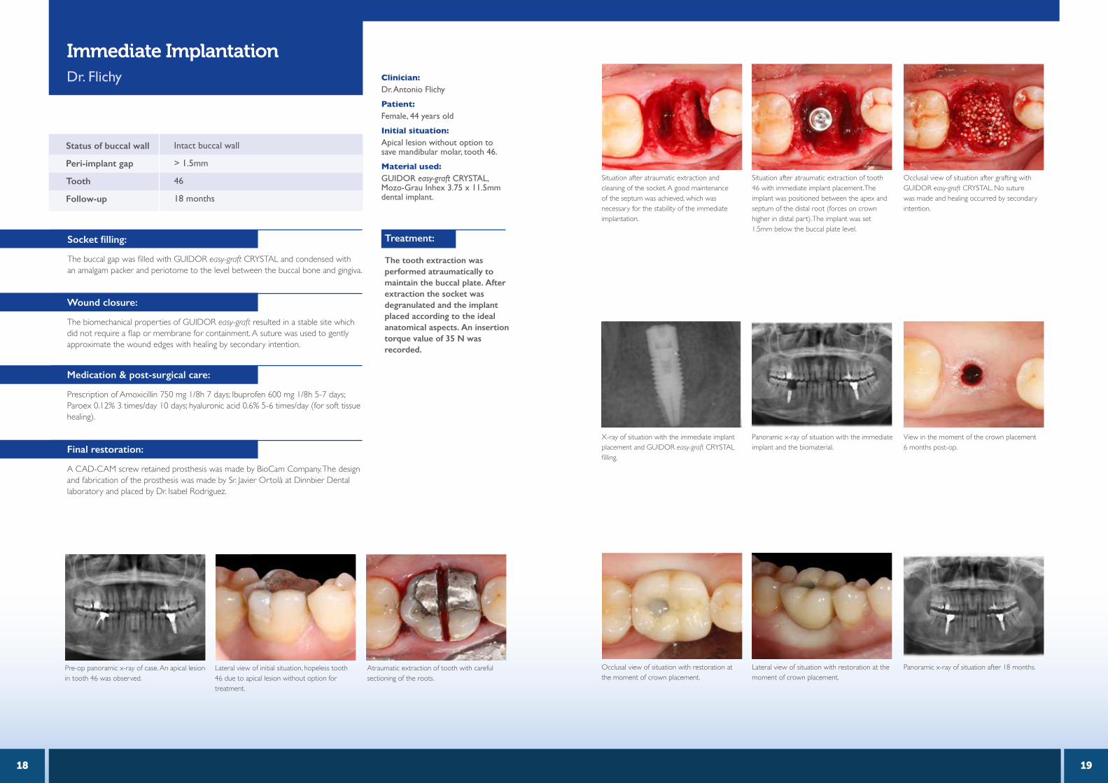
The tooth extraction was performed atraumatically to maintain the buccal plate. After extraction the socket was degranulated and the implant placed according to the ideal anatomical aspects. An insertion torque value of 35 N was recorded.
Clinician:Dr. Antonio Flichy
Patient:Female, 44 years old
Initial situation: Apical lesion without option to save mandibular molar, tooth 46.
Material used:GUIDOR easy-graft CRYSTAL, Mozo-Grau Inhex 3.75 x 11.5mm dental implant.
Socket filling:
The buccal gap was filled with GUIDOR easy-graft CRYSTAL and condensed with an amalgam packer and periotome to the level between the buccal bone and gingiva.
Wound closure:
The biomechanical properties of GUIDOR easy-graft resulted in a stable site which did not require a flap or membrane for containment. A suture was used to gently approximate the wound edges with healing by secondary intention.
Medication & post-surgical care:
Prescription of Amoxicillin 750 mg 1/8h 7 days; Ibuprofen 600 mg 1/8h 5-7 days; Paroex 0.12% 3 times/day 10 days; hyaluronic acid 0.6% 5-6 times/day (for soft tissue healing).
Status of buccal wall
Peri-implant gap
Tooth
Follow-up
Intact buccal wall
> 1.5mm
46
18 months
Situation after atraumatic extraction and cleaning of the socket. A good maintenance of the septum was achieved, which was necessary for the stability of the immediate implantation.
X-ray of situation with the immediate implant placement and GUIDOR easy-graft CRYSTAL filling.
Occlusal view of situation with restoration at the moment of crown placement.
Situation after atraumatic extraction of tooth 46 with immediate implant placement. The implant was positioned between the apex and septum of the distal root (forces on crown higher in distal part). The implant was set 1.5mm below the buccal plate level.
Panoramic x-ray of situation with the immediate implant and the biomaterial.
Lateral view of situation with restoration at the moment of crown placement.
Occlusal view of situation after grafting with GUIDOR easy-graft CRYSTAL. No suture was made and healing occurred by secondary intention.
View in the moment of the crown placement 6 months post-op.
Panoramic x-ray of situation after 18 months.Pre-op panoramic x-ray of case. An apical lesion in tooth 46 was observed.
Lateral view of initial situation, hopeless tooth 46 due to apical lesion without option for treatment.
Atraumatic extraction of tooth with careful sectioning of the roots.
Final restoration:
A CAD-CAM screw retained prosthesis was made by BioCam Company. The design and fabrication of the prosthesis was made by Sr. Javier Ortolà at Dinnbier Dental laboratory and placed by Dr. Isabel Rodriguez.
20 21
Immediate ImplantationDr. Kakar
Treatment:
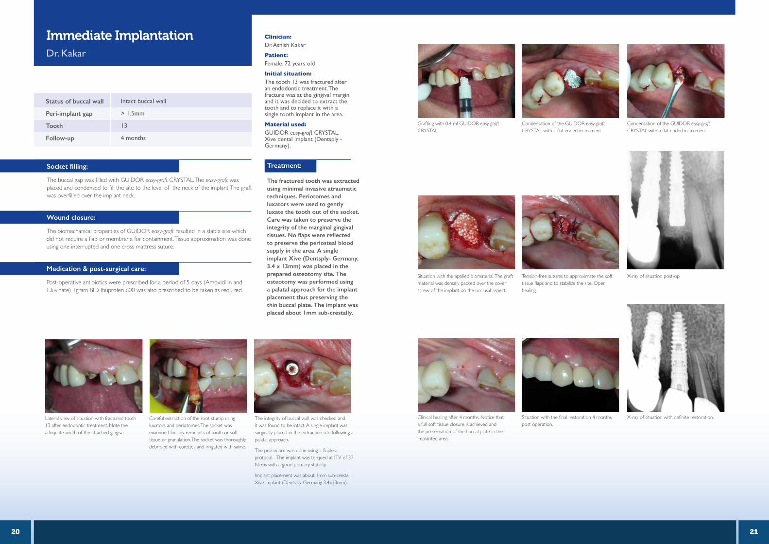
The fractured tooth was extracted using minimal invasive atraumatic techniques. Periotomes and luxators were used to gently luxate the tooth out of the socket. Care was taken to preserve the integrity of the marginal gingival tissues. No flaps were reflected to preserve the periosteal blood supply in the area. A single implant Xive (Dentsply- Germany, 3.4 x 13mm) was placed in the prepared osteotomy site. The osteotomy was performed using a palatal approach for the implant placement thus preserving the thin buccal plate. The implant was placed about 1mm sub-crestally.
Clinician:Dr. Ashish Kakar
Patient:Female, 72 years old
Initial situation: The tooth 13 was fractured after an endodontic treatment. The fracture was at the gingival margin and it was decided to extract the tooth and to replace it with a single tooth implant in the area.
Material used:GUIDOR easy-graft CRYSTAL, Xive dental implant (Dentsply - Germany).
Socket filling:
The buccal gap was filled with GUIDOR easy-graft CRYSTAL. The easy-graft was placed and condensed to fill the site to the level of the neck of the implant. The graft was overfilled over the implant neck.
Wound closure:
The biomechanical properties of GUIDOR easy-graft resulted in a stable site which did not require a flap or membrane for containment. Tissue approximation was done using one interrupted and one cross mattress suture.
Medication & post-surgical care:
Post-operative antibiotics were prescribed for a period of 5 days (Amoxicillin and Cluvinate) 1gram BID. Ibuprofen 600 was also prescribed to be taken as required.
Status of buccal wall
Peri-implant gap
Tooth
Follow-up
Intact buccal wall
> 1.5mm
13
4 months
Grafting with 0.4 ml GUIDOR easy-graft CRYSTAL.
Situation with the applied biomaterial. The graft material was densely packed over the cover screw of the implant on the occlusal aspect.
Clinical healing after 4 months. Notice that a full soft tissue closure is achieved and the preservation of the buccal plate in the implanted area.
Condensation of the GUIDOR easy-graft CRYSTAL with a flat ended instrument.
Tension-free sutures to approximate the soft tissue flaps and to stabilize the site. Open healing.
Situation with the final restoration 4 months post operation.
Condensation of the GUIDOR easy-graft CRYSTAL with a flat ended instrument.
X-ray of situation post-op.
X-ray of situation with definite restoration.Lateral view of situation with fractured tooth 13 after endodontic treatment. Note the adequate width of the attached gingiva.
Careful extraction of the root stump using luxators and periotomes. The socket was examined for any remnants of tooth or soft tissue or granulation. The socket was thoroughly debrided with curettes and irrigated with saline.
The integrity of buccal wall was checked and it was found to be intact. A single implant was surgically placed in the extraction site following a palatal approach.
The procedure was done using a flapless protocol. The implant was torqued at ITV of 37 Ncms with a good primary stability.
Implant placement was about 1mm sub-crestal, Xive Implant (Dentsply-Germany, 3.4x13mm).
22 23
Immediate ImplantationDr. Leventis
Treatment:
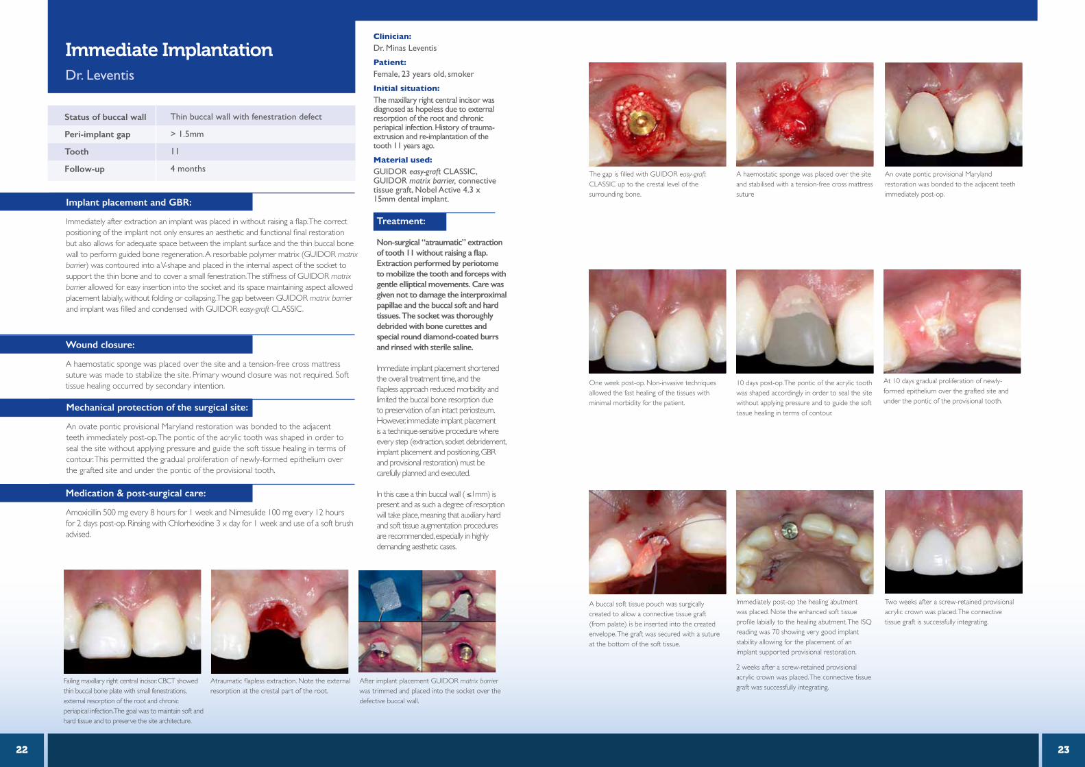
Non-surgical “atraumatic” extraction of tooth 11 without raising a flap. Extraction performed by periotome to mobilize the tooth and forceps with gentle elliptical movements. Care was given not to damage the interproximal papillae and the buccal soft and hard tissues. The socket was thoroughly debrided with bone curettes and special round diamond-coated burrs and rinsed with sterile saline. Immediate implant placement shortened the overall treatment time, and the flapless approach reduced morbidity and limited the buccal bone resorption due to preservation of an intact periosteum. However, immediate implant placement is a technique-sensitive procedure where every step (extraction, socket debridement, implant placement and positioning, GBR and provisional restoration) must be carefully planned and executed. In this case a thin buccal wall ( ≤1mm) is present and as such a degree of resorption will take place, meaning that auxiliary hard and soft tissue augmentation procedures are recommended, especially in highly demanding aesthetic cases.
Clinician:Dr. Minas Leventis
Patient:Female, 23 years old, smoker
Initial situation: The maxillary right central incisor was diagnosed as hopeless due to external resorption of the root and chronic periapical infection. History of trauma-extrusion and re-implantation of the tooth 11 years ago.
Material used:GUIDOR easy-graft CLASSIC, GUIDOR matrix barrier, connective tissue graft, Nobel Active 4.3 x 15mm dental implant.Implant placement and GBR:
Immediately after extraction an implant was placed in without raising a flap. The correct positioning of the implant not only ensures an aesthetic and functional final restoration but also allows for adequate space between the implant surface and the thin buccal bone wall to perform guided bone regeneration. A resorbable polymer matrix (GUIDOR matrix barrier) was contoured into a V-shape and placed in the internal aspect of the socket to support the thin bone and to cover a small fenestration. The stiffness of GUIDOR matrix barrier allowed for easy insertion into the socket and its space maintaining aspect allowed placement labially, without folding or collapsing. The gap between GUIDOR matrix barrier and implant was filled and condensed with GUIDOR easy-graft CLASSIC.
Status of buccal wall
Peri-implant gap
Tooth
Follow-up
Thin buccal wall with fenestration defect
> 1.5mm
11
4 months
Wound closure:
A haemostatic sponge was placed over the site and a tension-free cross mattress suture was made to stabilize the site. Primary wound closure was not required. Soft tissue healing occurred by secondary intention.
Mechanical protection of the surgical site:
An ovate pontic provisional Maryland restoration was bonded to the adjacent teeth immediately post-op. The pontic of the acrylic tooth was shaped in order to seal the site without applying pressure and guide the soft tissue healing in terms of contour. This permitted the gradual proliferation of newly-formed epithelium over the grafted site and under the pontic of the provisional tooth.
Medication & post-surgical care:
Amoxicillin 500 mg every 8 hours for 1 week and Nimesulide 100 mg every 12 hours for 2 days post-op. Rinsing with Chlorhexidine 3 x day for 1 week and use of a soft brush advised.
The gap is filled with GUIDOR easy-graft CLASSIC up to the crestal level of the surrounding bone.
10 days post-op. The pontic of the acrylic tooth was shaped accordingly in order to seal the site without applying pressure and to guide the soft tissue healing in terms of contour.
At 10 days gradual proliferation of newly-formed epithelium over the grafted site and under the pontic of the provisional tooth.
A haemostatic sponge was placed over the site and stabilised with a tension-free cross mattress suture
One week post-op. Non-invasive techniques allowed the fast healing of the tissues with minimal morbidity for the patient.
Immediately post-op the healing abutment was placed. Note the enhanced soft tissue profile labially to the healing abutment. The ISQ reading was 70 showing very good implant stability allowing for the placement of an implant supported provisional restoration.
2 weeks after a screw-retained provisional acrylic crown was placed. The connective tissue graft was successfully integrating.
An ovate pontic provisional Maryland restoration was bonded to the adjacent teeth immediately post-op.
A buccal soft tissue pouch was surgically created to allow a connective tissue graft (from palate) is be inserted into the created envelope. The graft was secured with a suture at the bottom of the soft tissue.
Two weeks after a screw-retained provisional acrylic crown was placed. The connective tissue graft is successfully integrating.
Failing maxillary right central incisor. CBCT showed thin buccal bone plate with small fenestrations, external resorption of the root and chronic periapical infection. The goal was to maintain soft and hard tissue and to preserve the site architecture.
Atraumatic flapless extraction. Note the external resorption at the crestal part of the root.
After implant placement GUIDOR matrix barrier was trimmed and placed into the socket over the defective buccal wall.
24 25
Immediate ImplantationDr. Flichy
Treatment:
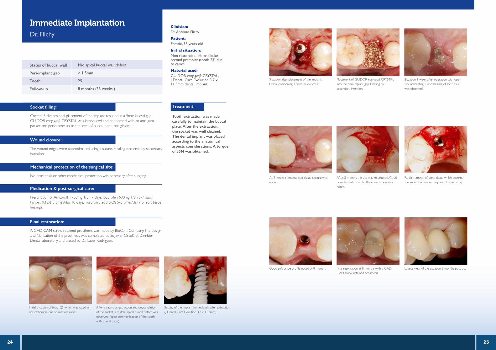
Tooth extraction was made carefully to maintain the buccal plate. After the extraction, the socket was well cleaned. The dental implant was placed according to the anatomical aspects considerations. A torque of 35N was obtained.
Clinician:Dr. Antonio Flichy
Patient:Female, 38 years old
Initial situation: Non restorable left maxibular second premolar (tooth 25) due to caries.
Material used:GUIDOR easy-graft CRYSTAL, J Dental Care Evolution 3.7 x 11.5mm dental implant.
Socket filling:
Correct 3 dimensional placement of the implant resulted in a 5mm buccal gap. GUIDOR easy-graft CRYSTAL was introduced and condensed with an amalgam packer and periotome up to the level of buccal bone and gingiva.
Wound closure:
The wound edges were approximated using a suture. Healing occurred by secondary intention.
Status of buccal wall
Peri-implant gap
Tooth
Follow-up
Mid apical buccal wall defect
> 1.5mm
25
8 months (33 weeks )
Situation after placement of the implant. Palatal positioning 1.5mm below crest.
At 2 weeks complete soft tissue closure was noted.
Good soft tissue profile noted at 8 months.
Placement of GUIDOR easy-graft CRYSTAL into the peri-implant gap. Healing by secondary intention.
After 5 months the site was re-entered. Good bone formation up to the cover screw was noted.
Final restoration at 8 months with a CAD-CAM screw retained prosthesis.
Situation 1 week after operation with open wound healing. Good healing of soft tissue was observed.
Partial removal of bone tissue which covered the implant screw, subsequent closure of flap.
Lateral view of the situation 8 months post op.
Initial situation of tooth 25 which was rated as not restorable due to massive caries.
After atraumatic extraction and degranulation of the socket, a middle apical buccal defect was observed (apex communication of the tooth with buccal plate).
Setting of the implant immediately after extraction (J Dental Care Evolution 3.7 x 11.5mm).
Mechanical protection of the surgical site:
No prosthesis or other mechanical protection was necessary after surgery.
Medication & post-surgical care:
Prescription of Amoxicillin 750mg 1/8h 7 days; Ibuprofen 600mg 1/8h 5-7 days; Paroex 0.12% 3 times/day 10 days; hyaluronic acid 0.6% 5-6 times/day (for soft tissue healing).
Final restoration:
A CAD-CAM screw retained prosthesis was made by BioCam Company. The design and fabrication of the prosthesis was completed by Sr. Javier Ortolà at Dinnbier Dental laboratory and placed by Dr. Isabel Rodriguez.
26 27
Immediate ImplantationDr. Kakar
Treatment:
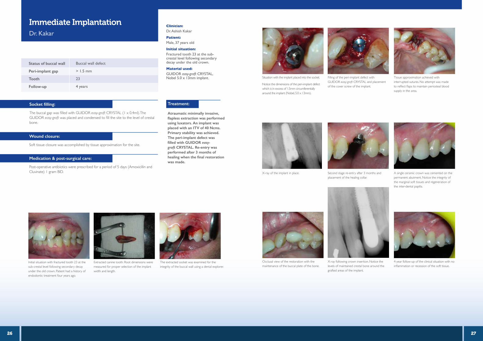
Atraumatic minimally invasive, flapless extraction was performed using luxators. An implant was placed with an ITV of 40 Ncms.Primary stability was achieved. The peri-implant defect was filled with GUIDOR easy-graft CRYSTAL. Re-entry was performed after 3 months of healing when the final restoration was made.
Clinician:Dr. Ashish Kakar
Patient:Male, 37 years old
Initial situation: Fractured tooth 23 at the sub-crestal level following secondary decay under the old crown.
Material used:GUIDOR easy-graft CRYSTAL, Nobel 5.0 x 13mm implant.
Socket filling:
The buccal gap was filled with GUIDOR easy-graft CRYSTAL (1 x 0.4ml). The GUIDOR easy-graft was placed and condensed to fill the site to the level of crestal bone.
Wound closure:
Soft tissue closure was accomplished by tissue approximation for the site.
Status of buccal wall
Peri-implant gap
Tooth
Follow-up
Buccal wall defect
> 1.5 mm
23
4 years
Situation with the implant placed into the socket.
Notice the dimensions of the peri-implant defect which is in excess of 1.5mm circumferentially around the implant (Nobel, 5.0 x 13mm).
X-ray of the implant in place.
Occlusal view of the restoration with the maintenance of the buccal plate of the bone.
Filling of the peri-implant defect with GUIDOR easy-graft CRYSTAL and placement of the cover screw of the implant.
Second stage re-entry after 3 months and placement of the healing collar.
X-ray following crown insertion. Notice the levels of maintained crestal bone around the grafted areas of the implant.
Tissue approximation achieved with interrupted sutures. No attempt was made to reflect flaps to maintain periosteal blood supply in the area.
A single ceramic crown was cemented on the permanent abutment. Notice the integrity of the marginal soft tissues and regeneration of the inter-dental papilla.
4-year follow-up of the clinical situation with no inflammation or recession of the soft tissue.
Initial situation with fractured tooth 23 at the sub-crestal level following secondary decay under the old crown. Patient had a history of endodontic treatment four years ago.
Extracted canine tooth. Root dimensions were measured for proper selection of the implant width and length.
The extracted socket was examined for the integrity of the buccal wall using a dental explorer.
Medication & post-surgical care:
Post-operative antibiotics were prescribed for a period of 5 days (Amoxicillin and Cluvinate) 1 gram BID.
28 29
Dr. Flichy
Treatment:
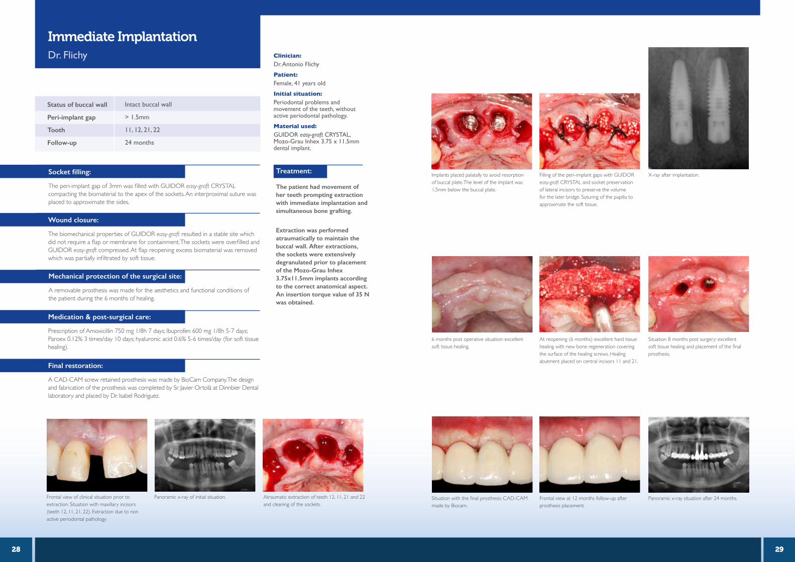
The patient had movement of her teeth prompting extraction with immediate implantation and simultaneous bone grafting.
Extraction was performed atraumatically to maintain the buccal wall. After extractions, the sockets were extensively degranulated prior to placement of the Mozo-Grau Inhex 3.75x11.5mm implants according to the correct anatomical aspect. An insertion torque value of 35 N was obtained.
Clinician:Dr. Antonio Flichy
Patient:Female, 41 years old
Initial situation: Periodontal problems and movement of the teeth, without active periodontal pathology.
Material used:GUIDOR easy-graft CRYSTAL, Mozo-Grau Inhex 3.75 x 11.5mm dental implant.
Socket filling:
The peri-implant gap of 3mm was filled with GUIDOR easy-graft CRYSTAL compacting the biomaterial to the apex of the sockets. An interproximal suture was placed to approximate the sides.
Wound closure:
The biomechanical properties of GUIDOR easy-graft resulted in a stable site which did not require a flap or membrane for containment. The sockets were overfilled and GUIDOR easy-graft compressed. At flap reopening excess biomaterial was removed which was partially infiltrated by soft tissue.
Mechanical protection of the surgical site:
A removable prosthesis was made for the aesthetics and functional conditions of the patient during the 6 months of healing.
Medication & post-surgical care:
Prescription of Amoxicillin 750 mg 1/8h 7 days; Ibuprofen 600 mg 1/8h 5-7 days; Paroex 0.12% 3 times/day 10 days; hyaluronic acid 0.6% 5-6 times/day (for soft tissue healing).
Status of buccal wall
Peri-implant gap
Tooth
Follow-up
Intact buccal wall
> 1.5mm
11, 12, 21, 22
24 months
Implants placed palatally to avoid resorption of buccal plate. The level of the implant was 1.5mm below the buccal plate.
6 months post operative situation: excellent soft tissue healing.
Situation with the final prosthesis: CAD-CAM made by Biocam.
Filling of the peri-implant gaps with GUIDOR easy-graft CRYSTAL and socket preservation of lateral incisors to preserve the volume for the later bridge. Suturing of the papilla to approximate the soft tissue.
At reopening (6 months): excellent hard tissue healing with new bone regeneration covering the surface of the healing screws. Healing abutment placed on central incisors 11 and 21.
Frontal view at 12 months follow-up after prosthesis placement.
X-ray after implantation.
Situation 8 months post surgery: excellent soft tissue healing and placement of the final prosthesis.
Panoramic x-ray situation after 24 months.Frontal view of clinical situation prior to extraction. Situation with maxillary incisors (teeth 12, 11, 21, 22). Extraction due to non active periodontal pathology.
Panoramic x-ray of initial situation. Atraumatic extraction of teeth 12, 11, 21 and 22 and cleaning of the sockets.
Final restoration:
A CAD-CAM screw retained prosthesis was made by BioCam Company. The design and fabrication of the prosthesis was completed by Sr. Javier Ortolà at Dinnbier Dental laboratory and placed by Dr. Isabel Rodriguez.
Immediate Implantation
30 31
Immediate-delayed ImplantationDr. Leventis
Treatment & discussion:
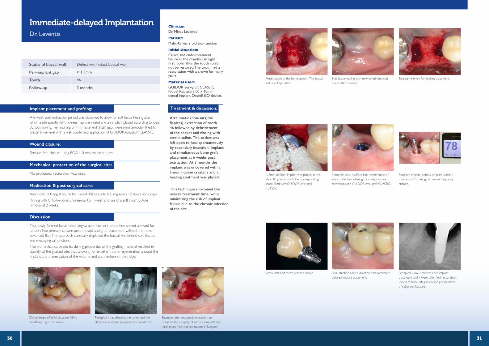
Atraumatic (non-surgical/flapless) extraction of tooth 46 followed by debridement of the socket and rinsing with sterile saline. The socket was left open to heal spontaneously by secondary intention. Implant and simultaneous bone graft placement at 6 weeks post extraction. At 3 months the implant was uncovered with a linear incision crestally and a healing abutment was placed.
This technique shortened the overall treatment time, while minimizing the risk of implant failure due to the chronic infection of the site.
Clinician:Dr. Minas Leventis
Patient:Male, 45 years old, non-smoker
Initial situation: Caries and endo-treatment failure at the mandibular right first molar thus the tooth could not be restored. The tooth had a restoration with a crown for many years.
Material used:GUIDOR easy-graft CLASSIC, Nobel Replace 5.00 x 10mm dental implant Osstell ISQ device.
Implant placement and grafting:
A 6 week post extraction period was observed to allow for soft tissue healing after which a site specific full-thickness flap was raised and an implant placed according to ideal 3D positioning. The resulting 3mm (mesial and distal) gaps were simultaneously filled to crestal bone level with a well condensed application of GUIDOR easy-graft CLASSIC.
Wound closure:
Tension-free closure using PGA 4-0 resorbable sutures.
Status of buccal wall
Peri-implant gap
Tooth
Follow-up
Defect with intact buccal wall
> 1.5mm
46
3 months
Mechanical protection of the surgical site:
No provisional restoration was used.
Medication & post-surgical care:
Amoxicillin 500 mg, 8 hourly for 1 week. Nimesulide 100 mg, every 12 hours for 2 days.Rinsing with Chlorhexidine 3 times/day for 1 week and use of a soft brush. Suture removal at 2 weeks.
Preservation of the bone septum. The buccal wall was kept intact.
Soft tissue healing with new keratinized soft tissue after 6 weeks.
Surgical re-entry for implant placement.
A 5mm x10mm implant was placed at the ideal 3D position with the corresponding space filled with GUIDOR easy-graft CLASSIC.
3 months post-op. Excellent preservation of site architecture utilizing minimally invasive techniques and GUIDOR easy-graft CLASSIC.
Excellent implant stability (implant stability quotient of 78) using resonance frequency analysis.
Screw retained metal-ceramic crown. Final situation after extraction and immediate-delayed implant placement.
Periapical x-ray 3 months after implant placement and 1 week after final restoration. Excellent bone integration and preservation of ridge architecture.
Discussion
The newly-formed keratinized gingiva over the post-extraction socket allowed for tension-free primary closure post implant and graft placement without the need advanced flap. This approach coronally displaced the buccal keratinized soft tissues and mucogingival junction. The biomechanical in situ hardening properties of the grafting material resulted in stability of the grafted site, thus allowing for excellent bone regeneration around the implant and preservation of the volume and architecture of the ridge.
Clinical image of initial situation, failing mandibular right first molar.
Periapical x-ray showing the caries and the chronic inflammation around the mesial root.
Situation after atraumatic extraction to preserve the integrity of surrounding soft and hard issues (root sectioning, use of luxators).
32 33
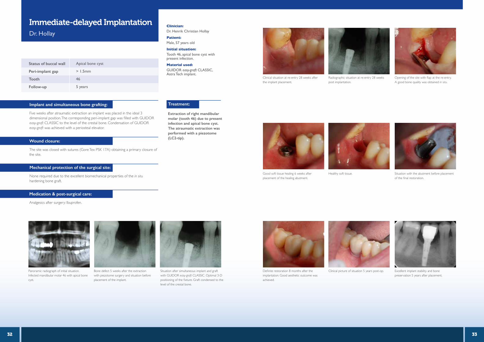
Immediate-delayed ImplantationDr. Hollay
Treatment:
Extraction of right mandibular molar (tooth 46) due to present infection and apical bone cyst. The atraumatic extraction was performed with a piezotome (LC2-tip).
Clinician:Dr. Henrik Christian Hollay
Patient:Male, 57 years old
Initial situation: Tooth 46, apical bone cyst with present infection.
Material used:GUIDOR easy-graft CLASSIC, Astra Tech implant.
Implant and simultaneous bone grafting:
Five weeks after atraumatic extraction an implant was placed in the ideal 3 dimensional position. The corresponding peri-implant gap was filled with GUIDOR easy-graft CLASSIC to the level of the crestal bone. Condensation of GUIDOR easy-graft was achieved with a periosteal elevator.
Wound closure:
The site was closed with sutures (Gore Tex PSK 17A) obtaining a primary closure of the site.
Status of buccal wall
Peri-implant gap
Tooth
Follow-up
Apical bone cyst
> 1.5mm
46
5 years
Clinical situation at re-entry 28 weeks after the implant placement.
Good soft tissue healing 6 weeks after placement of the healing abutment.
Definite restoration 8 months after the implantation. Good aesthetic outcome was achieved.
Radiographic situation at re-entry 28 weeks post implantation.
Healthy soft tissue.
Clinical picture of situation 5 years post-op.
Opening of the site with flap at the re-entry. A good bone quality was obtained in situ.
Situation with the abutment before placement of the final restoration.
Excellent implant stability and bone preservation 5 years after placement.
Panoramic radiograph of initial situation. Infected mandibular molar 46 with apical bone cyst.
Bone defect 5 weeks after the extraction with piezotome surgery and situation before placement of the implant.
Situation after simultaneous implant and graft with GUIDOR easy-graft CLASSIC. Optimal 3-D positioning of the fixture. Graft condensed to the level of the crestal bone.
Mechanical protection of the surgical site:
None required due to the excellent biomechanical properties of the in situ hardening bone graft.
Medication & post-surgical care:
Analgesics after surgery: Ibuprofen.
34 35
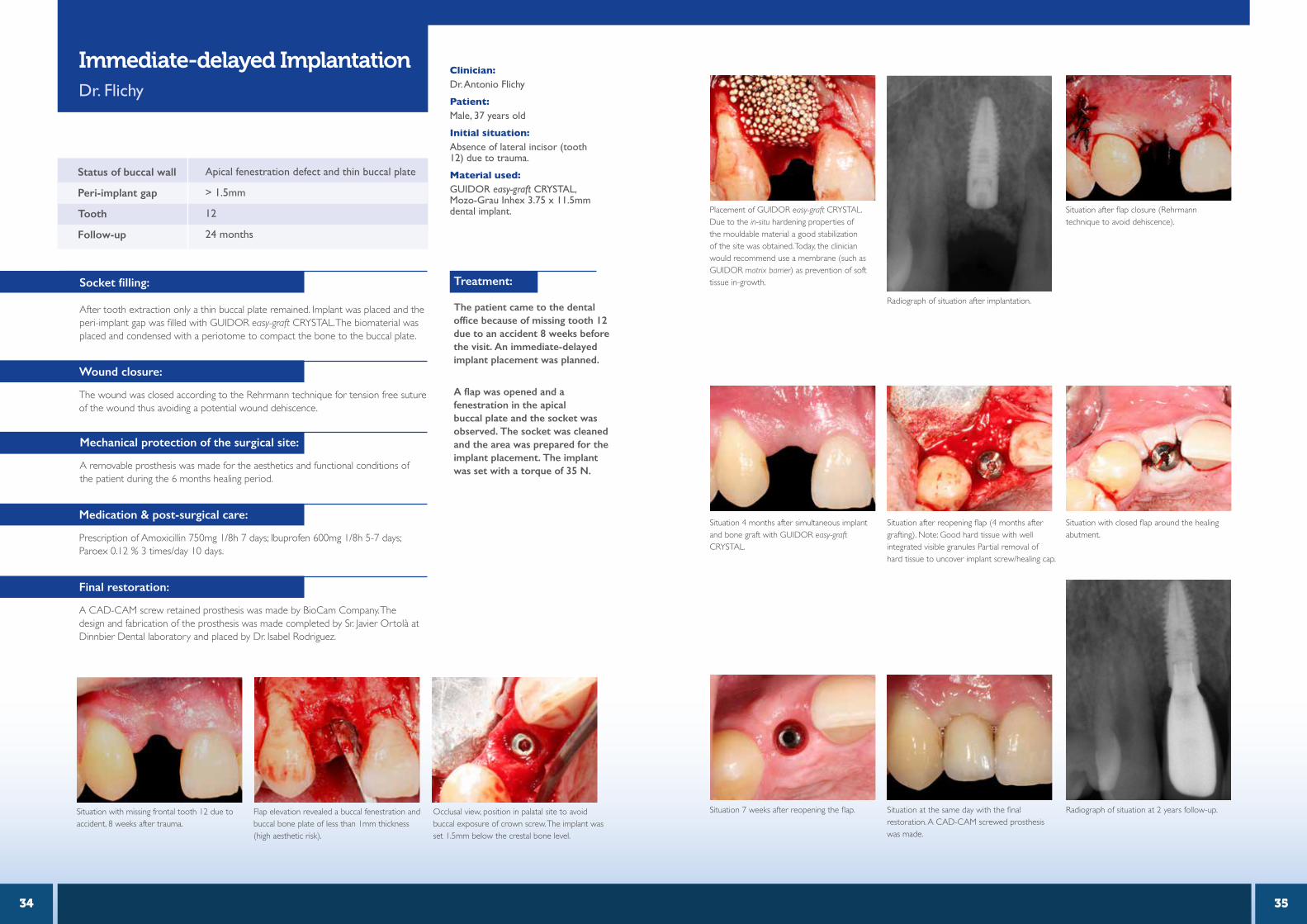
Immediate-delayed ImplantationDr. Flichy
Treatment:
The patient came to the dental office because of missing tooth 12 due to an accident 8 weeks before the visit. An immediate-delayed implant placement was planned.
A flap was opened and a fenestration in the apical buccal plate and the socket was observed. The socket was cleaned and the area was prepared for the implant placement. The implant was set with a torque of 35 N.
Clinician:Dr. Antonio Flichy
Patient:Male, 37 years old
Initial situation: Absence of lateral incisor (tooth 12) due to trauma.
Material used:GUIDOR easy-graft CRYSTAL, Mozo-Grau Inhex 3.75 x 11.5mm dental implant.
Socket filling: After tooth extraction only a thin buccal plate remained. Implant was placed and the peri-implant gap was filled with GUIDOR easy-graft CRYSTAL. The biomaterial was placed and condensed with a periotome to compact the bone to the buccal plate.
Wound closure:
The wound was closed according to the Rehrmann technique for tension free suture of the wound thus avoiding a potential wound dehiscence.
Status of buccal wall
Peri-implant gap
Tooth
Follow-up
Apical fenestration defect and thin buccal plate
> 1.5mm
12
24 months
Placement of GUIDOR easy-graft CRYSTAL. Due to the in-situ hardening properties of the mouldable material a good stabilization of the site was obtained. Today, the clinician would recommend use a membrane (such as GUIDOR matrix barrier) as prevention of soft tissue in-growth.
Situation 4 months after simultaneous implant and bone graft with GUIDOR easy-graft CRYSTAL.
Situation 7 weeks after reopening the flap.
Radiograph of situation after implantation.
Situation after reopening flap (4 months after grafting). Note: Good hard tissue with well integrated visible granules Partial removal of hard tissue to uncover implant screw/healing cap.
Situation at the same day with the final restoration. A CAD-CAM screwed prosthesis was made.
Situation after flap closure (Rehrmann technique to avoid dehiscence).
Situation with closed flap around the healing abutment.
Radiograph of situation at 2 years follow-up.Situation with missing frontal tooth 12 due to accident, 8 weeks after trauma.
Flap elevation revealed a buccal fenestration and buccal bone plate of less than 1mm thickness (high aesthetic risk).
Occlusal view, position in palatal site to avoid buccal exposure of crown screw. The implant was set 1.5mm below the crestal bone level.
Mechanical protection of the surgical site:
A removable prosthesis was made for the aesthetics and functional conditions of the patient during the 6 months healing period.
Medication & post-surgical care:
Prescription of Amoxicillin 750mg 1/8h 7 days; Ibuprofen 600mg 1/8h 5-7 days; Paroex 0.12 % 3 times/day 10 days.
Final restoration:
A CAD-CAM screw retained prosthesis was made by BioCam Company. The design and fabrication of the prosthesis was made completed by Sr. Javier Ortolà at Dinnbier Dental laboratory and placed by Dr. Isabel Rodriguez.
36 37
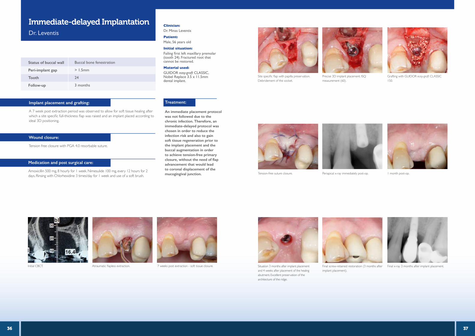
Immediate-delayed ImplantationDr. Leventis
Clinician:Dr. Minas Leventis
Patient:Male, 56 years old
Initial situation: Failing first left maxillary premolar (tooth 24). Fractured root that cannot be restored.
Material used:GUIDOR easy-graft CLASSIC, Nobel Replace 3.5 x 11.5mm dental implant.
Status of buccal wall
Peri-implant gap
Tooth
Follow-up
Buccal bone fenestration
> 1.5mm
24
3 months
Treatment:
An immediate placement protocol was not followed due to the chronic infection. Therefore, an immediate-delayed protocol was chosen in order to reduce the infection risk and also to gain soft tissue regeneration prior to the implant placement and the buccal augmentation in order to achieve tension-free primary closure, without the need of flap advancement that would lead to coronal displacement of the mucogingival junction.
Implant placement and grafting:
A 7 week post extraction period was observed to allow for soft tissue healing after which a site specific full-thickness flap was raised and an implant placed according to ideal 3D positioning.
Wound closure:
Tension free closure with PGA 4.0 resorbable suture.
Medication and post surgical care:
Amoxicillin 500 mg, 8 hourly for 1 week. Nimesulide 100 mg, every 12 hours for 2 days. Rinsing with Chlorhexidine 3 times/day for 1 week and use of a soft brush.
Initial CBCT. Atraumatic flapless extraction. 7 weeks post extraction - soft tissue closure.
Site specific flap with papilla preservation. Debridement of the socket.
Tension-free suture closure.
Situation 3 months after implant placement and 4 weeks after placement of the healing abutment. Excellent preservation of the architecture of the ridge.
Precise 3D implant placement. ISQ measurement (60).
Periapical x-ray immediately post-op.
Final screw-retained restoration (3 months after implant placement).
Grafting with GUIDOR easy-graft CLASSIC 150.
1 month post-op.
Final x-ray 3 months after implant placement.
38 39
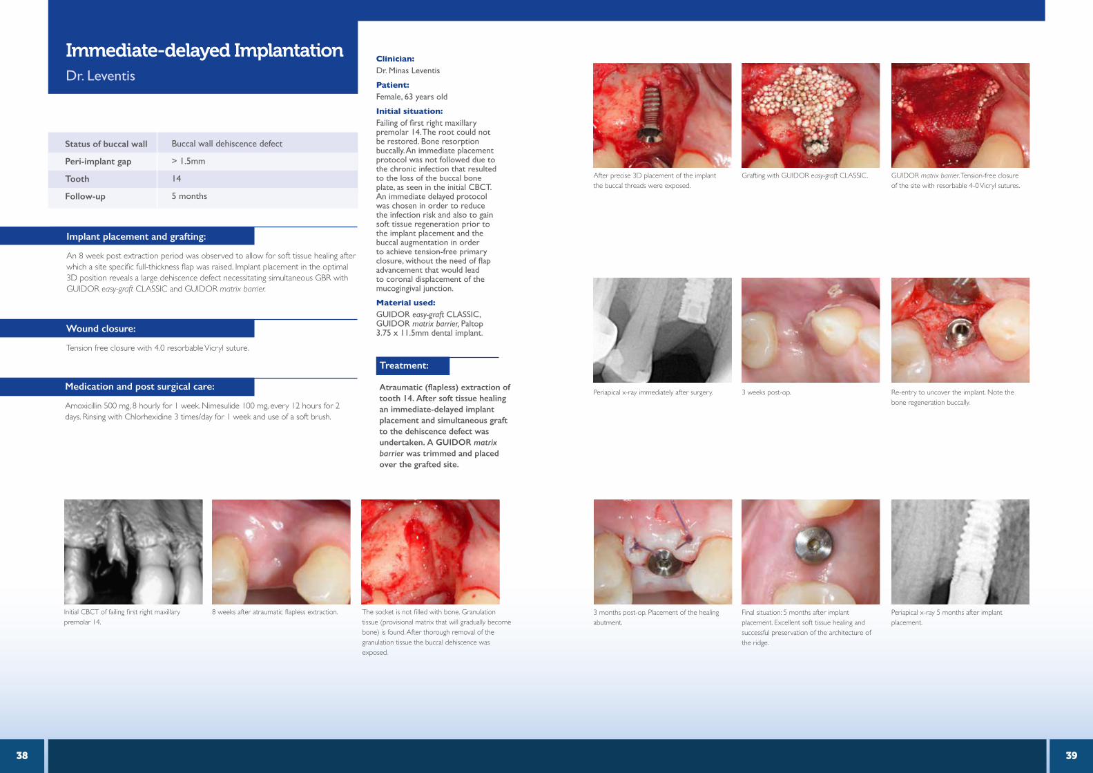
Immediate-delayed ImplantationDr. Leventis
Clinician:Dr. Minas Leventis
Patient:Female, 63 years old
Initial situation: Failing of first right maxillary premolar 14. The root could not be restored. Bone resorption buccally. An immediate placement protocol was not followed due to the chronic infection that resulted to the loss of the buccal bone plate, as seen in the initial CBCT. An immediate delayed protocol was chosen in order to reduce the infection risk and also to gain soft tissue regeneration prior to the implant placement and the buccal augmentation in order to achieve tension-free primary closure, without the need of flap advancement that would lead to coronal displacement of the mucogingival junction.
Material used:GUIDOR easy-graft CLASSIC, GUIDOR matrix barrier, Paltop 3.75 x 11.5mm dental implant.
Status of buccal wall
Peri-implant gap
Tooth
Follow-up
Buccal wall dehiscence defect
> 1.5mm
14
5 months
Treatment:
Atraumatic (flapless) extraction of tooth 14. After soft tissue healing an immediate-delayed implant placement and simultaneous graft to the dehiscence defect was undertaken. A GUIDOR matrix barrier was trimmed and placed over the grafted site.
Implant placement and grafting:
An 8 week post extraction period was observed to allow for soft tissue healing after which a site specific full-thickness flap was raised. Implant placement in the optimal 3D position reveals a large dehiscence defect necessitating simultaneous GBR with GUIDOR easy-graft CLASSIC and GUIDOR matrix barrier.
Wound closure:
Tension free closure with 4.0 resorbable Vicryl suture.
Medication and post surgical care:
Amoxicillin 500 mg, 8 hourly for 1 week. Nimesulide 100 mg, every 12 hours for 2 days. Rinsing with Chlorhexidine 3 times/day for 1 week and use of a soft brush.
Initial CBCT of failing first right maxillary premolar 14.
8 weeks after atraumatic flapless extraction. The socket is not filled with bone. Granulation tissue (provisional matrix that will gradually become bone) is found. After thorough removal of the granulation tissue the buccal dehiscence was exposed.
After precise 3D placement of the implant the buccal threads were exposed.
Periapical x-ray immediately after surgery.
Grafting with GUIDOR easy-graft CLASSIC.
3 weeks post-op.
GUIDOR matrix barrier. Tension-free closure of the site with resorbable 4-0 Vicryl sutures.
Re-entry to uncover the implant. Note the bone regeneration buccally.
3 months post-op. Placement of the healing abutment.
Final situation: 5 months after implant placement. Excellent soft tissue healing and successful preservation of the architecture of the ridge.
Periapical x-ray 5 months after implant placement.
40 41
Disclaimer
Medical science is dynamic and constantly advancing. The presented information is accurate to the best of the authors’ knowledge and reflects current knowledge at the time of publication (2015), but we cannot guarantee its correctness and completeness. The document has been written for a professional audience that is able to place the information in the proper context and to assess the risks and advantages of the procedures and methods presented by the authors where they deviate from the traditional schools of thought.
It must be taken into account that indications differ for each patient. Treatment success significantly depends on multiple biological and medical factors as well as on adequate preliminary and follow-up treatment. The authors and the company Sunstar Suisse SA can therefore not guarantee the success of treatment with the suggested treatments.
Any liability for material or immaterial damage arising from the use (or disuse) of this information is excluded. The GUIDOR matrix barrier, GUIDOR easy-graft CLASSIC and GUIDOR easy-graft CRYSTAL instructions for use leaflets, which are authoritative regarding therapeutic information, must be observed. Each user is asked to study the instructions for use in detail.
Cited References
Araujo M G and Lindhe J: Dimensional ridge alterations following tooth extraction. An experimental study in the dog J Clin Periodontol (2005) 32(2): 212-8.
Artzi Z, Weinreb M, Givol N, Rohrer M D, Nemcovsky C E, Prasad H S and Tal H: Biomaterial resorption rate and healing site morphology of inorganic bovine bone and beta-tricalcium phosphate in the canine: a 24-month longitudinal histologic study and morphometric analysis Int J Oral Maxillofac Implants (2004) 19(3): 357-68.
Botticelli D, Berglundh T, Buser D and Lindhe J: The jumping distance revisited Clinical Oral Implants Research (2003) 14(1): 35-42.
Botticelli D, Berglundh T and Lindhe J: The influence of a biomaterial on the closure of a marginal hard tissue defect adjacent to implants Clinical Oral Implants Research (2004a) 15(3): 285-292.
Botticelli D, Berglundh T and Lindhe J: The influence of a biomaterial on the closure of a marginal hard tissue defect adjacent to implants. An experimental study in the dog Clin Oral Implants Res (2004b) 15(3): 285-92.
Branemark P-I: Osseointegration and its experimental background The Journal of prosthetic dentistry (1983) 50(3): 399-410.
Buser D, Martin W and Belser U C: Optimizing esthetics for implant restorations in the anterior maxilla: anatomic and surgical considerations The International journal of oral & maxillofacial implants (2004) 19: 43-61.
Chen S T and Buser D: Esthetic outcomes following immediate and early implant placement in the anterior maxilla—a systematic review Int J Oral Maxillofac Implants (2014) 29(Suppl): 186-215.
Chen S T, Wilson Jr T G and Hammerle C: Immediate or early placement of implants following tooth extraction: review of biologic basis, clinical procedures, and outcomes Int J Oral Maxillofac Implants (2004) 19(19): 12-25.
Elian N, Cho S, Froum S, Smith R B and Tarnow D P: A simplified socket classification and repair technique Pract Proced Aesthet Dent (2007) 19(2): 99.
Esposito M, Grusovin M G, Polyzos I P, Felice P and Worthington H V: Timing of implant placement after tooth extraction: immediate, immediate-delayed or delayed implants? A Cochrane systematic review European journal of oral implantology (2010a) 3(3): 189-205.
Esposito M, Piattelli M, Pistilli R, Pellegrino G and Felice P: Sinus lift with guided bone regeneration or anorganic bovine bone: 1-year post-loading results of a pilot randomised clinical trial Eur J Oral Implantol (2010b) 3(4): 297-305.
Fugazzotto P A: The role of guided tissue regenerative therapy in today’s clinical practice. JIACD (2011) 3(1): 33-45.
LinksITI, International Team for Implantology: http://www.iti.org/
EAO, European Association for Osseointegration: http://www.eao.org/
EDI, European Association of Dental Implantologists: http://www.bdizedi.org/
ReferencesDisclaimer
Hämmerle C H F, Araujo M and Simion M: Osteology Consensus Report Evidence-based knowledge on the biology and treatment of extraction sockets Clin. Oral. Impl. Res. (2012) 23(Suppl.5): 80-82.
Huynh-Ba G, Pjetursson B E, Sanz M, Cecchinato D, Ferrus J, Lindhe J and Lang N P: Analysis of the socket bone wall dimensions in the upper maxilla in relation to immediate implant placement Clinical oral implants research (2010) 21(1): 37-42.
Jensen S S and Terheyden H: Bone augmentation procedures in localized defects in the alveolar ridge: clinical results with different bone grafts and bone-substitute materials Int J Oral Maxillofac Implants (2009) 24 Suppl: 218-36.
Jofre J, Valenzuela D, Quintana P and Asenjo-Lobos C: Protocol for immediate implant replacement of infected teeth Implant dentistry (2012) 21(4): 287-294.
Koh R U, Rudek I and Wang H-L: Immediate implant placement: positives and negatives Implant Dentistry (2010) 19(2): 98-108.
Leventis M, Fairbairn P J and Horowitz M: Extraction Site Preservation Using an In-Situ Hardening Alloplastic Bone Graft Substitute Compendium (2014a) 35(4): 11-13.
Leventis M D, Fairbairn P, Dontas I, Faratzis G, Valavanis K D, Khaldi L, Kostakis G and Eleftheriadis E: Biological Response to β-Tricalcium Phosphate/Calcium Sulfate Synthetic Graft Material: An Experimental Study Implant dentistry (2014b) 23(1): 37-43.
Lundgren D, Mathisen T and Gottlow J: The development of a bioresorbable barrier for guided tissue regeneration J Swed Dent Assoc (1994a) 86(13): 741-756.
Lundgren D, Sennerby L, Falk H, Friberg B and Nyman S: The use of a new bioresorbable barrier for guided bone regeneration in connection with implant installation. Case reports Clin Oral Implants Res (1994b) 5(3): 177-84.
Martin W, Morton D, Buser D, Buser D, Belser U and Wismeijer D: Diagnostic factors for esthetic risk assessment ITI Treatment Guide (2006) 1: 11-20.
Nickenig H J, Rothamel D, Berger C, Bonsmann M, Cacaci C, Drysdale C, Fairbairn P, Happe A, Kobler P, Konstantinovic V, Reinhardt S, Tetsch J, Tomkiewicz W, Wagner W, Wojtowicz A, Vizethum F and Ozyuvaci H: Avoiding implant malpositioning EDI Journal (2014) 10(2/2014): 14-26.
Rosen P S: The Combined Ue of Allograft and a Polylactic Acid Barrier for GTR and GBR Efforts: 2 Case Reports JIACD (2010) 2(7): 55-62.
42 43
Ruffieux K, Kohli M, Benner K-U, D’Avenia F, Fairbairn P J, Friedrich D, Glaser R, Greschak J, Heuckmann K-H, Hollay H-C, Huber A, Neumeyer S, Neumeyer-Wühr S, Schmidlin P, Schug J, Trödhan A and Weber F E Bone Augmentation - Material Science and Clinical Manual easy-graft / easy-graft CRYSTAL (2010) Schlieren, DS Dental.
Sanz I, Garcia-Gargallo M, Herrera D, Martin C, Figuero E and Sanz M: Surgical protocols for early implant placement in post extraction sockets: a systematic review Clinical oral implants research (2012) 23(s5): 67-79.
Schmidlin P R, Nicholls F, Kruse A, Zwahlen R A and Weber F E: Evaluation of moldable, in situ hardening calcium phosphate bone graft substitutes Clin Oral Implants Res (2013) 24(2): 149-57.
Schropp L, Wenzel A, Kostopoulos L and Karring T: Bone healing and soft tissue contour changes following single-tooth extraction: a clinical and radiographic 12-month prospective study Int J Periodontics Restorative Dent (2003) 23(4): 313-23.
Schug J, Kirste M, Huber A, Hollay H C, Troedhan A and Leventis M D (2013). Post Extraction Alveolar Ridge Preservation; Scientific Background, Minimally Invasive Treatment Protocols and Experts Reports using Alloplastic Biomaterials.
Tarnow D P and Chu S J: Human histologic verification of osseointegration of an immediate implant placed into a fresh extraction socket with excessive gap distance without primary flap closure, graft, or membrane: a case report The International journal of periodontics & restorative dentistry (2010) 31(5): 515-521.
Trisi P, Rao W, Rebaudi A and Fiore P: Histologic effect of pure-phase beta-tricalcium phosphate on bone regeneration in human artificial jawbone defects International Journal of Periodontics and Restorative Dentistry (2003) 23(1): 69-78.
Troedhan A, Schlichting I, Kurrek A and Wainwright M: Primary implant stability in augmented sinuslift-sites after completed bone regeneration: a randomized controlled clinical study comparing four subantrally inserted biomaterials Scientific reports (2014) 4.
Vignoletti F, Matesanz P, Rodrigo D, Figuero E, Martin C and Sanz M: Surgical protocols for ridge preservation after tooth extraction. A systematic review Clin Oral Implants Res (2012) 23 Suppl 5: 22-38.
Wang H-L and Boyapati L: “PASS” principles for predictable bone regeneration Implant Dentistry (2006) 15(1): 8-17.
References

Presented as a service to Dentistry by Sunstar GUIDOR® - Guiding Oral Rehabilitaton
Sunstar Suisse S.A.Route de Pallatex 15CH-1163 Etoywww.guidor.com
GUIDOR and easy-graft are trademarks of Sunstar Suisse S.A. in the U.S. and in other countries SALM
-BO
O-0
03 (E
N) V
2.1 E
ditio
n da
te 1
0.10.1
5