Performance Supercapacitor Electrode Materials Electronic ... · 1 Electronic Supplementary...
16
1 Electronic Supplementary Information (ESI) Graphene Quantum Dots doped Polyaniline Nanofiber as High Performance Supercapacitor Electrode Materials Sanjoy Mondal, Utpal Rana and Sudip Malik* Polymer Science Unit, Indian Association for the Cultivation of Science, 2A & 2B Raja S.C. Mullick Road., Jadavpur, Kolkata – 700032, India. Table of content 1) Synthesis of GO.....................................................................................................S2 2) Synthesis of GQDs and GQDP composites in Table S1........................................S3 3) Characterization of GQD and GQDP composite...................................................S4 4) Electrochemical experiments.................................................................................S5 5) UV-vis and PL study of GQD...............................................................................S6 6) XRD study of Graphite, GO and GQD.................................................................S7 7) FTIR study of GQDP composites..........................................................................S8 8) XPS study of N1s, O1s and C1s............................................................................S9 9) Powder XRD of GQDP composites....................................................................S10 10) Raman study of GQDP composites....................................................................S11 11) CV plot of GQDP10 composite with different scan rate....................................S12 12) Specific capacitance of GQDP composite..........................................................S13 13) Impedance study................................................................................................ S14 14) Literature review.................................................................................................S15 15) References...........................................................................................................S16 Electronic Supplementary Material (ESI) for ChemComm. This journal is © The Royal Society of Chemistry 2015
Transcript of Performance Supercapacitor Electrode Materials Electronic ... · 1 Electronic Supplementary...

1
Electronic Supplementary Information (ESI)
Graphene Quantum Dots doped Polyaniline Nanofiber as High
Performance Supercapacitor Electrode MaterialsSanjoy Mondal, Utpal Rana and Sudip Malik*
Polymer Science Unit, Indian Association for the Cultivation of Science,
2A & 2B Raja S.C. Mullick Road., Jadavpur, Kolkata – 700032, India.
Table of content
1) Synthesis of GO.....................................................................................................S2
2) Synthesis of GQDs and GQDP composites in Table S1........................................S3
3) Characterization of GQD and GQDP composite...................................................S4
4) Electrochemical experiments.................................................................................S5
5) UV-vis and PL study of GQD...............................................................................S6
6) XRD study of Graphite, GO and GQD.................................................................S7
7) FTIR study of GQDP composites..........................................................................S8
8) XPS study of N1s, O1s and C1s............................................................................S9
9) Powder XRD of GQDP composites....................................................................S10
10) Raman study of GQDP composites....................................................................S11
11) CV plot of GQDP10 composite with different scan rate....................................S12
12) Specific capacitance of GQDP composite..........................................................S13
13) Impedance study................................................................................................ S14
14) Literature review.................................................................................................S15
15) References...........................................................................................................S16
Electronic Supplementary Material (ESI) for ChemComm.This journal is © The Royal Society of Chemistry 2015
2
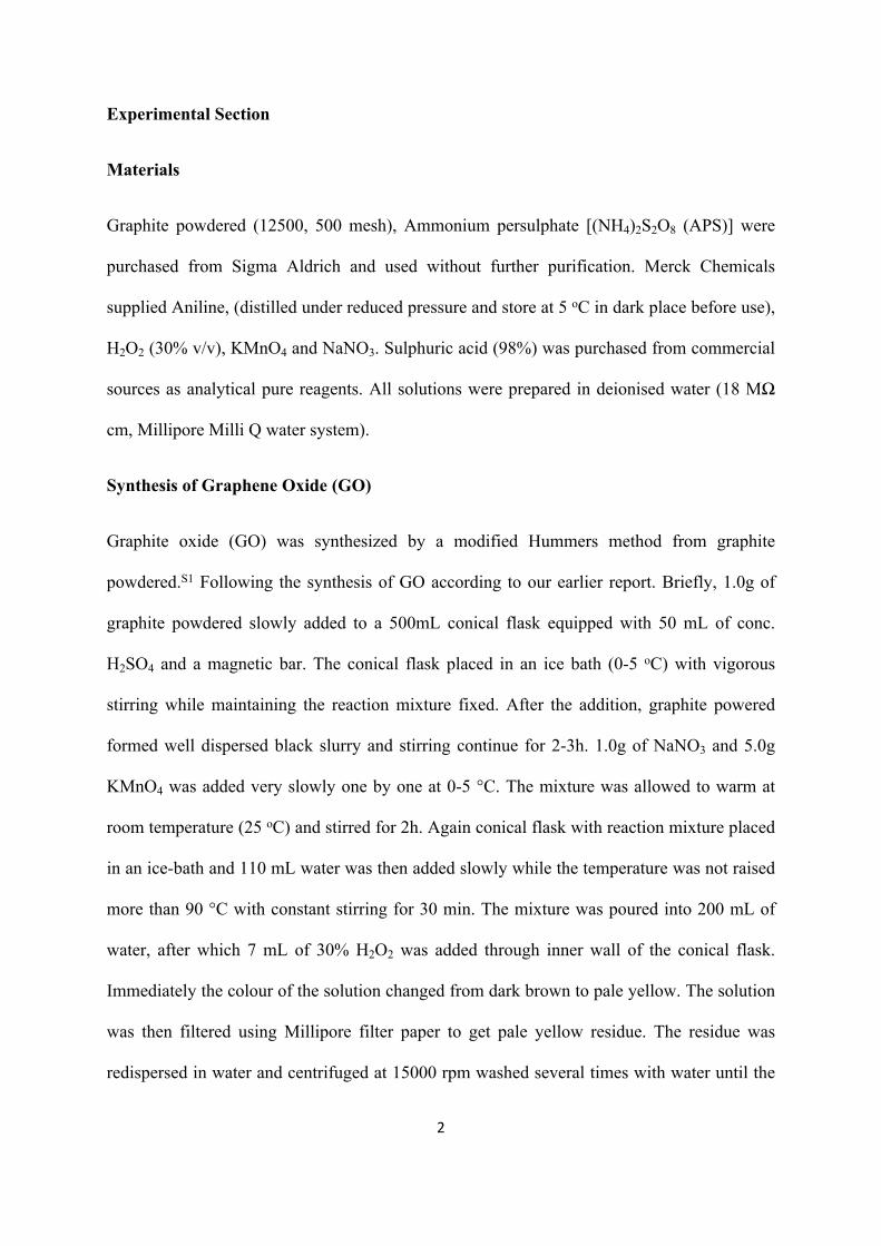
Experimental Section
Materials
Graphite powdered (12500, 500 mesh), Ammonium persulphate [(NH4)2S2O8 (APS)] were
purchased from Sigma Aldrich and used without further purification. Merck Chemicals
supplied Aniline, (distilled under reduced pressure and store at 5 oC in dark place before use),
H2O2 (30% v/v), KMnO4 and NaNO3. Sulphuric acid (98%) was purchased from commercial
sources as analytical pure reagents. All solutions were prepared in deionised water (18 MΩ
cm, Millipore Milli Q water system).
Synthesis of Graphene Oxide (GO)
Graphite oxide (GO) was synthesized by a modified Hummers method from graphite
powdered.S1 Following the synthesis of GO according to our earlier report. Briefly, 1.0g of
graphite powdered slowly added to a 500mL conical flask equipped with 50 mL of conc.
H2SO4 and a magnetic bar. The conical flask placed in an ice bath (0-5 oC) with vigorous
stirring while maintaining the reaction mixture fixed. After the addition, graphite powered
formed well dispersed black slurry and stirring continue for 2-3h. 1.0g of NaNO3 and 5.0g
KMnO4 was added very slowly one by one at 0-5 °C. The mixture was allowed to warm at
room temperature (25 oC) and stirred for 2h. Again conical flask with reaction mixture placed
in an ice-bath and 110 mL water was then added slowly while the temperature was not raised
more than 90 °C with constant stirring for 30 min. The mixture was poured into 200 mL of
water, after which 7 mL of 30% H2O2 was added through inner wall of the conical flask.
Immediately the colour of the solution changed from dark brown to pale yellow. The solution
was then filtered using Millipore filter paper to get pale yellow residue. The residue was
redispersed in water and centrifuged at 15000 rpm washed several times with water until the

3
* All specific capacitance are at 1 A/g current density
Composites Aniline[mmol]
GQD[wt%]
GQD:Ani(w/w)
APS[mmol]
Yield(mg)
Cs[F /g]*
GQDP0 100 mg[1.1]
0 mg ------ 250 27 205.7
GQDP5 100 mg[1.1]
5 mg 0.05: 1 250 91 332.89
GQDP10 100 mg[1.1]
10 0.1:1 250 96 1044.34
GQDP15 100 mg[1.1]
15 0.15:1 250 103 776.67
GQDP20 100 mg[1.1]
20 0.2:1 250 106 319.34
pH of the solution was neutral. The resultant solid material was dried in vacuum at 25 oC and
stored in the ambient environment to get pure GO.
Synthesis of Graphene Quantum Dots (GQD)
Synthesized flake shaped GO was dispersed in water (4 mg/mL) with sonication followed by
stirring. 40 mL of 30% H2O2 was subsequently added to 5 mL of GO suspension. The
resulting mixture reacted at 90 oC with vigorous stirring for 12h. Unreacted H2O2 and water
was removed by rotary evaporation at 60oC. Ethanol was added as a poor solvent to
precipitate out and wash the GQD. Finally GQD was purified by dialysis.
Table S1: Preparation of GQD/PANI composite with different GQD weight ratio

4
Characterization of GQD and GQDP composite
Surface morphology as well as over all morphology of GQDP composite were characterized
by FESEM (JEOL, JSM 6700F) instrument operating at 5 kV. To reduce the surface
potential, samples were coated with platinum for 90 s. TEM imaging of GQDP composites
carried by HRTEM (JEOL, 2010EX) instrument, an accelerating voltage of 200 kV and.
Small amount of water dispersed samples were spread on a 200 mesh Cu-grid coated with a
holey carbon support AFM topologies of synthesized GQDs was recorded using atomic force
microscopy (Veeco, model AP0100) in noncontact mode at a tip resonance frequency of 300
kHz. The well dispersed water sample spread on freshly cleaved mica surface at ambient
conditions. UV-vis spectroscopy of GQDs and GQDP composites were done by Agilent
(model 8453) UV-vis spectrophotometer. Fluorescence spectra of GQDs obtain by using
Horiba Jobin Yvon Fluoromax 3 instrument. The FTIR spectroscopy was carried out in a
FTIR-8400S instrument (Shimadzu) using KBr pellets. Raman studies were performed using
a Raman triple spectrometer (model T-64000, Horiba-Jobin Yvon) fitted with a synapse
detector. The samples were excited with a 514.5 nm laser (Spectra Physics, model Stabilite
2017). Powered WXRD analysis were performed by using a Bruker AXS diffractometer (D8
advance) using CuKα radiation (λ=1.54 Å), a generator voltage of 40 kV and a current of 40
mA. XPS study was performed by using a focused monochromatized A1 Kα X-ray source
(1486.8 eV) in Omicron Nano-Technology 0571 XPS instrument.

5
Cs =𝑖 × Δt
ΔV × m (F/g)
𝐸 = 𝐶𝑠 × ∆𝑉2 7.2
(W h /kg)
…………………… (1)
…………………… (2)
…………………… (3)𝑃 = 𝐸 × 3600 𝑡⁄ (W /kg)
Electrochemical measurements
All electrochemical experiments like cyclic voltammogram (CV), galvanostatic
charge/discharge (GCD) and electrochemical impedance spectroscopy (EIS) study was done
by a CHI6087E electrochemical workstation (CHI, USA) by a conventional three-electrode
system. In present study modified glassy carbon electrode (GCE) as a working electrode, a
saturated calomel electrode (SCE) as the reference electrode, Pt wire electrode as the
auxiliary electrode and 0.5 M H2SO4 as a electrolyte were used. Prior to use the GCE (3 mm
in diameter, surface area of 0.07 cm2) were carefully polished with 1, 0.3, and 0.05 μm
alumina powder and sequentially washed through water and ethanol with sonication at room
temperature until a mirror finish was obtained. The specific capacitance (Cs), energy density,
(E) and power density (P) were calculated from the discharge curve based on equation (1), (2)
and (3) respectively.
Where, ‘i’ = current (A); ‘𝞓t’ = discharge time (s), ‘𝞓V’ = voltage windows (V) and ‘m’= mass of GQDP composites (g).

6
200 300 400 500 600 700
198 nm
UV-vis
Wavelength (nm)
504 nm (ex= 420 nm) PL Intensity (a.u.)
PL
Abs.
Inte
nsity
(a.u
.)
UV-vis and PL study of GQDs
Fig.S1: UV-vis and PL spectra of water well dispersed GQD.

7
5 10 15 20 25 30 35 40
26.7111.24In
tens
ity (a
.u.)
Graphite GO
8.61
2 (degree)
GQD
XRD study of Graphite, GO and GQD
Fig.S2: XRD pattern of synthesized graphite, GO and GQDs

8
500 1000 1500 2000 2500 3000 3500 4000
% T
rans
mitt
ance
GQDP10
1300
GQDP15
GQDP20
1140
800 3432
14961580
Wavenumber (cm-1)
GQDP5
FTIR study
Fig.S3: FTIR spectra of synthesized GQDP composites

9
100 200 300 400 500 600 700
532.5
400
285 GQDP10
O 1s
C 1s
N 1s
In
tens
ity (a
.u.)
Binding energy (eV)392 394 396 398 400 402 404 406 408
(400.2 eV)
(399.36 eV)
Inte
nsity
(a.u
.)
Binding energy (eV)
(389.64 eV)
-NH-
N
=N-
N 1s
279 282 285 288 291 294 297
C 1s
(289.3 eV)C=O
(285.4 eV)C-O/ C-N
(284.2 eV)C=C/C-CIn
tens
ity (a
.u.)
Binding energy (eV)
525 528 531 534 537 540 543
533.5 eV(531.1 eV)
O 1s
Binding energy (eV)
Inte
nsity
(a.u
.) C=O C-O/C-OH
(a) (b)
(c) (d)
XPS study of GQDP composites
Fig.S4 XPS study of GQDP10 composite (a) whole scan (b) deconvolution spectra of N1s,
(c) O1s and (d) C1s.
The C1s (~285 eV) peak of GQDP10 composite deconvolate into three Gaussian
sub-peaks position at ~284.2 eV (C=C/C-C), 285.4 eV (C-O/C-N) and 289.3 eV
(C=O) in Fig.S3. Again deconvolate N1s peak shown in Fig.S4 that contains two sub-
peaks at ~531.1 eV (C=O) and 533.5 eV (C-O/ C-OH).

10
10 15 20 25 30 35 40
GQDP5
2 (degree)
GQDP10
GQDP15
GQDP2020.3 25.5
Inte
nsity
(a.u
.)
XRD study of GQDP composites
Fig.S5 Powder XRD study of synthesized GQDP composites.

11
500 1000 1500 2000 2500 3000
GQDP5
Raman Shift (cm-1)
GQDP10
GQDP15
Ram
an In
tens
ity (a
.u.)
GQDP20
GD
Raman Study of GQDP composites
Fig.S6 Raman study of synthesized GQDP composite under 514 nm laser
Raman spectra of GQDP composites shows GQDs are involve for PANI formation as
a soft doping materials. All GQDP composites have D and G band at 1355 and 1580 cm-1
respectively. D band related to defects or edge areas and G band corresponds to the vibration
of sp2-hybridized carbon. GQDs have some fluorescence behaviour which makes noise in
region of 2000-3000cm-1. In all composites have higher ID/IG ratio (<1.0) suggested that
GQDs intercalation with PANI.

12
0 100 200 300 400 500-16
-12
-8
-4
0
4
8
12
16
Anodic current Cathodic current
Scan rate (mV/s)
Curr
ent (
mA)
-0.2 0.0 0.2 0.4 0.6 0.8-16
-12
-8
-4
0
4
8
12
16
Curr
ent (
mA)
Potential (V) vs Ag/AgCl
10 mV 20 mV 50 mV 100 mV 200 mV 300 mV 400 mV 500 mV
CV plot of GQDP10 composite with different scan rate
Fig.S7 Scan dependent CV study of GQDP10 composite in 0.5 M H2SO4 electrolyte by 3
electrode cell at 25 oC

13
GQDP5 GQDP10 GQDP15 GQDP200
200
400
600
800
1000
Sp. c
apac
itanc
e (F/
g)
GQDP composite
Specific capacitance of GQDP composite
Fig.S8: Specific capacitance value of different GQDP composites.

14
Impedance study
Fig.S9 Impedance study of GQDP composites.
0 200 400 600 800 10000
200
400
600
800
1000
-Zim
g (oh
m)
GQDP5 GQDP10 GQDP15 GQDP20
Zreal (ohm)

15
Previous report about supercapacitor
Materials Preparation method Current density Cs (F/g) Ref Remark
GQD/PANI Chemical oxidative polymerization
1 A/g 1044 This work 1. Simple solution process to make GQD/PANI composites.
2. Good Specific capacitance value with small voltage drop
rGO-PANI Chemical oxidative polymerization
1 A/g 533 S2 Specific capacitances value comparable less
Graphene-PANI paper
Electro-polymerization 1 A/g 763 S3 Polymerization technique is different with low specific capacitance
value
PANI Chemical-Vapor-Induced in Situ Polymerization
1 A/g 2136 S4 Electrode fabrication processes is really different and laborious.
Porous nanorod PANI-graphene
Chemical oxidative polymerization
1 A/g 878.57 S5 Specific capacitances value comparable less
GQDs/PANI Electrochemical polymerization 15.0 A/cm-2 667.5 F/ cm-
2-S6 Separately, PANI as positive active
material and GQDs as negative active material are used

16
References
S1 W. S. Hummers, R. E Offeman, J. Am. Chem. Soc., 1958, 80, 1339.
S2. W. Chen, R. B. Rakhi and H. N. Alshareef, Nanoscale, 2013, 5, 4134.
S3. H. P. Cong, X.-C. Ren, P. Wang and S.-H. Yu, Energy Environ. Sci., 2013, 6, 1185.
S4. X. Li, L. Yang, Y. Lei, L. Gu, and D. Xiao, ACS Appl. Mater. Interfaces, 2014, 6, 19978.
S5. L. Hu, J. Tu, S. Jiao, J. Hou, H. Zhu and D. J. Fray, Phys.Chem. Chem. Phys., 2012, 14, 15652.
S6. W. Liu, X. Yan, J. Chen, Y. Feng and Q. Xueb, Nanoscale, 2013, 5, 6053.