
Performance of disposable endoscopic forceps ac - … disposable biopsy forceps made using simple...
9
Copyright © 2019 The Korean Association of Internal Medicine This is an Open Access article distributed under the terms of the Creative Commons Attribution Non-Commercial License (http://creativecommons.org/licenses/ by-nc/4.0/) which permits unrestricted noncommercial use, distribution, and reproduction in any medium, provided the original work is properly cited. pISSN 1226-3303 eISSN 2005-6648 http://www.kjim.org ORIGINAL ARTICLE Korean J Intern Med 2019;34:530-538 https://doi.org/10.3904/kjim.2017.191 1 Digestive Disease Center, 2 Department of Pathology, CHA Bundang Medical Center, CHA University, Seongnam; 3 Interventional Research Center, M.I. Tech Co. Ltd., Pyeongtaek, Korea Received : May 27, 2017 Revised : October 24, 2017 Accepted : November 2, 2017 Correspondence to Joo Young Cho, M.D. Digestive Disease Center, CHA Bundang Medical Center, CHA University, 59 Yatap-ro, Bun- dang-gu, Seongnam 13496, Korea Tel: +82-31-780-5641 Fax: +82-31-780-5219 E-mail: [email protected] *These authors contributed equally to this work. Background/Aims: Recently, to lower the production costs and risk of infection, new disposable biopsy forceps made using simple manufacturing techniques have been introduced. However, the effects of the manufacturing techniques are unclear. The aim of this study was to evaluate which types of biopsy forceps could obtain good-quality specimens according to the manufacturing techniques. Methods: By using an in vitro nitrile glove popping model, we compared the pop- ping ability among eight different disposable biopsy forceps (one pair of biopsy forceps with cups made by a cutting method [cutting forceps], four pairs of biopsy forceps with cups made by a pressing method [pressing forceps], and three pairs of biopsy forceps with cups made using a injection molding method [molding forceps]). Using an in vivo swine model, we compared the penetration depth and quality of specimen among the biopsy forceps. Results: In the in vitro model, the molding forceps provided a significantly high- er popping rate than the other forceps (cutting forceps, 25.0%; pressing forceps, 17.5%; and molding forceps, 41.7%; p = 0.006). In the in vivo model, the cutting and pressing forceps did not provide larger specimens, deeper biopsy specimen, and higher specimen adequacy than those obtained using the molding forceps ( p = 0.2631, p = 0.5875, and p = 0.2147, respectively). However, the molding forceps showed significantly more common crush artifact than the others (cutting for- ceps, 0%; pressing forceps, 5.0%; and molding forceps, 43.3%; p = 0.0007). Conclusions: The molding forceps provided lower performance than the cutting and pressing forceps in terms of crush artifact. Keywords: Biopsy; Instruments; Endoscopy; Disposable equipment Performance of disposable endoscopic forceps ac- cording to the manufacturing techniques Chang-Il Kwon 1,* , Gwangil Kim 2,* , Jong Pil Moon 3 , Ho Yun 3 , Weon Jin Ko 1 , Joo Young Cho 1 , and Sung Pyo Hong 1 INTRODUCTION In the field of gastrointestinal endoscopy, the develop- ment of new devices and related programs has led to the advancement of diagnostic methods at an exponential rate, but direct histopathological examination is still the most important and accurate diagnostic method. Therefore, the quality of endoscopic biopsy specimen is crucial for a successful histological diagnosis. The basic concept of biopsy forceps has not changed much, but various types of biopsy forceps with different designs and materials are widely used [1]. Previous reports indi- cated that the quality of biopsy specimens depends on the shape and size of the forceps cups [2-5]. However, as endoscopes are becoming slimmer, smaller-sized cups have become more widely used, since larger-sized cups limit the diameter of the working channel. This resulted in technological advancements, including changing the
Transcript of Performance of disposable endoscopic forceps ac - … disposable biopsy forceps made using simple...

Copyright © 2019 The Korean Association of Internal MedicineThis is an Open Access article distributed under the terms of the Creative Commons Attribution Non-Commercial License (http://creativecommons.org/licenses/by-nc/4.0/) which permits unrestricted noncommercial use, distribution, and reproduction in any medium, provided the original work is properly cited.
pISSN 1226-3303eISSN 2005-6648
http://www.kjim.org
ORIGINAL ARTICLE
Korean J Intern Med 2019;34:530-538https://doi.org/10.3904/kjim.2017.191
1Digestive Disease Center, 2Department of Pathology, CHA Bundang Medical Center, CHA University, Seongnam; 3Interventional Research Center, M.I. Tech Co. Ltd., Pyeongtaek, Korea
Received : May 27, 2017Revised : October 24, 2017Accepted : November 2, 2017
Correspondence to Joo Young Cho, M.D.Digestive Disease Center, CHA Bundang Medical Center, CHA University, 59 Yatap-ro, Bun-dang-gu, Seongnam 13496, KoreaTel: +82-31-780-5641Fax: +82-31-780-5219E-mail: [email protected]
*These authors contributed equally to this work.
Background/Aims: Recently, to lower the production costs and risk of infection, new disposable biopsy forceps made using simple manufacturing techniques have been introduced. However, the effects of the manufacturing techniques are unclear. The aim of this study was to evaluate which types of biopsy forceps could obtain good-quality specimens according to the manufacturing techniques.Methods: By using an in vitro nitrile glove popping model, we compared the pop-ping ability among eight different disposable biopsy forceps (one pair of biopsy forceps with cups made by a cutting method [cutting forceps], four pairs of biopsy forceps with cups made by a pressing method [pressing forceps], and three pairs of biopsy forceps with cups made using a injection molding method [molding forceps]). Using an in vivo swine model, we compared the penetration depth and quality of specimen among the biopsy forceps.Results: In the in vitro model, the molding forceps provided a significantly high-er popping rate than the other forceps (cutting forceps, 25.0%; pressing forceps, 17.5%; and molding forceps, 41.7%; p = 0.006). In the in vivo model, the cutting and pressing forceps did not provide larger specimens, deeper biopsy specimen, and higher specimen adequacy than those obtained using the molding forceps (p = 0.2631, p = 0.5875, and p = 0.2147, respectively). However, the molding forceps showed significantly more common crush artifact than the others (cutting for-ceps, 0%; pressing forceps, 5.0%; and molding forceps, 43.3%; p = 0.0007).Conclusions: The molding forceps provided lower performance than the cutting and pressing forceps in terms of crush artifact.
Keywords: Biopsy; Instruments; Endoscopy; Disposable equipment
Performance of disposable endoscopic forceps ac-cording to the manufacturing techniques Chang-Il Kwon1,*, Gwangil Kim2,*, Jong Pil Moon3, Ho Yun3, Weon Jin Ko1, Joo Young Cho1, and Sung Pyo Hong1
INTRODUCTION
In the field of gastrointestinal endoscopy, the develop-ment of new devices and related programs has led to the advancement of diagnostic methods at an exponential rate, but direct histopathological examination is still the most important and accurate diagnostic method. Therefore, the quality of endoscopic biopsy specimen is crucial for a successful histological diagnosis. The basic
concept of biopsy forceps has not changed much, but various types of biopsy forceps with different designs and materials are widely used [1]. Previous reports indi-cated that the quality of biopsy specimens depends on the shape and size of the forceps cups [2-5]. However, as endoscopes are becoming slimmer, smaller-sized cups have become more widely used, since larger-sized cups limit the diameter of the working channel. This resulted in technological advancements, including changing the
531
Kwon CI, et al. Performance of endoscopic forceps
www.kjim.orghttps://doi.org/10.3904/kjim.2017.191
shape of cups from oval to elongated shape, processing the edges of cups in a serrated or toothed-jaw cutting shape, or inserting a stabilizing needle or spike between cups, using more precise processing methods [1].
Meanwhile, with reports indicating cross-contamina-tion and spread of infection by endoscopic accessories, the reusable biopsy forceps have been replaced by the disposable ones [6-8]. However, the replacement process was not easy because of the fixed insurance payment system in some countries. There have been even some cases where disposable biopsy forceps were reprocessed to be reused several times as an interim measure. The biggest technical issue regarding reusing disposable biopsy forceps is that it can cause deformation of the forceps, not to mention cross-contamination. Unlike reusable biopsy forceps, which are made to endure ex-posure to sterilants or disinfectants, disposable biopsy forceps are easily changed or deformed when damaged by reprocessing. As such, the working channel of the en-doscope can be damaged, which would incur high repair costs or various unexpected problems in biopsy sam-pling. To resolve these issues, disposable biopsy forceps with lower production costs were required to overcome the limitations of the insurance payment system. Many countries, including the United States, used this meth-od until 2000, when the change was triggered by the U.S. Food and Drug Administration during the presentation of a guideline to regulate single-use devices. Currently, professional companies are commissioned to perform a more thorough reprocessing, or cheaper disposable biopsy forceps are used only once and then discarded [9,10].
The disposable biopsy forceps do not have more func-tional problems than the reusable ones, but they do have various types of technical issues affecting cost-effective-ness. To reduce the production costs, either less costly methods could be used instead of precision manufactur-ing methods for reusable biopsy forceps, or low-priced materials instead of main materials could be used. The most essential problem in terms of the technical aspects or production costs is the cup portion that catches and obtains specimens. As one of the manufacturing meth-ods of this cup portion, the injection molding method has become widely used. It has a very short processing time and a capability of mass production, with lead to cheap prices. However, these low-priced disposable bi-
opsy forceps are currently being widely used without a proper evaluation of the quality of specimens obtained, biopsy performance, and complication rate in compari-son with the other types of forceps.
The first aim of this study was to evaluate which types of disposable biopsy forceps could obtain better-quality specimens according to the manufacturing techniques, including the injection molding method. The second aim was to identify which test could be used to accurate-ly evaluate the performance of biopsy forceps.
METHODS
An in vitro nitrile glove popping model and an in vivo swine model were used to compare performance among different disposable biopsy forceps in this proof-of-con-cept study. The animal study was approved by the Insti-tutional Animal Care Committee of Medi Kinetics Co. Ltd. (Pyeongtaek, Korea), a contract research organiza-tion for pre-clinical testing, prior to its commencement of this study (MK-IACUC # 160919-001).
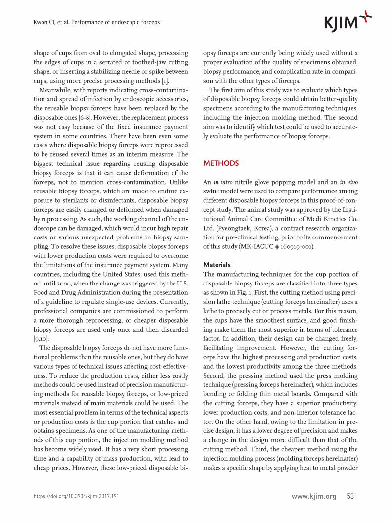
MaterialsThe manufacturing techniques for the cup portion of disposable biopsy forceps are classified into three types as shown in Fig. 1. First, the cutting method using preci-sion lathe technique (cutting forceps hereinafter) uses a lathe to precisely cut or process metals. For this reason, the cups have the smoothest surface, and good finish-ing make them the most superior in terms of tolerance factor. In addition, their design can be changed freely, facilitating improvement. However, the cutting for-ceps have the highest processing and production costs, and the lowest productivity among the three methods. Second, the pressing method used the press molding technique (pressing forceps hereinafter), which includes bending or folding thin metal boards. Compared with the cutting forceps, they have a superior productivity, lower production costs, and non-inferior tolerance fac-tor. On the other hand, owing to the limitation in pre-cise design, it has a lower degree of precision and makes a change in the design more difficult than that of the cutting method. Third, the cheapest method using the injection molding process (molding forceps hereinafter) makes a specific shape by applying heat to metal powder
532 www.kjim.org https://doi.org/10.3904/kjim.2017.191
The Korean Journal of Internal Medicine Vol. 34, No. 3, May 2019
in a designated mold. Having the most superior pro-ductivity among the three methods, it can be mass-pro-duced at a very low cost. However, it has a very coarse metal surface, crude design, and the possibility of hav-ing an ejector pin mark left while removing it from the mold. It also has a very high error rate in production and a very low degree of precision. A secondary precise sur-face finishing can be performed to obtain better quality, but not to avoid an increase in the production costs.
Generally, the cost of producing the cups of the cut-ting forceps is approximately 10 times more than that of the other two methods. The costs per unit of the cups of the pressing and molding forceps are almost same. However, the two methods require mold making, which costs twice more for the pressing forceps than for the molding forceps. In addition, in the lifetime of the mold of the pressing forceps, bending or the folding thin met-al boards can easily induce stress to the mold. Therefore, pressing forceps require more development/higher pro-
duction costs because more-frequent mold replacement is needed than that for the molding forceps.
The following eight different disposable biopsy for-ceps with similar closed cup diameters (2.2 to 2.4 mm) were evaluated: A, cutting forceps, FB-230K, EndoJaw (Olympus Co., Tokyo, Japan); B, pressing forceps, Ra-dial Jaw 4 (Boston Scientific Corp., Natick, MA, USA); C, pressing forceps, DBF-2.4-160-S, Captura (Cook Medical Inc., Winston-Salem, NC, USA); D, molding forceps, BFO160P, Endo-Upex (Upex-Med Corp., Any-ang, Korea); E, molding forceps, FM-EF0003, ClearBite (Finemedix Co. Ltd., Daegu, Korea); F, pressing forceps, BIO1-C4-23-180, EndoBite (Medwork GmbH, Aisch, Germany); G, pressing forceps, IC-FCN23S, Core-For-ceps (Incore Co. Ltd., Seoul, Korea); and H, molding forceps, BF160, Optimos (Taewoong Medical Co. Ltd., Gimpo, Korea). The characteristics of the different for-ceps models are presented in Table 1.
Samples ofcups area
Manufacturing techniques Cutting forceps Pressing forceps Molding forceps
Precise processing bya precision lathe
Smooth surface (red circle)Minimal tolerance factor
Relatively high productivityRelatively low production costs
Highest productivityLowest production costs
High production costsLow productivity
Lower degree of precisionLimitation of modification
Rough surfaceEjector pin mark (red circle)High error rate in producing
Press technique with a thinmetal board (red circles)
Injection molding processwith metal powderCharacteristics
Advantage
Disadvantage
Figure 1. Characteristics of disposable biopsy forceps according to the manufacturing techniques. Cutting forceps, made us-ing a precision lathe technique, are characterized by the smoothest surface and the best finishing. Pressing forceps, made of a thin metal board with a press molding technique, are characterized by higher productivity and lower production cost than cutting forceps. Molding forceps, made with an injection molding process, are characterized by the highest productivity and lowest production cost. However, it has a coarse surface and crude design. Red circles present the typical characteristic areas of each type.
533
Kwon CI, et al. Performance of endoscopic forceps
www.kjim.orghttps://doi.org/10.3904/kjim.2017.191
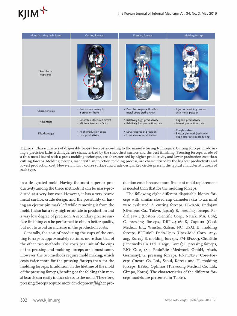
In vivo nitrile glove popping testThis is a commonly used test to determine the grasp-ing ability of biopsy forceps. The tips of air-filled nitrile gloves (Nitrisoft, Kossan Latex Industries Sdn. Bhd., Klang, Malaysia), which are used during endoscopic examination, were grasped and pulled with biopsy for-ceps to determine if there was any cutting or tearing of the gloves (Fig. 2). Cutting and tearing are not generally distinguished when assessing grasping ability, but the cutting phenomenon refers to popping with a sharp cut of the part grasped inside the cups of the biopsy forceps, while tearing phenomenon refers to popping
with a large tear of the surrounding areas and not just a cut of the part grasped inside the cups. These two are not accurately distinguishable, and a better test result is not necessarily indicative of a better grasping abili-ty, which is one weakness of the test. Furthermore, cups with smoothly processed surface may be slippery when attempting to grasp the gloves and thus may not create any popping. This is a very simple, widely used meth-od, but determining whether it can successfully access the performance of biopsy forceps is highly important. Thus, this test was included in this study to determine its objective accuracy. Using four biopsy forceps of each type, 20 popping tests, five times for each forceps, were performed. The biopsy forceps were tested in a random order by a tester (W.J.K.) who was blinded to the type of the biopsy forceps.
In vivo swine studyTwo mini-pigs (Sus scrofa; mean age, 14 months; mean body weight, 30.2 kg) were used for the in vivo swine study. Before this study, the pigs were fasted for > 24 hours, except for water given ad libitum. The pigs were pre-sedated using an intramuscular injection of atro-pine sulfate (0.04 mg/kg), xylazine (2 mg/kg), and tilet-amine-zolazepam (5 mg/kg). Then, the pigs were intu-bated, and general anesthesia was achieved using 0.5% to 2% isoflurane through the endotracheal tube with 70% nitrous oxide and 30% oxygen provided by a venti-lator. For endoscopic examination, the pigs were placed on their left lateral sides on a table.
Using a forward endoscope (GIF-Q260J, Olympus Co.), an expert endoscopist (W.J.K.) who was blinded to
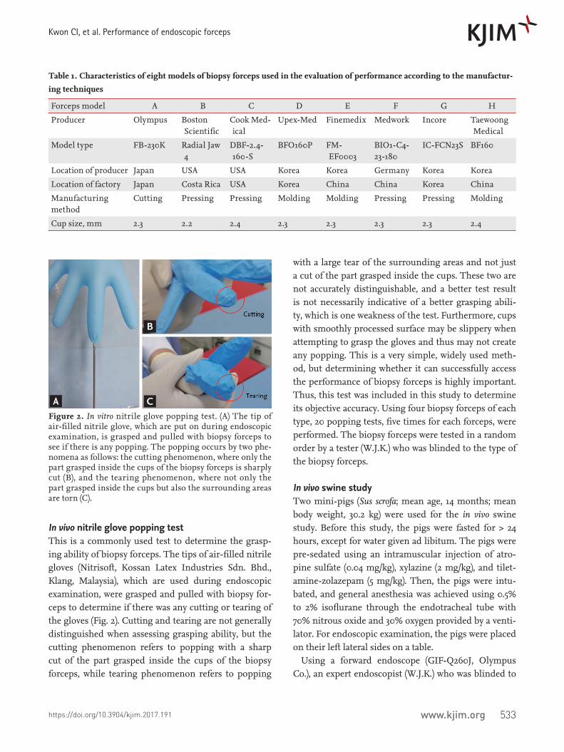
Table 1. Characteristics of eight models of biopsy forceps used in the evaluation of performance according to the manufactur-ing techniques
Forceps model A B C D E F G H
Producer Olympus Boston Scientific
Cook Med-ical
Upex-Med Finemedix Medwork Incore Taewoong Medical
Model type FB-230K Radial Jaw 4
DBF-2.4-160-S
BFO160P FM-EF0003
BIO1-C4-23-180
IC-FCN23S BF160
Location of producer Japan USA USA Korea Korea Germany Korea Korea
Location of factory Japan Costa Rica USA Korea China China Korea China
Manufacturing method
Cutting Pressing Pressing Molding Molding Pressing Pressing Molding
Cup size, mm 2.3 2.2 2.4 2.3 2.3 2.3 2.3 2.4
Figure 2. In vitro nitrile glove popping test. (A) The tip of air-filled nitrile glove, which are put on during endoscopic examination, is grasped and pulled with biopsy forceps to see if there is any popping. The popping occurs by two phe-nomena as follows: the cutting phenomenon, where only the part grasped inside the cups of the biopsy forceps is sharply cut (B), and the tearing phenomenon, where not only the part grasped inside the cups but also the surrounding areas are torn (C).
A
B
C

534 www.kjim.org https://doi.org/10.3904/kjim.2017.191
The Korean Journal of Internal Medicine Vol. 34, No. 3, May 2019
the type of the biopsy forceps performed the endoscop-ic examination with biopsy sampling. Using two pairs of biopsy forceps of each type, five specimens were ob-tained from the greater curvature side of the antrum in one swine and five specimens from the opposite lesser curvature side of the antrum in the other swine (Fig. 3).
Histopathological examinationAll biopsy specimens were examined by an experienced gastrointestinal pathologist (G.K.) who was blinded to the type of the biopsy forceps tested. Each specimen was analyzed for maximal diameter (measured in millime-ter), depth of acquired specimen (lamina propria, mus-cularis mucosa, or submucosa), adequacy for diagnosis (adequate or inadequate; general adequacy of the speci-mens for diagnosis by the experienced gastrointestinal pathologist), and crush artifact (none, mild, moderate, or severe; severe crush artifact can affect the diagnosis) (Fig. 4) [5].
The same technique was used for the preparation of the histology slides for tissues obtained by all forceps. The best specimen from each set was used for statistical analysis. Specimens were fixed with 10% neutral-buff-ered formalin and processed routinely in a Thermo Shandon Exelsior ES tissue processor (Thermo Fisher Scientific, Waltham, MI, USA). Paraffin-embedded tis-sues were sectioned on a microtome at 4 µm and mount-
ed on the glass slides for hematoxylin and eosin stain-ing.
Statistical analysisThe mean ± standard deviation (SD), and range were used to summarize the data for continuous variables and percentages for categorical variables. The Kruskal-Wal-lis test was used to analyze the difference of specimens between the groups. A p value of < 0.05 was considered significant. Statistical analysis was performed with the IBM SPSS Statistics version 21.0.0 software (IBM Co., Armonk, NY, USA).
RESULTS
In vivo nitrile glove popping testBetween the tested types of forceps, there was a sig-nificant difference in popping rate (p < 0.001). Type D (molding forceps) and type F forceps (pressing forceps) had dominant popping rates (85% and 55%, respectively). Between the three types of manufacturing techniques, the molding forceps provided a significantly higher bursting rate than the other forceps (cutting forceps, 25.0%; pressing forceps, 17.5%; and molding forceps, 41.7%; p = 0.006) (Table 2).
Figure 4. Representative images of crush artifacts classified as grade 0 to 3 (H&E, × 200). (A) Grade 0, no crush artifact; (B) grade 1, mild crush artifact; (C, D) grade 2, moderate crush artifact; and grade 3, severe crush artifact that can affect diagnosis. The arrows present the compression areas that were used for classifying the degree of crush artifact, and red dot circles present the parts of tearing tissues.
A
C
B
DFigure 3. Endoscopic examination with biopsy samplings. Five specimens were obtained from the greater curvature side of the antrum in one swine (A, B), and five specimens were obtained from the opposite lesser curvature side of the antrum in the other swine (C, D).
A
C
B
D
535
Kwon CI, et al. Performance of endoscopic forceps
www.kjim.orghttps://doi.org/10.3904/kjim.2017.191
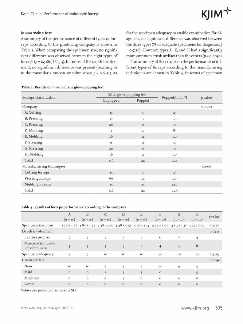
In vivo swine testA summary of the performance of different types of for-ceps according to the producing company is shown in Table 3. When comparing the specimen size, no signifi-cant difference was observed between the eight types of forceps (p = 0.5180) (Fig. 5). In terms of the depth involve-ment, no significant difference was present (reaching % to the muscularis mucosa or submucosa; p = 0.6431). As
for the specimen adequacy to enable examination for di-agnosis, no significant difference was observed between the three types (% of adequate specimens for diagnosis; p = 0.5219). However, types D, E, and H had a significantly more common crush artifact than the others (p = 0.0030).
The summary of the results on the performance of dif-ferent types of forceps according to the manufacturing techniques are shown in Table 4. In terms of specimen
Table 2. Results of in vitro nitrile glove popping test
Forceps classificationNitril glove popping test
Popped/total, % p valueUnpopped Popped
Company < 0.001
A, Cutting 15 5 25
B, Pressing 17 3 15
C, Pressing 20 0 0
D, Molding 3 17 85
E, Molding 16 4 20
F, Pressing 9 11 55
G, Pressing 20 0 0
H, Molding 16 4 20
Total 116 44 27.5
Manufacturing techniques 0.006
Cutting forceps 15 5 25
Pressing forceps 66 14 17.5
Molding forceps 35 25 41.7
Total 116 44 27.5
Table 3. Results of forceps performance according to the company
A(n = 10)
B(n = 10)
C(n = 10)
D(n = 10)
E(n = 10)
F(n = 10)
G(n = 10)
H(n = 10)
p value
Specimen size, mm 3.70 ± 1.22 3.85 ± 1.44 4.48 ± 1.16 3.48 ± 0.53 4.05 ± 1.13 4.24 ± 1.09 4.03 ± 1.37 3.85 ± 0.91 0.5180
Depth involvement 0.6431
Lamina propria 7 7 7 5 8 6 7 4
Muscularis mucosa or submucosa
3 3 3 5 2 4 3 6
Specimen adequacy 9 9 10 10 10 10 10 10 0.5219
Crush artifact 0.0030
None 10 10 9 5 7 10 9 5
Mild 0 0 1 4 3 0 1 5
Moderate 0 0 0 1 0 0 0 0
Severe 0 0 0 0 0 0 0 0
Values are presented as mean ± SD.
536 www.kjim.org https://doi.org/10.3904/kjim.2017.191
The Korean Journal of Internal Medicine Vol. 34, No. 3, May 2019
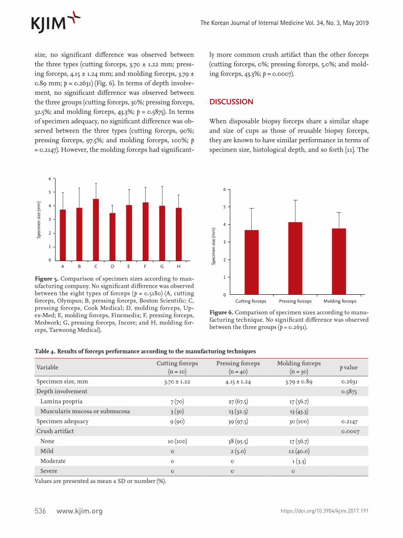
size, no significant difference was observed between the three types (cutting forceps, 3.70 ± 1.22 mm; press-ing forceps, 4.15 ± 1.24 mm; and molding forceps, 3.79 ± 0.89 mm; p = 0.2631) (Fig. 6). In terms of depth involve-ment, no significant difference was observed between the three groups (cutting forceps, 30%; pressing forceps, 32.5%; and molding forceps, 43.3%; p = 0.5875). In terms of specimen adequacy, no significant difference was ob-served between the three types (cutting forceps, 90%; pressing forceps, 97.5%; and molding forceps, 100%; p = 0.2147). However, the molding forceps had significant-
ly more common crush artifact than the other forceps (cutting forceps, 0%; pressing forceps, 5.0%; and mold-ing forceps, 43.3%; p = 0.0007).
DISCUSSION
When disposable biopsy forceps share a similar shape and size of cups as those of reusable biopsy forceps, they are known to have similar performance in terms of specimen size, histological depth, and so forth [11]. The
Spec
imen
size
(mm
)
6
5
4
3
2
1
0A B C D E F G H
Figure 5. Comparison of specimen sizes according to man-ufacturing company. No significant difference was observed between the eight types of forceps (p = 0.5180) (A, cutting forceps, Olympus; B, pressing forceps, Boston Scientific; C, pressing forceps, Cook Medical; D, molding forceps, Up-ex-Med; E, molding forceps, Finemedix; F, pressing forceps, Medwork; G, pressing forceps, Incore; and H, molding for-ceps, Taewoong Medical).
Spec
imen
size
(mm
)
6
5
4
3
2
1
0Cutting forceps Pressing forceps Molding forceps
Figure 6. Comparison of specimen sizes according to manu-facturing technique. No significant difference was observed between the three groups (p = 0.2631).
Table 4. Results of forceps performance according to the manufacturing techniques
VariableCutting forceps
(n = 10)Pressing forceps
(n = 40)Molding forceps
(n = 30)p value
Specimen size, mm 3.70 ± 1.22 4.15 ± 1.24 3.79 ± 0.89 0.2631
Depth involvement 0.5875
Lamina propria 7 (70) 27 (67.5) 17 (56.7)
Muscularis mucosa or submucosa 3 (30) 13 (32.5) 13 (43.3)
Specimen adequacy 9 (90) 39 (97.5) 30 (100) 0.2147
Crush artifact 0.0007
None 10 (100) 38 (95.5) 17 (56.7)
Mild 0 2 (5.0) 12 (40.0)
Moderate 0 0 1 (3.3)
Severe 0 0 0
Values are presented as mean ± SD or number (%).

537
Kwon CI, et al. Performance of endoscopic forceps
www.kjim.orghttps://doi.org/10.3904/kjim.2017.191
performance of biopsy forceps can vary depending on the manufacturing technique of cups, but simple tests such as the glove popping test have been commonly used to assess the performance of biopsy forceps. This is the first study to investigate if there is any difference in performance between the disposable biopsy forceps based on manufacturing techniques and to determine the accuracy of the glove popping test. We showed for the first time that a better result of the glove popping test does not necessarily mean better performance, and that crush artifact, but not specimen size and adequacy, can vary considerably depending on the manufacturing technique of cups.
As mentioned in the introduction section, accesso-ries and endoscopes have had continuous and steady technological advancements. Further development has been restrained by the transition from reusable to disposable accessories. Biopsy forceps are the simplest among endoscopic accessories, with their basic func-tion being specimen obtainment. Therefore, they have advanced in the direction of reducing production cost, rather than technological development. The transition from reusable to disposable biopsy forceps was also a very important issue for manufacturers, and the pro-duction cost could not be reduced without consider-ing the performance of biopsy forceps. For this reason, making disposable biopsy forceps in the same way as reusable biopsy forceps, such as the cutting method using precision lathe technique, was highly inefficient in terms of production cost. For this reason, there was only one company that manufactured cutting forceps. Consequently, pressing and molding forceps were in-troduced sequentially to continue the effort to reduce the production costs. Many third-party companies and major companies in Korea continue to develop new dis-posable biopsy forceps, but most of them focus on low-priced molding forceps. The reality is that the emphasis is predominantly placed on the user’s convenience and the design of the handle rather than the performance of cups. Although some institutions are using the glove popping test to assess the biopsy forceps objectively, the actual association between this test and the performance of forceps has not been reported yet.
The problem of the glove popping test, as mentioned earlier, is that the test result can be good not only when cutting is done accurately with good performance (Fig.
2B) but also when the glove is torn due to the coarse sur-face of the cups (Fig. 2C). As can be seen in Table 2, which shows the results of the glove popping test, the results of the molding forceps were generally good. However, these results bear little similarity when compared with the results of performance shown in Tables 3 and 4. The assumption that the tearing phenomenon by rough sur-face of cups would be the mechanism of glove popping by the molding forceps can be made based on the fact that crush artifact was more frequent than in the other groups. However, because of the very high popping rate in type D forceps, there was a significant bias that in-creased the popping rate of the entire molding forceps. Nevertheless, we think that evaluating the performance of biopsy forceps using the popping test is not accurate because we did not find any differences between the groups. We would like to argue that using the glove pop-ping test alone is not recommended when assessing the performance of biopsy forceps.
Crush artifact, which does not occur when the speci-men grasped inside the cups cuts cleanly, refers to a se-rious compression in the middle part when some of the tissues outside the cups are torn apart because of poor grip ability. As can be seen in Fig. 4, crush artifact can be graded depending on the extent of base tissue, which has not been grasped but has been pulled out without being cut cleanly from the tissue. A severe grade means that the compression is serious enough to affect the di-agnosis [5]. As shown in Table 4, fortunately, none of the three groups had severe crush artifacts, but crush arti-facts were most common with the molding forceps. This means that during an endoscopic biopsy, complications may occur depending on the type of biopsy forceps used and the condition of the base tissue. This also implies that when performing biopsy of deep ulcer or relative-ly thin duodenum or colon, the specimen may not cut cleanly and some of the base tissue can be torn, causing complications, such as bleeding or perforation. A pre-vious report that only compared pressing forceps also reported a 6% to 7% incidence of crush artifact, which was similar to our results [5]. This report also explained that little crush artifact means lack of tissue compres-sion with a good tissue fenestration due to good grip ability of the cups.
The limitations of this study are as follows: (1) in vitro and in vivo animal study; (2) small sample size of biopsy
538 www.kjim.org https://doi.org/10.3904/kjim.2017.191
The Korean Journal of Internal Medicine Vol. 34, No. 3, May 2019
specimens per type of biopsy forceps; (3) performed gas-tric biopsies only; (4) assessing performance simply ac-cording to the manufacturing techniques of cups, given that other parts of biopsy forceps can also affect the per-formance; (5) lack of subtype analysis because not many types of cups were used; (6) lack of analysis of biopsy-in-duced performance complications such as immediate bleeding; and (7) the adequacy of biopsy specimen and crush artifact may depend on the pulling force of the endoscopist. The participation of two or more endosco-pists is recommended for a well-designed future study.
In conclusion, this was the first study to compare the performance of biopsy forceps according to manufac-turing techniques. We found that the molding forceps provided lower performance than the cutting and press-ing forceps in terms of crush artifact. In addition, the simple glove popping test should not be recommended for the evaluation of performance of biopsy forceps.
Conflict of interestJong Pil Moon and Ho Yun are research workers at the Interventional Research Center of M.I.Tech Co. Ltd. which is developing products related to the research be-ing reported. The others have no financial conflicts of interest.
AcknowledgmentsChang-Il Kwon received research support for this study from the Industry & Energy and the National Center of Efficacy Evaluation for the Development of Health Products Targeting Digestive Disorders (NCEED).
REFERENCES
1. Tytgat GN, Ignacio JG. Technicalities of endoscopic biop-sy. Endoscopy 1995;27:683-688.
2. Kaneko E, Kumagai J, Honda N, Nakamura S, Kino I. Evaluation of the new giant-biopsy forceps in the diagno-sis of mucosal and submucosal gastric lesions. Endosco-py 1983;15:322-326.
3. Ladas SD, Tsamouri M, Kouvidou C, Raptis SA. Effect of forceps size and mode of orientation on endoscop-ic small bowel biopsy evaluation. Gastrointest Endosc 1994;40:51-55.
4. Danesh BJ, Burke M, Newman J, Aylott A, Whitfield P, Cotton PB. Comparison of weight, depth, and diagnostic adequacy of specimens obtained with 16 different biopsy forceps designed for upper gastrointestinal endoscopy. Gut 1985;26:227-231.
5. Sussman DA, Deshpande AR, Shankar U, et al. Compar-ison of performance characteristics of oval cup forceps versus serrated jaw forceps in gastric biopsy. Dig Dis Sci 2016;61:2338-2343.
6. O'Connor HJ, Axon AT. Gastrointestinal endoscopy: in-fection and disinfection. Gut 1983;24:1067-1077.
7. Birnie GG, Quigley EM, Clements GB, Follet EA, Watkin-son G. Endoscopic transmission of hepatitis B virus. Gut 1983;24:171-174.
8. Fireman Z. Biopsy forceps: reusable or disposable? J Gas-troenterol Hepatol 2006;21:1089-1092.
9. U.S. Food and Drug Administration. Enforcement prior-ities for single-use devices reprocessed by third parties and hospitals [Internet]. Rockville (MD): FDA, 2000 [cited 2018 Jan 3]. Available from: https://www.fda.gov/medical-devices/deviceregulationandguidance/guidancedocu-ments/ucm107164.htm.
10. Alfa MJ, Castillo J. Impact of FDA policy change on the reuse of single-use medical devices in Michigan hospi-tals. Am J Infect Control 2004;32:337-341.
11. Yang R, Naritoku W, Laine L. Prospective, randomized comparison of disposable and reusable biopsy forceps in gastrointestinal endoscopy. Gastrointest Endosc 1994;40:671-674.
KEY MESSAGE
1. The performance of disposable biopsy forceps can vary depending on the manufacturing tech-nique for their cups.
2. The glove popping test, commonly used to as-sess the performance of biopsy forceps, does not represent the exact performance.
3. In terms of crush artifact, the molding forceps provided lower performance than the cutting and pressing forceps.