Duodenal Hematoma after upper Gastrointestinal Endoscopy ...
Performance measures for lower gastrointestinal endoscopy:a European Society of Gastrointestinal Endoscopy (ESGE) QualityImprovement Initiative
Authors
Michal F. Kaminski1, 2, 3, Siwan Thomas-Gibson4, Marek Bugajski1, 2,
Michael Bretthauer3, 5, Colin J. Rees6, Evelien Dekker7, Geir Hoff3, 8, 9,
Rodrigo Jover10, Stepan Suchanek11, Monika Ferlitsch12, John
Anderson13, Thomas Roesch14, Rolf Hultcranz15, Istvan Racz16,
Ernst J. Kuipers17, Kjetil Garborg3, James E. East18, Maciej Rupinski1, 2,
Birgitte Seip19, Cathy Bennett20, Carlo Senore21, Silvia Minozzi21,
Raf Bisschops22, Dirk Domagk23, Roland Valori24, Cristiano Spada25,
Cesare Hassan26, Mario Dinis-Ribeiro27, 28, Matthew D. Rutter29, 30
Institutions
1 Department of Gastroenterology, Hepatology and Oncology,
Medical Center for Postgraduate Education, Warsaw, Poland
2 Department of Gastroenterological Oncology and Department
of Cancer Prevention, The Maria Sklodowska-Curie Memorial
Cancer Center and Institute of Oncology, Warsaw, Poland
3 Department of Health Management and Health Economics,
Institute of Health and Society, University of Oslo, and
Department of Transplantation Medicine, KG Jebsen Center for
Colorectal Cancer Research, Oslo University Hospital, Oslo,
Norway
4 Wolfson Unit for Endoscopy, St. Mark’s Hospital, Harrow, and
Imperial College, London, UK
5 Department of Transplantation Medicine, Oslo University
Hospital, Oslo, Norway
6 South Tyneside NHS Foundation Trust, South Tyneside, United
Kingdom
7 Department of Gastroenterology and Hepatology, Academic
Medical Center, University of Amsterdam, Amsterdam, The
Netherlands
8 Department of Research and Development, Telemark Hospital,
Skien, Norway
9 Cancer Registry of Norway, Oslo, Norway
10 Unidad de Gastroenterologia, Hospital General Universitario de
Alicante, Alicante, Spain
11 Department of Internal Medicine, First Faculty of Medicine,
Charles University, Military University Hospital, Prague, Czech
Republic
12 Department of Medicine III, Division of Gastroenterology and
Hepatology, Medical University of Vienna, Vienna, Austria
13 Gloucestershire Hospitals NHS Foundation Trust, Cheltenham
General Hospital, Gloucestershire, UK
14 Department of Interdisciplinary Endoscopy, University Hospital
Hamburg-Eppendorf, Germany
15 Karolinska Institute and Karolinska University Hospital,
Stockholm, Sweden
16 Department of Internal Medicine and Gastroenterology, Petz
Aladar County and Teaching Hospital, Györ, Hungary
17 Department of Gastroenterology and Hepatology, Erasmus MC
University Medical Center, Rotterdam, The Netherlands
18 Translational Gastroenterology Unit, John Radcliffe Hospital,
University of Oxford, Oxford, UK
19 Department of Gastroenterology, Vestfold Hospital Trust,
Tønsberg, Norway
20 Centre for Technology Enabled Research, Faculty of Health and
Life Sciences, Coventry University, Coventry, UK
21 CPO Piemonte, AOU Città della Salute e della Scienza, Turin,
Italy
22 Department of Gastroenterology and Hepatology, University
Hospital Leuven and KU Leuven, Leuven, Belgium
23 Department of Internal Medicine, Joseph’s Hospital,
Warendorf, Germany
24 Department of Gastroenterology, Gloucestershire Hospitals
NHS Foundation Trust, Gloucestershire, UK
25 Digestive Endoscopy Unit, Agostino Gemelli University
Hospital, Rome, Italy
26 Nuovo Regina Margherita Hospital, Rome, Italy
27 Center for Health Technology and Services Research
(CINTESIS), Faculty of Medicine, University of Porto, Portugal
28 Servicio de Gastroenterologia, Instituto Portugues de
Oncologia Francisco Gentil, Porto, Portugal
29 Department of Gastroenterology, University Hospital of North
Tees, Stockton-on-Tees, Cleveland, UK
30 School of Medicine, Durham University, UK
Bibliography
DOI http://dx.doi.org/10.1055/s-0043-103411 Published online: 7.3.2017 | Endoscopy 2017; 49 © Georg Thieme Verlag KG Stuttgart · New YorkISSN 0013-726X
This article is published simultaneously in the journals
Endoscopy and the United European Gastroenterology Journal.
Copyright 2017 © Georg Thieme Verlag KG and
© by the United European Gastroenterology
Corresponding author
Michal F. Kaminski, MD PhD, Department of Gastroenterological
Oncology, Institute of Oncology, Roentgen Street 5,
02-781 Warsaw, Poland
Fax: +48-22-5463067
Guideline
Kaminski Michal F et al. Performance measures for … Endoscopy 2017; 49
IntroductionThe European Society of Gastrointestinal Endoscopy (ESGE) andUnited European Gastroenterology (UEG) have identified quali-ty of endoscopy as a major priority. We described our rationalefor this priority in a recent manuscript that also addressed themethodology of the current quality initiative process [1].
Because of the variation in physicians’ performance and theintroduction of nationwide colorectal cancer (CRC) screeningprograms, lower gastrointestinal (LGI) endoscopy was the firstarea of endoscopy to address quality [2–4]. Over more than adecade, several potential measures of quality in LGI endoscopyhave been identified. In consequence, many professional socie-ties have published recommendations on performance meas-ures for LGI endoscopy [5–7]. These recommendations arehowever numerous (44 different performance measures) [5–7], country specific, and not always evidence based, which haslimited their wider adoption in Europe.
The aim of the ESGE LGI working group was to identify ashort list of key performance measures for LGI endoscopy thatwere widely applicable to endoscopy services throughout Eur-ope. This list would ideally consist of performance measureswith the following requirements: proven impact on significant
clinical outcomes or quality of life; a well-defined, reliable, andsimple method/approach for measurement; susceptibility forimprovement; and application to all levels of endoscopy servi-ces.
This paper reports the agreed list of key performance meas-ures for LGI endoscopy and describes the methodological pro-cess applied in the development of these measures.
MethodologyWe previously described the multistep process for producingsuch performance measures [1]. In brief, at the United Europe-an Gastroenterology Week in 2014, we used a modified Delphiconsensus process to develop quality measures in the followingdomains: pre-procedure, completeness of procedure, identifi-cation of pathology, management of pathology, complications,procedure numbers, patient experience, and post-procedure[1, 8, 9]. We decided to have one or two key performance meas-ures for each quality domain.
In order to identify key performance measures, we first cre-ated a list of all possible performance measures for LGI endos-copy through email correspondence and teleconferences thattook place between December 5, 2014 and February 7, 2015.All possible performance measures that were identified by thisprocess were then structured using the PICO framework (whereP stands for Population/Patient; I for Intervention/Indicator; Cfor Comparator/Control, and O for Outcome) to inform sear-ches for available evidence to support the performance meas-ures. This process resulted in 38 PICOs. Detailed literature sear-ches were performed by an expert team of methodologists andyielded results for 29 PICOs (see Supporting Information; avail-able online). Working group members also identified additionalarticles relevant for the performance measures in question.
The PICOs and the clinical statements derived from thesewere adapted or omitted during iterative rounds of commentsand suggestions from the working group members during theDelphi process. The evolution and adaptation of the differentPICOs and clinical statements during the Delphi process can bereviewed in the Supporting Information. The domain addres-sing the competence of endoscopists’ quality (including proce-dure numbers), along with its associated PICOs and clinicalstatements, was moved for future initiatives.
In total, working group members participated in a maximumof three rounds of voting to agree on performance measures inpredefined domains and their respective thresholds, as discus-
ABSTRACTThe European Society of Gastrointestinal Endoscopy and United Eu-
ropean Gastroenterology present a short list of key performance
measures for lower gastrointestinal endoscopy. We recommend
that endoscopy services across Europe adopt the following seven
key performance measures for lower gastrointestinal endoscopy
for measurement and evaluation in daily practice at a center and
endoscopist level:
1 Rate of adequate bowel preparation (minimum standard 90%);
2 Cecal intubation rate (minimum standard 90%); 3 Adenoma de-
tection rate (minimum standard 25%); 4 Appropriate polypectomy
technique (minimum standard 80%); 5 Complication rate (mini-
mum standard not set); 6 Patient experience (minimum standard
not set); 7 Appropriate post-polypectomy surveillance recommen-
dations (minimum standard not set).
Other identified performance measures have been listed as less rel-
evant based on an assessment of their importance, scientific ac-
ceptability, feasibility, usability, and comparison to competing
measures.
ABBREVIATIONS
ADR adenoma detection rateASGE American Society for Gastrointestinal EndoscopyCI confidence intervalCRC colorectal cancerEPAGE European Panel on the Appropriateness of
Gastrointestinal EndoscopyESGE European Society of Gastrointestinal EndoscopyFIT fecal immunochemical testFOBT fecal occult blood testISFU Importance, Scientific acceptability, Feasibility,
UsabilityLGI lower gastrointestinal tractLST laterally spreading tumorPICO population/patient; intervention/indicator;
comparator/control; outcomePDR polyp detection rateQIC Quality Improvement CommitteeUEG United European Gastroenterology
Kaminski Michal F et al. Performance measures for … Endoscopy 2017; 49
Guideline
sed below. Statements were discarded if agreement was notreached over the three voting rounds. The agreement that isgiven for the different statements refers to the last votinground in the Delphi process. The key performance measureswere distinguished from the minor performance measuresbased on the ISFU criteria (Importance, Scientific acceptability,Feasibility, Usability, and comparison with competing meas-ures), and expressed by mean voting scores.
The performance measures are displayed in boxes under therelevant quality domain. Each box describes the performancemeasure, the level of agreement during the modified Delphiprocess, the grading of available evidence (the evidence wasgraded according to the Grading of Recommendations Assess-ment, Development and Evaluation [GRADE] system) [10], howthe performance measure should be measured, and recom-mendations supporting its adoption. The boxes further list themeasurement of agreement (scores), the desired threshold,and suggestions on how to deal with underperformance.
The minimum number needed to assess whether the thresh-old for a certain performance measure is reached can be calcu-lated by estimating the 95% confidence intervals (CIs) aroundthe predefined threshold for different sample sizes [8, 9, 11].For the sake of practicality and to simplify implementation andauditing, we suggest that at least 100 consecutive procedures(or all, if < 100 performed) should be measured to assess a per-formance measure. Continuous monitoring should however bethe preferred method of measurement.
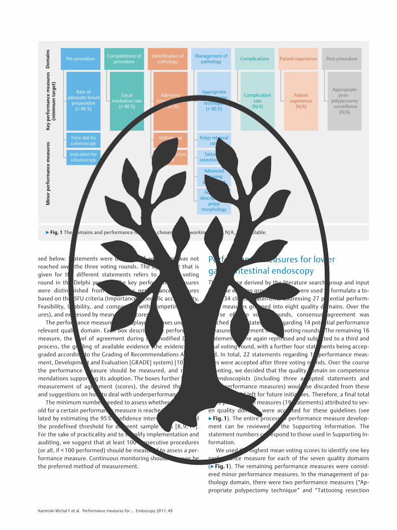
Performance measures for lowergastrointestinal endoscopyThe evidence derived by the literature search group and inputfrom the working group members were used to formulate a to-tal of 34 clinical statements addressing 27 potential perform-ance measures grouped into eight quality domains. Over thecourse of two voting rounds, consensus agreement wasreached for 18 statements regarding 14 potential performancemeasures (agreement in both voting rounds). The remaining 16statements were again rephrased and subjected to a third andfinal voting round, with a further four statements being accep-ted. In total, 22 statements regarding 18 performance meas-ures were accepted after three voting rounds. Over the courseof voting, we decided that the quality domain on competenceof endoscopists (including three accepted statements andthree performance measures) would be discarded from theseguidelines and left for future initiatives. Therefore, a final totalof 15 performance measures (19 statements) attributed to sev-en quality domains were accepted for these guidelines (see
▶Fig. 1). The entire process of performance measure develop-ment can be reviewed in the Supporting Information. Thestatement numbers correspond to those used in Supporting In-formation.
We used the highest mean voting scores to identify one keyperformance measure for each of the seven quality domains(▶Fig. 1). The remaining performance measures were consid-ered minor performance measures. In the management of pa-thology domain, there were two performance measures (“Ap-propriate polypectomy technique” and “Tattooing resection
Dom
ains
Key
perf
orm
ance
mea
sure
s(m
inim
um ta
rget
)M
inor
per
form
ance
mea
sure
s
Pre-procedure Completeness of procedure
Identification of pathology
Management of pathology Complications Patient experience Post-procedure
Rate of adequate bowel
preparation(≥ 90 %)
Cecal intubation rate
(≥ 90 %)
Adenoma detection rate
(≥ 25 %)
Appropriate polypectomy
technique (≥ 80 %)
Time slot for colonoscopy
Withdrawal time
Polyp retrieval rate
Indication for colonoscopy
Polyp detection rate
Tattooing resection sites
Advanced imaging
assessment
Adequate description of
polyp morphology
Complication rate
(N/A)
Patient experience
(N/A)
Appropriate post-
polypectomy surveillance
(N/A)
▶ Fig. 1 The domains and performance measures chosen by the working group.N/A, not available.
Kaminski Michal F et al. Performance measures for … Endoscopy 2017; 49

sites”) that had similar voting scores. We decided to select “Ap-propriate polypectomy technique” as the key performancemeasure for this domain, based on its wider usability and betterfeasibility.
All performance measures were deemed valuable by theworking group members and were obtained after a rigorousprocess, as described above. From a practical viewpoint, it mayhowever be desirable to implement the key performance meas-ures first in units that are not monitoring any performancemeasures at this time. Once a culture of quality measurement(with the aim of improving practice, outcomes, and patient ex-perience) is accepted and software is available, the minor per-formance measures may then further aid the monitoring ofquality in LGI endoscopy. The use of appropriate endoscopy re-porting systems is key to facilitate data retrieval on identifiedperformance measures [12].
All of the performance measures are presented below usingthe descriptive framework developed by the Quality Improve-ment Committee (QIC) and a short summary of the evidencefor the ISFU criteria. The performance measures are listed ac-cording to the domain to which they were attributed (for asummary, see ▶Fig. 1).
1 Domain: Pre-procedureKey per-
formance
measure
Rate of adequate bowel preparation
Description The percentage of patients with an adequatelyprepared bowel
Domain Pre-procedure
Category Process
Rationale It has been shown that the quality of bowel prepara-tion affects the rates of cecal intubation and adeno-ma detectionInadequate bowel preparation results in increasedcosts and inconvenience as the examination has tobe rescheduled or alternative investigations have tobe organized
Construct Denominator: Patients undergoing colonoscopyNumerator: Patients in the denominator with ade-quate bowel preparation (assessed with a validatedscale, preferably the Boston Bowel Preparation Scale[BBPS; score≥6], Ottawa Scale [score≤7],Aronchick Scale [excellent, good or fair])Exclusions: Emergency colonoscopiesCalculation: Proportion (%)Level of analysis: Service and individual levelFrequency: Continuous monitoring using novelendoscopy reporting systems [12] should be thepreferred approach; an alternative approach is ayearly audit of a sample of 100 consecutive LGIendoscopies
Key per-
formance
measure
Rate of adequate bowel preparation
Standards Minimum standard:≥90%Target standard:≥95%Bowel preparation quality, assessed using a valida-ted scale, such as the BBPS, the Ottawa Scale, or theAronchick Scale, should be included in everycolonoscopy reportIf the minimum standard is not reached, analysis ofthe factors influencing bowel preparation should beperformed on a service level (information given topatients, dietary restrictions, cleansing agent used,colonoscopy timing)After evaluation and adjustment, close monitoringshould be performed with a further audit within 6months
Consensusagreement
100%
PICO 1.1–1.2 (see Supporting Information)
Evidencegrading
Moderate quality evidence
The acceptance of this performance measure is based on agree-ment with the following statements:▪ In patients undergoing screening or diagnostic colonoscopy,
bowel preparation quality should be recorded using a vali-dated scale with high intraobserver reliability. (Statementnumber N1.1) Agreement: 100%
▪ A service should have a minimum of ≥90% procedures and atarget of ≥95% procedures with adequate bowel prepara-tion, assessed using a validated scale with high intraobserverreliability. (N1.2) Agreement: 100%
The quality of bowel preparation is important for the efficacy ofcolonoscopy. As pointed out in the ESGE guidelines on bowelpreparation for colonoscopy [13], the quality of bowel prepara-tion is associated with two other important performance meas-ures for colonoscopy, namely adenoma detection rate (ADR)and cecal intubation rate [14]. Suboptimal bowel preparationresults in further costs and inconvenience because the exami-nation has to be repeated or an alternative examination has tobe arranged [15].
To determine the scientific acceptability of measuring bowelpreparation quality, we focused on the performance of differ-ent bowel preparation scales and the quantification of ade-quacy of bowel preparation. There were no direct comparisonsof performance between the bowel preparation scales (seeSupporting Information). Three bowel preparation scales haveundergone comprehensive validation and have shown suffi-cient validity and reliability: the Boston Bowel Preparation Scale(BBPS) [16], the Ottawa Scale [17], and the Aronchick Scale[18]. The BBPS is the most thoroughly validated scale andshould be the preferred one [19]. There were no significant dif-ferences between intermediate and high quality bowel prepa-ration (regardless of the scale used) in terms of the detectionrates for adenomas or advanced adenomas (see Supporting In-
Kaminski Michal F et al. Performance measures for … Endoscopy 2017; 49
Guideline

formation) [20]. Therefore, adequate bowel preparation maybe defined as: BBPS ≥6, Ottawa Scale ≤7, or Aronchick Scale ex-cellent, good, or fair. The adoption of validated scales for bowelpreparation quality assessment has been proven to be feasiblein routine practice [21].
The proposed minimum (≥90%) and target standard (≥95%)rates of adequate bowel preparation were based on values re-ported in recent population-based studies [22–24] and on ran-domized clinical trials of split-dose bowel cleansing regimens[25, 26], respectively.
Minor
perform-
ance
measure
Time slot allotted for colonoscopy
Description Time allotted for each colonoscopy in dailyschedule
Domain Pre-procedure
Category Structure
Rationale Colonoscopy needs adequate time allocated for theentire procedure (including discussion with the pa-tient, sedation, insertion, withdrawal, and therapy)Time pressure due to inadequate time slots mayimpair colonoscopy quality
Construct Denominator: Number of colonoscopies scheduledin an outpatient colonoscopy list (session)Numerator: Outpatient colonoscopy list (session)working hoursExclusions: Emergency colonoscopyCalculation: Average time length (minutes)Level of analysis: Service levelFrequency: Two-yearly check of booking log
Standards Minimum standard: 30 minutes for clinical andprimary screening colonoscopy; 45 minutes forcolonoscopy following positive fecal occult bloodtestingTarget standard: no target standard setIf the minimum standard is not reached, a systematicapproach to schedule modification shouldbe applied
Consensusagreement
100%
PICO 1.3 (see Supporting Information)
Evidencegrading
No evidence
The acceptance of this performance measure is based on agree-ment with the following statement:▪ Colonoscopy needs adequate time allocated for insertion,
withdrawal, and therapy. Routine colonoscopy should be al-located a minimum of 30 minutes. Colonoscopies followingpositive fecal occult blood testing should be allocated aminimum of 45 minutes to allow for therapeutic interven-tion. (N1.3) Agreement: 100%
There is some evidence that productivity pressure may nega-tively affect the quality of colonoscopy [27]. Although it hasbeen shown that working behind schedule is not associatedwith lower ADRs [28], the effect of a very tight schedule on co-lonoscopy performance is unknown (see Supporting Informa-tion). The working group members suggested that 30 minutesand 45 minutes are minimum times that should be allotted forroutine colonoscopy and colonoscopy after positive fecal occultblood testing (longer time to accommodate high prevalence oflarge polyps), respectively. These values correspond well withmean total procedure times for colonoscopy reported in recentstudies [29, 30].
Minor
perform-
ance
measure
Indication for colonoscopy
Description The colonoscopy report should include an explicitindication for the procedure, categorized accordingto existing guidelines on appropriate use of colonos-copy (the ASGE or the EPAGE II guidelines)
Domain Pre-procedure
Category Process
Rationale Colonoscopies with an appropriate indication areassociated with higher diagnostic yield for relevantlesions than colonoscopies without an appropriateindication
Construct Denominator: All colonoscopies performedNumerator: Colonoscopies with appropriate and“uncertain” indication (according to ASGE orEPAGE II)Exclusions: NoneCalculation: Proportion (%)Level of analysis: Service levelFrequency: Continuous monitoring using novelendoscopy reporting systems [12] should be thepreferred approach; an alternative approach isa yearly audit of a sample of 100 consecutive LGIendoscopies
Kaminski Michal F et al. Performance measures for … Endoscopy 2017; 49

Minor
perform-
ance
measure
Indication for colonoscopy
Standards Minimum standard:≥85%Target standard:≥95%All reports from colonoscopies performed shouldinclude an appropriate indication according to theASGE or EPAGE II guidelinesWhen performed for screening, the colonoscopyreport should state this and it must be ensured thatthe subject meets the criteria for screeningA colonoscopy reporting system with a drop-downmenu for indication is ideal to ensure properrecording of the indication and later auditingIf the minimum standard is not met, a systematicapproach to validate the appropriateness of colonos-copies should be applied (i. e. validation of appro-priateness before colonoscopy scheduling)After evaluation and adjustment, close monitoringshould be performed with a further audit within6 months
Consensusagreement
93.8%
PICO 1.4 (see Supporting Information)
Evidencegrading
Moderate quality evidence
The acceptance of this performance measure is based on agree-ment with the following statement:▪ For audit purposes, the colonoscopy report should include
an explicit indication for the procedure, categorized accord-ing to existing guidelines on appropriateness of colonoscopyuse. (N1.4) Agreement: 93.8%
Appropriate referrals for colonoscopy may help to optimize theuse of limited resources and protect patients from the potentialharms of unnecessary invasive procedures. Colonoscopies withan appropriate indication are associated with significantly high-er diagnostic yields for cancer and other relevant lesions thancolonoscopies without an appropriate indication [31–34]. TheAmerican Society for Gastrointestinal Endoscopy (ASGE) andthe European Panel on the Appropriateness of GastrointestinalEndoscopy (EPAGE) II guidelines on the appropriateness of colo-noscopy use [35, 36] consistently show 67%–96% sensitivityand 13%–40% specificity for the detection of relevant findings(see Supporting Information) [31–34].
The proposed minimum standard of appropriate indicationfor colonoscopy (≥85%) was based on values achieved in stud-ies from academic and non-academic centers over the last 5years [32, 33,37]. The use of appropriate endoscopy reportingsystems with a drop-down menu for indication is key to facili-tate data acquisition for this performance measure [12].
2 Domain: Completeness of procedureKey per-
formance
measure
Cecal intubation rate
Description The percentage of colonoscopies reaching andvisualizing the whole cecum and its landmarks
Domain Completeness of procedure
Category Process
Rationale Whole bowel examination is a prerequisite forcomplete and reliable inspection of the mucosa insearch of lesionsA low cecal intubation rate is associated with anincreased risk of interval colorectal cancerIncomplete colonoscopy leads to increased costs andinconvenience as the examination has to be repeated
Construct Denominator: All screening or diagnostic colonos-copiesNumerator: Procedures in the denominator thatreport reaching the cecum (documented in writtenform and by photo/video)Exclusions:▪ Therapeutic procedures with no indication to
reach the cecum▪ Emergency colonoscopiesCalculation: Proportion (%)Level of analysis: Service and endoscopist levelFrequency: Continuous monitoring using novelendoscopy reporting systems [12] should be thepreferred approach; an alternative approach is ayearly audit of a sample of 100 consecutive LGIendoscopies
Standards Minimum standard:≥90%Target standard:≥95%Cecal intubation, meaning complete visualizationof the whole cecum and its landmarks, should bedocumented in a written report, as well as with photoor video documentationIf the minimum standard is not reached for an individ-ual endoscopist, additional training should be offeredIf the minimum standard is not reached on a servicelevel, an audit to determine the cause should beperformedAfter evaluation and adjustment, close monitoringshould be performed with a further audit within6 months
Consensusagreement
97.9%
PICO 2.1–2.3 (see Supporting Information)
Evidencegrading
Moderate quality evidence
Kaminski Michal F et al. Performance measures for … Endoscopy 2017; 49
Guideline
The acceptance of this performance measure is based on agree-ment with the following statements:▪ Complete colonoscopy requires cecal intubation with
complete visualization of the whole cecum and itslandmarks. (N2.1) Agreement: 100%
▪ A service should have a minimum unadjusted cecal intuba-tion rate of≥90% and a target rate of≥95% as a measure ofthe completeness of colonoscopy examination. (N2.2)Agreement: 93.8%
▪ Complete colonoscopy (cecal intubation) should be docu-mented both in written form and in a photo or video report.(N2.3) Agreement: 100%
Cecal intubation is a prerequisite for complete visualization ofthe colorectum. Cecal intubation must be confirmed with pho-to or video documentation. Clear cecal image documentation isassociated with a higher polyp detection rate (PDR) [38]. Forthe purpose of colorectal neoplasia detection, terminal ileumintubation is useful only to confirm completion of the colonos-copy when classic cecal landmarks are not confidently seen[39].
Failed cecal intubation results in further costs and inconveni-ence as the examination must be rescheduled or an alternativeinvestigation organized. A cecal intubation rate <80% is asso-ciated with significantly higher risks of proximal and distal in-terval CRCs when compared with higher completion rates [40].Adjustment of the cecal intubation rate for inadequate bowelpreparation or impassable strictures makes the measurementless feasible and harbors the risk of gaming. In recent large pop-ulation-based studies, unadjusted cecal intubation rates alwaysexceeded 90% and were usually above 95% [22, 41–45]. Theeffect of raising the target standard beyond the minimum of95% is uncertain.
3 Domain: Identification of pathologyKey per-
formance
measure
Adenoma detection rate (ADR)
Description Percentage of colonoscopies with at least oneadenoma identified
Domain Identification of pathology
Category Process
Rationale ADR reflects adequate inspection of the bowelmucosaADR is associated with interval CRC and CRC death,with improvement in the ADR lowering the risk forCRC and CRC death
Key per-
formance
measure
Adenoma detection rate (ADR)
Construct Denominator: All colonoscopies in patients aged50 years or olderNumerator: Procedures in the denominator in whichat least one adenoma was identifiedExclusions:▪ Emergency colonoscopy▪ Endoscopy with a specific therapeutic indication,
including work-up of a previously detected lesionor follow-up of disease activity in inflammatorybowel disease
Calculation: Proportion (%)Level of analysis: Service and endoscopist levelFrequency: Continuous monitoring using novelendoscopy reporting systems [12] should be thepreferred approach; an alternative approach is ayearly audit of a sample of 100 consecutive LGIendoscopies
Standards Minimum standard:≥25%Target standard: no current target standard definedADR should be monitored in all settings (screeningand out-patient), which requires routine access tohistopathology reportsIf the minimum standard is not met by an individualendoscopist, appropriate feedback followed by acompetence assessment (with special considerationof withdrawal time and technique) should be givenIf the minimum standard is not met on a service level,comprehensive training for the center leader shouldbe considered
Consensusagreement
100%
PICO 3.1–3.4 (see Supporting Information)
Evidencegrading
Moderate to high quality evidence
The acceptance of this performance measure is based on agree-ment with the following statement:▪ Adenoma detection rate should be used as a measure of
adequate inspection at screening or diagnostic colonoscopyin patients aged 50 years or more. (N3.1) Agreement: 100%
The detection and removal of adenomas, which are major pre-cursor lesions for CRC, is seen as a key aspect of CRC preven-tion. However, there is a wide variation between endoscopistsin terms of their skills at detecting adenomas, expressed as theADR [22, 43, 46–48]. ADR has been inversely associated withthe risk of interval CRC [46] and CRC death [47]. A similar rela-tionship with the incidence of distal interval CRC was confirmedfor flexible sigmoidoscopy screening [49]. Of note, the detec-tion rate of serrated polyps has been shown to strongly corre-late with the ADR [43]. Although ADR is considered a surrogatefor meticulous inspection of the colorectal mucosa, the correla-tion with other important, but non-neoplastic, findings hasnever been studied.
Kaminski Michal F et al. Performance measures for … Endoscopy 2017; 49

Several interventions, including education, creating aware-ness, feedback, and benchmarking on colonoscopy quality,have all helped to improve the ADR [50–53]. Recently, it hasbeen shown that an improved ADR translates to risk reductionsfor interval CRC and death, which closes the quality improve-ment loop [54].
It has been postulated that ADR has an inherent limitation ofnot measuring the total number of adenomas detected [41]. Apotentially more accurate measure, namely number of adeno-mas per colonoscopy, has been proposed, but this was provennot to be superior to ADR in a recent study [55].
It is challenging to set the standards for ADR, especially inpopulations enriched with fecal occult blood test (FOBT)-posi-tive patients. In a primary colonoscopy screening setting, a 1%increase in ADR predicted a 3% decrease in the risk of intervalCRC within the observed ADR range of 7.35%–52.5% [47]. Inanother study, an ADR above 24.6% was associated with a re-duced risk of interval CRC and subsequent death [54]. In recentpopulation-based studies, a proposed minimum standard ADRof 25% was met by the majority of endoscopists [22, 47, 51]. Infecal immunochemical test (FIT) positive-enriched populations,the minimum standard may need to be higher; however, the ex-act value is yet to be established.
Minor per-
formance
measure
Withdrawal time
Description Time spent on withdrawal of the endoscope fromcecum to anal canal and inspection of the entirebowel mucosa at negative (no biopsy or therapy)screening or diagnostic colonoscopy
Domain Identification of pathology
Category Process
Rationale A mean withdrawal time of 6 minutes or longer wasassociated with higher ADRs and lower intervalcancer rates as compared to shorter withdrawaltimes
Construct Withdrawal time is measured from cecum to analsphincterDenominator: Number of negative (no biopsy/therapy) screening or diagnostic colonoscopiesNumerator: Sum of withdrawal time in colonoscopiesincluded in the numeratorExclusions:▪ Emergency colonoscopy▪ Incomplete colonoscopyCalculation: Mean time in minutesLevel of analysis: Endoscopist levelFrequency: Measured only if the ADR is insufficient,using a sample of 100 consecutive colonoscopies
Minor per-
formance
measure
Withdrawal time
Standards Minimum standard: mean 6 minutesTarget standard: mean 10 minutesTime can be measured by different methods:stopwatch operated by a nurse, time stamp onphotodocumentation of the cecum and rectum,length of video recording, or external device (thisrequires inclusion of the withdrawal time in thecolonoscopy report)Withdrawal time should be measured only when theADR is insufficientFeedback on mean withdrawal time should be givento endoscopists
Consensusagreement
87.5%
PICO 3.6 (see Supporting Information)
Evidencegrading
Moderate quality evidence
The acceptance of this performance measure is based on agree-ment with the following statement:▪ A mean withdrawal time of at least 6 minutes should be used
as a supportive measure of adequate identification of pa-thology at negative screening or diagnostic colonoscopy.(N3.6) Agreement: 87.5%
Colonoscope withdrawal time provides information about thetime that endoscopists spend identifying pathology. A meanwithdrawal time of > 6 minutes has been associated with higherADRs [56]. Although the association between withdrawal timeand ADR was not observed in all studies [57], a recent largepopulation-based analysis confirmed the positive relation be-tween these two measures, with a 3.6% absolute increase inADR per minute increase in withdrawal time [24]. Importantly,the latter study also showed an inverse association betweenmean withdrawal time and the incidence of interval CRC [24].The observed association was not linear and the risk of intervalCRC leveled off at a mean withdrawal time of 8 minutes (themost significant difference was observed for the 6-minute cut-off). In another study, an increase in mean withdrawal time be-yond 10 minutes had minimal effect on ADR [58]. Therefore,the minimum standard mean withdrawal time of 6 minutesand the target standard of 10 minutes are quite well defined.
Monitoring withdrawal time or institution policy on withdra-wal time above a certain threshold showed inconsistent effectson ADRs [59–61]. The explanation could be that the variationin withdrawal technique is more important than the withdrawaltime [62]. Therefore, it appears that the withdrawal time is par-ticularly useful as a supportive tool when the observed ADR isless than the minimum standard of 25% [63].
Kaminski Michal F et al. Performance measures for … Endoscopy 2017; 49
Guideline

Minor per-
formance
measure
Polyp detection rate (PDR)
Description Percentage of colonoscopies in patients aged 50 yearsor older in which at least one polyp was identified
Domain Identification of pathology
Category Process
Rationale PDR reflects adequate inspection of bowel mucosaPDR correlates with ADR and polypectomy rate isweakly associated with interval CRC risk
Construct Denominator: All screening and diagnostic colonos-copies in patients aged 50 years or olderNumerator: Procedures in the denominator with atleast one polyp identifiedExclusions:▪ Emergency colonoscopy▪ Endoscopy with a specific therapeutic indication,
including work-up of a previously detected lesionor follow-up of disease activity in inflammatorybowel disease
Calculation: Proportion (%)Level of analysis: Service and endoscopist levelFrequency: Continuous monitoring using novelendoscopy reporting systems [12] should be thepreferred approach; an alternative approach is ayearly audit of a sample of 100 consecutive LGIendoscopies
Standards Minimum standard: 40%Target standard: no current target standard definedPDR is an approximation of ADR and should only beused when there is limited access to histopathologyreports; however, caution is needed because PDR issusceptible to gamingIf the minimum standard is not met, there should bean attempt to obtain histopathology reports andcalculate the ADR
Consensusagreement
84.6%
PICO 3.1 (see Supporting Information)
Evidencegrading
Low quality evidence.
The acceptance of this performance measure is based on agree-ment with the following statement:▪ Polyp detection rate should be used as a measure of ade-
quate inspection at screening or diagnostic colonoscopy inpatients aged 50 years or more. (N3.5) Agreement: 84.6%
PDR is a surrogate for ADR and is more feasible to measure as itdoes not require histological verification. In some studies, PDRhas been shown to correlate well with ADR [64–66]; however,in others the correlation was poor for polyps in the distal colo-rectum [67, 68]. In one study, polypectomy rates of at least 25%were associated with a significantly lower risk of proximal inter-val CRC [40]. In a recent study, PDR was found to be non-infer-ior to ADR in predicting the risk of interval CRC [55]. With anaverage adenoma to polyp detection quotient of 0.64, the
minimum standard PDR was estimated at 40%, which corre-sponds with an ADR of 25% [66]. The detection of adenomasand non-neoplastic polyps are however associated, which mayinflate the PDR [67]. The use of PDR instead of ADR could there-fore be considered if there is limited availability of histopathol-ogy data, accepting the potential risks of gaming. We note thatthe increased pressure on quality may force endoscopists to de-tect and remove non-neoplastic lesions that would otherwisebe undetected so as to inflate the rate of detection of “so-called” polyps.
4 Domain: Management of pathologyKey per-
formance
measure
Appropriate polypectomy technique
Description Adequate resection technique of colorectal polypsincludes biopsy forceps removal of polyps≤3mm insize, and snare (cold or with diathermy) polypecto-my for larger polyps. Polyp size estimated by endos-copists has to be included in the endoscopy report
Domain Management of pathology
Category Process
Rationale Inappropriate polypectomy technique increases therisk of incomplete polyp removalIncomplete polyp removal leads to further costs andinconvenience as the examination has to be repeatedIncomplete polyp removal is also considered tocontribute to the development of interval CRCs
Construct Denominator: Polyps > 3mm in size removed atcolonoscopy (polyp size estimated by endoscopist)Numerator: Polyps in the denominator removedwith snare polypectomy (cold or with diathermy)Exclusions: NoneCalculation: proportion (%)Level of analysis: Service and endoscopistFrequency: Continuous monitoring using novelendoscopy reporting systems [12] should be thepreferred approach; an alternative approach is ayearly audit of a sample of 100 consecutive LGI en-doscopies
Standards Minimum standard: ≥80%Target standard: ≥90%Colonoscopy reports must include information onpolyp resection techniqueIf the minimum standard is not met, the rate ofcomplete polyp resection should be measured andfeedback should be given to the endoscopist orservice. Additional training on basic polypectomytechnique should be consideredAfter evaluation and adjustment, close monitoringshould be performed with a further audit within6 months
Consensusagreement
93.3%
PICO 4.6 (see Supporting Information)
Evidencegrading
Low quality evidence
Kaminski Michal F et al. Performance measures for … Endoscopy 2017; 49
The acceptance of this performance measure is based on agree-ment with the following statement:▪ Adequate resection technique of small and diminutive colo-
rectal polyps includes biopsy forceps removal of polyps ≤3mm in size and snare polypectomy for larger polyps. (N4.6)Agreement: 93.3%
Incomplete polypectomy is considered the cause for up to25% of interval CRCs [69, 70]. Incomplete resection of polyps5–20mm in size varies from 6.5% to 22.7% among endos-copists [71]; however, completeness of polyp resection isconsidered challenging to measure, and statements regard-ing this topic have not reached agreement in the currentDelphi process (see Supporting Information).
Biopsy forceps resection of polyps 4–5mm in size or largerhas been shown to be inferior to snare techniques, with regardto completeness of resection [72, 73]. Therefore, the appropri-ate resection technique for colorectal polyps includes biopsyforceps removal of polyps ≤3mm in size, and snare (cold orwith diathermy) polypectomy for larger polyps. Despite this, ina recent large cohort study, it was demonstrated that 28.2% oflesions ≥5mm in size were resected using biopsy forceps in-stead of a snare technique [74]. Contrary to this, in a largestudy from the UK, over 90% of polyps larger than 3mm in sizewere removed using a snare [75].
There are insufficient data to set the minimum and targetstandards reliably, but the proposed values for the use of ap-propriate polypectomy techniques of ≥80% and ≥90%, respec-tively, seem relatively easy to achieve.
Minor per-
formance
measure
Tattooing resection sites
Description In patients undergoing removal of colorectal non-pedunculated lesions 20mm in size or larger, or withsuspicious macroscopic features regardless of size,the resection site should be tattooed to improve fu-ture re-location of the resection site
Domain Management of pathology
Category Process
Rationale Facilitates detection of the post-polypectomy site atsurveillance colonoscopy or surgical resection
Construct Tattooing the resection site of the abovementionedlesions should be applied in all cases. A service mustprovide appropriate equipmentDenominator: Colonoscopies with removal of non-pedunculated lesions 20mm in size or larger, or withsuspicious macroscopic features regardless of sizeNumerator: Procedures in the denominator wherethe resection site was marked with a tattooExclusions: NoneCalculation: Proportion (%)Level of analysis: Service levelFrequency: Continuous monitoring using novelendoscopy reporting systems [12] should be thepreferred approach; an alternative approach is a3-yearly audit of all colonoscopies performed over a3-month period
Minor per-
formance
measure
Tattooing resection sites
Standards Minimum standard: UnknownTarget standard: 100%Every endoscopy report for procedures whereremoval of the abovementioned lesions wasperformed should include written information ontattooing the resection siteIf tattooing is not performed in all cases, feedbackshould be given to the service and all endoscopists
Consensusagreement
93.3%
PICO 4.5 (see Supporting Information)
Evidencegrading
Very low quality evidence
The acceptance of this performance measure is based on agree-ment with the following statement:▪ In patients undergoing removal of colorectal lesions with a
depressed component (0-IIc, according to the Paris classifi-cation) or non-granular or mixed-type laterally spreadingtumors, located between the ascending and the sigmoidcolon, the resection site should be tattooed to improvefuture re-location of the resection site. (N4.1) Agreement:93.3%
Colorectal lesions with a depressed component and non-granu-lar or mixed-type laterally spreading tumors (LSTs) harbor an in-creased risk of malignancy [76–78]. Therefore, the site ofendoscopic removal of these lesions often needs to be re-loca-ted to identify recurrence or to guide surgical management. Ithas been shown that tattooing significantly shortens the timeto re-locate the resection site on endoscopy [79]. There is how-ever no evidence that tattooing the resection site increases therate of re-location of lesions (see Supporting Information). Pre-operative tattooing using prepacked kits was proven to be avery effective method of tumor localization in laparoscopic sur-gery [80]. Moreover, some studies have shown that tattooingimproves lymph node yield and facilitates the harvesting of sus-picious lymph nodes during colorectal surgery [81, 82].
Although the accepted statement focused only on lesionswith an increased risk of malignancy, for audit purposes it willbe much more feasible to track the tattooing of resection sitesfor all lesions larger than 20mm in size. These lesions are fre-quently removed piecemeal, which increases the risk of recur-rence [83], and have a considerable risk of malignancy [84].The minimum standard for tattooing resection sites is un-known.
Kaminski Michal F et al. Performance measures for … Endoscopy 2017; 49
Guideline

Minor per-
formance
measure
Polyp retrieval rate
Description Percentage of polyps removed that were retrievedfor histopathology
Domain Management of pathology
Category Process
Rationale The retrieval of polyps is required forhistopathological diagnosis and is a prerequisite forrecommendations on proper post-polypectomysurveillance interval
Construct Denominator: Polypectomies of polyps > 5mmNumerator: Polyps in the denominator that wereretrieved for histopathology examinationExclusions: Removal of diminutive polyps (≤5mm)Calculation: Proportion (%)Level of analysis: Service and endoscopist levelFrequency: Continuous monitoring using novelendoscopy reporting systems [12] should be thepreferred approach; an alternative approach is ayearly audit of a sample of 100 consecutive LGIendoscopies
Standards Minimum standard: ≥90%Target standard: ≥95%Colonoscopy reports must include information onnon-retrieval of non-diminutive polypsIf the minimum standard is not reached, feedbackshould be given on the importance of thisperformance measure
Consensusagreement
86.7%
PICO no PICO (see Supporting Information)
Evidencegrading
Very low quality evidence
The acceptance of this performance measure is based on agree-ment with the following statement:▪ The non-diminutive polyp retrieval rate should be moni-
tored. A service should have a polyp retrieval rate of ≥90%.(N4.2) Agreement: 86.7%
The retrieval of polyps after endoscopic resection is a “sine quanon” requirement for histopathology examination. Histopa-thology examination guides further management includingpost-polypectomy surveillance. Diminutive polyps (≤5mm insize) harbor a very low risk of cancer or advanced histologyand are considered amenable for a resect-and-discard policyfollowing in vivo optical diagnosis under strictly controlled con-ditions [85]. Furthermore, diminutive polyps are frequently re-moved using biopsy forceps, which makes their retrieval quitestraightforward.
It has therefore been decided to monitor only the retrieval ofpolyps larger than 5mm in size. Their retrieval is not only moreimportant from the clinical perspective but also technicallymore difficult because it requires the transected polyp to besuctioned into a trap, ensnared, or grasped using a Roth net,so that it can be removed together with the endoscope
[86, 87]. Even though the need for polyp retrieval seems ob-vious, it is unknown what the effect of substandard retrieval ison repeat colonoscopy rates or the appropriateness of recom-mended post-polypectomy surveillance.
The proposed minimum standard (≥90%) and target stand-ard (≥95%) for polyp retrieval rate were based on values re-ported in recent large studies [41, 45, 88, 89]. Polyp retrievalrate seems feasible to measure and is amenable for improve-ment through education and competitive feedback [90].
Minor per-
formance
measure
Advanced imaging assessment
Description In patients undergoing removal of colorectal lesionswith a depressed component (0-IIc, according to theParis classification) or non-granular or mixed-typelaterally spreading tumors (LSTs), conventional orvirtual chromoendoscopy should be used to improvedelineation of the lesion margins and to predict thepotential depth of invasion
Domain Management of pathology
Category Process
Rationale Polyps with a depressed component (0-IIc) and non-granular or mixed type LSTs harbor a higher risk ofsubmucosal invasionSuch polyps frequently have indistinct borders, there-fore better margin delineation is warrantedImproved delineation and prediction of deep invasionmay optimize management of these lesions
Construct Advanced imaging assessment should always beused before an attempt to remove the abovemen-tioned lesions. A service offering removal of thesetypes of lesions must provide dedicated equipmentDenominator: Colonoscopies with removal of le-sions with a depressed component (0-IIc) or non-granular or mixed-type LSTsNumerator: Procedures in the denominator wherevirtual or conventional chromoendoscopy was usedto improve delineation of the lesion margins (de-scribed in the report)Exclusions: NoneCalculation: Proportion (%)Level of analysis: Service and endoscopistFrequency: Continuous monitoring using novelendoscopy reporting systems [12] should be thepreferred approach; an alternative approach is a3-yearly audit of all colonoscopies performed over a3-month period
Standards Minimum standard: UnknownTarget standard: 100%If the target standard is not met, feedback on theappropriate use of advanced imaging assessment iswarrantedAt a service level, the availability of equipment shouldbe analyzed and facilitatedAfter evaluation and adjustment, close monitoringshould be performed with a further audit within6 months
Consensusagreement
93.3%
PICO 4.4 (see Supporting Information)
Kaminski Michal F et al. Performance measures for … Endoscopy 2017; 49
Minor per-
formance
measure
Advanced imaging assessment
Evidencegrading
No evidence
The acceptance of this performance measure is based on agree-ment with the following statement:▪ In patients undergoing removal of colorectal lesions with a
depressed component (0-IIc, according to the Paris classi-fication) or non-granular or mixed-type laterally spreadingtumors, conventional or virtual chromoendoscopy should beused to improve delineation of lesion margins and predictpotential depth of invasion. (N4.4) Agreement: 93.3%
In 2014, the ESGE issued guidelines on advanced endoscopicimaging for the detection and differentiation of colorectal neo-plasia in which it suggested the use of advanced endoscopicimaging for margin assessment and prediction of deep submu-cosal invasion in lesions with a depressed component (0-IIc) ornon-granular or mixed-type LSTs [85]. The quality of evidencesupporting these recommendations was considered very lowand moderate for margin delineation and assessment of depthof submucosal invasion, respectively. Since then no new evi-dence with clinically relevant endpoints for the patients (in-complete resection, interrupted procedure, cancer detection)has been published to further support its use (see SupportingInformation).
The availability, feasibility, and minimum standard of ad-vanced imaging use, particularly in the community setting, areunknown. Colonoscopy services should set up structured mon-itoring and initiate audit to generate further evidence for ad-vanced imaging.
Minor per-
formance
measure
Adequate description of polyp morphology
Description The Paris classification should be routinely used todescribe the morphology of non-pedunculatedlesions identified at colonoscopy
Domain Management of pathology
Category Process
Rationale The Paris classification is a helpful tool to assess therisk of invasionWhen polyp description is adequate, removal ofpolyps harboring suspicious features is likely to beavoided
Minor per-
formance
measure
Adequate description of polyp morphology
Construct Denominator: Colonoscopies with removal ofnon-pedunculated lesionsNumerator: Procedures in the denominator wherethe Paris classification was used to describe lesionsExclusions: NoneCalculation: Proportion (%)Level of analysis: Service and endoscopistFrequency: Continuous monitoring using novelendoscopy reporting systems [12] should be thepreferred approach; an alternative approach is a3-yearly audit of all colonoscopies performed overa 3-month period
Standards Minimum standard: UnknownTarget standard: 100%Written colonoscopy reports should include a lesiondescription based on the Paris classificationIf the target standard is not met, feedback onadequate description of polyp morphology iswarrantedAfter evaluation and adjustment, close monitoringshould be performed with a further audit within 6months.
Consensusagreement
84.6%
PICO 3.9 (see Supporting Information)
Evidencegrading
Very low quality evidence
The acceptance of this performance measure is based on agree-ment with the following statement:▪ The Paris classification should be routinely used to describe
the morphology of non-polypoid lesions identified at colo-noscopy. (N4.5) Agreement: 84.6%
The Paris classification was developed with the aim of standar-dizing the terminology of superficial colorectal lesion morphol-ogy [76]. It divided lesions into two main groups: polypoid andnon-polypoid, further defining four subtypes of the latter. Al-though its use is widely endorsed, it has never been fully valida-ted. Recent studies have shown only moderate interobserveragreement for the Paris classification, even among experts[91, 92]. More importantly, short training sessions are not suffi-cient to improve the agreement, suggesting that refinement ofthe classification is needed [91]. Adoption of the classificationin the community setting is unknown. The introduction of theParis classification did however have two important effects: itraised awareness of subtle colorectal lesions among Westernendoscopists [93] and helped to predict submucosal invasionof colorectal lesions before their removal [78, 93].
In light of the lack of better classifications, the Paris classifi-cation should be routinely used to describe the morphology ofnon-polypoid lesions identified at colonoscopy and its usageshould be monitored. No minimum standard for this key per-formance measure was defined because of lack of evidence.
Kaminski Michal F et al. Performance measures for … Endoscopy 2017; 49
Guideline
5 Domain: ComplicationsKey per-
formance
measure
Complication rate
Description Percentage of patients in which complications(immediate, 7-day readmission rate, and 30-daymortality rate) occur after screening, diagnostic, ortherapeutic colonoscopy
Domain Complications
Category Outcome
Rationale Monitoring the rate of complications after screening,diagnostic, and therapeutic colonoscopy is importantto assess the safety of procedures, to identify possibletargets for improvement, and to allow accurate in-formed consent of patients
Construct Record the following parameters:▪ Early complications, adverse events, and harms▪ 7-day readmission rate (30-day readmission rates,
where there are reliable registries and sufficientresources)
▪ 30-day mortality rateAssessment should be done using a reliable methodthat allows identification of immediate and delayedcomplications, such as:▪ Direct contact (e. g. telephone call) with the
patient▪ Analysis of hospital records (readmission rate)▪ Analysis of registries (readmission rate and
mortality rate)Denominator: All colonoscopiesNumerator: Procedures in the denominator with acomplication registered (separately for early, 7-dayreadmission [30– day readmission, where there arereliable registries and sufficient resources], and30-day mortality)Exclusions: NoneCalculation: Proportion (%) (separate for eachparameter)Level of analysis: ServiceFrequency: Yearly for all colonoscopies performedat a service level
Standards Minimum standard:≤0.5% for 7-day readmissionrate, standards not set for 30-day mortality rate orimmediate complication rateTarget standard: no target standard setEndoscopic reporting systems should allow thereporting of early (in-hospital) complications,including the type of complication, description ofany action relating to the complication (need fortransfusion, hospitalization, or prolonged hospitali-zation; surgery; death; need for endoscopic re-inter-vention), and time from endoscopic procedure toonset of the complicationRegular morbidity and mortality conferences areencouraged to assess the causes of any complicationsand to discuss solutions to avoid them
Consensusagreement
93.8%
PICO 5.1–5.2 (see Supporting Information)
Evidencegrading
Low quality evidence
The acceptance of this performance measure is based on agree-ment with the following statement:▪ In patients undergoing colonoscopy, a 6-day readmission
rate and 30-day mortality rate should be monitored using areliable system. (N5.1) Agreement: 93.8%
The rate of complications, adverse events, and harms are im-portant outcome measures of colonoscopy performance.Some studies and guidelines have reported rates for specificcomplications such as perforation, bleeding, or sedation-relat-ed cardiopulmonary adverse events [6, 45, 94–96]. Thesespecific outcomes are however difficult to compare across ser-vices because they are infrequent, have variable definitions,and depend on case mix. For feasibility reasons, we propose tomeasure adverse outcomes, as defined in previous studies [97–100], to give an overall rate of complications and to drill downinto specific outcomes only if the standard is not met.
The definitions of complications are of paramount impor-tance because the differences between major and minor com-plications or between minor complications and routine eventsencountered during the course of the procedure can be vague.The all-cause 30-day mortality rate is certainly well defined andimportant to measure. In large clinical or administrative data-bases, the rate of all-cause 30-day mortality has been estima-ted at 0.07% (1 in 1500) [95–97, 100–102] and the colonosco-py-specific mortality at more than 10 times lower (1 in 15000or lower) [95, 96, 102, 103]. Although all-cause 30-day mortal-ity rates would be impossible to compare across services, alldeaths should be discussed during morbidity and mortalityconferences [104]. The LGI working group members decidedthat, although the accepted statement focused on the 6-dayreadmission rate, this should be changed to a 7-day readmis-sion rate in order to make it more comparable with the pub-lished literature. The 7-day or 30-day hospital admission/read-mission rate is a well-defined and objective way to track latecomplications of colonoscopy [95–97, 99, 100].
Late complications represent over half of all colonoscopy-associated complications [98]. Furthermore, the 6-day read-mission rate was shown to predict 30-day all-cause mortality[99]. The reported all-cause 7-day and 30-day hospital admis-sion/readmission rates were 0.5% [99] and 1.1%–3.8%, respec-tively [95, 97, 100] (0.5% for colonoscopy-specific readmissionrates) [95]. Therefore, the minimum standard of 0.5% seemsacceptable for 7-day overall or 30-day colonoscopy-specificreadmission rates.
The early complication rate (diagnosed immediately duringthe procedure or before patient discharge) is relatively easy tomeasure using appropriate endoscopy reporting systems [12].The definition of an early complication is however more chal-lenging and, in the view of the working group, should only in-clude complications that result in one of the following: (i)lengthening of the hospital stay; (ii) unscheduled further endo-scopic procedure; or (iii) emergency intervention, includingblood transfusion or surgery [6].
Reliable recording of all colonoscopy complications is a ma-jor concern [98]. A direct telephone call with a patient [101], a-nalysis of hospital records [100], and analysis of administrativedata claims [97, 100] have all been used for this purpose, but it
Kaminski Michal F et al. Performance measures for … Endoscopy 2017; 49

is uncertain which method is the most feasible and reliable (seeSupporting Information) [98].
6 Domain: Patient experienceKey per-
formance
measure
Patient experience
Description Patient experience during and after colonoscopy andsigmoidoscopy should be routinely measured andself-reported by patients using validated scales
Domain Patient experience
Category Outcome
Rationale Colonoscopy can be an unpleasant experience.Moreover, there are considerable differencesbetween endoscopists and between differentsedation modalities with regards to patient-reportedpain and discomfortPatient experience and its improvement is crucial forthe acceptance of procedures
Construct Denominator: All colonoscopiesNumerator: Procedures in the denominator in whichpatient experience was measured using a validatedscale (the Global Rating Scale, the Gastronet, orothers)Exclusions: Emergency colonoscopiesCalculation: Proportion (%)Level of analysis: Individual endoscopist and serviceFrequency: Continuous monitoring using novelendoscopy reporting systems [12] should be thepreferred approach; an alternative approach is ayearly audit of a sample of 100 consecutive LGIendoscopies
Standards Minimum standard: UnknownTarget standard:≥90%Currently there is no standard approach to measuringpatient experience: different questionnaires are avail-able and their comparative performance is unclear.Ideally, patient experience should be self-reportedusing a standardized and validated reporting methodAudits should be performed on both service andindividual endoscopist level to assess patient-reported outcomesIn case of substandard results (for example if oneendoscopist performs worse than others in the sameservice), additional training and feedback should beconsidered
Consensusagreement
93.8%
PICO 7.1–7.4 (see Supporting Information)
Evidencegrading
Very low quality evidence
The acceptance of this performance measure is based on agree-ment with the following statements:▪ Patient experience during and after unsedated or moder-
ately sedated colonoscopy or sigmoidoscopy should be rou-tinely measured. (N7.1) Agreement: 93.8%
▪ Patient experience with colonoscopy or sigmoidoscopyshould be self-reported by a patient using a validated scale.(N7.2) Agreement: 93.8%
Colonoscopy may be perceived to be a painful and embarras-sing procedure and this perception hampers patient participa-tion in screening programs, adherence to surveillance recom-mendations, and even diagnostic work-up for large bowelsymptoms [105–107]. Although sedation may decrease painduring colonoscopy, it does not eliminate it [108], has little ef-fect on post-procedure pain [22], and increases the risk of com-plications [109]. Therefore, monitoring patient experience, in-cluding intra- and post-procedure pain levels, is crucial.
Monitoring patient experience is feasible, yet it is not univer-sal and no standardized approach exists. The two most widelyused and validated questionnaires for assessing patient experi-ence are the Global Rating Scale [110, 111] and the Gastronet[22, 108, 112–115]. Patient coverage and response rates variedacross services from less than 80% to over 90% [22, 116, 117]and sustained compliance is a concern [116]. Of note, there ispoor to moderate correlation between physician- or nurse-re-corded and patient-reported pain levels, therefore the lattermeasure should be the preferred one [118]. The two main vali-dated scales for pain assessment are a Visual Analog Scale and4-point Verbal Rating Scale. Three studies have shown similarsensitivities for these scales (see Supporting Information)[119–121].
7 Domain: Post-procedureKey per-
formance
measure
Appropriate post-polypectomy surveillance
recommendations
Description Adherence to post-polypectomy surveillancerecommendations should be monitored and thereason for deviation from national/Europeanguidelines should always be provided
Domain Post-procedure
Category Process
Rationale Post-polypectomy surveillance recommendationsreflect the best evidence-based balance betweenbenefit and harmToo frequent surveillance wastes resources andexposes patients to complications of an invasiveprocedureToo infrequent surveillance may limit the effective-ness of surveillance
Kaminski Michal F et al. Performance measures for … Endoscopy 2017; 49
Guideline
Key per-
formance
measure
Appropriate post-polypectomy surveillance
recommendations
Construct This performance measure takes into account notonly patients’ adherence to the recommendationsbut also whether there were any written recom-mendations (letter to the patient or the patient’sgeneral practitioner)Denominator: Patients who underwent colorectalpolypectomyNumerator: Patients in the denominator who re-ceived proper (national or European) surveillancerecommendationsExclusions: Reason provided for deviation from theactual surveillance recommendationsCalculation: Proportion (%)Level of analysis: Service and individual endoscopistFrequency: Continuous monitoring using novelendoscopy reporting systems [12] should be thepreferred approach; an alternative approach is ayearly audit of a sample of 100 consecutive LGI en-doscopies
Standards Minimum standard: no standard definedTarget standard:≥95%All endoscopists should follow national or Europeanguidelines for post-polypectomy surveillance andany deviation from these guidelines should be clear-ly statedWhen no written recommendation is given, thisshould be treated as a missing recommendationEndoscopic reporting systems should contain dataabout surveillance recommendations issued to thepatientIf there is suboptimal performance, an automatedsystem that issues surveillance recommendationsfrom the endoscopy database and reminders to thepatients should be considered
Consensusagreement
93.8%
PICO No PICO (see Supporting Information)
Evidencegrading
Low quality evidence
The acceptance of this performance measure is based on agree-ment with the following statement:▪ Adherence to post-polypectomy surveillance recommen-
dations should be monitored. The reason for deviation fromnational/European guidelines should always be provided.(N8.1) Agreement: 93.8%
Patients who have had adenomas removed are believed to be atincreased risk of developing new adenomas or cancer in the fu-ture [122–124]. In order to mitigate this risk, professional so-cieties recommend patients undergo colonoscopy surveillancedepending on age, comorbidity, and adenoma characteristics[125, 126]. Surveillance intervals recommended in the guide-lines represent the best evidence-based balance between thebenefits (protection against CRC) and harms (too frequent in-vasive examinations) of subsequent colonoscopies.
Adherence to these recommendations is key to the efficacyand efficiency of colonoscopy surveillance. Unfortunately, stud-ies from the Netherlands and Canada have shown that less than30% of patients who have undergone adenoma removal receiveappropriate surveillance [127, 128]. One of the key reasons forinappropriate surveillance is inappropriate recommendationsgiven by gastroenterologists, surgeons, or primary care physi-cians [129, 130]. The adherence of physicians to the post-poly-pectomy surveillance recommendations could be relatively ea-sily monitored using modern endoscopy reporting systems[12]. Any deviation from guideline recommendations shouldbe clearly stated in the reporting system, with the rationale forthis provided.
No minimum standard for this key performance measurewas defined because of lack of evidence.
General conclusions, research priorities,and future prospectsThis paper describes a short list of key performance measuresfor LGI endoscopy that have the best evidence-based impacton clinical outcomes, while being feasible to measure and sus-ceptible to improvement.
The systematic process of development of these key per-formance measures revealed broad variation in the availableevidence between the performance measures in different qual-ity domains. Although the domains of completeness of proce-dure, identification of pathology, and pre-procedure have rela-tively robust scientific support, others, such as management ofpathology and patient experience, are rather understudied. In-deed, these two quality domains were listed among the key re-search priorities by the ESGE research committee and are con-sidered key research questions by the LGI working group (see
▶Table 1) [131].The other notable feature of the identified performance
measures is that the evidence behind them comes almost ex-clusively from the field of CRC prevention and early detection.Although performance measures from the pre-procedure andcompleteness of procedure domains are largely universal, per-formance measures within the identification of pathology,management of pathology, and post-procedure domains arenot applicable outside of the CRC screening/surveillancesetting. Further research on these topics is warranted (see ▶Ta-ble1).
The first step now is to implement these key performancemeasures in endoscopy practice throughout Europe. We encou-rage individual endoscopists, as well as heads of endoscopyunits, to start implementation of the performance measureswithout delay. Implementing performance measures is impor-tant to identify services and individual endoscopists with sub-standard levels of performance. The aim is not to penalize theseendoscopists or services but to have a tool to improve the qual-ity of endoscopy. Feedback and benchmarking of colonoscopyperformance measures are usually sufficient to positively influ-ence the overall quality of colonoscopy [54, 132]. If the provi-sion of such information turns out to be insufficient to promote
Kaminski Michal F et al. Performance measures for … Endoscopy 2017; 49

improvement, the next step is to provide assistance and addi-tional training [50, 52].
At a service level, the implementation of key performancemeasures may well require investment in hardware to accom-modate a more efficient auditing process. We want to encou-rage hospital management to support the implementation ofthese performance measures in their endoscopy services. Wethink that, in an era where general hospital accreditation hasbecome increasingly important, hospital administrations willbe more susceptible to support such actions. Moreover, weowe it to our patients to overcome individual or financial barri-ers to ensure that endoscopy services are of the highest qualityand to set research priorities to gather data that will inform thenext generation of performance measures.
Supporting informationThe detailed literature searches performed by an expert teamof methodologists, as well as evolution and adaptation of thedifferent PICOs and clinical statements during the Delphi vot-ing process can be viewed in Supporting Information on theESGE website.
online content viewable at: http://www.esge.com/perform-ance-measures-for-lower-gastrointestinal-endoscopy.html
AcknowledgmentsThe authors gratefully acknowledge the contributions from: Dr.Stuart Gittens, ECD Solutions in the development and runningof the web platform; Iwona Escreet and all at Hamilton Servicesfor project administrative support; the Scottish Intercollegiate
Guidelines Network for hosting the critical appraisal module;EuropaColon for their support. Michal F. Kaminski, Marek Bu-gajski, Michael Bretthauer, Kjetil Garborg, and Geir Hoff aresupported by a grant Pol-Nor/204233/30/2013 from the Pol-ish–Norwegian Research Programme. Michael Bretthauer issupported by Top Researcher Grants of the Norwegian CancerSociety and the Norwegian Research Council. UEG supplied co-funding and additional project governance to this endeavor.
Competing interests
M. Kaminski receives speaker’s and teaching fees from Olym-pus Poland. M. Bretthauer receives funds from Thieme Verlagfor editorial work for Endoscopy. C. Rees’s department receivesresearch funding from Olympus Medical, ARC Medical, AquilantEndoscopy, Almirall, and Cook (from 2010 to present). E. Dek-ker’s department has received research support and loanequipment from Olympus Europe (for the last 10 years). J. E.East has received research support and speaker’s fee fromOlympus (from June 2014 to present); research support andconsultancy fees from Cosmo Technologies (from January2014 to present). C. Bennett owns and works for SystematicResearch Ltd; and received a consultancy fee from ESGE to pro-vide scientific, technical, and methodological expertise for thepresent project. C. Senore’s department receives PillCam Co-lon devices from Covidien-Given for study conduct, and loanerFuse systems from EndoChoice. R. Bisschops has received:speaker’s fees from Covidien (2009–2014) and Fujifilm(2013); speaker’s fee and hands-on training sponsorship fromOlympus Europe (2013–2014); speaker’s fee and research sup-port from Pentax Europe; and an editorial fee from Thieme Ver-
▶Table 1 Areas for further research.
Domain Key research questions
1 Pre-procedure What kind of intervention improves the rate of adequate bowel preparation?What is the appropriate time that should be allotted for screening and diagnostic colonoscopies?
2 Completeness of procedure What is the diagnostic yield (and interval cancer rate) relative to increasing cecal intubation rate?What is the benefit of cecal intubation documented within a written report only or within a written andphoto report?
3 Identification of pathology What is the target standard for adenoma detection rate?What performance measure reflects the identification of pathology outside the CRC screening/surveillancesetting?
4 Management of pathology What is the most reliable and feasible method of measuring completeness of polyp removal?What is the effectiveness of add-on techniques/scales (chromoendoscopy/Paris classification/tattooing resectionsites) in the management of pathology?
5 Complications What is the most reliable and feasible method to monitor complication rates?Does monitoring help to reduce complication rates?
6 Patient experience What is the most reliable and feasible method to monitor patient experience?How can patient experience with colonoscopy be optimized?
7 Post-procedure What are the optimal surveillance intervals following removal of colorectal polyps?What is the effect of monitoring appropriate post-polypectomy surveillance recommendations on adherence tosurveillance colonoscopy?
CRC, colorectal cancer.
Kaminski Michal F et al. Performance measures for … Endoscopy 2017; 49
Guideline

lag as co-editor of Endoscopy. R. Valori is a director of QualitySolutions for Healthcare, a company providing consultancy forimproving quality in healthcare, and of AnderVal Ltd., a compa-ny providing endoscopy skills training. C. Spada has receivedtraining support from Given Imaging (2013 and 2014). C. Has-san has received equipment on loan from Fujinon, Olympus,EndoChoice, and Medtronic; consultancy fees from Medtronic,Alpha-Wasserman, Norgine, and EndoChoice. M. Dinis-Ribeiroreceives funds from Thieme Verlag for editorial work for Endos-copy; his department has received support from Olympus for ateaching protocol (from August 2014 to July 2015). M. D. Rut-ter’s department receives research funding from Olympus for acolitis surveillance trial (2014 to present). J. Anderson, M. Bu-gajski, D. Domagk, M. Ferlitsch, K. Garborg, G. Hoff, R. Hult-crantz, R. Jover, E. J. Kuipers, I. Racz, S. Thomas-Gibson, T.Rösch, M. Rupinski, B. Seip, and S. Suchanek have no compet-ing interests.
References
[1] Rutter MD, Senore C, Bisschops R et al. The European Society of Gas-trointestinal Endoscopy Quality Improvement Initiative: developingperformance measures. Endoscopy 2016; 48: 81–89
[2] Minoli G, Meucci G, Prada A et al. Quality assurance and colonoscopy.Endoscopy 1999; 31: 522–527
[3] Ball JE, Osbourne J, Jowett S et al. Quality improvement programme toachieve acceptable colonoscopy completion rates: prospective beforeand after study. BMJ 2004; 329: 665–667
[4] Rex DK, Bond JH, Winawer S et al. Quality in the technical perform-ance of colonoscopy and the continuous quality improvement pro-cess for colonoscopy: recommendations of the U.S. Multi-SocietyTask Force on Colorectal Cancer. Am J Gastroenterol 2002; 97: 1296–1308
[5] Valori R, Rey JF, Atkin WS et al. European guidelines for quality assur-ance in colorectal cancer screening and diagnosis. First Edition–Quality assurance in endoscopy in colorectal cancer screening anddiagnosis. Endoscopy 2012; 44: (Suppl. 03): SE88– SE105
[6] Rembacken B, Hassan C, Riemann JF et al. Quality in screening colo-noscopy: position statement of the European Society of Gastrointes-tinal Endoscopy (ESGE). Endoscopy 2012; 44: 957–968
[7] Rex DK, Petrini JL, Baron TH et al. Quality indicators for colonoscopy.Am J Gastroenterol 2006; 101: 873–885
[8] Bisschops R, Areia M, Coron E et al. Performance measures for uppergastrointestinal endoscopy: a European Society of GastrointestinalEndoscopy (ESGE) Quality Improvement Initiative. Endoscopy 2016;48: 843–864
[9] Bisschops R, Areia M, Coron E et al. Performance measures for uppergastrointestinal endoscopy: A European Society of GastrointestinalEndoscopy quality improvement initiative. United European Gastro-enterol J 2016; 4: 629–656
[10] Guyatt GH, Oxman AD, Vist GE et al. GRADE: an emerging consensuson rating quality of evidence and strength of recommendations. BMJ2008; 336: 924–926
[11] Do A, Weinberg J, Kakkar A et al. Reliability of adenoma detection rateis based on procedural volume. Gastrointest Endosc 2013; 77: 376–380
[12] Bretthauer M, Aabakken L, Dekker E et al. Reporting systems in gas-trointestinal endoscopy: Requirements and standards facilitatingquality improvement: European Society of Gastrointestinal Endos-
copy position statement. United European Gastroenterol J 2016; 4:172–176
[13] Hassan C, Bretthauer M, Kaminski MF et al. Bowel preparation for co-lonoscopy: European Society of Gastrointestinal Endoscopy (ESGE)guideline. Endoscopy 2013; 45: 142–150
[14] Froehlich F, Wietlisbach V, Gonvers JJ et al. Impact of colonic cleansingon quality and diagnostic yield of colonoscopy: the European Panel ofAppropriateness of Gastrointestinal Endoscopy European multicenterstudy. Gastrointest Endosc 2005; 61: 378–384
[15] Rex DK, Imperiale TF, Latinovich DR et al. Impact of bowel preparationon efficiency and cost of colonoscopy. Am J Gastroenterol 2002; 97:1696–1700
[16] Calderwood AH, Jacobson BC. Comprehensive validation of the Bos-ton Bowel Preparation Scale. Gastrointest Endosc 2010; 72: 686–692
[17] Rostom A, Jolicoeur E. Validation of a new scale for the assessment ofbowel preparation quality. Gastrointest Endosc 2004; 59: 482–486
[18] Aronchick CA, Lipshutz WH, Wright SH et al. A novel tableted purga-tive for colonoscopic preparation: efficacy and safety comparisonswith Colyte and Fleet Phospho-Soda. Gastrointest Endosc 2000; 52:346–352
[19] Parmar R, Martel M, Rostom A et al. Validated scales for colon cleans-ing: a systematic review. Am J Gastroenterol 2016; 111: 197–204(quiz 205)
[20] Clark BT, Protiva P, Nagar A et al. Quantification of adequate bowelpreparation for screening or surveillance colonoscopy in men. Gas-troenterology 2016; 150: 396–405 (quiz e14–e15)
[21] Calderwood AH, Logan JR, Zurfluh M et al. Validity of a web-basededucational program to disseminate a standardized bowel prepara-tion rating scale. J Clin Gastroenterol 2014; 48: 856–861
[22] Bretthauer M, Kaminski MF, Loberg M et al. Population-based colo-noscopy screening for colorectal cancer: a randomized clinical trial.JAMA Intern Med 2016; 176: 894–902
[23] Calderwood AH, Schroy PC3rd, Lieberman DA et al. Boston BowelPreparation Scale scores provide a standardized definition of ade-quate for describing bowel cleanliness. Gastrointest Endosc 2014; 80:269–276
[24] Shaukat A, Rector TS, Church TR et al. Longer withdrawal time isassociated with a reduced incidence of interval cancer after screeningcolonoscopy. Gastroenterology 2015; 149: 952–957
[25] Zorzi M, Valiante F, Germana B et al. Comparison between differentcolon cleansing products for screening colonoscopy. A noninferioritytrial in population-based screening programs in Italy. Endoscopy2016; 48: 223–231
[26] Radaelli F, Paggi S, Hassan C et al. Split-dose preparation for colonos-copy increases adenoma detection rate: a randomised controlled trialin an organised screening programme. Gut 2017; 66: 270–277
[27] Whitson MJ, Bodian CA, Aisenberg J et al. Is production pressure jeo-pardizing the quality of colonoscopy? A survey of U.S. endoscopists'practices and perceptions Gastrointest Endosc 2012; 75: 641–648
[28] Condiotte AM, Robertson DJ, Blodgett C et al. “Running late” andadenoma detection – is there an association? Endoscopy 2015; 47:232–237
[29] Jain D, Goyal A, Zavala S. Predicting colonoscopy time: a quality im-provement initiative. Clin Endosc 2016; 49: 555–559
[30] Moritz V, Holme O, Leblanc M et al. An explorative study from theNorwegian Quality Register Gastronet comparing self-estimated ver-sus registered quality in colonoscopy performance. Endosc Int Open2016; 4: E326– E332
[31] Hassan C, Di Giulio E, Marmo R et al. Appropriateness of the indicationfor colonoscopy: systematic review and meta-analysis. J Gastrointes-tin Liver Dis 2011; 20: 279–286
Kaminski Michal F et al. Performance measures for … Endoscopy 2017; 49

[32] Gimeno Garcia AZ, Gonzalez Y, Quintero E et al. Clinical validation ofthe European Panel on the Appropriateness of GastrointestinalEndoscopy (EPAGE) II criteria in an open-access unit: a prospectivestudy. Endoscopy 2012; 44: 32–37
[33] Mangualde J, Cremers MI, Vieira AM et al. Appropriateness of outpa-tient gastrointestinal endoscopy in a non-academic hospital. World JGastrointest Endosc 2011; 3: 195–200
[34] Carrion S, Marin I, Lorenzo-Zuniga V et al. [Appropriateness of colo-noscopy indications according to the new EPAGE II criteria]. Gastro-enterol Hepatol 2010; 33: 484–489
[35] Appropriate use of gastrointestinal endoscopy. American Society forGastrointestinal Endoscopy. Gastrointest Endosc 2000; 52: 831–837
[36] Juillerat P, Peytremann-Bridevaux I, Vader JP et al. Appropriateness ofcolonoscopy in Europe (EPAGE II). Presentation of methodology, gen-eral results, and analysis of complications. Endoscopy 2009; 41: 240–246
[37] Eskeland SL, Dalen E, Sponheim J et al. European Panel on the Appro-priateness of Gastrointestinal Endoscopy II guidelines help in select-ing and prioritizing patients referred to colonoscopy - -a quality con-trol study. Scand J Gastroenterol 2014; 49: 492–500
[38] Thoufeeq MH, Rembacken BJ. Meticulous cecal image documentationat colonoscopy is associated with improved polyp detection. EndoscInt Open 2015; 3: E629– E633
[39] Neilson LJ, Bevan R, Panter S et al. Terminal ileal intubation and biopsyin routine colonoscopy practice. Expert Rev Gastroenterol Hepatol2015; 9: 567–574
[40] Baxter NN, Sutradhar R, Forbes SS et al. Analysis of administrativedata finds endoscopist quality measures associated with postcolono-scopy colorectal cancer. Gastroenterology 2011; 140: 65–72
[41] Lee TJ, Rutter MD, Blanks RG et al. Colonoscopy quality measures:experience from the NHS Bowel Cancer Screening Programme. Gut2012; 61: 1050–1057
[42] Belderbos TD, Grobbee EJ, van Oijen MG et al. Comparison of cecalintubation and adenoma detection between hospitals can provide in-centives to improve quality of colonoscopy. Endoscopy 2015; 47:703–709
[43] Zorzi M, Senore C, Da Re F et al. Detection rate and predictive factorsof sessile serrated polyps in an organised colorectal cancer screeningprogramme with immunochemical faecal occult blood test: theEQuIPE study (Evaluating Quality Indicators of the Performance ofEndoscopy). Gut 2000: DOI: 10.1136/gutjnl-2015-310587
[44] Jover R, Zapater P, Polania E et al. Modifiable endoscopic factors thatinfluence the adenoma detection rate in colorectal cancer screeningcolonoscopies. Gastrointest Endosc 2013; 77: 381 –389 e1
[45] Gavin DR, Valori RM, Anderson JT et al. The national colonoscopy au-dit: a nationwide assessment of the quality and safety of colonoscopyin the UK. Gut 2013; 62: 242–249
[46] Kaminski MF, Regula J, Kraszewska E et al. Quality indicators for colo-noscopy and the risk of interval cancer. NEJM 2010; 362: 1795–1803
[47] Corley DA, Jensen CD, Marks AR et al. Adenoma detection rate andrisk of colorectal cancer and death. NEJM 2014; 370: 1298–1306
[48] van Rijn JC, Reitsma JB, Stoker J et al. Polyp miss rate determined bytandem colonoscopy: a systematic review. Am J Gastroenterol 2006;101: 343–350
[49] Rogal SS, Pinsky PF, Schoen RE. Relationship between detection ofadenomas by flexible sigmoidoscopy and interval distal colorectalcancer. Clin Gastroenterol Hepatol 2013; 11: 73–78
[50] Kaminski MF, Anderson J, Valori R et al. Leadership training to improveadenoma detection rate in screening colonoscopy: a randomisedtrial. Gut 2016; 65: 616–624
[51] Brenner H, Altenhofen L, Kretschmann J et al. Trends in adenoma de-tection rates during the first 10 years of the German screening colo-noscopy program. Gastroenterology 2015; 149: 356–366 e1
[52] Coe SG, Crook JE, Diehl NN et al. An endoscopic quality improvementprogram improves detection of colorectal adenomas. Am J Gastroen-terol 2013; 108: 219–226 (quiz 227)
[53] Corley DA, Jensen CD, Marks AR. Can we improve adenoma detectionrates? A systematic review of intervention studies Gastrointest En-dosc 2011; 74: 656–665
[54] Kaminski MF, Rupinski M, Wieszczy P et al. Effect of adenoma detec-tion rate improvement on the risk of colorectal cancer and death.Gastroenterology 2015; 148: S189
[55] Kaminski MF, Wieszczy P, Kolacz A et al. Comparison of quality meas-ures for detection of neoplasia at screening colonoscopy. Gastroin-testinal Endoscopy 2016; 83: AB527
[56] Barclay RL, Vicari JJ, Doughty AS et al. Colonoscopic withdrawal timesand adenoma detection during screening colonoscopy. NEJM 2006;355: 2533–2541
[57] Moritz V, Bretthauer M, Ruud HK et al. Withdrawal time as a qualityindicator for colonoscopy - a nationwide analysis. Endoscopy 2012;44: 476–481
[58] Lee TJ, Blanks RG, Rees CJ et al. Longer mean colonoscopy withdrawaltime is associated with increased adenoma detection: evidence fromthe Bowel Cancer Screening Programme in England. Endoscopy 2013;45: 20–26
[59] Sawhney MS, Cury MS, Neeman N et al. Effect of institution-widepolicy of colonoscopy withdrawal time > or = 7 minutes on polyp de-tection. Gastroenterology 2008; 135: 1892–1898
[60] Barclay RL, Vicari JJ, Greenlaw RL. Effect of a time-dependent colono-scopic withdrawal protocol on adenoma detection during screeningcolonoscopy. Clin Gastroenterol Hepatol 2008; 6: 1091–1098
[61] Vavricka SR, Sulz MC, Degen L et al. Monitoring colonoscopy with-drawal time significantly improves the adenoma detection rate andthe performance of endoscopists. Endoscopy 2016; 48: 256–262
[62] Lee RH, Tang RS, Muthusamy VR et al. Quality of colonoscopy with-drawal technique and variability in adenoma detection rates (with vi-deos). Gastrointest Endosc 2011; 74: 128–134
[63] Rutter MD, Chilton A, Patnick J. Monitoring colonoscopy withdrawaltimes remains important. Endoscopy 2013; 45: 73
[64] Patel NC, Islam RS, Wu Q et al. Measurement of polypectomy rate byusing administrative claims data with validation against the adenomadetection rate. Gastrointest Endosc 2013; 77: 390–394
[65] Williams JE, Holub JL, Faigel DO. Polypectomy rate is a valid qualitymeasure for colonoscopy: results from a national endoscopy data-base. Gastrointest Endosc 2012; 75: 576–582
[66] Francis DL, Rodriguez-Correa DT, Buchner A et al. Application of aconversion factor to estimate the adenoma detection rate from thepolyp detection rate. Gastrointest Endosc 2011; 73: 493–497
[67] Atia MA, Patel NC, Ratuapli SK et al. Nonneoplastic polypectomy dur-ing screening colonoscopy: the impact on polyp detection rate, ade-noma detection rate, and overall cost. Gastrointest Endosc 2015; 82:370–375.e1
[68] Boroff ES, Gurudu SR, Hentz JG et al. Polyp and adenoma detectionrates in the proximal and distal colon. Am J Gastroenterol 2013; 108:993–999
[69] Robertson DJ, Lieberman DA, Winawer SJ et al. Colorectal cancerssoon after colonoscopy: a pooled multicohort analysis. Gut 2014; 63:949–956
[70] le Clercq CM, Bouwens MW, Rondagh EJ et al. Postcolonoscopy colo-rectal cancers are preventable: a population-based study. Gut 2014;63: 957–963
[71] Pohl H, Srivastava A, Bensen SP et al. Incomplete polyp resectionduring colonoscopy-results of the complete adenoma resection(CARE) study. Gastroenterology 2013; 144: 74–80 e1
Kaminski Michal F et al. Performance measures for … Endoscopy 2017; 49
Guideline
[72] Kim JS, Lee BI, Choi H et al. Cold snare polypectomy versus cold for-ceps polypectomy for diminutive and small colorectal polyps: a ran-domized controlled trial. Gastrointest Endosc 2015; 81: 741–747
[73] Lee CK, Shim JJ, Jang JY. Cold snare polypectomy vs. cold forceps po-lypectomy using double-biopsy technique for removal of diminutivecolorectal polyps: a prospective randomized study. Am J Gastroen-terol 2013; 108: 1593–1600
[74] Britto-Arias M, Waldmann E, Jeschek P et al. Forceps versus snarepolypectomies in colorectal cancer screening: are we adhering to theguidelines? Endoscopy 2015; 47: 898–902
[75] Din S, Ball AJ, Taylor E et al. Polypectomy practices of sub-centimeterpolyps in the English Bowel Cancer Screening Programme. Surg En-dosc 2015; 29: 3224–3230
[76] Kudo S, Lambert R, Allen JI et al. Nonpolypoid neoplastic lesions of thecolorectal mucosa. Gastrointest Endosc 2008; 68: S3– S47
[77] Lambert R, Kudo SE, Vieth M et al. Pragmatic classification of super-ficial neoplastic colorectal lesions. Gastrointest Endosc 2009; 70:1182–1199
[78] Moss A, Bourke MJ, Williams SJ et al. Endoscopic mucosal resectionoutcomes and prediction of submucosal cancer from advanced colo-nic mucosal neoplasia. Gastroenterology 2011; 140: 1909–1918
[79] Wang R, Wang Y, Li D et al. Application of carbon nanoparticles tomark locations for re-inspection after colonic polypectomy. Surg En-dosc 2016; 30: 1530–1533
[80] Park JW, Sohn DK, Hong CW et al. The usefulness of preoperative co-lonoscopic tattooing using a saline test injection method with pre-packaged sterile India ink for localization in laparoscopic colorectalsurgery. Surg Endosc 2008; 22: 501–505
[81] Bartels SA, van der Zaag ES, Dekker E et al. The effect of colonoscopictattooing on lymph node retrieval and sentinel lymph node mapping.Gastrointest Endosc 2012; 76: 793–800
[82] Kang J, Park HS, Kim IK et al. Effect of preoperative colonoscopic tat-tooing on lymph node harvest in T1 colorectal cancer. Int J ColorectalDis 2015; 30: 1349–1355
[83] Belderbos TD, Leenders M, Moons LM et al. Local recurrence afterendoscopic mucosal resection of nonpedunculated colorectal lesions:systematic review and meta-analysis. Endoscopy 2014; 46: 388–402
[84] Zafar A, Mustafa M, Chapman M. Colorectal polyps: when should wetattoo? Surg Endosc 2012; 26: 3264–3266
[85] Kaminski MF, Hassan C, Bisschops R et al. Advanced imaging for de-tection and differentiation of colorectal neoplasia: European Societyof Gastrointestinal Endoscopy (ESGE) Guideline. Endoscopy 2014; 46:435–457
[86] Deenadayalu VP, Rex DK. Colon polyp retrieval after cold snaring.Gastrointest Endosc 2005; 62: 253–256
[87] Ye F, Feng Y, Lin J. Retrieval of colorectal polyps following snare poly-pectomy: Experience of the multiple-suction technique in 602 cases.Int J Colorectal Dis 2008; 23: 431–436
[88] Fernandes C, Pinho R, Ribeiro I et al. Risk factors for polyp retrievalfailure in colonoscopy. United European Gastroenterol J 2015; 3:387–392
[89] Komeda Y, Suzuki N, Sarah M et al. Factors associated with failedpolyp retrieval at screening colonoscopy. Gastrointest Endosc 2013;77: 395–400
[90] Belderbos TD, van Oijen MG, Moons LM et al. The “golden retriever”study: improving polyp retrieval rates by providing education andcompetitive feedback. Gastrointest Endosc 2016; 83: 596–601
[91] van Doorn SC, Hazewinkel Y, East JE et al. Polyp morphology: an in-terobserver evaluation for the Paris classification among internationalexperts. Am J Gastroenterol 2015; 110: 180–187
[92] Aziz Aadam A, Wani S, Kahi C et al. Physician assessment and man-agement of complex colon polyps: a multicenter video-based surveystudy. Am J Gastroenterol 2014; 109: 1312–1324
[93] Soetikno R, Friedland S, Kaltenbach T et al. Nonpolypoid (flat and de-pressed) colorectal neoplasms. Gastroenterology 2006; 130: 566–576 (quiz 588–589)
[94] Fisher DA, Maple JT, Ben-Menachem T et al. Complications of colo-noscopy. Gastrointest Endosc 2011; 74: 745–752
[95] Levin TR, Zhao W, Conell C et al. Complications of colonoscopy in anintegrated health care delivery system. Ann Intern Med 2006; 145:880–886
[96] Rabeneck L, Paszat LF, Hilsden RJ et al. Bleeding and perforation afteroutpatient colonoscopy and their risk factors in usual clinical practice.Gastroenterology 2008; 135: 1899–1906 1906.e1
[97] Warren JL, Klabunde CN, Mariotto AB et al. Adverse events after out-patient colonoscopy in the Medicare population. Ann Intern Med2009; 150: 849–857 w152
[98] Adler A, Lieberman D, Aminalai A et al. Data quality of the Germanscreening colonoscopy registry. Endoscopy 2013; 45: 813–818
[99] Sarkar S, Geraghty J, Moore AR et al. A multicentre study to determinethe incidence, demographics, aetiology and outcomes of 6-dayemergency readmission following day-case endoscopy. Eur J Gastro-enterol Hepatol 2012; 24: 1438–1446
[100] Saraste D, Martling A, Nilsson PJ et al. Complications after colonos-copy and surgery in a population-based colorectal cancer screeningprogramme. J Med Screen 2016; 23: 135–140
[101] Ko CW, Riffle S, Michaels L et al. Serious complications within 30days of screening and surveillance colonoscopy are uncommon. ClinGastroenterol Hepatol 2010; 8: 166–173
[102] Ko CW, Dominitz JA. Complications of colonoscopy: magnitude andmanagement. Gastrointest Endosc Clin N Am 2010; 20: 659–671
[103] Reumkens A, Rondagh EJ, Bakker CM et al. Post-colonoscopy com-plications: a systematic review, time trends, and meta-analysis ofpopulation-based studies. Am J Gastroenterol 2016; 111: 1092–1101
[104] Tapper EB, Leffler DA. The Morbidity and Mortality Conference inGastroenterology and Hepatology: an important cornerstone of pa-tient safety and optimal care. Gastroenterology 2016; 150: 19–23
[105] McLachlan SA, Clements A, Austoker J. Patients' experiences and re-ported barriers to colonoscopy in the screening context–a systema-tic review of the literature. Patient Educ Couns 2012; 86: 137–146
[106] Denters MJ, Schreuder M, Depla AC et al. Patients' perception of co-lonoscopy: patients with inflammatory bowel disease and irritablebowel syndrome experience the largest burden. Eur J GastroenterolHepatol 2013; 25: 964–972
[107] Pylvanainen K, Kairaluoma M, Mecklin JP. Compliance and satisfac-tion with long-term surveillance in Finnish HNPCC families. FamCancer 2006; 5: 175–178
[108] Seip B, Bretthauer M, Dahler S et al. Patient satisfaction with on-de-mand sedation for outpatient colonoscopy. Endoscopy 2010; 42:639–646
[109] Wernli KJ, Brenner AT, Rutter CM et al. Risks associated with anes-thesia services during colonoscopy. Gastroenterology 2016; 150:888–894 (quiz e18)
[110] Sint Nicolaas J, de Jonge V, Korfage IJ et al. Benchmarking patientexperiences in colonoscopy using the Global Rating Scale. Endos-copy 2012; 44: 462–472
[111] de Jonge V, Sint Nicolaas J, Lalor EA et al. A prospective audit of pa-tient experiences in colonoscopy using the Global Rating Scale: acohort of 1,187 patients. Can J Gastroenterol 2010; 24: 607–613
[112] Hoff G, Bretthauer M, Huppertz-Hauss G et al. The Norwegian Gas-tronet project: Continuous quality improvement of colonoscopy in14 Norwegian centres. Scand J Gastroenterol 2006; 41: 481–487
[113] Holme O, de Lange T, Stallemo A et al. Routine vs. on-demand an-algesia in colonoscopy: a randomized clinical trial. Endoscopy 2016;48: 823–828
Kaminski Michal F et al. Performance measures for … Endoscopy 2017; 49

[114] Moritz V, Bretthauer M, Holme O et al. Time trends in quality indi-cators of colonoscopy. United European Gastroenterol J 2016; 4:110–120
[115] Kaminski MF, Kraszewska E, Rupinski M et al. Design of the PolishColonoscopy Screening Program: a randomized health servicesstudy. Endoscopy 2015; 47: 1144–1150
[116] Seip B, Bretthauer M, Dahler S et al. Sustaining the vitality of colo-noscopy quality improvement programmes over time. Experiencefrom the Norwegian Gastronet programme. Scand J Gastroenterol2010; 45: 362–369
[117] Ghanouni A, Plumb A, Hewitson P et al. Patients' experience of co-lonoscopy in the English Bowel Cancer Screening Programme.Endoscopy 2016; 48: 232–240
[118] Rostom A, Ross ED, Dube C et al. Development and validation of anurse-assessed patient comfort score for colonoscopy. GastrointestEndosc 2013; 77: 255–261
[119] Skovlund E, Flaten O. Response measures in the acute treatment ofmigraine. Cephalalgia 1995; 15: 519–522 ; discussion 450–451
[120] Skovlund E, Bretthauer M, Grotmol T et al. Sensitivity of pain ratingscales in an endoscopy trial. Clin J Pain 2005; 21: 292–296
[121] Breivik EK, Bjornsson GA, Skovlund E. A comparison of pain ratingscales by sampling from clinical trial data. Clin J Pain 2000; 16: 22–28
[122] Cottet V, Jooste V, Fournel I et al. Long-term risk of colorectal cancerafter adenoma removal: a population-based cohort study. Gut 2012;61: 1180–1186
[123] Martinez ME, Baron JA, Lieberman DA et al. A pooled analysis of ad-vanced colorectal neoplasia diagnoses after colonoscopic polypec-tomy. Gastroenterology 2009; 136: 832–841
[124] Loberg M, Kalager M, Holme O et al. Long-term colorectal-cancermortality after adenoma removal. NEJM 2014; 371: 799–807
[125] Atkin WS, Valori R, Kuipers EJ et al. European guidelines for qualityassurance in colorectal cancer screening and diagnosis. First Edi-tion–Colonoscopic surveillance following adenoma removal. Endos-copy 2012; 44: (Suppl. 03): SE151– SE163
[126] Hassan C, Quintero E, Dumonceau JM et al. Post-polypectomy colo-noscopy surveillance: European Society of Gastrointestinal Endos-copy (ESGE) Guideline. Endoscopy 2013; 45: 842–851
[127] van Heijningen EM, Lansdorp-Vogelaar I, Steyerberg EW et al. Ad-herence to surveillance guidelines after removal of colorectal ade-nomas: a large, community-based study. Gut 2015; 64: 1584–1592
[128] Schreuders E, Sint Nicolaas J, de Jonge V et al. The appropriateness ofsurveillance colonoscopy intervals after polypectomy. Can J Gastro-enterol 2013; 27: 33–38
[129] Mysliwiec PA, Brown ML, Klabunde CN et al. Are physicians doing toomuch colonoscopy? A national survey of colorectal surveillance afterpolypectomy Ann Intern Med 2004; 141: 264–271
[130] Boolchand V, Olds G, Singh J et al. Colorectal screening after poly-pectomy: a national survey study of primary care physicians. AnnIntern Med 2006; 145: 654–659
[131] Rees C, Ngu WS, Regula J et al. European Society of GastrointestinalEndoscopy – Establishing the key unanswered research questionswithin gastrointestinal endoscopy. Endoscopy 2016; 48: 1–11
[132] Kiefe CI, Allison JJ, Williams OD et al. Improving quality improvementusing achievable benchmarks for physician feedback: a randomizedcontrolled trial. JAMA 2001; 285: 2871–2879
Kaminski Michal F et al. Performance measures for … Endoscopy 2017; 49
Guideline