Percutaneous transcatheter closure of a mitral...
5
174 INTRODUCTION Paravalvular leak (PVL) may occur when there is an incomplete seal between a prosthetic valve and the surrounding cardiac tissue after surgical valve replacement. Redo surgery for PVL is associated with an increased risk of a recurrent leak, mor- bidity, and mortality. 1 Transcatheter closure of PVL is an evolving therapeutic option, particularly for patients who are non-surgical candidates and those who decline another surgical procedure. Percuta- neous transcatheter closure of PVL is a techni- cally challenging procedure and is not taken up in few centres. We describe our initial experience with PVL closure using a Cocoon device in a patient who had undergone double-valve replacement. CASE REPORT A 66-year-old man presented to the cardiology outpatient department, with exertional dyspnoea (New York Heart Association functional Class II). 2 He had undergone double-valve replacement with Edwards's Life Sciences bio-prosthetic valves two years ago for infective endocarditis affecting both aortic and mitral valves. Transthoracic two di- mensional (2D-) echocardiography showed mod- erate mitral paravalvular regurgitation. Transoesophageal echocardiography (TEE) showed a defect 5 mm in diameter and in the 9 0 '- clock position. Coronary angiography revealed normal epicardial coronary arteries and left ven- tricular angiography showed moderate degree of mitral insufficiency. After explaining the available treatment options with the patient, he was taken up for percutaneous closure of the defect. Left ventricular angiogram was performed under general anaesthesia using a pig-tail catheter. The para-mitral valvular leak was seen in right anterior oblique (RAO) position at 30 0 (Figure 1A). Transseptal puncture was done using a Mullins sheath and a Brockenbrough needle and position within the left atrium confirmed using contrast in- jection, pressure measurement, and oxygen satu- ration. After the left atrium was accessed, 5000 IU unfractionated heparin was administered to Case Report: Percutaneous transcatheter closure of a mitral paravalvular leak via anterograde approach without arteriovenous loop in a patient with double valve replacement M. Boochi Babu, 1 D. Rajasekhar, 1 V. Vanajakshamma, 1 A. Chandra, 2 M. Hanumantha Rao, 3 M.L. Sreenivasa Kumar 1 Departments of 1 Cardiology, 2 Cardiothoracic & Vascular Surgery, 3 Anaesthesiology, Sri Venkateswara Institute of Medical Sciences, Tirupati ABSTRACT Paravalvular leak (PVL) is a rare but potentially serious complication of surgically implanted prosthetic heart valves. Patients who have PVL can be asymptomatic or present with haemolysis, heart failure, or endocarditis. The gold- standard treatment for prosthetic-valve PVL is surgery to repair or replace the valve but carries higher operative risk compared to the initial procedure. In view of this, percutaneous transcatheter closure of PVL is becoming an increas- ingly attractive alternative strategy. In this report, we describe a case of mitral PVL in a patient with double-valve replacement, which was successfully closed using Cocoon duct occluder device (Vascular Innovations Co., Ltd, Thai- land) via ante-grade approach, guided by transesophageal echocardiography (TEE) and fluoroscopy and without complete arterio-venous loop. Boochi Babu M, Rajasekhar D, Vanajakshamma V, Chandra A, Rao MH, Sreenivasa Kumar ML. Percutaneous transcatheter closure of a mitral paravalvular leak via anterograde approach without arteriovenous loop in a patient with double valve replacement. J Clin Sci Res 2013;2:174-8. Corresponding author: Dr. M.L. Sreenivasa Kumar, Assistant Professor, Department of Cardiology, Sri Venkateswara Institute of Medical Sciences, Tirupati, India. e-mail: [email protected] Received: 25 March, 2013. Percutaneous transcatheter closure of a mitral paravalvular leak Boochi Babu et al
Transcript of Percutaneous transcatheter closure of a mitral...
174
INTRODUCTIONParavalvular leak (PVL) may occur when there isan incomplete seal between a prosthetic valve andthe surrounding cardiac tissue after surgical valvereplacement. Redo surgery for PVL is associatedwith an increased risk of a recurrent leak, mor-bidity, and mortality.1 Transcatheter closure of PVLis an evolving therapeutic option, particularly forpatients who are non-surgical candidates and thosewho decline another surgical procedure. Percuta-neous transcatheter closure of PVL is a techni-cally challenging procedure and is not taken up infew centres. We describe our initial experience withPVL closure using a Cocoon device in a patientwho had undergone double-valve replacement.
CASE REPORTA 66-year-old man presented to the cardiologyoutpatient department, with exertional dyspnoea(New York Heart Association functional Class II).2He had undergone double-valve replacement withEdwards's Life Sciences bio-prosthetic valves two
years ago for infective endocarditis affecting bothaortic and mitral valves. Transthoracic two di-mensional (2D-) echocardiography showed mod-erate mitral paravalvular regurgitation.Transoesophageal echocardiography (TEE)showed a defect 5 mm in diameter and in the 90'-clock position. Coronary angiography revealednormal epicardial coronary arteries and left ven-tricular angiography showed moderate degree ofmitral insufficiency. After explaining the availabletreatment options with the patient, he was takenup for percutaneous closure of the defect.Left ventricular angiogram was performed undergeneral anaesthesia using a pig-tail catheter. Thepara-mitral valvular leak was seen in right anterioroblique (RAO) position at 300 (Figure 1A).Transseptal puncture was done using a Mullinssheath and a Brockenbrough needle and positionwithin the left atrium confirmed using contrast in-jection, pressure measurement, and oxygen satu-ration. After the left atrium was accessed, 5000IU unfractionated heparin was administered to
Case Report:Percutaneous transcatheter closure of a mitral paravalvular leak viaanterograde approach without arteriovenous loop in a patient with double
valve replacementM. Boochi Babu,1 D. Rajasekhar,1 V. Vanajakshamma,1 A. Chandra,2 M. Hanumantha Rao,3
M.L. Sreenivasa Kumar1
Departments of 1Cardiology, 2Cardiothoracic & Vascular Surgery, 3Anaesthesiology,Sri Venkateswara Institute of Medical Sciences, Tirupati
ABSTRACTParavalvular leak (PVL) is a rare but potentially serious complication of surgically implanted prosthetic heart valves.Patients who have PVL can be asymptomatic or present with haemolysis, heart failure, or endocarditis. The gold-standard treatment for prosthetic-valve PVL is surgery to repair or replace the valve but carries higher operative riskcompared to the initial procedure. In view of this, percutaneous transcatheter closure of PVL is becoming an increas-ingly attractive alternative strategy. In this report, we describe a case of mitral PVL in a patient with double-valvereplacement, which was successfully closed using Cocoon duct occluder device (Vascular Innovations Co., Ltd, Thai-land) via ante-grade approach, guided by transesophageal echocardiography (TEE) and fluoroscopy and withoutcomplete arterio-venous loop.Boochi Babu M, Rajasekhar D, Vanajakshamma V, Chandra A, Rao MH, Sreenivasa Kumar ML. Percutaneoustranscatheter closure of a mitral paravalvular leak via anterograde approach without arteriovenous loop in a patientwith double valve replacement. J Clin Sci Res 2013;2:174-8.
Corresponding author: Dr. M.L. Sreenivasa Kumar, Assistant Professor, Department of Cardiology, Sri VenkateswaraInstitute of Medical Sciences, Tirupati, India. e-mail: [email protected]
Received: 25 March, 2013.
Percutaneous transcatheter closure of a mitral paravalvular leak Boochi Babu et al
175
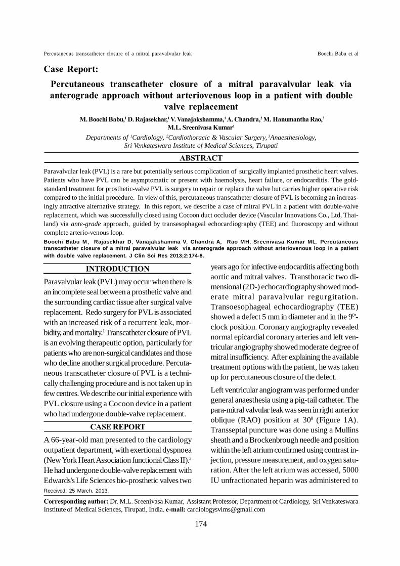
avoid intracardiac, valvular, or intracatheter throm-bosis during the procedure. A Judkins right (JR)diagnostic catheter was advanced through theMullins sheath into the left atrium through which a0.035- inch 260 - cm Terumo floppy Glide wire(Terumo Medical Corp., Somerset, New Jersey )was passed to cross the defect and then throughthe aortic prosthesis until it reaches the proximaldescending aorta (Figure 1B). The Glide wire wasthen exchanged for an Amplatz stiff wire (CookMedical Inc., Bloomington, Indiana). The Mullinssheath was then removed and replaced with the8-French Cook sheath (Cook Medical Inc.,Bloomington, Indiana) which was advanced be-yond the PVL (Figure 1C) into the left ventricle
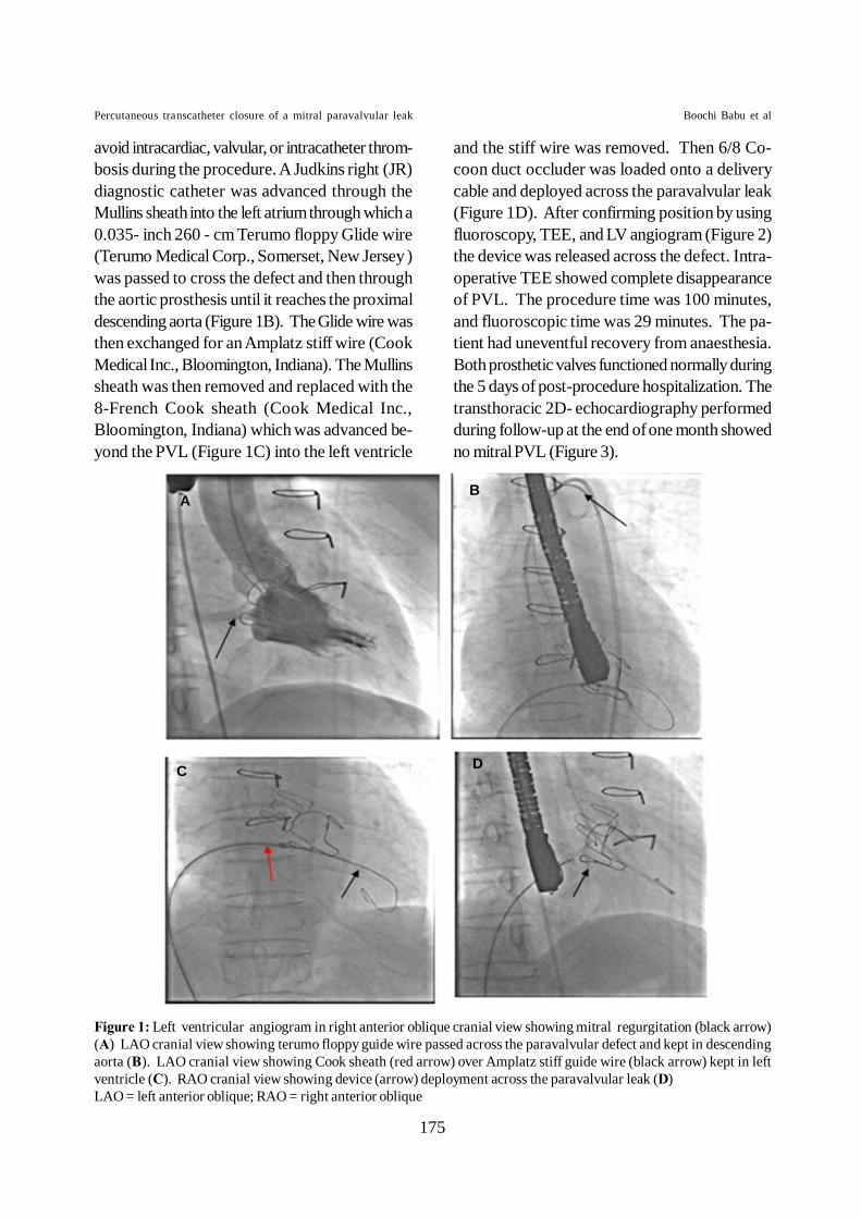
and the stiff wire was removed. Then 6/8 Co-coon duct occluder was loaded onto a deliverycable and deployed across the paravalvular leak(Figure 1D). After confirming position by usingfluoroscopy, TEE, and LV angiogram (Figure 2)the device was released across the defect. Intra-operative TEE showed complete disappearanceof PVL. The procedure time was 100 minutes,and fluoroscopic time was 29 minutes. The pa-tient had uneventful recovery from anaesthesia.Both prosthetic valves functioned normally duringthe 5 days of post-procedure hospitalization. Thetransthoracic 2D- echocardiography performedduring follow-up at the end of one month showedno mitral PVL (Figure 3).
Figure 1: Left ventricular angiogram in right anterior oblique cranial view showing mitral regurgitation (black arrow)(A) LAO cranial view showing terumo floppy guide wire passed across the paravalvular defect and kept in descendingaorta (B). LAO cranial view showing Cook sheath (red arrow) over Amplatz stiff guide wire (black arrow) kept in leftventricle (C). RAO cranial view showing device (arrow) deployment across the paravalvular leak (D)LAO = left anterior oblique; RAO = right anterior oblique
Percutaneous transcatheter closure of a mitral paravalvular leak Boochi Babu et al
BA
C D

176
DISCUSSION
PVL is a relatively rare but serious complicationof surgical replacement of mitral and aortic valves.Most of the PVLs are small, asymptomatic, andfollow a benign clinical course. Larger PVLs canoccur in 1% to 5% of patients with serious clinicalconsequences like heart failure, endocarditis andhaemolysis.3 Most of these clinically significantPVLs occur in association with prosthetic mitralvalves.4
Though redo surgery is usually recommended inadult patients with symptomatic PVLs, it is asso-ciated with a mortality rate approaching 16%.1 Inaddition, there is an increased recurrence of leaks.5Percutaneous transcatheter closure of PVLs wasdescribed first in 1992 using a double-umbrelladevice (Rashkind device).6 The technique hasslowly evolved over the following years, as an at-tractive alternative to redo-surgery in patients withappropriately suitable defects. The initial technicalsuccess rate has ranged from 60% to 90% and aneed for repeat intervention has been reported inup to 40% of cases.2
Transcatheter closure of mitral PVLs can beachieved using three different approaches depend-
ing on the location of the defect and its dimen-sions; preferred access; and experience of theoperator. The ante-grade approach via the femo-ral vein is the most common technique as it pro-vides easy access to the lateral defects and some-times to medial defects (like in the present case)after transseptal puncture. This technique avoidslarge arterial sheaths used for retrograde arterialtechnique. The retrograde transfemoral arterial ap-proach is a suitable alternative, providing benefitsin some cases like large defects with significantgradients across the PVL, that interfere withantegrade access against the shunt flow. The thirdapproach is retrograde transapical that providesdirect access to a mitral PVL located anywherearound the mitral annulus and is more useful inpatients with both mitral and aortic prostheticvalves. The percutaneous transcatheter closureof mitral PVL using ante-grade technique is morecomplex than that of aortic PVL since it is neces-sary to form a complete AV loop with a long guidewire over which the delivery sheath is advanced.In patients with mitral PVL who also have aorticvalve prosthesis, the guide wire can interfere withthe aortic prosthesis and complicate the proce-dure. Hence, the transapical approach is preferred
Figure 2: Left ventricular angiogram in right ante-rior oblique cranial view showing no residual leakacross the device.
Figure 3: Transthoracic echocardiography apical four-cham-ber view showing device (arrow) and no paravalvular leakacross the prosthetic mitral valve after device implantationin systole.
Percutaneous transcatheter closure of a mitral paravalvular leak Boochi Babu et al
177
by some interventional cardiologists in patients withboth mitral and aortic prosthesis.
The Cocoon duct occluder, used in transcatheterclosure of patent ductus arteriosus is made up ofnitinol wire covered with platinum using nanofusiontechnology. As our patient had two prostheticvalves there was a risk of leaflet impingement dueto the location of the PVL, We chose Cocoonductal occluder which is more uniform with a singleretention disk of limited size unlike a septaloccluder with two large retention disks, reducingthe risk of interfering with neighbouring prostheticvalve leaflet function.
Until now, very few cases of mitral paravalvularleaks were closed across the world using trans-catheter percutaneous technique in patients whohad undergone double-valve replacement.7-11 Tothe best of our knowledge, this is the first reportof percutaneous device deployment for closure ofa mitral paravalvular leak from India via ante-gradeapproach in a patient who had undergone double-valve replacement without the construction of com-plete arterio venous wire loop, and the first casein which Cocoon duct occluder was used for thatpurpose. (An internet search of Google scholar,PubMed, Cochrane databases for the terms"Double valve replacement; Percutaneous Clo-sure; Mitral Paravalvular Leak; Retrograde Ap-proach without a wire loop; Cocoon ductoccluder" did not yield any results of case reportstill to date).In spite of its progress, percutaneous transcatheterprosthetic PVL closures are one of the most chal-lenging procedures facing interventional cardiolo-gists today. The major challenge associated withtranscatheter PVL closure is the lack of devicesthat are specifically designed to meet the anatomicand physiologic properties of PVLs. Till today,there are no devices approved by the United Statesof America (USA) Food and Drug Administration(FDA) for the closure of PVLs specifically. Hence,the devices that have been approved by the FDA
for the closure of other cardiovascular defects (e.g.,patent ductus arteriosus, ventricular septal defect,and atrial septal defect) are used in an off-labelfashion for the closure of PVLs. In future, devel-opment of low profile, specific and adaptive de-vices that can conform to the variety of shapes ofthese defects is likely to materialize and will in-crease the procedural frequency and success.
ACKNOWLEDGEMENTThis procedure was done with the financial sup-port of Sri Venkateswara Pranadana Schemeof Sri Venkateswara Insitute of Medical Sciencesand Tirumala Tirupati Devasthanams, Tirupati.
REFERENCES1. Echevarria JR, Bernal JM, Rabasa JM, Morales D,
Revilla Y, Revuelta JM. Reoperation forbioprosthetic valve dysfunction. A decade of clini-cal experience. Eur J Cardiothorac Surg1991;5:523-6.
2. Fang JC, O'Gara PT. The history and physical ex-amination: An evidence-based approach. In: BonowRO, Mann DL, Zipes DP, Libby P, editors.Braunwald's heart disease: a textbook of cardiovas-cular disease. 7th edition. Philadelphia: ElsevierSaunders;2012.p.107-25.
3. Pate GE, Al Zubaidi A, Chandavimol M, ThompsonCR, Munt BI, Webb JG. Percutaneous closure ofprosthetic paravalvular leaks: case series and re-view. Catheter Cardiovasc Interv 2006;68:528-33.
4. Safi AM, Kwan T, Afflu E, Al Kamme A, SalciccioliL. Paravalvular regurgitation: a rare complicationfollowing valve replacement surgery. Angiology2000;51:479-87.
5. Expositor V, García-Camarero T, Bernal JM, ArnáizE, Sarralde A, Garcia I, et al. Repeat mitral valvereplacement: 30-years' experience. Rev Esp Cardiol2009;62:929-32.
6. Hourihan M, Perry SB, Mandell VS, Keane JF, RomeJJ, Bittl JA, et al. Transcatheter umbrella closure ofvalvular and paravalvular leaks. J Am Coll Cardiol1992;20:1371-7.
7. Shapira Y, Hirsch R, Kornowski R, Hasdai D, AssaliA, Vaturi M, et al. Percutaneous closure ofperivalvular leaks with Amplatzer occluders: feasi-bility safety, and short tem results. J Heart ValveDis 2007;16:305-13.
Percutaneous transcatheter closure of a mitral paravalvular leak Boochi Babu et al

178
8. Lasorda DM, Mohsin JC. Percutaneous closure ofperivalvular mitral regurgitation with an Amplatzeroccluder device in a patient with both prostheticmitral and aortic valves. J Interv Cardiol2008;21:190-5.
9. Nietlispach F, Johnson M, Moss RR, Wijesinghe N,Gurvitch R, Tay EL, et al. Transcatheter closure ofparavalvular defects using a purpose-specificoccluder. JACC Cardiovasc Intv 2010;3:759-65.
10. Ruiz CE, Jelnin V, Kronzon I, Dudiy Y, Del Valle-Fernandez R, Einhorn BN, et al Clinical outcomes inpatients undergoing percutaneous closure ofperiprosthetic paravalvular leaks. J Am Coll Cardiol2011;58:2210-7.
11. Moreno R, Sanchez Recalde A, Lopez Fernández T,Moreno-Gomez I, Mesa JM, Lopez-Sendon JL. Per-cutaneous closure of mitral paravalvular leaks inpatients with aortic Valve prostheses. Rev EspCardiol (Engl Ed) 2012;65:768-9.
Percutaneous transcatheter closure of a mitral paravalvular leak Boochi Babu et al