Percutaneous Coronary Intervention in Spontaneous Coronary … · Percutaneous Coronary...
6
CASE REPORT Percutaneous Coronary Intervention in Spontaneous Coronary Artery Dissection: Role of Intravascular Ultrasound Ankur Kalra • Avin Aggarwal • Rachel Kneeland • Jay H. Traverse To view enhanced content go to www.cardiologytherapy-open.com Received: June 12, 2014 / Published online: August 20, 2014 Ó The Author(s) 2014. This article is published with open access at Springerlink.com ABSTRACT Spontaneous coronary artery dissection (SCAD) is a rare, life-threatening condition that usually manifests as an acute myocardial infarction. Diagnosing SCAD with conventional coronary angiogram can be challenging, particularly if the true lumen is severely narrowed. Our case highlights the challenges in performing successful percutaneous coronary intervention (PCI) in patients with SCAD. Intravascular ultrasound can prove to be a pivotal tool in the diagnosis and successful management of such cases by establishing the anatomic site of dissection, and confirming stent placement in the true lumen following PCI. Keywords: Cardiology; False lumen; Intravascular ultrasound; Percutaneous coronary intervention; Postpartum dissection; Spontaneous coronary artery dissection INTRODUCTION Spontaneous coronary artery dissection (SCAD) is a life-threatening disorder that usually affects young women. A high index of suspicion is necessary for diagnosing SCAD, as the classic longitudinal radiolucent intimal flap may not be seen in every patient on angiography. Additional tomographic imaging techniques like intravascular ultrasound (IVUS), therefore, play a crucial role in establishing the right diagnosis. In addition, IVUS is also important in ensuring appropriate positioning of wires and catheters in the true lumen during intervention, and satisfactory stent deployment. Here, we present a case that highlights the challenges in Electronic supplementary material The online version of this article (doi:10.1007/s40119-014-0029-4) contains supplementary material, which is available to authorized users. A. Kalra (&) Á J. H. Traverse Minneapolis Heart Institute at Abbott Northwestern Hospital, Minneapolis, MN, USA e-mail: [email protected]; [email protected] A. Aggarwal Department of Medicine, Hennepin County Medical Center, Minneapolis, MN, USA R. Kneeland Arizona College of Osteopathic Medicine, Midwestern University, Glendale, AZ, USA J. H. Traverse Department of Cardiology, University of Minnesota Medical School, Minneapolis, MN, USA Cardiol Ther (2014) 3:61–66 DOI 10.1007/s40119-014-0029-4
Transcript of Percutaneous Coronary Intervention in Spontaneous Coronary … · Percutaneous Coronary...
CASE REPORT
Percutaneous Coronary Intervention in SpontaneousCoronary Artery Dissection: Role of IntravascularUltrasound
Ankur Kalra • Avin Aggarwal • Rachel Kneeland • Jay H. Traverse
To view enhanced content go to www.cardiologytherapy-open.comReceived: June 12, 2014 / Published online: August 20, 2014� The Author(s) 2014. This article is published with open access at Springerlink.com
ABSTRACT
Spontaneous coronary artery dissection (SCAD)
is a rare, life-threatening condition that usually
manifests as an acute myocardial infarction.
Diagnosing SCAD with conventional coronary
angiogram can be challenging, particularly if
the true lumen is severely narrowed. Our case
highlights the challenges in performing
successful percutaneous coronary intervention
(PCI) in patients with SCAD. Intravascular
ultrasound can prove to be a pivotal tool in
the diagnosis and successful management of
such cases by establishing the anatomic site of
dissection, and confirming stent placement in
the true lumen following PCI.
Keywords: Cardiology; False lumen;
Intravascular ultrasound; Percutaneous
coronary intervention; Postpartum dissection;
Spontaneous coronary artery dissection
INTRODUCTION
Spontaneous coronary artery dissection (SCAD)
is a life-threatening disorder that usually affects
young women. A high index of suspicion is
necessary for diagnosing SCAD, as the classic
longitudinal radiolucent intimal flap may not
be seen in every patient on angiography.
Additional tomographic imaging techniques
like intravascular ultrasound (IVUS), therefore,
play a crucial role in establishing the right
diagnosis. In addition, IVUS is also important in
ensuring appropriate positioning of wires and
catheters in the true lumen during intervention,
and satisfactory stent deployment. Here, we
present a case that highlights the challenges in
Electronic supplementary material The onlineversion of this article (doi:10.1007/s40119-014-0029-4)contains supplementary material, which is available toauthorized users.
A. Kalra (&) � J. H. TraverseMinneapolis Heart Institute at Abbott NorthwesternHospital, Minneapolis, MN, USAe-mail: [email protected];[email protected]
A. AggarwalDepartment of Medicine, Hennepin CountyMedical Center, Minneapolis, MN, USA
R. KneelandArizona College of Osteopathic Medicine,Midwestern University, Glendale, AZ, USA
J. H. TraverseDepartment of Cardiology, University of MinnesotaMedical School, Minneapolis, MN, USA
Cardiol Ther (2014) 3:61–66
DOI 10.1007/s40119-014-0029-4
performing successful percutaneous coronary
intervention (PCI) in patients with SCAD.
Informed consent was obtained from the
patient for publication of this case report and
associated images.
CASE
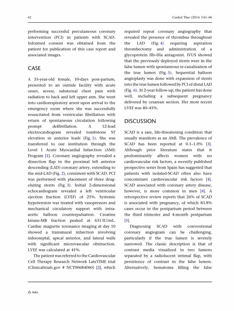
A 35-year-old female, 10-days post-partum,
presented to an outside facility with acute
onset, severe, substernal chest pain with
radiation to back and left upper arm. She went
into cardiorespiratory arrest upon arrival to the
emergency room where she was successfully
resuscitated from ventricular fibrillation with
return of spontaneous circulation following
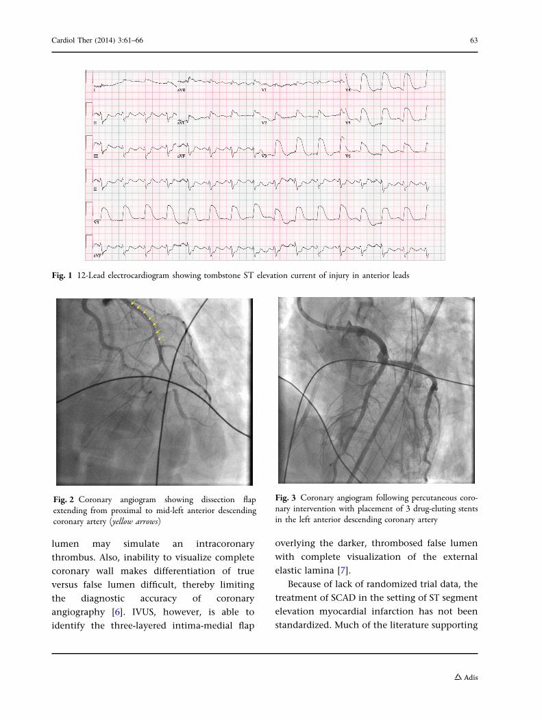
prompt defibrillation. A 12-lead
electrocardiogram revealed tombstone ST
elevation in anterior leads (Fig. 1). She was
transferred to our institution through the
Level 1 Acute Myocardial Infarction (AMI)
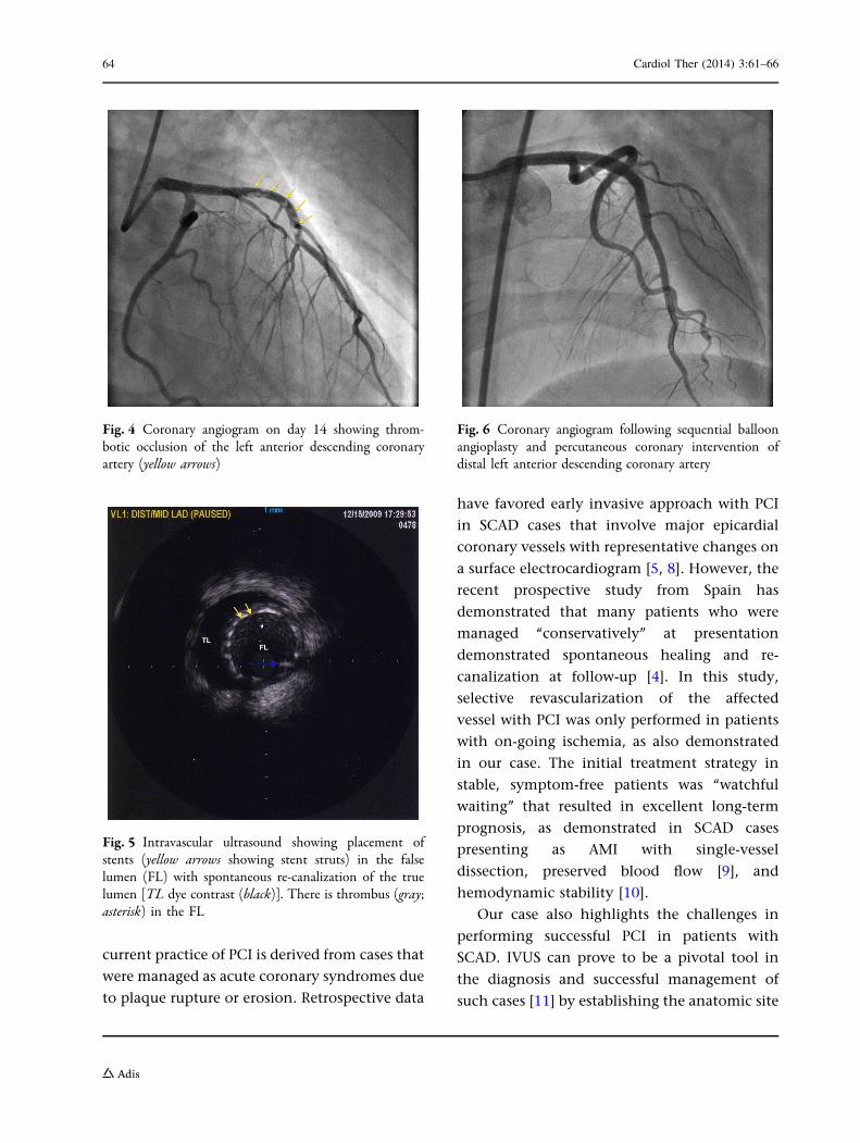
Program [1]. Coronary angiography revealed a
dissection flap in the proximal left anterior
descending (LAD) coronary artery, extending to
the mid-LAD (Fig. 2), consistent with SCAD. PCI
was performed with placement of three drug-
eluting stents (Fig. 3). Initial 2-dimensional
echocardiogram revealed a left ventricular
ejection fraction (LVEF) of 25%. Systemic
hypotension was treated with vasopressors and
mechanical circulatory support with intra-
aortic balloon counterpulsation. Creatine
kinase-MB fraction peaked at 631 IU/mL.
Cardiac magnetic resonance imaging at day 10
showed a transmural infarction involving
inferoseptal, apical anterior, and lateral walls
with significant microvascular obstruction.
LVEF was calculated at 41%.
The patient was referred to the Cardiovascular
Cell Therapy Research Network LateTIME trial
(Clinicaltrials.gov # NCT00684060) [2], which
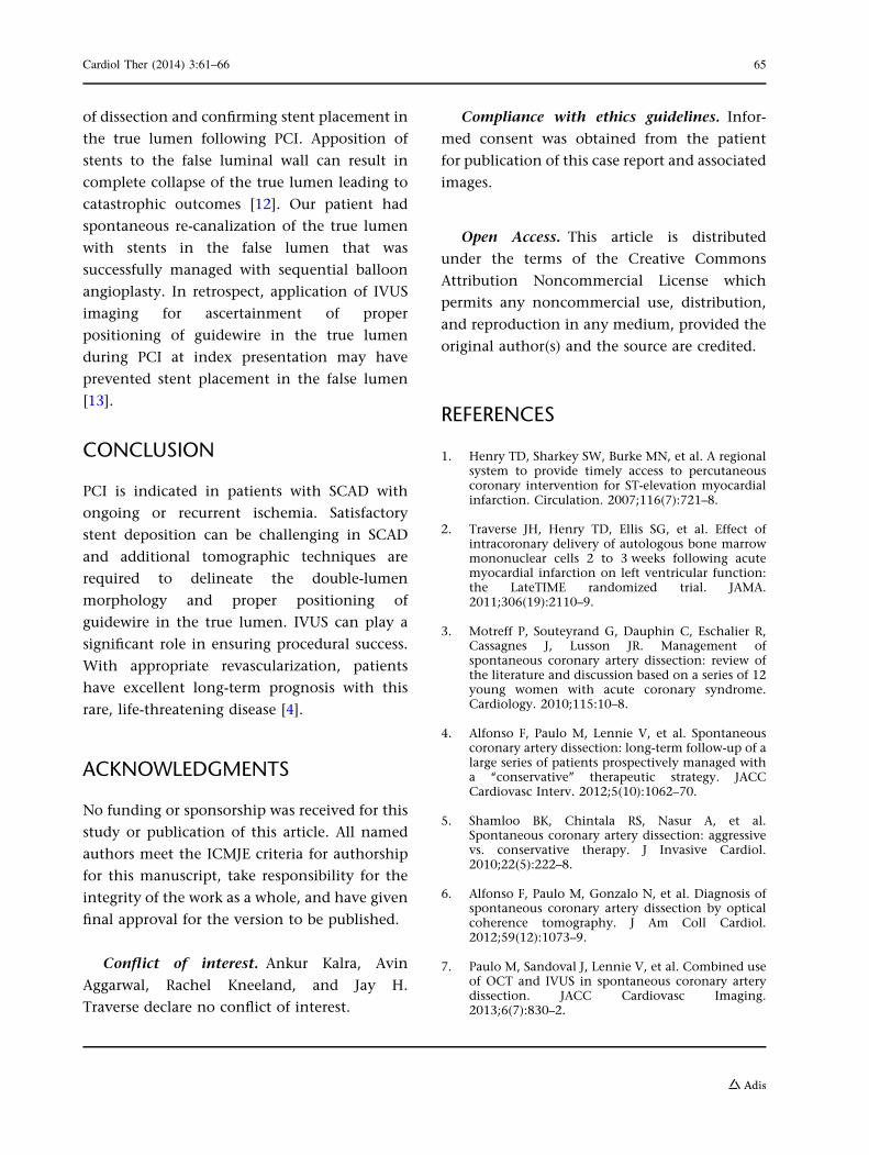
required repeat coronary angiography that
revealed the presence of thrombus throughout
the LAD (Fig. 4) requiring aspiration
thrombectomy and administration of a
glycoprotein IIb–IIIa antagonist. IVUS showed
that the previously deployed stents were in the
false lumen with spontaneous re-canalization of
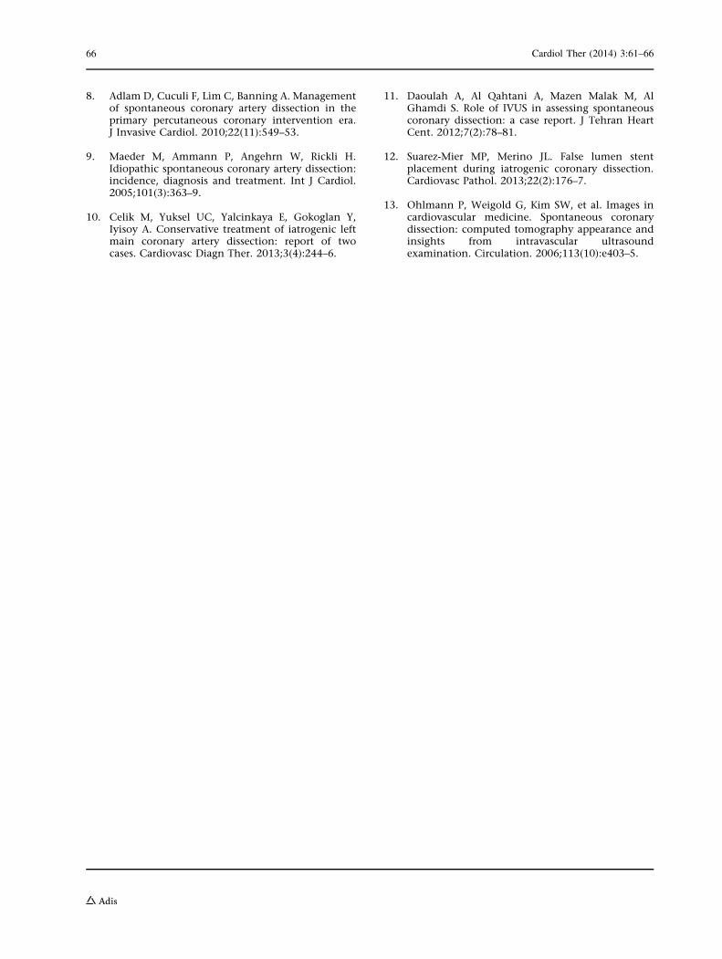
the true lumen (Fig. 5). Sequential balloon
angioplasty was done with expansion of stents
into the true lumen followed by PCI of distal LAD
(Fig. 6). At 2-year follow-up, the patient has done
well, including a subsequent pregnancy
delivered by cesarean section. Her most recent
LVEF was 40–45%.
DISCUSSION
SCAD is a rare, life-threatening condition that
usually manifests as an AMI. The prevalence of
SCAD has been reported at 0.1–1.0% [3].
Although prior literature states that it
predominantly affects women with no
cardiovascular risk factors, a recently published
prospective series from Spain has suggested that
patients with isolated-SCAD often also have
concomitant cardiovascular risk factors [4].
SCAD associated with coronary artery disease,
however, is more common in men [4]. A
retrospective review reports that 26% of SCAD
is associated with pregnancy, of which 83.8%
cases occur in the postpartum period between
the third trimester and 4-month postpartum
[5].
Diagnosing SCAD with conventional
coronary angiogram can be challenging,
particularly if the true lumen is severely
narrowed. The classic description is that of
contrast media visualized in two lumens
separated by a radiolucent intimal flap, with
persistence of contrast in the false lumen.
Alternatively, hematoma filling the false
62 Cardiol Ther (2014) 3:61–66
lumen may simulate an intracoronary
thrombus. Also, inability to visualize complete
coronary wall makes differentiation of true
versus false lumen difficult, thereby limiting
the diagnostic accuracy of coronary
angiography [6]. IVUS, however, is able to
identify the three-layered intima-medial flap
overlying the darker, thrombosed false lumen
with complete visualization of the external
elastic lamina [7].
Because of lack of randomized trial data, the
treatment of SCAD in the setting of ST segment
elevation myocardial infarction has not been
standardized. Much of the literature supporting
Fig. 1 12-Lead electrocardiogram showing tombstone ST elevation current of injury in anterior leads
Fig. 2 Coronary angiogram showing dissection flapextending from proximal to mid-left anterior descendingcoronary artery (yellow arrows)
Fig. 3 Coronary angiogram following percutaneous coro-nary intervention with placement of 3 drug-eluting stentsin the left anterior descending coronary artery
Cardiol Ther (2014) 3:61–66 63
current practice of PCI is derived from cases that
were managed as acute coronary syndromes due
to plaque rupture or erosion. Retrospective data
have favored early invasive approach with PCI
in SCAD cases that involve major epicardial
coronary vessels with representative changes on
a surface electrocardiogram [5, 8]. However, the
recent prospective study from Spain has
demonstrated that many patients who were
managed ‘‘conservatively’’ at presentation
demonstrated spontaneous healing and re-
canalization at follow-up [4]. In this study,
selective revascularization of the affected
vessel with PCI was only performed in patients
with on-going ischemia, as also demonstrated
in our case. The initial treatment strategy in
stable, symptom-free patients was ‘‘watchful
waiting’’ that resulted in excellent long-term
prognosis, as demonstrated in SCAD cases
presenting as AMI with single-vessel
dissection, preserved blood flow [9], and
hemodynamic stability [10].
Our case also highlights the challenges in
performing successful PCI in patients with
SCAD. IVUS can prove to be a pivotal tool in
the diagnosis and successful management of
such cases [11] by establishing the anatomic site
Fig. 4 Coronary angiogram on day 14 showing throm-botic occlusion of the left anterior descending coronaryartery (yellow arrows)
Fig. 5 Intravascular ultrasound showing placement ofstents (yellow arrows showing stent struts) in the falselumen (FL) with spontaneous re-canalization of the truelumen [TL dye contrast (black)]. There is thrombus (gray;asterisk) in the FL
Fig. 6 Coronary angiogram following sequential balloonangioplasty and percutaneous coronary intervention ofdistal left anterior descending coronary artery
64 Cardiol Ther (2014) 3:61–66
of dissection and confirming stent placement in
the true lumen following PCI. Apposition of
stents to the false luminal wall can result in
complete collapse of the true lumen leading to
catastrophic outcomes [12]. Our patient had
spontaneous re-canalization of the true lumen
with stents in the false lumen that was
successfully managed with sequential balloon
angioplasty. In retrospect, application of IVUS
imaging for ascertainment of proper
positioning of guidewire in the true lumen
during PCI at index presentation may have
prevented stent placement in the false lumen
[13].
CONCLUSION
PCI is indicated in patients with SCAD with
ongoing or recurrent ischemia. Satisfactory
stent deposition can be challenging in SCAD
and additional tomographic techniques are
required to delineate the double-lumen
morphology and proper positioning of
guidewire in the true lumen. IVUS can play a
significant role in ensuring procedural success.
With appropriate revascularization, patients
have excellent long-term prognosis with this
rare, life-threatening disease [4].
ACKNOWLEDGMENTS
No funding or sponsorship was received for this
study or publication of this article. All named
authors meet the ICMJE criteria for authorship
for this manuscript, take responsibility for the
integrity of the work as a whole, and have given
final approval for the version to be published.
Conflict of interest. Ankur Kalra, Avin
Aggarwal, Rachel Kneeland, and Jay H.
Traverse declare no conflict of interest.
Compliance with ethics guidelines. Infor-
med consent was obtained from the patient
for publication of this case report and associated
images.
Open Access. This article is distributed
under the terms of the Creative Commons
Attribution Noncommercial License which
permits any noncommercial use, distribution,
and reproduction in any medium, provided the
original author(s) and the source are credited.
REFERENCES
1. Henry TD, Sharkey SW, Burke MN, et al. A regionalsystem to provide timely access to percutaneouscoronary intervention for ST-elevation myocardialinfarction. Circulation. 2007;116(7):721–8.
2. Traverse JH, Henry TD, Ellis SG, et al. Effect ofintracoronary delivery of autologous bone marrowmononuclear cells 2 to 3 weeks following acutemyocardial infarction on left ventricular function:the LateTIME randomized trial. JAMA.2011;306(19):2110–9.
3. Motreff P, Souteyrand G, Dauphin C, Eschalier R,Cassagnes J, Lusson JR. Management ofspontaneous coronary artery dissection: review ofthe literature and discussion based on a series of 12young women with acute coronary syndrome.Cardiology. 2010;115:10–8.
4. Alfonso F, Paulo M, Lennie V, et al. Spontaneouscoronary artery dissection: long-term follow-up of alarge series of patients prospectively managed witha ‘‘conservative’’ therapeutic strategy. JACCCardiovasc Interv. 2012;5(10):1062–70.
5. Shamloo BK, Chintala RS, Nasur A, et al.Spontaneous coronary artery dissection: aggressivevs. conservative therapy. J Invasive Cardiol.2010;22(5):222–8.
6. Alfonso F, Paulo M, Gonzalo N, et al. Diagnosis ofspontaneous coronary artery dissection by opticalcoherence tomography. J Am Coll Cardiol.2012;59(12):1073–9.
7. Paulo M, Sandoval J, Lennie V, et al. Combined useof OCT and IVUS in spontaneous coronary arterydissection. JACC Cardiovasc Imaging.2013;6(7):830–2.
Cardiol Ther (2014) 3:61–66 65
8. Adlam D, Cuculi F, Lim C, Banning A. Managementof spontaneous coronary artery dissection in theprimary percutaneous coronary intervention era.J Invasive Cardiol. 2010;22(11):549–53.
9. Maeder M, Ammann P, Angehrn W, Rickli H.Idiopathic spontaneous coronary artery dissection:incidence, diagnosis and treatment. Int J Cardiol.2005;101(3):363–9.
10. Celik M, Yuksel UC, Yalcinkaya E, Gokoglan Y,Iyisoy A. Conservative treatment of iatrogenic leftmain coronary artery dissection: report of twocases. Cardiovasc Diagn Ther. 2013;3(4):244–6.
11. Daoulah A, Al Qahtani A, Mazen Malak M, AlGhamdi S. Role of IVUS in assessing spontaneouscoronary dissection: a case report. J Tehran HeartCent. 2012;7(2):78–81.
12. Suarez-Mier MP, Merino JL. False lumen stentplacement during iatrogenic coronary dissection.Cardiovasc Pathol. 2013;22(2):176–7.
13. Ohlmann P, Weigold G, Kim SW, et al. Images incardiovascular medicine. Spontaneous coronarydissection: computed tomography appearance andinsights from intravascular ultrasoundexamination. Circulation. 2006;113(10):e403–5.
66 Cardiol Ther (2014) 3:61–66