Percutaneous Contoured Locking Plate Fixation of...
5
Percutaneous Contoured Locking Plate Fixation of the Pilon Fracture: Surgical Technique Thomas Lee, DPM, 1 Neal M. Blitz, DPM, FACFAS, 2 and Shannon M. Rush, DPM, FACFAS 3 Key Words: ankle fracture, distal tibial fracture, locking plate, percutaneous, Pilon fracture P reservation of the soft tissue is of particular impor- tance in the care of pilon fractures. Improper soft tissue management has been attributed to high rates of non- union, nerve injury, and failures (1–12). Traditional open reduction internal fixation (ORIF) of pilon fractures al- lows for direct visualization of the fracture(s) but is criticized for the large exposure and periosteal stripping. As a result, this approach has been blamed for high nonunion rates as well as failure of the soft tissue to accommodate implants (1, 5, 8, 13). External fixation has also been used but has not demonstrated much advantage to internal fixation and requires significant postoperative care. Also, external fixation has a higher incidence of malunion (6, 14) and has inherent risks for pin-tract infections (14, 15). In an attempt to limit the iatrogenic soft tissue injury (5, 6, 7), some surgeons have used a limited ORIF concentrating on restoration of the articular surface combined with ring or hybrid external fixation. The “2-stage” technique combines the benefits of both external and internal fixation, and is thought to limit soft tissue related complications by allowing soft tissue re- covery time before the introduction of internal fixation. Initial temporary external fixation combined with lim- ited exposures for internal fixation (2-stage technique) at a later date allows for less disruption of the soft tissue envelope, leading to a decrease in complications associ- ated with traditional open approaches. This may be achieved through relatively “small incisions” extending from the pole of the malleolus to the metaphyseal frac- ture(s), although these incisions can still be quite long, especially when significant articular reconstruction and/or bone grafting is required. Other descriptions in- clude “limited approach” or “minimally invasive”; and this differs from a true percutaneous approach. With the advent of locking plate technology, surgeons have successfully managed a variety of fractures though smaller incisions to introduce the plate(s)—the percutane- ous approach. Unlike traditional plating methods, locking plates do not rely on frictional forces between the plate/bone interface to achieve compression and stability. This allows for less damage to the periosteal blood supply, which may theoretically decrease the incidence of delayed or nonunion, soft tissue complications, and possibly secondary loss of fixation (16, 17). A recent study by Salton et al (18) dem- onstrated no major complications and only 4 minor soft tissue complications in a series of 19 patients treated with a limited incision and percutaneous medial plate fixation. More recently available contoured locking plates with mul- tiple options for metaphyseal screw placement through the “percutaneous” incision allow for the surgeon to achieve stability through this access point. With percutaneous lock- ing plates, the incision may be even smaller than used with the traditional 2-stage technique. Technique Initial operative management may involve a single-stage approach (Figure 1) or the 2-stage approach, depending of fracture pattern and degree of edema (Figures 2 to 4). The percutaneous locking plate approach is probably best suited for closed noncomminuted metadiaphyseal pilon fractures with minimal articular surface involvement and when bone grafting of the metaphysis is not necessary (Figure 1, A, Figure 2, and Figure 5). Injury computed tomography (CT) is often obtained to determine the extent of articular damage and intra-articular fractures (Figure 1, B, and 2, B, Figure 6). If a 2-stage ap- proach is used, a medially based external fixator is preferred (allows lateral access to a concomitant fibular fracture, if Address correspondence to: Neal M. Blitz, DPM, FACFAS, Chief of Foot Surgery, Department of Orthopaedic Surgery, Bronx-Lebanon Hos- pital Center, 1650 Selwyn Avenue 3H, Bronx, NY 10457. E-mail: nealblitz@ yahoo.com. 1 Attending Podiatric Surgeon, Kaiser Foundation Hospital, Vallejo, CA. 2 Chief of Foot Surgery, Department of Orthopaedic Surgery, Bronx- Lebanon Hospital Center, Bronx, NY. 3 Attending Podiatric Surgeon, Camino Medical Group, Podiatry De- partment, Mountain View, CA. Financial Disclosure: None reported. Conflict of Interest: None reported. Copyright © 2008 by the American College of Foot and Ankle Surgeons 1067-2516/08/4706-0020$34.00/0 doi:10.1053/j.jfas.2008.06.009 598 THE JOURNAL OF FOOT & ANKLE SURGERY
Transcript of Percutaneous Contoured Locking Plate Fixation of...

Percutaneous Contoured Locking PlateFixation of the Pilon Fracture: SurgicalTechnique
Thomas Lee, DPM,1 Neal M. Blitz, DPM, FACFAS,2 and Shannon M. Rush, DPM, FACFAS3
Key Words: ankle fracture, distal tibial fracture, locking plate, percutaneous, Pilon fracture
Preservation of the soft tissue is of particular impor-tance in the care of pilon fractures. Improper soft tissuemanagement has been attributed to high rates of non-union, nerve injury, and failures (1–12). Traditional openreduction internal fixation (ORIF) of pilon fractures al-lows for direct visualization of the fracture(s) but iscriticized for the large exposure and periosteal stripping.As a result, this approach has been blamed for highnonunion rates as well as failure of the soft tissue toaccommodate implants (1, 5, 8, 13). External fixation hasalso been used but has not demonstrated much advantageto internal fixation and requires significant postoperativecare. Also, external fixation has a higher incidence ofmalunion (6, 14) and has inherent risks for pin-tractinfections (14, 15). In an attempt to limit the iatrogenicsoft tissue injury (5, 6, 7), some surgeons have used alimited ORIF concentrating on restoration of the articularsurface combined with ring or hybrid external fixation.The “2-stage” technique combines the benefits of bothexternal and internal fixation, and is thought to limit softtissue related complications by allowing soft tissue re-covery time before the introduction of internal fixation.
Initial temporary external fixation combined with lim-ited exposures for internal fixation (2-stage technique) ata later date allows for less disruption of the soft tissueenvelope, leading to a decrease in complications associ-ated with traditional open approaches. This may beachieved through relatively “small incisions” extending
Address correspondence to: Neal M. Blitz, DPM, FACFAS, Chief ofFoot Surgery, Department of Orthopaedic Surgery, Bronx-Lebanon Hos-pital Center, 1650 Selwyn Avenue 3H, Bronx, NY 10457. E-mail: [email protected].
1Attending Podiatric Surgeon, Kaiser Foundation Hospital, Vallejo, CA.2Chief of Foot Surgery, Department of Orthopaedic Surgery, Bronx-
Lebanon Hospital Center, Bronx, NY.3Attending Podiatric Surgeon, Camino Medical Group, Podiatry De-
partment, Mountain View, CA.Financial Disclosure: None reported.Conflict of Interest: None reported.Copyright © 2008 by the American College of Foot and Ankle Surgeons
1067-2516/08/4706-0020$34.00/0doi:10.1053/j.jfas.2008.06.009598 THE JOURNAL OF FOOT & ANKLE SURGERY
from the pole of the malleolus to the metaphyseal frac-ture(s), although these incisions can still be quite long,especially when significant articular reconstructionand/or bone grafting is required. Other descriptions in-clude “limited approach” or “minimally invasive”; andthis differs from a true percutaneous approach.
With the advent of locking plate technology, surgeonshave successfully managed a variety of fractures thoughsmaller incisions to introduce the plate(s)—the percutane-ous approach. Unlike traditional plating methods, lockingplates do not rely on frictional forces between the plate/boneinterface to achieve compression and stability. This allowsfor less damage to the periosteal blood supply, which maytheoretically decrease the incidence of delayed or nonunion,soft tissue complications, and possibly secondary loss offixation (16, 17). A recent study by Salton et al (18) dem-onstrated no major complications and only 4 minor softtissue complications in a series of 19 patients treated with alimited incision and percutaneous medial plate fixation.More recently available contoured locking plates with mul-tiple options for metaphyseal screw placement through the“percutaneous” incision allow for the surgeon to achievestability through this access point. With percutaneous lock-ing plates, the incision may be even smaller than used withthe traditional 2-stage technique.
Technique
Initial operative management may involve a single-stageapproach (Figure 1) or the 2-stage approach, depending offracture pattern and degree of edema (Figures 2 to 4). Thepercutaneous locking plate approach is probably best suited forclosed noncomminuted metadiaphyseal pilon fractures withminimal articular surface involvement and when bone graftingof the metaphysis is not necessary (Figure 1, A, Figure 2, andFigure 5). Injury computed tomography (CT) is often obtainedto determine the extent of articular damage and intra-articularfractures (Figure 1, B, and 2, B, Figure 6). If a 2-stage ap-proach is used, a medially based external fixator is preferred
(allows lateral access to a concomitant fibular fracture, if
present) to stabilize the fractures as well as maintain length(Figure 3, A, and Figure 5, B). In some cases, the articularplafond may be percutaneously fixated and reduced with a
FIGURE 2 A 56-year-old femalewith comminuted pilon and fibulafracture, treated with the 2-stageapproach. (A) Injury radiographs. Alarge butterfly fragment of the tibialplafond is present. (B) Injury CT.The plafond is comminuted withthe major fracture line in the coro-nal plane. Note comminution of lat-eral plafond.
percutaneous screw across the major fracture line, although
VOLUME 47
definitive internal fixation should await soft tissue recovery(Figure 3, B). CT of the initial reduction allows the surgeon toassess the quality of the articular reduction before the second
FIGURE 1 A 24-year-old malewith isolated left pilon fracture,treated in a single stage. (A) Injuryx-rays demonstrate single articularfracture traversing the metaphysis.(B) Injury CT further reveals ob-liquely oriented fracture line, almostparallel with the anteromedial tibialsurface, making this fracture pat-tern amenable to medial plate fixa-tion. Fracture involves approxi-mately 50% of the tibial plafondwith some minor comminution ofthe distal lateral tibia. (C) Fluoro-scopic series demonstrating lock-ing plate application. After the plateis positioned, a nonlocking screwproximal to the fracture is placed tobuttress the plate against the dis-tally based fracture to reduce thefracture. It is important to fluoro-scopically evaluate the reduction inmultiple planes before introducingfixation distally into the plate. Ana-tomic reduction is achieved.
stage of percutaneously plating (Figure 3, C, and Figure 6).
, NUMBER 6, NOVEMBER/DECEMBER 2008 599

Plate fixation of the pilon component should be per-formed when soft tissue edema is decreased. The fibularfracture is fixated with standard plating techniques andnot the focus of this techniques manuscript. The piloncomponent is percutaneously fixated with a contouredlocking plate. The medial tibial plate is introduced
FIGURE 3 Same patient as Figure 2, demonstrating temporary statemporary medial external fixator. (B) Radiographs with external fixathalf pin in the talar neck is located very close to the ankle joint capsuinfected. A delta configuration medial external fixator is a more applaced anteriolaterally (arrow), though not necessary to be placed dis placed once the soft tissues have recovered and the external fixatemporary reduction.
FIGURE 4 Same patient as Figures 2 and 3. (A) Intraoperative pholocking plate fixation. Percutaneous incision for medial locking platethis incision supraperiosteally along distal medial border of the tnonlocking) screws. (B) Weight-bearing radiographs approximatelydated in good alignment.
through an approximately 3.5-cm anteromedial incision,
600 THE JOURNAL OF FOOT & ANKLE SURGERY
or longer depending on surgeon skill level or comfort(Figure 4, A). The plate is then directed supraperiosteallyacross the metaphyseal fracture. Proximally the plate isfixated first with a single nonlocking screw (via a stabincision) to use the plate as a buttress to translate thedistal periarticular fracture segment laterally into ana-
tion with an external fixator. (A) Clinical photograph demonstratingplace. The medial monorail used in this case was not ideal becaused is at risk for development of a septic joint should this pin becomeriate choice. In this case, a periarticular percutaneous screw was
the temporary external fixations setting. Ideally, definitive fixationremoved. (C) CT after external fixation allows for evaluation of the
ph after the external fixator is removed and ready for percutaneousmedial to the tibialis anterior tendon. The plate is inserted through
Stab incisions are created for placement of proximal (locking oronths from injury. The articular component is completely consoli-
bilizaion inle, anpropuringtor is
tograto is
ibia.12 m
tomic position, in line with the tibial shaft (Figure 1, C).

Many countered locking distal tibial plates have multipleparallel options for screw placement at the distal end ofthe plate. Periarticular screws are then placed though theexisting anteromedial percutaneous incision and stab in-cisions are created for percutaneous placement of diaph-yseal locking screws.
Postoperative care includes keeping the patient non–weight bearing for 8 weeks or longer depending on theclinical situation. A removable cam walker may beplaced to allow for early range of motion, all dependingon the on the degree of comminution and stability of theconstruct. Figure 4, B, and Figure 7 demonstrate a healedpilon fracture treated with a medially contoured percuta-neous locking plate. The ability to initiate weight bearingis determined by radiographic evidence of healing andshould not be dictated by the method of reduction—
FIGURE 6 Same patient as Fig-ure 5. CT (with 3D reconstruction)following temporary external fixa-tion allows for planning of hardwareplacement. Tibial plafond has 3major fragments.
percutaneous or open.
VOLUME 47
Summary
Using a “true” percutaneous approach combined withnewer generation locking plates for the treatment ofcertain pilon fractures may decrease complications asso-ciated with disruption of the soft tissue envelope andassociated osseous complications and allow for quickerreturn to function. This technique may prove to be ad-vantageous when compared to more traditional openmethods. The percutaneous locking plate approach isprobably best suited for closed noncomminuted metadi-aphyseal pilon fractures with minimal articular surfaceinvolvement and when bone grafting of the metaphysis isnot necessary. This approach is not ideal for every pilonfracture and surgeons should have the significant experi-ence with pilon fracture management before attempting
FIGURE 5 Open pilon and fibulafracture, treated with the 2-stageapproach. (A) Injury radiographs.Displaced fracture, with corre-sponding skin laceration at medialaspect of metadiaphyseal spike.(B) Status post wash-out and deltaframe external fixation. Acceptablealignment is achieved until defini-tive fixation is placed.
this approach.
, NUMBER 6, NOVEMBER/DECEMBER 2008 601
References
1. Blauth M, Bastian L, Krettek C, Knop C, Evans S. Surgical options forthe treatment of severe tibial pilon fractures: a study of three tech-niques. J Orthop Trauma 15(3):153–160, 2001.
2. Koulouvaris P, Stafilas K, Kalos N, Mavrodontidis A, Mitsionis G,Xenakis T. A comparison of treatment methods for pilon fractures.J Bone Joint Surg Br 86-B(supp II):173, 2004.
3. Borens O, Kloen P, Richmond J, Roederer G, Levine DS, Helfet DL.Minimally invasive treatment of pilon fractures with a low profileplate: preliminary results in 17 cases. Arch Orthop Trauma Surg Sep2, 2006. [Epub ahead of print.]
4. Allgower R. Fractures of the lower end of the tibia into the ankle joint.
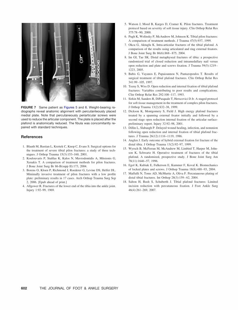
FIGURE 7 Same patient as Figures 5 and 6. Weight-bearing ra-diographs reveal anatomic alignment with percutanteously placedmedial plate. Note that percutaneously periarticular screws wereused to reduce the articular component. The plate is placed after theplafond is anatomically reduced. The fibula was concomitantly re-paired with standard techniques.
Injury 1:92–99, 1969.
602 THE JOURNAL OF FOOT & ANKLE SURGERY
5. Watson J, Moed B, Karges D, Cramer K. Pilon fractures. Treatmentprotocol based on severity of soft tissue injury. Clin Orthop Relat Res375:78–90, 2000.
6. Pugh K, Wolinsky P, McAndrew M, Johnson K. Tibial pilon fractures:A comparison of treatment methods. J Trauma 47(5):937, 1999.
7. Okcu G, Aktuglu K. Intra-articular fractures of the tibial plafond. Acomparison of the results using articulated and ring external fixators.J Bone Joint Surg Br 86(6):868–875, 2004.
8. Im GI, Tae SK. Distal metaphyseal fractures of tibia: a prospectiverandomized trial of closed reduction and intramedullary nail versusopen reduction and plate and screws fixation. J Trauma 59(5):1219–1223, 2005.
9. Babis G, Vayanos E, Papaioannou N, Pantazopoulos T. Results ofsurgical treatment of tibial plafond fractures. Clin Orthop Relat Res341:99–105, 1997.
10. Teeny S, Wiss D. Open reduction and internal fixation of tibial plafondfractures: Variables contributing to poor results and complications.Clin Orthop Relat Res 292:108–117, 1993.
11. Sirkin M, Sanders R, DiPasquale T, Herscovici D Jr. A staged protocolfor soft tissue management in the treatment of complex pilon fractures.J Orthop Trauma 13(2):S32–38, 1999.
12. Dickson K, Montgomery S, Field J. High energy plafond fracturestreated by a spanning external fixator initially and followed by asecond stage open reduction internal fixation of the articular surface-preliminary report. Injury 32:92–98, 2001.
13. Dillin L, Slabaugh P. Delayed wound healing, infection, and nonunionfollowing open reduction and internal fixation of tibial plafond frac-tures. J Trauma 26(12):1116–1119, 1986.
14. Anglen J. Early outcome of hybrid external fixation for fracture of thedistal tibia. J Orthop Trauma 13(2):92–97, 1999.
15. Wyrsch B, McFerran M, McAndrew M, Limbird T, Harper M, John-son K, Schwartz H. Operative treatment of fractures of the tibialplafond. A randomized, prospective study. J Bone Joint Surg Am78(11):1646–57, 1996.
16. Egol K, Kubiak E, Fulkerson E, Kummer F, Koval K. Biomechanicsof locked plates and screws. J Orthop Trauma 18(8):488–93, 2004.
17. Maffulli N, Toms AD, McMurtie A, Oliva F. Percutaneous plating ofdistal tibial fractures. Int Orthop 28(3):159–62, 2004.
18. Salton H, Rush S, Schuberth J. Tibial plafond fractures: Limitedincision reduction with percutaneous fixation. J Foot Ankle Surg
46(4):261–269, 2007.