Percutaneous Biliary Drainage: A12-year ExperiencePercutaneous Biliary Drainage: A 12-year...
14
J KAU: Med. Sci., Vol. 9, pp. 27-40 (1421 A.H. / 2001 A.D.) Correspondence & reprint requests to: Dr. Mohamed M. Rawas, Department of Radiology, King Khalid Na- tional Guard Hospital, P.O. Box 9515, Jeddah 21423, Saudi Arabia. Accepted for publication: 27 September 2000. Received: 26 January 2000. 27 Percutaneous Biliary Drainage: A12-year Experience MOHAMED M. RAWAS, FRCP(C) Department of Radiology, King Khalid National Guard Hospital, Jeddah, Saudi Arabia ABSTRACT. To report 12 years (108 patients, 273 procedures) experience and clinical outcome of percutaneous biliary drainage, with and without stent ap- plication, in benign and malignant obstruction. A retrospective study of pa- tients who presented biliary obstruction and who had percutaneous biliary drainage, with or without stent application. The technical success of external biliary drainage (EBD), dual biliary drainage (DBD) and stent application was 97%, 91%, and 98%. EBD patients with obstructive jaundice and cholangitis (36) showed good clinical outcome in 67% and was poor in 33%. EBD pa- tients with obstructive jaundice, (39) only showed good clinical outline in 62%, fair in 31% and poor in 7%. The mean potency for soft stents is 32.7 and 7.4 months in benign and malignant strictures. The mean potency for metallic stents is 26.8 and 4.5 months in benign and malignant strictures. Adequate la- boratory results were not obtained in (33) patients whose general condition im- proved after (EBD). The complication rate is 11% for (EBD) and (DBD), 20% for all stents. The 30-day mortality for (EBD) and (DBD) is 3% and none was noted post stent application. Percutaneous biliary drainage, with or without stent application, is useful in the diagnosis and management of patients with malignant or benign biliary tract obstruction. Keywords: Percutaneous, Cholangiogram, Biliary drainage. Introduction Percutaneous biliary drainage is a known interventional radiology procedure. The in- itial description of the technique appeared in the late 1970s and early 1980s. Since then, there have been many developments that included internal biliary drainage and
Transcript of Percutaneous Biliary Drainage: A12-year ExperiencePercutaneous Biliary Drainage: A 12-year...

27Percutaneous Biliary Drainage: A 12-year experienceJ KAU: Med. Sci., Vol. 9, pp. 27-40 (1421 A.H. / 2001 A.D.)
Correspondence & reprint requests to: Dr. Mohamed M. Rawas, Department of Radiology, King Khalid Na-tional Guard Hospital, P.O. Box 9515, Jeddah 21423, Saudi Arabia.
Accepted for publication: 27 September 2000. Received: 26 January 2000.
27
Percutaneous Biliary Drainage: A12-year Experience
MOHAMED M. RAWAS, FRCP(C)
Department of Radiology, King Khalid National Guard Hospital,Jeddah, Saudi Arabia
ABSTRACT. To report 12 years (108 patients, 273 procedures) experience andclinical outcome of percutaneous biliary drainage, with and without stent ap-plication, in benign and malignant obstruction. A retrospective study of pa-tients who presented biliary obstruction and who had percutaneous biliarydrainage, with or without stent application. The technical success of externalbiliary drainage (EBD), dual biliary drainage (DBD) and stent application was97%, 91%, and 98%. EBD patients with obstructive jaundice and cholangitis(36) showed good clinical outcome in 67% and was poor in 33%. EBD pa-tients with obstructive jaundice, (39) only showed good clinical outline in62%, fair in 31% and poor in 7%. The mean potency for soft stents is 32.7 and7.4 months in benign and malignant strictures. The mean potency for metallicstents is 26.8 and 4.5 months in benign and malignant strictures. Adequate la-boratory results were not obtained in (33) patients whose general condition im-proved after (EBD). The complication rate is 11% for (EBD) and (DBD), 20%for all stents. The 30-day mortality for (EBD) and (DBD) is 3% and none wasnoted post stent application. Percutaneous biliary drainage, with or withoutstent application, is useful in the diagnosis and management of patients withmalignant or benign biliary tract obstruction.
Keywords: Percutaneous, Cholangiogram, Biliary drainage.
Introduction
Percutaneous biliary drainage is a known interventional radiology procedure. The in-itial description of the technique appeared in the late 1970s and early 1980s. Sincethen, there have been many developments that included internal biliary drainage and
Mohamed M. Rawas28
different stent applications. This article reports a 12-year experience of EBD, DBD,and different stent applications. The clinical outcome of the procedures, as well as oth-er parameters, are discussed and compared with the available literature.
Materials and Methods
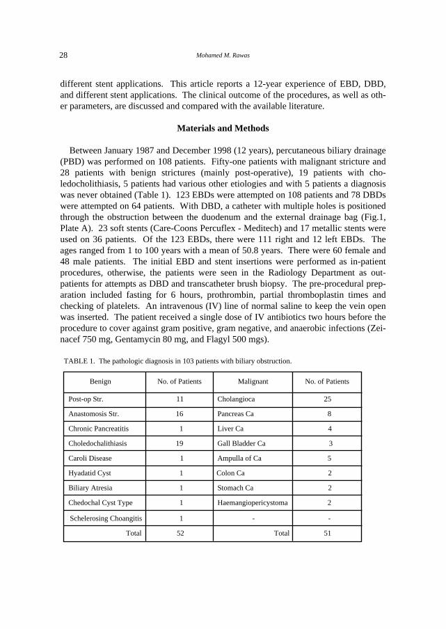
Between January 1987 and December 1998 (12 years), percutaneous biliary drainage(PBD) was performed on 108 patients. Fifty-one patients with malignant stricture and28 patients with benign strictures (mainly post-operative), 19 patients with cho-ledocholithiasis, 5 patients had various other etiologies and with 5 patients a diagnosiswas never obtained (Table 1). 123 EBDs were attempted on 108 patients and 78 DBDswere attempted on 64 patients. With DBD, a catheter with multiple holes is positionedthrough the obstruction between the duodenum and the external drainage bag (Fig.1,Plate A). 23 soft stents (Care-Coons Percuflex - Meditech) and 17 metallic stents wereused on 36 patients. Of the 123 EBDs, there were 111 right and 12 left EBDs. Theages ranged from 1 to 100 years with a mean of 50.8 years. There were 60 female and48 male patients. The initial EBD and stent insertions were performed as in-patientprocedures, otherwise, the patients were seen in the Radiology Department as out-patients for attempts as DBD and transcatheter brush biopsy. The pre-procedural prep-aration included fasting for 6 hours, prothrombin, partial thromboplastin times andchecking of platelets. An intravenous (IV) line of normal saline to keep the vein openwas inserted. The patient received a single dose of IV antibiotics two hours before theprocedure to cover against gram positive, gram negative, and anaerobic infections (Zei-nacef 750 mg, Gentamycin 80 mg, and Flagyl 500 mgs).
TABLE 1. The pathologic diagnosis in 103 patients with biliary obstruction.
Benign
No. of Patients Malignant No. of Patients
Post-op Str.
Anastomosis Str.
Chronic Pancreatitis
Choledochalithiasis
Caroli Disease
Hyadatid Cyst
Biliary Atresia
Chedochal Cyst Type
Schelerosing Choangitis
Total
11 Cholangioca 25
16 Pancreas Ca 8
1 Liver Ca 4
19 Gall Bladder Ca 3
1 Ampulla of Ca 5
1 Colon Ca 2
1 Stomach Ca 2
1 Haemangiopericystoma 2
1 - -
52 Total 51

29Percutaneous Biliary Drainage: A 12-year experience
The procedures were done under fluoroscopic guidance. Pethidine 50 mg IV andPhenergan 50 mg IM were used for sedation and analgesia. Local anaesthetic of 1%Zylocaine without adrenaline was infiltrated locally at the skin puncture site. Three pe-diatric patients, aged 1, 5, and 9 years, were done under general anaesthesia. A per-cutaneous transhepatic cholangiogram (PTC) is obtained first by using 21 Chiba needle.After biliary ducts visualisation, cannulation of segmental or sub-segmental duct (usual-ly posterior segment of the right lobe) was performed (Fig.1) using Hawkins biliary set(Cook). The EBD catheter (9Fr Kifa) was then shaped using boiled water and intro-duced. On one side, a hole was made close to the catheter tip. The catheter was ad-vanced to above the obstruction site for external drainage.
Fig.1. Plate A: Showing DBD catheter with multi side holes is positioned throught biliary ducts to bowel.The other end is connected to the external drainage bag. Note the catheter cannulation through a pos-terior subsegmental bile duct. Plate B: Showing final cholangiogram after soft stent insertion in-patient with anastomotic stricture. Plate C: Showing complete obstruction at cho-ledochoduondenostomy. Plate D: Striker stent in place. Place E: Final cholangiogram.
E
B
C
D
A

Mohamed M. Rawas30
Attempts, to achieve DBD, were performed after a few days to weeks of externaldrainage. This may be obtained after 1 to 4 attempts. In some patients, DBD was ob-tained at the time of EBD. Balloon dilatation of the stricture and stent insertion wasdone within one week of DBD. Transcatheter brush biopsy was done at the time ofDBD if no diagnosis was obtained by other means (surgery and fine needle aspirationbiopsy).
All patients not going for DBD had ampullary balloon dilatation (10 mm diameter)or mechanical stone crushing a few days after initial EBD. Some patients had onlyEBD, after which they had a surgical operation, referred back to original hospital orwere lost to follow-up. Patients were assessed for improvement in their general condi-tion, cholangitis, and laboratory parameters. Complications and mortality are dis-cussed.
The results of EBD and DBD were classified as Good, Fair, and Poor. Good resultsmean more than 50% reduction in serum bilirubin, reduction in alkaline phosphataseand SGOT, improved general condition (itching, malaise, appetite, skin and eye col-our). Fair results mean less than 50% reduction in serum bilirubin, with some re-duction in alkaline phosphatase and SGOT and some improvement in general condi-tion. Poor results mean no significant change observed in laboratory parameters orgeneral condition. For cholangitis, good results mean resolution of fever and chills,with white blood count close to normal range. Poor results mean no recovery fromcholangitis was observed.
Results
All patients were subjected to EBD. Sixty-four patients moved to the second step ofDBD. Forty stents were applied in 36 patients (Table 2). Other procedures includedampullary balloon dilatation in 9 patients for choledocholithiasis. One of these patientsneeded mechanical stone crushing due to large size of the stones. Three other patientshad 4 mechanical stone crushing only. Transcatheter brush biopsies were performed in16 patients.
Most of the EBD approaches were right-sided, 11/123 procedures. The left-sided ap-proach was performed in 12/123 procedures. In 2 patients, the left EBD (does not ap-pear in (Tables 2 & 3) and was initially planned and performed to avoid right hydatidcyst and tumour invaded right lobe respectively. The other 10 left EBDs were due tofailed right EBDs in 5 patients, isolated left ductal system due to hilar malignancy in 5patients, and 1 of these patients was done twice. The left EBD procedures were alltechnically successful. This is probably due to readily accessible ductal systems fromanterior sub costal approach.

31Percutaneous Biliary Drainage: A 12-year experience
The total number of failed EBD and DBD procedures was 11/201 with technical suc-cess of 190/201 (95%). The right EBD technical success was 119/123 (97%). Thefour failed procedures (Table 3) were with a 4-year child with Caroli disease. The sec-ond patient was obstructed dilated biliary tree but predominantly on the left side, withminimal right ductal dilatation. The third and fourth patients had hilar cho-langiocarcinoma with dilated left ductal system. In all 4 patients, left EBDs were suc-cessful (Table 3). Three patients went on to have stent application through the left side
TABLE 3. Reasoned failure of EBD and DBD procedures and patient outcome.
No.
1.
2.
3.
4.
5.
6.
7.
8.
9.
10.
11.
Pathology
Caroli
Anastomotic str.
Cholangio Ca.
Cholangio Ca.
Cholangio Ca.
Post-op str
Post-op str
Post-op str
Post-op str
Cholangio Ca.
Cholangio Ca.
No. of Attempts
2
1
2
1
3
3
3
3
3
1
4
Reason of Failure
Multiple stricture
Minumal dilatation
Technical
Acute angle
Silk ligature
Silk ligature
Silk ligature
Silk ligature
Tight stricture
Tight stricture
Tight stricture
Patient Outcome
(L) DBD
(L) Stricker stn.
(L) Soft stn.
(L) Soft stn.
(L) DBD
Surgery
Surgery
Surgery
Surgery
Surgery (Utube)
Right EBD extensive tumour
RIGHT
EBD
RIGHT
DBD
TABLE 2. Different percutaneous transhepatic biliary procedures performed in 108 patients.
Procedures No. of Procedures
EBD
DBD
Stents:
a) Percuflex Carey-Coons
b) Metallic: 1. Percuflex Carey-Coons
2. Gianturco-Rosch Z
3. Wall
Brush biopsy
Dilatation of ampulla
Mechanical crushing of stones
Stricture dilatation and splinting
123
78
40
23
7
5
5
16
9
5
2

Mohamed M. Rawas32
and 1 patient had left EBD. The clinical outcome of EBD in patients with and withoutcholangitis is detailed (Table 4 & 5).
The DBD technical success was 71/78 (91%). Seven failed procedures (Table 3)were encountered. In 1 patient, acute angle between entry point to biliary tree and stric-ture site was believed to be the reason. This patient had a successful DBD from the leftside. Three patients with silk ligature found around the CHD at operation. The other 3patient failures were due to tight sticture. Multiple attempts were made in two of them.The third patient had only one attempt after which a U tube was inserted surgically.
The most commonly encountered pathology in this series was benign surgery-relatedstricture (27 patients). Patients came for biliary drainage after the original surgery(post-operative strictures) or post-surgical anastomosis for ductal injury (anastomoticstricture) (Table 1). 8/11 post-operative stricture patients presented post-open chol-ecystectomy and 3/11 post-laparoscopic cholecystectomy. The anastomotic strictureswere 11/16 choledocho-jejunostomy, 3/16 choledocho-duendenostomy, 1/16 chol-edocho-choledochal anastomosis (liver transplant), and 1/16 hepatico-jejunostomy.
The patients presented 1 week to 5-years after the operation. The length of time wasrelated to severity of the injury, i.e., patients with CHD ligature presented within the
TABLE 4. Result of EBD in patients with jaundice and cholangitis.
Correlates
Anastomatic str.
Post-op str.
Choledocolithiasis
Caroli disease
Biliary atresia
Schlerosing cholangitis
Cholangio Ca.
Ampullary Ca.
Liver Ca.
Pancreatic Ca.
Stomach Ca.
No diagnosis
TOTAL
No.
9
2
5
1
1
1
19
9
2
1
2
1
15
2
36
Good
7
0
4
0
1
0
12/19 (63%)
7
2
0
1
1
11/15 (73%)
1
24/36 (67%)
Poor
2
2
1
1
0
1
7/19 (37%)
2
0
1
1
0
4/15 (27%)
1
12/36 (33%)

33Percutaneous Biliary Drainage: A 12-year experience
TABLE 5. Results of EBD in patients with jaundice.
No.
4
4
8
1
1
18
6
4
3
2
2
1
1
19
2
39
Anastomotic str.
Post-op str..
Choledocolithiasis
Chronic pancreatitis
Hydatid cyst
Cholangio Ca.
Pancreatic Ca.
Liver Ca.
Ampulla Ca.
Colon Ca.
Gall Baldder Ca.
Lymphoma
No diagnosis
TOTAL
Good
2 4
7
1
0
14/18 (78%)
4
1
0
2
1
0 1
9/19 (47%)
1
24/39 (62%)
Fair
0
0
1 0
1
2/18 (11%)
1
3
3
0
1
1
0
9/19 (47%)
1
12/39 (31%)
Poor
2
0
0 0
0
2/18 (11%)
1
0
0
0
0
0
0
1/19 (5%)
1
3/39 (7%)
first two weeks after the operation. Patients with adequate surgical anastomosis maypresent many years after with anastomotic strictures.
In patients presenting with cholangitis (Table 4), the benign obstructions (mainlyanastomotic stricture and choledocho-lithiasis) showed good response of cholangitisresolution in 63%. The 2 patients who presented post-operation did not respond anddied of septic shock. The patients suffered from CHD transection and choledocho-jejunostomy for pancreatic carcinoma respectively. In patients with cholangitis andmalignant obstructions, the response of cholangitis resolution was generally good(73%), particularly in cholangio-carcinoma and ampullary carcinoma.
The patients who came to EBD due to jaundice only and no cholangitis show betterresults in benign obstruction (78% good results) versus (47% in malignant obtruction(Table 5).
Seven benign strictures received soft stents (12 & 14 Fr) (Fig. 1, Plate B). The rangeof potency was 7-84 months and a mean of 32.7 months. Thirteen benign strictures re-ceived metallic stents (Gianturco-Rosch Z, Stricker, Wall) . (Fig. 1, Plates C, D, E)shows the range of potency is 3-96 months, with a mean of 26.8 months. One of these
Mohamed M. Rawas34
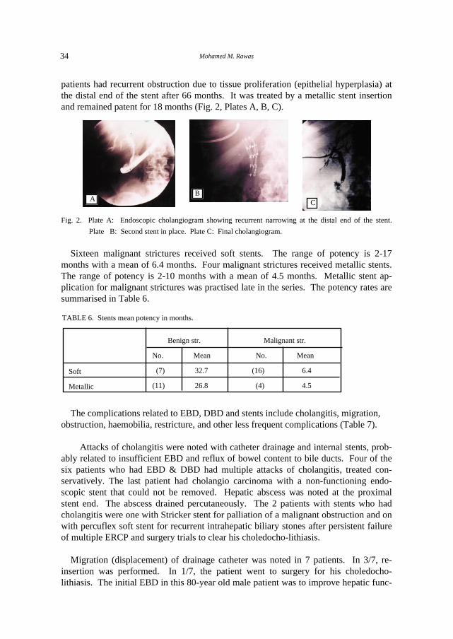
patients had recurrent obstruction due to tissue proliferation (epithelial hyperplasia) atthe distal end of the stent after 66 months. It was treated by a metallic stent insertionand remained patent for 18 months (Fig. 2, Plates A, B, C).
Fig. 2. Plate A: Endoscopic cholangiogram showing recurrent narrowing at the distal end of the stent.
Plate B: Second stent in place. Plate C: Final cholangiogram.
Sixteen malignant strictures received soft stents. The range of potency is 2-17months with a mean of 6.4 months. Four malignant strictures received metallic stents.The range of potency is 2-10 months with a mean of 4.5 months. Metallic stent ap-plication for malignant strictures was practised late in the series. The potency rates aresummarised in Table 6.
The complications related to EBD, DBD and stents include cholangitis, migration, obstruction, haemobilia, restricture, and other less frequent complications (Table 7).
Attacks of cholangitis were noted with catheter drainage and internal stents, prob-ably related to insufficient EBD and reflux of bowel content to bile ducts. Four of thesix patients who had EBD & DBD had multiple attacks of cholangitis, treated con-servatively. The last patient had cholangio carcinoma with a non-functioning endo-scopic stent that could not be removed. Hepatic abscess was noted at the proximalstent end. The abscess drained percutaneously. The 2 patients with stents who hadcholangitis were one with Stricker stent for palliation of a malignant obstruction and onwith percuflex soft stent for recurrent intrahepatic biliary stones after persistent failureof multiple ERCP and surgery trials to clear his choledocho-lithiasis.
Migration (displacement) of drainage catheter was noted in 7 patients. In 3/7, re-insertion was performed. In 1/7, the patient went to surgery for his choledocho-lithiasis. The initial EBD in this 80-year old male patient was to improve hepatic func-
TABLE 6. Stents mean potency in months.
Soft
Metallic
Benign str.
No. Mean
(7) 32.7
(11) 26.8
Malignant str.
No. Mean
(16) 6.4
(4) 4.5
A CB
35Percutaneous Biliary Drainage: A 12-year experience
tion and general condition before surgery. Reinsertion was not necessary. In 1/7, dis-placement and reinsertion occurred twice (76-year old male with metastatic pancreaticcarcinoma). Reinsertion of displaced catheter failed in 1/7 patients with chlerosingcholangitis. The last patient showed displaced catheter to right subphrenic space withcontinued bile drainage from the subphrenic bilima. The patient died before re-insertion.
Migration was noted in a patient with a soft stent. The anchoring silk threads wereinadvertently cut surgically. The stent was lost in the stool. Another patient had his Zstents migrate upward from the stricture site. Both patients received metallic stents fortheir anastomotic strictures.
The catheter obstruction and subsequent interruption of drainage was noted and newcatheter insertion was performed in the patient with Caroli disease. The second patienthad catheter obstruction during a long EBD period ( 4 months) in a patient anastomoticstricture awaiting stenting. The two obstructed stents were both soft type and 10 and17 months of follow-up. Four patients had haemobilia related to EBD and DBD; 2from the right and 2 from the left side catheters. One patient had severe haemobiliaduring attempts of the left DBD. The manipulation was stopped and the catheterclamped for 24 hours. The left DBD was achieved the next day through the anastomot-ic stricture. The second patient had transient haemobilia after achieved the left DBD.
TABLE 7. Complications related to EBD, DBD, and stents.
*Balloon Dilatation Only
Complication
Migration
Obstruction
Cholangitis
Haemobilia
Recurrent stricture
Broken balloon catheter
Bile leak at insertion site
Death (30-day mortality)
Septicaemia
Bile peritonitis
Haemoperitoneum
TOTAL
Morbidity
Mortality
EBD & DBD
7
2
6
4
2*
1
-
6
-
-
-
28/201 (14%)
11%
3%
Stents
2
2
2
-
1
-
1
-
-
-
-
28/201 (14%)
20%

Mohamed M. Rawas36
Spontaneous resolution was noted. The other 2 patients had transient haemobilia afterthe right EBD, with spontaneous resolution. In all cases, no further radiological or sur-gical intervention was needed.
Two patients had recurrent strictures. Both were anastomotic strictures and were di-lated with 10mm diameter balloon. The stricture site was splinted by nelaton 16FRcatheter for three months. The strictures recurred after 5 and 21 months respectively.Both were re-drained and stented by soft stents. One patient with a metallic stent hadrecurrent obstruction at the distal stent end and required another metallic stent applica-tion (Fig. 2). The dilatation balloon was detached from the catheter shaft in one pa-tient. The balloon was manipulated to the duodenum where it was expelled naturallyper rectum. Bile leek at insertion site was noted in 1 patient after the left side soft stentplacement. This was due to an inadequate stent position, with the proximal end of thestent close to the liver surface (extraductal). The stent was repositioned.
Thirty-day mortalities were seen in 2/6 patients after EBD. Both had cholangitis andsepticaemia post-surgery (open cholecystectomy with CHD transection, choledocho-jejunostomy for pancreatic cancer). Revised surgery was attempted after EBD. Bothdied 5 and 6 days after EBD in septic shock. One had cholangio carcinoma, the otheranastomotic stricture. One patient out of six had cholangitis and sepsis progressed toseptic shock and death in 9 days, despite right EBD. The last patient was the one withhydatid syst and left EBD. He died from sepsis after 19 days. The hydatid cyst wastreated surgically and was communicating to the biliary tree. The 3 patients who hadoperations after EBD are not true 30-days post-procedural mortality.
Balloon dilatation of ampulla was done in 9/19 patients with choledocholithiasis. In4/9 patients, the check cholangiogram 4-14 days later showed no biliary stones. All 4patients had previously failed endoscopic papillotomy and manipulation. Five out ofnine patients had no previous endoscopic intervention. One patient had mechanicalcrushing of larger than 8 mm stones. Four out of nine patients had normal cholangio-gram 3-20 days later. One out of nine patients went to surgery for persistent stones.
Ten out of nineteen patients with choledocho-lithiasis had EBD but no balloon dil-atation of the ampulla. Three out of ten had stone mechanical crushing. Two out ofthree showed no stones on cholangiogram 3 and 9 days later. One out of three showedpersistent fragments and went to surgery. One patient of this group had previouslyfailed endoscopic papilotomy and manipulation. Seven out of ten patients had surgery5-21 days post EBD. Two out of seven had additional reasons for surgery (post-op bil-iary fistula and choledocho-colonic fistula). One out of seven patients of this group hadprevious endoscopic intervention.
The conclusion of the experience with choledocho-lithiasis is that balloon dilatationof the ampulla with or without previous papillotomy seems to be effective in clearingstones of 8 mm or less in size. Larger stones can be mechanically crushed before bal-loon dilatation. EBD can be useful pre-operatively in patients with choledocholithiasis.

37Percutaneous Biliary Drainage: A 12-year experience
A B
Transatheter biliary brush biopsy was performed in 16 patients for diagnosis (Fig. 3,Plates A, B). Ten out of sixteen biopsies showed malignancy. Seven of those werecholangio carcinoma, 1 ampullary carcinoma, 1 recurrent ampullary carcinoma, and 1post whipple recurrent pancreatic carcinoma. Four out of sixteen biopsies showed nomalignancy and were proven benign stricture. In 2/16 biopsies, no diagnosis could beobtained and proved by FNA to have cholangio carcinom and pancreatic carcinoma re-spectively.
Fig. 3. Plate A: Cholangiogram via EBD catheter with tip at the stricture site. Plate B: The brushbiopsy is within the stricture.
Discussion
Percutaneous biliary drainage is a known procedure. It was initially described in thelate 1970s and early 1980s[1]. Experience has accumulated over the last two decades,with further evolution of its techniques, results and complications[2,3,4]. The technicalsuccess of EBD was reported to be above 90%[2,3]. The overall biliary decompressionsuccess in this patient series is 100%. The 4 patients who failed right EBD procedureswere successfully drained through the left EBD. Adequate biliary decompression wasachieved in all patients.
The clinical results of biliary drainage were judged using criteria resembling that ofGazzangia et al[3]. It was noted in the results presented here that 24/36 of the patientswith jaundice and cholangitis responded well to EBD along with other supportivemeasures, such as IV antibiotics. This supports the data of other authors[2,3,4]. In pa-tients with jaundice only, the results obtained in this series indicated that 62% weregood and 31% had a fair outcome which is slightly less than that of Gazzaniga[3]
(81.5% and 15% respectively).
The complication of 14% in this series compared favourably to 15.4% of Gazzaniga[3]. No haemoperitoneum, bile peritonitis or drainage-induced septicaemia was noted.This is probably due to selective cannulisation of segmental ducts peripheral in the liverand avoiding enteral duct punctures in the liver hilum.

Mohamed M. Rawas38
The true 30-day procedure-related death noted in the series was 1.5%. The recordedmortalities were related to EBDs being ineffective in altering the course of the originaldisease.
Percutaneous metallic stent application is a known form of management for benignbiliary strictures[5-8]. The potency rate is reported at 87% at 1 year and 68.7% at 3years[5,6]. During the period of available follow-up on our patients, the mean potencyrate was 26.8 months (Table 6). Further follow-up, unfortunately, cannot be obtained.Of the patients who were referred back to their original hospitals after having stent in-sertion, only 1/13 patients needed re-intervention and a second stent insertion, due toepithelial hyperplasia and stenosis at the distal end of the stent after 66 months. Thehigh mean potency rate of soft stents in benign strictures in this series (32.7 months) ismainly due to the large stent size (12-14F). The known potency rate of 10F stents is afew months to one-year[9]. When our patients were present with cholangitis, con-servative management was implemented. This was successful in most patients. Thestent was replaced only after percutaneous or endoscopic cholangiogram proved ob-structed stent.
The treatment strategy adopted of internal stent insertion for benign strictures is mul-ti-factorial. My initial experience with 2 patients showed recurrent stricture after re-peated balloon dilatation and external catheter stenting (16F) for 4 months. Patient ac-ceptance of such treatment (long external stenting and repeated interventions) was quitediscouraging. Approximately half of our patients are referred from remote hospitals. Ithas been shown from previous experience that patients are subjected to increased dis-comfort and the treatment may continue with dwelling catheter in place for 6 to 12months[7,10,11]. The internal soft stent used in this series remains in place as long as itis patent. When obstructed, it can be removed and/or replaced, if needed, endo-scopically or percutaneous.
Our mean potency rate for soft stents is 6.4 months. This agrees with the previouslyreported experience[9]. Other authors have reported occlusion rates of 6 - 23%[12,13].The implementation of expendable metallic stents in patients with malignant biliarystricture is a well-established method of palliation[5-15]. The reported potency variedfrom 5.8 to 7 months and occlusion occurred in 7-24%. The mean potency rate of 4.5months in this series needs to be looked at with caution. The number of stents is onlyfour and patient follow-up was only 2 -10 months. Further experience and longer pe-riod of follow-up will most likely improve the potency rate.
In conclusion, percutaneous biliary drainage in skilled hands is a very useful methodin the management of obstructive jaundice, with and without cholangitis. Metallicstents are a useful tool in palliation of malignant biliary disease and show promising re-sults in benign stricture. Large size soft stents remain a good method to stent beingstrictures and can be easily removed or replaced. Balloon dilatation of the ampulla,with or without mechanical stone crushing, has been effective in choledocho-lithiasis.
39Percutaneous Biliary Drainage: A 12-year experience
Transcatheter brush biopsy of biliary stricture is a good method, added to FNA and sur-gery, to obtain tissue diagnosis in biliary tract obstruction.
References
[1] Ferrucci JT, Freeny PC, Stark DD, Foley WD, Mueller PR, May G, Burhenne HJ. Advances inhepatobiliary radiology. Radiology 1988; 168(2): 319-338.
[2] Ishikawa Y, Oishi I, Miyai M, Kishimoto T, Miyamura S, Sagayama T, Morigaki T, YamashitaT, Itoh N. Percutaneous transhepatic drainage: experience in 100 cases. J Clin Gastroenterol 1980;2(3): 305-314.
[3] Gazzaniga GM, Faggioni A, Bandanza G, Filauro M. Percutaneous transhepatic biliary drainage:twelve years experience. Hepatogastroenterol 1990; 37(5): 517-523.
[4] Akiyama H, Okazaki T, Takashima I, Satoh S, Ryan T, Iwamori S, Hidaka T, Morio K, OkuharaT. Percutaneous treatments for biliary disease. Radiology 1990; 176(1): 25-30.
[5] Coons H. Metallic stents for the treatment of biliary obstruction: a report of 100 cases. CardiovascIntervent Radiology 1992; 15(6): 367-374.
[6] Maccioni F, Rossi M, Salvatori FM, Ricci P, Bezzi M, Rossi P. Metallic stents in benign biliarystrictures: three-year follow-up. Cardiovasc Intervent Radiology 1992; 15(6): 360-366.
[7] Rossi P, Bezzi M, Salvatori FM, Maccioni F, Porcaro ML. Recurrent benign biliary strictures:management with self-expanding metallic stents. Radiology 1990; 175(3): 661-665.
[8] Adam A. Metallic biliary endoprostheses. Cardiovasc Intervent Radiology 1994; 17(3): 127-132.[9] Mygind T, Itennild V. Expandable metallic endoprotheses for biliary obstruction. Acta Radiologica
1993; 34(3): 252-257.[10] Moore AV Jr., Illescas FF, Mills RR, Wertman DE, Heaston DK, Newman GE, Zuger JH, Sal-
mon RB, Dunnick NR. Percutaneous dilation of benign biliary strictures. Radiology 1987; 163(3):625-628.
[11] Williams HJ, Bender CE, May GR. Benign post-operative biliary strictures: dilatation with flu-oroscopic guidance. Radiology 1987; 163(3): 629-634.
[12] Kaufman SL. Percutaneous palliation of unresectable pancreatic cancer. Surg Clin N Am 1995; 75(5): 989-999.
[13] Lammer J, Neumayer K. Biliary drainage endoprostheses: experience with 201 placements. Radiol-ogy 1986; 159(3): 625-629.
[14] LaBerge JM, Doherty M, Gordon RL, Ring EJ. Hilar malignancy: treatment with an expandablemetallic transhepatic biliary stent. Radiology 1990; 177(3): 793-797.
[15] Lammer J, Klein GE, Kleiner R, Hausegger K, Einspieler R. Obstructive jaundice: use of expand-able metal endoprosthesis for biliary drainage. Work in Progress. Radiology 1990; 177(3): 789-792.
Mohamed M. Rawas40
bK'« �ö� s� W��«d*« b�J�« «uM� �e� ÎU�U� ±≤ �d��
�«Ë� bL�� bL��, wM�u�« �d�K� b�U� pK*« vHA��� , WF�_« r��
W��uF��« WO�dF�« WJKL*« − �b����
w� W�d�d?��« �bzU?H�«Ë U?Î�U� ±≤ �d?�?� W�«�� q?O�?�� - ÆhK�?��*«W?OK�«b�« WKO?7u?��« Ë� �uM�« s� Í�«d?� �e� W?OKL?� ≤∑≥ Ë i�d?� ±∞∏ÆYO?�)«Ë b?O?L(« Í�«d*« �«b?��ô«Ë oO?OC?��« v{d?� w� p��, ,t�Ëb�Ës�c�« Í�«d*« �«b?��ô«Ë oO?O?C?��« v{d* W?O?�U?�d?�?�« W?�«�� ¡«d?�≈ -Ë√ ÊËb� «u?N?��« s�c�«Ë W�b?�J�« «u?MIK� Í�«d*« �e?��« W?OKL?F� «u?F?C?�Ë√ w��U?)« �e?�?K� ÎU?OM� W?I�d?D�« ÕU?$ ZzU?�M�« X?�?��√ ÆW?OK�«� WK?O?7u?��Æw�«u?????�?�« vK?� %π∏ Ë %π± Ë %π∑ wK?�«b�« q?O????7u?????�?�« Ë√ ÃË�e*«≥∂® »U??N?��U� »u??�?B?*« Í�«d*« �«b?��ô« v?{d* Íd�d?�?�« s�?�??��«v{d* Íd�d��« s��?��« Æ%≥≥ w� ÎUHOF{Ë %∂∑ w� ΫbO?� ÊU� ©ÎUC�d�w� Ϋb?O?� ÊU?� ©ÎU?C�d?� ≥π® »U?N?��U� »u?�?B*« d?O?� Í�«d*« �«b?��ô«wH�d??B?�?�« qL?F�« j?�u?�??� Æ%∑w� ÎU?H??O?F??{Ë %≥± w� ÎUDO??��Ë %∂≤ «�«b�?�ô« w� ΫdN� ¥\µ Ë ≤∂\∑ ÊU?� WO�b?F*« WOK�«b?�« öO7u?�K�w� WO?�U� W?OKLF?� U�UO� vK� qB?�� r� Æw�«u��« vK� W?�O�?)«Ë �bO?L(«�e��« bF� v{d?*« ¡ôRN� W�UF�« W�U(« w� s�?% k�u� sJ�Ë i�d� ≥≥ÃË�e*«Ë w��U�K� %±± X�U?� �e��« WOKL?F� «bOI?F��« W�?�� Æw��U)«W��«d*« «uMIK� �e��« W?OKL� Ê≈ ÆWOK�«b�« öO?7u��« �«u�√ qJ� %≤∞Ëv{d* Ãö??F�«Ë h?O?�??A??��« w� �b?zU?� Ë� W??OK?�«� WKO??7u??�� Ë√ ÊËb�
Æ¡«u� b� vK� W�O�)«Ë �bOL(« W��«d*« «�«b��ô«