Peptic Ulcer Disease (PUD) - Med Study Group -...
12
Done By: Mona Makhamreh PBL Gastrointestinal System lecture #2 Today's lecture is mainly about Peptic Ulcer disease and manifestations of chronic liver disease. Peptic Ulcer Disease (PUD) Definition: loss of continuity of the mucosal lining covering any part of GI tract could be gastric mucosa, small intestine, or large intestine. However in PUD, the most common locations are the gastric mucosa and duodenal mucosa. Gastric peptic ulcer could be benign or malignant, duodenal peptic ulcer is usually benign. While any gastric lesion seen during endoscopy should be biopsied (because it could be malignant), while in duodenal ulcer that’s not always necessary because it's most probably due to H.Pylori. Duodenal Ulcers are most likely to be related to H.Pylori infection, while Gastric ulcerations could be due to H.Pylori, or due to NSAIDs, or even a stress ulcer. [Important point could be mentioned in the exam]: In the case of duodenal ulcer viewed in an endoscopy, the direct measure towards it is H.Pylori eradication regardless of the result because 95% it is due to H.Pylori. Pathogenesis : Protective factors vs. hostile factors
Transcript of Peptic Ulcer Disease (PUD) - Med Study Group -...

Done By: Mona Makhamreh
PBL Gastrointestinal System lecture #2
Today's lecture is mainly about Peptic Ulcer disease and
manifestations of chronic liver disease.
Peptic Ulcer Disease (PUD) Definition: loss of continuity of the mucosal lining covering
any part of GI tract could be gastric mucosa, small intestine, or
large intestine. However in PUD, the most common locations
are the gastric mucosa and duodenal mucosa.
Gastric peptic ulcer could be benign or malignant, duodenal
peptic ulcer is usually benign. While any gastric lesion seen
during endoscopy should be biopsied (because it could be
malignant), while in duodenal ulcer that’s not always necessary
because it's most probably due to H.Pylori.
Duodenal Ulcers are most likely to be related to H.Pylori
infection, while Gastric ulcerations could be due to H.Pylori, or
due to NSAIDs, or even a stress ulcer.
[Important point could be mentioned in the exam]: In the case
of duodenal ulcer viewed in an endoscopy, the direct measure
towards it is H.Pylori eradication regardless of the result
because 95% it is due to H.Pylori.
Pathogenesis :
Protective factors vs. hostile factors

Done By: Mona Makhamreh
From the figure above, you can identify the risk factors and
protective factors:
(H.Pylori, Gastric acid, Pepsin, NSAIDs) are the negative factors
(Bicarbonate, Prostaglandins, Mucus, and Blood flow to
mucosa) are the positive factors (protective)
If the negative factors are more than the Positive ones then
peptic ulcer forms.
This is a figure of the histopathology of a peptic ulcer. Notice
the loss in mucosal lining.

Done By: Mona Makhamreh
The causes of peptic ulcer disease include the following: The most important cause is the Infection with the bacteria Helicobacter pylori: There are areas where it is endemic while in other areas it isn't, Jordan is considered endemic with H.Pylori, more than 80%. So a family of 5, 4 out of the 5 will be H.Pylori infected.
Stress: Also known as a stress ulcer, seen in ICU patients, burnt patients, particularly any patient subjected to stress, especially during hospitalization. By stress it is not like the type experienced during examinations, it's more of a major stress like a trauma. Injury or death of mucus-producing cells. Excess acid production in the stomach. The hormone gastrin stimulates the production of acid in the stomach; therefore, any factors that increase gastrin production will in turn increase the production of stomach acid. An example of this is Gastrinoma (Zolinger-Ellison syndrome), a tumor that increases acid secretion, and usually the morphology of ulcers resulting from gastrinomas are multiple ulcerations that can be duodenal and gastric, but in unusual locations, such as pre-antrum or in antrum in the stomach. Such a case needs to test the Serum Gastrin Level.

Done By: Mona Makhamreh
Gastinoma tumor common locations are Pancreas particularly ampulla of Vater or in the duodenum, it is one of the carcinoid tumors. Drugs: NSAIDS mainly, so this should be considered when a patient is complaining of gastric pain. Corticosteroids, some say it doesn’t cause ulcers per se (alone) unless the mucosa itself was desrupted, yet it should be a consideration in the patient's history because if he had H.pylori and took steroids, this will increase the severity.
This diagram shows the percentage of factors contributing to duodenal and gastric ulcers. Notice how H.pylori is related to duodenal ulcers more than

Done By: Mona Makhamreh
gastric ulcers, in duodenal ulcers H.Pylori is mainly the cause (duodenal ulcer=H.pylori) They used to say: No acid no ulcer but this is no longer used, the more correct statement is: no H.pylori no ulcer, so ulcers aren’t only due to acidity.
H.pylori infection could cause the following in the gastric mucosa: Chronic gastritis – upper endoscopy shows an inflamed mucosa, nodular, and erythematous. 90% of cases Peptic ulcer disease – loss of mucosa not just inflamed. H.Pylori isn’t a benign condition, it could cause malignancy, particularly Maltoma which is mucosal cell lymphoma. could cause Reflux Oesophagitis. Non ulcer dyspepsia (gastric discomfort, gastric pain, nausea, non-specific symptom a patient could complain about it even if an upper GI endoscopy didn’t show any ulcers, this might be due to H.Pylori)
Done By: Mona Makhamreh
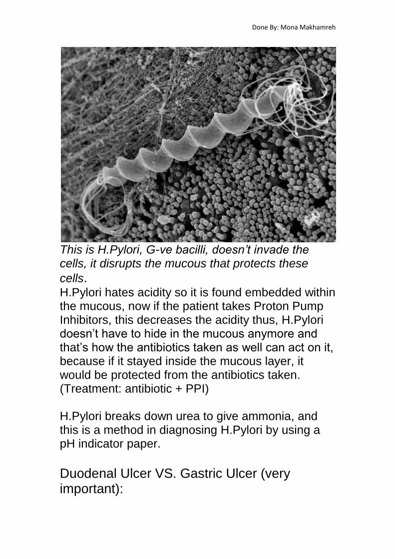
This is H.Pylori, G-ve bacilli, doesn’t invade the cells, it disrupts the mucous that protects these
cells. H.Pylori hates acidity so it is found embedded within the mucous, now if the patient takes Proton Pump Inhibitors, this decreases the acidity thus, H.Pylori doesn’t have to hide in the mucous anymore and that’s how the antibiotics taken as well can act on it, because if it stayed inside the mucous layer, it would be protected from the antibiotics taken. (Treatment: antibiotic + PPI) H.Pylori breaks down urea to give ammonia, and this is a method in diagnosing H.Pylori by using a pH indicator paper.
Duodenal Ulcer VS. Gastric Ulcer (very important):

Done By: Mona Makhamreh
1-Duodenal ulcer causes Fasting Pain, when a patient is fasting, that’s when the pain is felt while after a meal there is no pain. Gastric Ulcer however, once the patient eats, pain is felt so it is PostPrandial Pain. 2-Duodenal ulcers are more related to H.Pylori. - Male to female ratio—4:1 in duodenal ulcer, while it is 2:1 in gastric ulcer. - in general, Duodenal ulcer is more associated with blood group O, while Gastric with A.(this doesn’t mean that if you have blood group O you won’t develop gastric ulcer, it’s just in general). -In Smoking duodenal ulcer is as twice as common. Clinical Manifestations of peptic ulcer disease: -Gastric abdominal pain: depends if gastric ulcer or duodenal ulcer, these patients usually improve on antacids and PPIs (helps in diagnosis of PUD) -Nausea -Vomiting -Belching Complications:
Done By: Mona Makhamreh
• Upper G.I. bleeding (chronic or acute) which manifests as melena, hematemesis, coffee ground vomitus, or anemia. • Ulcers can perforate or penetrate the structures behind it like pancreas or splenic artery. It could also perforate to the peritoneal cavity causing peritonitis which is a surgical emergency. • Obstruction of G.I. tract, ulcer causes severe fibrosis which in turn causes gastric outlet obstruction, which manifests as vomiting and nausea because the ingested food isn’t passing in the GIT. Fibrosis eventually leads to pyloric stenosis.
Also Gastric carcinoma, not duodenal, since gastric is the one associated with malignant transformation. These bad complications are rarely seen due to early diagnosis and eradication of H.Pylori.
Diagnosis:
-Upper GI Endoscopy (the most used)
Culture (rarely used) – which helps in identifying the sensitivity of H.Pylori, because currently there is a problem of resistance in H.Pylori, because we used to treat it with triple therapy which contains Klacid (Clarithromycin), but now most of the H.pylori have Klacid resistance.

Done By: Mona Makhamreh
Urea Breath test (rapid urease test): performed during endoscopy, a superficial sample is taken and a pH indicator is used (positive or negative), if positive, you start giving treatment directly.
that’s the Urea breath test
Non-pharmacological Treatment of Peptic ulcer: 1-Avoid spicy food. 2-Avoid Alcohol. 3-Avoid Smoking. 4-Avoid heavy meals 5-Encourage small frequent low caloric meals. 6-Avoid ulcerating drugs e.g. NSAIDs (as they are contraindicated in PUD, he should stop them and use other measures of treatment-even if he had rheumatoid arthritis- or it will cause complications), corticosteroids. (smoking and NSAIDs are the most important)
PUD –Treatment
1) Proton pump inhibitor 2) H.pylori eradication:

Done By: Mona Makhamreh
Could be Triple therapy or quadruple therapy (2 antibiotics and PPI, some people add De-Nol which is Bismuth therapy) (The duration of therapy is not required form us because of the variations)
Examples on therapies: Amoxicillin, Klacid, PPI x2 Some add Metronidazole and Bismuth therapy There are many regimens for treatments, but bottom line is that if H.pylori was found, it must be eradicated to eradicate peptic ulcer disease. End of peptic ulcer disease.
Manifestations of Chronic Liver Disease
1) Hepatic encephalopathy (It is 4 grades, last one
ends in complete coma)
-due to accumulation of metabolites which have a toxic effect on the brain in the bloodstream. *Hepatic encephalopathy manifestations:

Done By: Mona Makhamreh
-Disturbance to sleep-wake cycle (1st
manifestation) the patient sleeps during day and stays awake during night. -Flapping tremor/Asterixis (also one of the early manifestations) 2) Fetor hepaticus: special odor in breath 3) Spider angiomas: dilatation of vessels due to high estrogen level, appear as a spider, usually on the anterior chest {healthy elderly could have spider angiomas normally but are small in number} 4) Palmar Erythema: also caused by increase in estrogen levels 5) Jaundice 6) Areas of Scratch marks on the patient due to itchiness which is caused by (icterus) deposition of bile salts on the skin which leads to irritation. 7) Anemia of chronic illness could be [normocytic normochromic anemia, could be microcytic (in case of upper GI bleeding), just know it causes anemia] 8) Lower limb edema due to decrease in albumin (made by the liver) hence decrease in oncotic pressure and the fluid moves to the interstitial spaces to cause edema 9) edema in the abdomen (ascites), which is responsive to periodic therapy at first but then starts to become refractory needing recurrent tapping to remove the fluids. 10) Spontaneous Bacterial Peritonitis (SBP): due to spontaneous translocation of E.Coli to the peritoneal cavity (not iatrogenic; not due to catheterization nor perforation of duodenal ulcer). Any patient with increasing abdominal girth/distention, think about SBP. 11) Esophageal Varices -important- (which causes massive fresh upper GI bleeding with high mortality

Done By: Mona Makhamreh
rate, diagnosed by endoscopy), it is an emergency, you can’t wait or the patient collapses. Also Hemorrhoids happen due to increase in portal hypertension. 12) Gynecomastia due to increase in estrogen and due to Spironolactone (aldosterone antagonist) for ascites which is a diuretic. 13) Finger clubbing (caused by different mechanisms)