Pelvic Pain: Physiatric Evaluation and Management
22
Pelvic Pain: Physiatric Evaluation and Management Pelvic Anatomy, Function, and Physical examination Jaclyn H. Bonder, MD Assistant Professor, Department of Rehabilitation Medicine Medical Director, Women’s Health Rehabilitation
Transcript of Pelvic Pain: Physiatric Evaluation and Management

Pelvic Pain: Physiatric Evaluation and Management
Pelvic Anatomy, Function, and Physical examination
Jaclyn H. Bonder, MD
Assistant Professor, Department of Rehabilitation Medicine
Medical Director, Women’s Health Rehabilitation
ANATOMY

The Boney Pelvis (Pelvic Girdle)
http://www.musclereleasetherapy.com/postural-balancing-2.html

ARS: What are the bony landmarks of the urogenital triangle?
• Pubic symphysis
• Bilateral Ischial tuberosities
• Bilateral ischial spines
• Coccyx
• Bilateral ischiopubic rami
Answers are in red

Poll: What are the bony landmarks of the urogenital triangle?

Bony Landmarks
• Urogential Triangle
– Pubic symphsis/arch
– Ischiopubic ramus
– Ischial tuberosity
• Anal triangle
– Ischial tuberosity
– Sacrotuberous
Ligament
– Coccyx
– Ischial spine

Joints and Ligaments
7
•Anterior & Posterior (LDL)
SI Ligament
•Sacrotuberous ligament
•Sacrospinous ligament
•Pubic symphysis
•Sacroiliac Joint
•Sacrococcygeal joint

Pelvic Floor Muscles: Superficial
8
A. Superficial & Deep transverse perineal
B. Bulbcavernosus/spongiosus
C. Ischiocavernosus
D. External Anal Sphincter (EAS)
E. Perineal body

9
Pelvic Floor Muscles: Levator Ani
Levator Ani
Puborectalis
Pubococcygeus
Iliococcygeus
PR
PR
PC
PC
IC
IC
IC
IC

Pelvic Floor Muscles: Deeper Layer
10
Piriformis
Obturator internus

More Anatomy – It never ends!

Nerves of the Pelvis
•Lumbosacral trunk
•Pudendal nerve
•Nerve to Levator Ani and Coccygeus
12
Pudendal Nerve: Anatomy

Nerve Anatomy continued: Abdominopelvic nerves

ARS:What are the nerves that provide sensory/cutaneous
innervation to this region?
1 (correct answer) 2 3 4
A = Pudendal Pudendal Pudendal Pudendal
B = Inferior cluneal Posterior femoral cutanueousnerve
Inferior cluneal Posterior femoral cutaneous n.
C = Obturator Obturator Medial cutaneousn. of thigh
Medial Cutaneous n. of thigh
D = Ilioinguinal + Genitofemoral
Iliohypogastric & Ilioinguinal
Iliohypogastric only Ilioinguinal + Genitofemoral
Physical Exam for Pelvic Pain
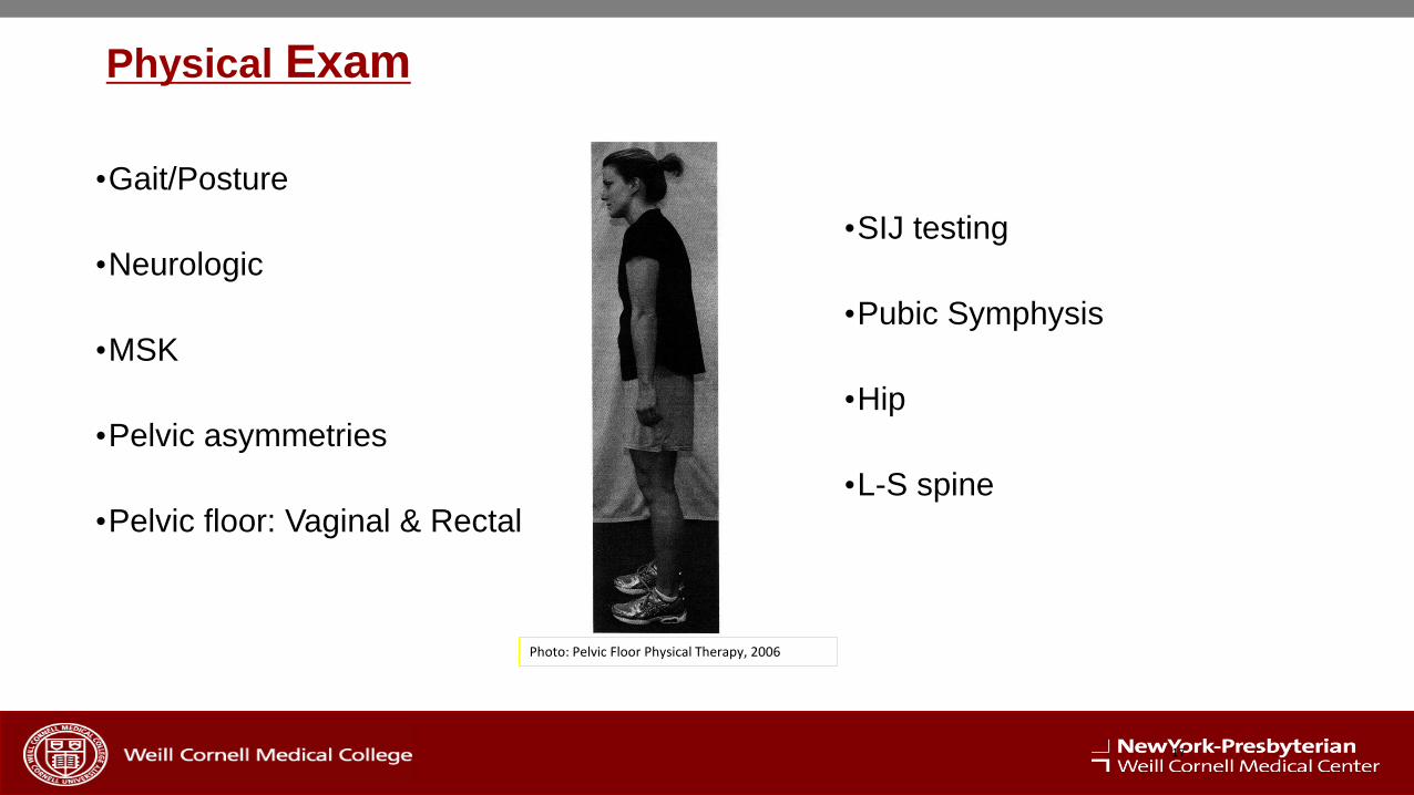
Physical Exam
•Gait/Posture
•Neurologic
•MSK
•Pelvic asymmetries
•Pelvic floor: Vaginal & Rectal
•SIJ testing
•Pubic Symphysis
•Hip
•L-S spine
17
Photo: Pelvic Floor Physical Therapy, 2006
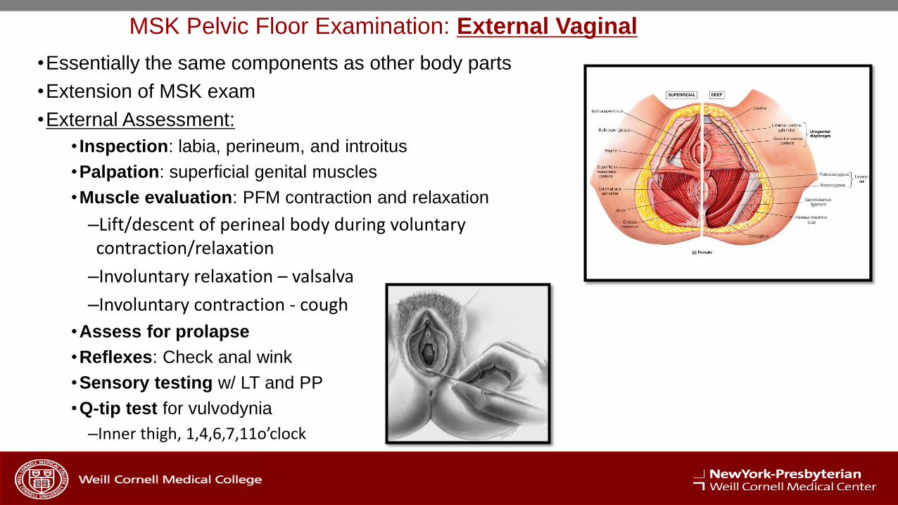
MSK Pelvic Floor Examination: External Vaginal
•Essentially the same components as other body parts
•Extension of MSK exam
•External Assessment:
• Inspection: labia, perineum, and introitus
•Palpation: superficial genital muscles
•Muscle evaluation: PFM contraction and relaxation
–Lift/descent of perineal body during voluntary contraction/relaxation
–Involuntary relaxation – valsalva
–Involuntary contraction - cough
•Assess for prolapse
•Reflexes: Check anal wink
•Sensory testing w/ LT and PP
•Q-tip test for vulvodynia
–Inner thigh, 1,4,6,7,11o’clock

•Palpate the muscles
–Superficial Genital muscles – DIP
–LAM – PIP @ 3-5 & 7-9 o’clock
–Compare L vs R
•Pain
•Quality of tone
•Muscle bulk
–Oburator Internus• Above arcus tendinous @ 3 & 9 o’clock
• ER hip into examiner’s hand
–Piriformis• Deep
• Bring knee to opposite shoulder
MSK Pelvic Floor Examination: Internal Vaginal
MSK Pelvic Floor Examination: Internal Vaginal
•Perform manual muscle testing
• Quality of contraction in 4 quadrants
–Left, right, anterior, posterior
• Strong hold
• Assess 5-10 quick flicks
• Compare right and left sides
• Modified Oxford Scale
–Poor inter-rater reliability
•Palpate ischial spine
–Tinel’s sign: pudendal n paresthesia
Grading of Strength (Modified from Laycock 2001)
• 0/5 - No contraction of muscles
• 1/5 - Flicker or pulsation is felt, no discernible lifting or
tightening
• 2/5 - Weak contraction, no discernible lifting or
tightening
• 3/5 - Moderate, some lifting of the posterior wall and
some tightening around the examiner's finger,
contraction is visible
• 4/5 - Good, elevation of the vaginal wall is felt against
resistance, drawing in of the perineum is felt, able to
hold for 5 or more seconds
• 5/5 - Strong resistance is felt, if 2 fingers are inserted,
fingers will be approximated, able to hold for with 10-
second hold
External
• Inspection:
–hemorrhoids, lesions
•Sensory testing
•Anal wink reflex
•Palpate the coccyx externally
Internal
•Sphincter exam–EAS
–IAS
–Resting tone and contraction
•Coccyx–Tenderness
–Position
–Mobility
•Ligaments - sacrococcygeal junction
•Muscle exam:–Coccygeus, OI and LAM
–Compare R and L
MSK Pelvic Floor Examination: Rectal

Function of Pelvic Floor Musculature
• Kegel 1950's –
– Support
– Sphincteric closure
– Sexual appreciation
• Herman & Wallace
– Above +
• Stabilization
• Sump Pump