Pelvic Intravenous Leiomyomatosis – Case Report
4
Pelvic Intravenous Leiomyomatosis – Case Report Leiomiomatose intravenosa pélvica – caso clínico Patricia Correia 1 Ana Castro 1 Anabela Rocha 2 Daniela Freitas 1 Cátia Carnide 1 Osvaldo Moutinho 1 1 Department of Gynecology and Obstetrics, Centro Hospitalar de Trás-os-Montes e Alto Douro (CHTMAD), Lordelo, Vila Real, Portugal 2 Department of Gynecology and Obstetrics, Centro Hospitalar de S. João, Porto, Portugal Rev Bras Ginecol Obstet 2016;38:412–415. Address for correspondence Patricia Correia, Resident Doctor, Avenida da Galiza, Ed. Miradouro, Bloco B, 5° Dto., 5050-222, Régua, Portugal (e-mail: [email protected]). Keywords ► intravenous leiomyomatosis ► uterine leiomyoma ► pelvic mass Abstract Introduction Intravenous leiomyomatosis is a benign and rare condition that can result in cardiac events with fatal outcomes when left untreated. Intravenous leio- myomatosis is probably underestimated because the diagnosis is easily missed. We present a case of an intravenous leiomyomatosis without extra-pelvic involvement, with a brief review of this pathology. Case Report 46-year-old woman submitted to hysterectomy and bilateral adnexec- tomy because of a pelvic mass detected in ultrasound. During the surgery, intravenous leiomyomatosis diagnosis was suspected. Pathological analysis confirmed this suspi- cion. Further imaging exams were performed without detecting any anomalies related to this condition. The patient remained with no evidence of disease after one year of follow-up. Conclusion Intravenous leiomyomatosis is a rare condition that can lead to serious complications. Early diagnosis followed by an appropriate treatment is very important to patient outcome, and underdiagnoses can be counteracted if the gynecologist is aware of this entity. Palavras-chave ► leiomiomatose intravenosa ► leiomioma uterino ► massa pélvica Resumo Introdução A leiomiomatose intravenosa é uma condição benigna, rara, que pode resultar em eventos cardíacos, podendo ser fatal quando não tratada. Esta patologia está provavelmente subestimada, uma vez que facilmente não é diagnosticada. Neste artigo, apresentamos um caso de leiomiomatose intravenosa sem envolvimento extrapélvico, com uma breve revisão da patologia. Relato de Caso Mulher de 46 anos de idade, submetida a histerectomia e anexecto- mia bilateral após detecção ecográfica de massa pélvica. Durante a cirurgia, houve a suspeita de leiomiomatose intravenosa, e o exame anátomo-patológico confirmou o diagnóstico. A paciente foi submetida a outros exames de imagem, não sendo detectada qualquer anomalia relacionada com a patologia. Após um ano de follow- up, a paciente manteve-se sem evidência de doença. Conclusão A leiomiomatose intravenosa é uma condição rara que pode levar a complicações graves. O diagnóstico precoce e o tratamento adequado são muito importantes para o prognóstico da paciente, e os subdiagnósticos podem ser evitados se o ginecologista estiver ciente dessa entidade. received February 15, 2016 accepted July 12, 2016 published online August 16, 2016 DOI http://dx.doi.org/ 10.1055/s-0036-1588002. ISSN 0100-7203. Copyright © 2016 by Thieme Publicações Ltda, Rio de Janeiro, Brazil Case Report THIEME 412
Transcript of Pelvic Intravenous Leiomyomatosis – Case Report
Pelvic Intravenous Leiomyomatosis – Case Report
Leiomiomatose intravenosa pélvica – caso clínico
Patricia Correia1 Ana Castro1 Anabela Rocha2 Daniela Freitas1 Cátia Carnide1 Osvaldo Moutinho1
1Department of Gynecology and Obstetrics, Centro Hospitalar deTrás-os-Montes e Alto Douro (CHTMAD), Lordelo, Vila Real, Portugal
2Department of Gynecology and Obstetrics, Centro Hospitalar de S.João, Porto, Portugal
Rev Bras Ginecol Obstet 2016;38:412–415.
Address for correspondence Patricia Correia, Resident Doctor,Avenida da Galiza, Ed. Miradouro, Bloco B, 5° Dto., 5050-222, Régua,Portugal (e-mail: [email protected]).
Keywords
► intravenousleiomyomatosis
► uterine leiomyoma► pelvic mass
Abstract Introduction Intravenous leiomyomatosis is a benign and rare condition that canresult in cardiac events with fatal outcomes when left untreated. Intravenous leio-myomatosis is probably underestimated because the diagnosis is easily missed. Wepresent a case of an intravenous leiomyomatosis without extra-pelvic involvement,with a brief review of this pathology.Case Report 46-year-old woman submitted to hysterectomy and bilateral adnexec-tomy because of a pelvic mass detected in ultrasound. During the surgery, intravenousleiomyomatosis diagnosis was suspected. Pathological analysis confirmed this suspi-cion. Further imaging exams were performed without detecting any anomalies relatedto this condition. The patient remained with no evidence of disease after one year offollow-up.Conclusion Intravenous leiomyomatosis is a rare condition that can lead to seriouscomplications. Early diagnosis followed by an appropriate treatment is very importantto patient outcome, and underdiagnoses can be counteracted if the gynecologist isaware of this entity.
Palavras-chave
► leiomiomatoseintravenosa
► leiomioma uterino► massa pélvica
Resumo Introdução A leiomiomatose intravenosa é uma condição benigna, rara, que poderesultar em eventos cardíacos, podendo ser fatal quando não tratada. Esta patologiaestá provavelmente subestimada, uma vez que facilmente não é diagnosticada. Nesteartigo, apresentamos um caso de leiomiomatose intravenosa sem envolvimentoextrapélvico, com uma breve revisão da patologia.Relato de Caso Mulher de 46 anos de idade, submetida a histerectomia e anexecto-mia bilateral após detecção ecográfica de massa pélvica. Durante a cirurgia, houve asuspeita de leiomiomatose intravenosa, e o exame anátomo-patológico confirmou odiagnóstico. A paciente foi submetida a outros exames de imagem, não sendodetectada qualquer anomalia relacionada com a patologia. Após um ano de follow-up, a paciente manteve-se sem evidência de doença.Conclusão A leiomiomatose intravenosa é uma condição rara que pode levar acomplicações graves. O diagnóstico precoce e o tratamento adequado são muitoimportantes para o prognóstico da paciente, e os subdiagnósticos podem ser evitadosse o ginecologista estiver ciente dessa entidade.
receivedFebruary 15, 2016acceptedJuly 12, 2016published onlineAugust 16, 2016
DOI http://dx.doi.org/10.1055/s-0036-1588002.ISSN 0100-7203.
Copyright © 2016 by Thieme PublicaçõesLtda, Rio de Janeiro, Brazil
Case ReportTHIEME
412
Introduction
Uterine leiomyomas affect 20–40% of women in reproductiveage.1 Occasionally, they occur with unusual growth patterns,including intravenous leiomyomatosis (IVL), diffuse perito-neal leiomyomatosis, benign metastasizing leiomyomas,retroperitoneal leiomyomas and parasitic leiomyomas.
Intravenous leiomyomatosis is a benign and rare condi-tion that, when left untreated, can result in cardiac eventswith fatal outcomes.2,3 Histologically, it is a benign smoothmuscle cell tumor growing within the uterine venous sys-tem, sometimes extending to the inferior vena cava and tothe right cardiac chambers. There are two main theoriesabout the etiology of this tumor: one contends that thetumor grows from the vein wall; the other suggests thatthe tumor is a uterine leiomyoma invading the vessels.4
Until 2010, there were 298 reported cases of IVL in theEnglish literature, and in 45% of these cases the tumorpenetrated to the inferior vena cava or reached the rightatrium.5 The occurrence of IVL is probably underestimatedbecause the diagnosis is easily missed.5 This reported caseemphasizes the importance of promptdiagnosis to perform acorrect treatment and follow-up, ensuring a minimized riskof recurrence.
Case Report
A 46-year-old woman, multigravida, parous 5, obese, with-out any history of pathologic events, who presented regularcycles, with abundant menses, and non-cyclic pelvic pain.She was referred to our hospital for gynecological consulta-tion because of a pelvic mass detected in a pelvic ultrasound.
The gynecological examination revealed a 12-week uteruswithout any other palpable masses. The transvaginal ultra-sound showed a uterus with augmented dimensions, withthe external limits deformed by multiple leiomyomas, thelarger of which measured 33 � 28 � 16 mm in intramural/subserous localization. Right adnexal area: complex hetero-
geneous mass, with a cystic bi-loculated component, mea-suring 100 � 91 � 52 mm, the major one measuring.measuring 67 � 46 mm, and two solid components with50 � 17 mm and 18 � 10 mm, respectively, without vascu-larization on Doppler exam. There were no ascites. Cancerantigen 125 (CA 125)was normal. The patient was submittedto an exploratory laparotomy.
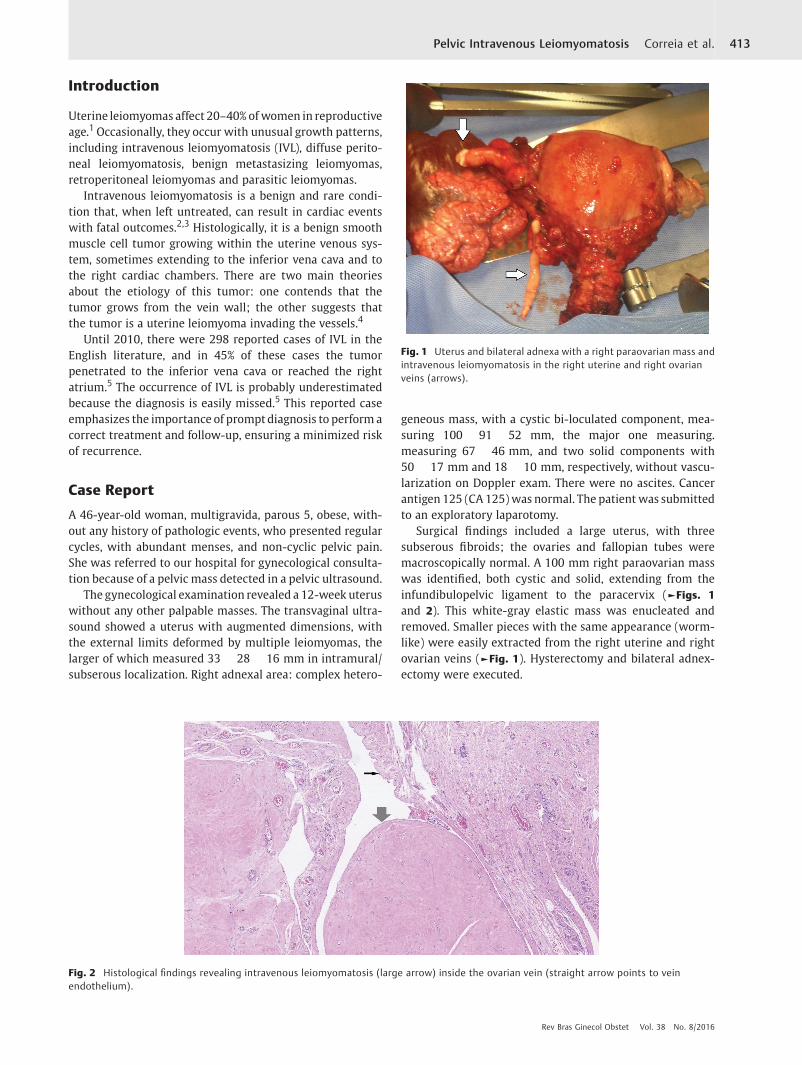
Surgical findings included a large uterus, with threesubserous fibroids; the ovaries and fallopian tubes weremacroscopically normal. A 100 mm right paraovarian masswas identified, both cystic and solid, extending from theinfundibulopelvic ligament to the paracervix (►Figs. 1
and 2). This white-gray elastic mass was enucleated andremoved. Smaller pieces with the same appearance (worm-like) were easily extracted from the right uterine and rightovarian veins (►Fig. 1). Hysterectomy and bilateral adnex-ectomy were executed.
Fig. 1 Uterus and bilateral adnexa with a right paraovarian mass andintravenous leiomyomatosis in the right uterine and right ovarianveins (arrows).
Fig. 2 Histological findings revealing intravenous leiomyomatosis (large arrow) inside the ovarian vein (straight arrow points to veinendothelium).
Rev Bras Ginecol Obstet Vol. 38 No. 8/2016
Pelvic Intravenous Leiomyomatosis Correia et al. 413
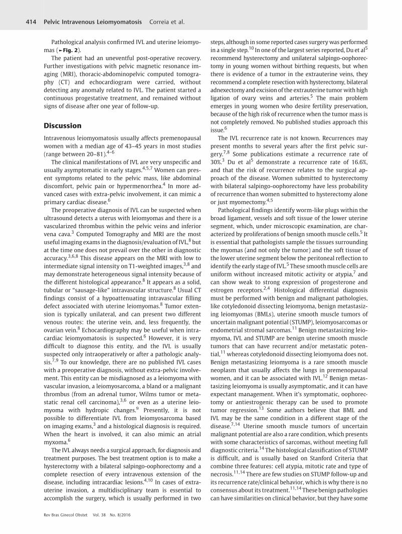
Pathological analysis confirmed IVL and uterine leiomyo-mas (►Fig. 2).
The patient had an uneventful post-operative recovery.Further investigations with pelvic magnetic resonance im-aging (MRI), thoracic-abdominopelvic computed tomogra-phy (CT) and echocardiogram were carried, withoutdetecting any anomaly related to IVL. The patient started acontinuous progestative treatment, and remained withoutsigns of disease after one year of follow-up.
Discussion
Intravenous leiomyomatosis usually affects premenopausalwomen with a median age of 43–45 years in most studies(range between 20–81).4–6
The clinical manifestations of IVL are very unspecific andusually asymptomatic in early stages.4,5,7 Women can pres-ent symptoms related to the pelvic mass, like abdominaldiscomfort, pelvic pain or hypermenorrhea.4 In more ad-vanced cases with extra-pelvic involvement, it can mimic aprimary cardiac disease.6
The preoperative diagnosis of IVL can be suspected whenultrasound detects a uterus with leiomyomas and there is avascularized thrombus within the pelvic veins and inferiorvena cava.3 Computed Tomography and MRI are the mostuseful imaging exams in the diagnosis/evaluation of IVL,8 butat the time one does not prevail over the other in diagnosticaccuracy.3,6,8 This disease appears on the MRI with low tointermediate signal intensity on T1-weighted images,3,8 andmay demonstrate heterogeneous signal intensity because ofthe different histological appearance.8 It appears as a solid,tubular or “sausage-like” intravascular structure.8 Usual CTfindings consist of a hypoattenuating intravascular fillingdefect associated with uterine leiomyomas.8 Tumor exten-sion is typically unilateral, and can present two differentvenous routes: the uterine vein, and, less frequently, theovarian vein.8 Echocardiography may be useful when intra-cardiac leiomyomatosis is suspected.6 However, it is verydifficult to diagnose this entity, and the IVL is usuallysuspected only intraoperatively or after a pathologic analy-sis.7,9 To our knowledge, there are no published IVL caseswith a preoperative diagnosis, without extra-pelvic involve-ment. This entity can be misdiagnosed as a leiomyoma withvascular invasion, a leiomyosarcoma, a bland or a malignantthrombus (from an adrenal tumor, Wilms tumor or meta-static renal cell carcinoma),3,6 or even as a uterine leio-myoma with hydropic changes.9 Presently, it is notpossible to differentiate IVL from leiomyosarcoma basedon imaging exams,3 and a histological diagnosis is required.When the heart is involved, it can also mimic an atrialmyxoma.6
The IVL always needs a surgical approach, for diagnosis andtreatment purposes. The best treatment option is to make ahysterectomy with a bilateral salpingo-oophorectomy and acomplete resection of every intravenous extension of thedisease, including intracardiac lesions.4,10 In cases of extra-uterine invasion, a multidisciplinary team is essential toaccomplish the surgery, which is usually performed in two
steps, although in some reported cases surgerywasperformedin a single step.10 In one of the largest series reported, Du et al5
recommend hysterectomy and unilateral salpingo-oophorec-tomy in young women without birthing requests, but whenthere is evidence of a tumor in the extrauterine veins, theyrecommend a complete resectionwith hysterectomy, bilateraladnexectomyand excisionof the extrauterine tumorwithhighligation of ovary veins and arteries.5 The main problememerges in young women who desire fertility preservation,because of the high risk of recurrence when the tumor mass isnot completely removed. No published studies approach thisissue.6
The IVL recurrence rate is not known. Recurrences maypresent months to several years after the first pelvic sur-gery.7,8 Some publications estimate a recurrence rate of30%.3 Du et al5 demonstrate a recurrence rate of 16.6%,and that the risk of recurrence relates to the surgical ap-proach of the disease. Women submitted to hysterectomywith bilateral salpingo-oophorectomy have less probabilityof recurrence than women submitted to hysterectomy aloneor just myomectomy.4,5
Pathological findings identify worm-like plugs within thebroad ligament, vessels and soft tissue of the lower uterinesegment, which, under microscopic examination, are char-acterized by proliferations of benign smooth muscle cells.5 Itis essential that pathologists sample the tissues surroundingthe myomas (and not only the tumor) and the soft tissue ofthe lower uterine segment below the peritoneal reflection toidentify the early stage of IVL.5 These smoothmuscle cells areuniform without increased mitotic activity or atypia,7 andcan show weak to strong expression of progesterone andestrogen receptors.2,4 Histological differential diagnosismust be performed with benign and malignant pathologies,like cotyledonoid dissecting leiomyoma, benign metastasiz-ing leiomyomas (BMLs), uterine smooth muscle tumors ofuncertainmalignant potential (STUMP), leiomyosarcomas orendometrial stromal sarcomas.11 Benign metastasizing leio-myoma, IVL and STUMP are benign uterine smooth muscletumors that can have recurrent and/or metastatic poten-tial,11whereas cotyledonoid dissecting leiomyoma does not.Benign metastasizing leiomyoma is a rare smooth muscleneoplasm that usually affects the lungs in premenopausalwomen, and it can be associated with IVL.12 Benign metas-tasizing leiomyoma is usually asymptomatic, and it can haveexpectant management. When it’s symptomatic, oophorec-tomy or antiestrogenic therapy can be used to promotetumor regression.13 Some authors believe that BML andIVL may be the same condition in a different stage of thedisease.7,14 Uterine smooth muscle tumors of uncertainmalignant potential are also a rare condition, which presentswith some characteristics of sarcomas, without meeting fulldiagnostic criteria.14 The histological classification of STUMPis difficult, and is usually based on Stanford Criteria thatcombine three features: cell atypia, mitotic rate and type ofnecrosis.11,14 There are few studies on STUMP follow-up andits recurrence rate/clinical behavior, which is why there is noconsensus about its treatment.11,14 Thesebenignpathologiescan have similarities on clinical behavior, but they have some
Rev Bras Ginecol Obstet Vol. 38 No. 8/2016
Pelvic Intravenous Leiomyomatosis Correia et al.414

important histological differences. Because of their rarity,there is no prevailing consensus about treatment and follow-up.
Antiestrogenic therapy, like tamoxifen, gonadotropin-re-leasing hormone agonists, letrozole or medroxyprogester-one, has been used to prevent tumor growth and reducetumor mass,5,6,15 but no efficacy has been demonstrated.
Because of the high recurrence risk, long term imagingfollow-up is a recommendedmeasure, usually TC orMRI,5,6,8
but no limit is set as to how long the surveillance shouldcontinue. There is also no consensus about the periodicity ofthe follow-up: some recommend it once a year or every sixmonths when complete resection is made,6 but othersrecommend a more closely spaced surveillance (every threeto six months).3
Although it was not possible to diagnose IVL before thesurgery, in this reported case it was possible to apply anadequate surgical treatment. In our opinion, based on thereview of the literature, women without birthing requestsand close to menopause should be submitted to hysterecto-my with a bilateral salpingo-oophorectomy, minimizing therecurrence risk (because of the estrogen-dependence of thetumor),4,5 especially when it is difficult to exclude extra-uterine disease. Once the histological diagnosis was made,different imaging studies were performed to exclude vascu-lar or heart disease, and to assure complete mass resection.After one year of follow-up, the patient remained asymp-tomatic, without any evidence of recurrence. Even withoutsigns of residual disease, based on some case reports,6,15 thepatient waskept under continuous progestative treatment inthe first year after the surgery to minimize the recurrencerisk; the treatment was suspended after this period. Thepatient will remain in close surveillance, with a yearly imagefollow-up for at least 10 years. This reported case is veryunique because IVL without extra-pelvic extension has re-ceived little attention in the reported literature.5 Du et al5
published the last review in the English literature onPubmed, indicating 298 cases of IVL reported until Au-gust 2010, 164 of which were without extra-pelvic involve-ment. Since that date to December 2015, using the key word“intravenous leiomyomatosis”, we found 81 papers includedby Pubmed, but they mainly report cases with heart orinferior vena cava involvement (65 articles). Only 8 articlesreport IVL cases without extra-pelvic involvement, with 35cases in total.
Thus,wecansay thatuntil 2015 therewereonly199reportedcases of intravenous leiomyomatosis without extra-pelvicinvolvement.
In conclusion, IVL is a rare condition that can lead toserious, even fatal, complications. This reported casewas notdiagnosed until surgery, but because of the surgeon’s suspi-cious, it was well conducted, with an exhaustive evaluation,allowing a proper treatment and follow-up.
Diagnosis coupled with an appropriate treatment is veryimportant to patient outcome, because it can reduce theprobability of recurrence. Underdiagnoses may be counter-acted if the gynecologist is aware of this entity.
References1 Wallach EE, Vlahos NF. Uterine myomas: an overview of develop-
ment, clinical features, and management. Obstet Gynecol 2004;104(2):393–406
2 Barksdale J, Abolhoda A, Saremi F. Intravenous leiomyomatosispresenting as acute Budd-Chiari syndrome. J Vasc Surg 2011;54(3):860–863
3 Fasih N, Prasad Shanbhogue AK,Macdonald DB, et al. Leiomyomasbeyond the uterus: unusual locations, rare manifestations. Radio-graphics 2008;28(7):1931–1948
4 Valdés Devesa V, Conley CR, Stone WM, Collins JM, Magrina JF.Update on intravenous leiomyomatosis: report of five patientsand literature review. Eur J Obstet Gynecol Reprod Biol 2013;171(2):209–213
5 Du J, Zhao X, GuoD, Li H, Sun B. Intravenous leiomyomatosis of theuterus: a clinicopathologic study of 18 cases, with emphasis onearly diagnosis and appropriate treatment strategies. Hum Pathol2011;42(9):1240–1246
6 Clay TD, Dimitriou J, McNally OM, Russell PA, Newcomb AE,Wilson AM. Intravenous leiomyomatosis with intracardiac exten-sion - a review of diagnosis and management with an illustrativecase. Surg Oncol 2013;22(3):e44–e52
7 Carr RJ, Hui P, Buza N. Intravenous leiomyomatosis revisited: anexperience of 14 cases at a single medical center. Int J GynecolPathol 2015;34(2):169–176
8 Bender LC, Mitsumori LM, Lloyd KA, Stambaugh LE III. AIRP bestcases in radiologic-pathologic correlation: intravenous leiomyo-matosis. Radiographics 2011;31(4):1053–1058
9 Clement PB, Young RH, Scully RE. Diffuse, perinodular, and otherpatterns of hydropic degeneration within and adjacent to uterineleiomyomas. Problems in differential diagnosis. Am J Surg Pathol1992;16(1):26–32
10 Wang J, Yang J, Huang H, et al. Management of intravenousleiomyomatosis with intracaval and intracardiac extension.Obstet Gynecol 2012;120(6):1400–1406
11 Ip PP, Tse KY, Tam KF. Uterine smooth muscle tumors other thanthe ordinary leiomyomas and leiomyosarcomas: a review ofselected variants with emphasis on recent advances and unusualmorphology that may cause concern for malignancy. Adv AnatPathol 2010;17(2):91–112
12 Lee HJ, Choi J, Kim KR. Pulmonary benign metastasizing leio-myoma associated with intravenous leiomyomatosis of the uter-us: clinical behavior and genomic changes supporting atransportation theory. Int J Gynecol Pathol 2008;27(3):340–345
13 Awonuga AO, Shavell VI, Imudia AN, Rotas M, Diamond MP,Puscheck EE. Pathogenesis of benign metastasizing leiomyoma:a review. Obstet Gynecol Surv 2010;65(3):189–195
14 Campbell JE, Knudtson JF, Valente PT, Robinson RD, Kost ER.Successful pregnancy following myomectomy for uterine smoothmuscle tumor of uncertainmalignant potential: A case report andreview of the literature. Gynecol Oncol Rep 2016;15:1–3
15 Barjot PJ, Refahi N, Berthet P, Delautre VD. Intravenous leiomyo-matosis of the uterus: a GnRH agonist utilisation before surgery.J Obstet Gynaecol 1998;18(5):492–493
Rev Bras Ginecol Obstet Vol. 38 No. 8/2016
Pelvic Intravenous Leiomyomatosis Correia et al. 415