PEDICLE SCREW FIXATION - Boolean Education
27
P EDICLE S CREW F IXATION I N F RACTURE OF T HORACO - LUMBAR S PINE Dr. Manvendra Swarup Sharma M.B.B.S. (Hons.), M.S. (Ortho.) D.N.B. (Ortho.)
Transcript of PEDICLE SCREW FIXATION - Boolean Education

PEDICLE SCREW FIXATION
IN FRACTURE OF
THORACO-LUMBAR SPINE
Dr. Manvendra Swarup Sharma
M.B.B.S. (Hons.), M.S. (Ortho.)
D.N.B. (Ortho.)
1
To Whom It May Concern
I do hereby declare that all references cited in the Thesis
and all the records have been solely prepared by me and it
has not been previously accepted for any higher degree. If
someone has done the same type of study for his own
purpose that is simply coincidental.
17/06/2011 Dr Manvendra Swarup Sharma
M.B.B.S. (Hons.), M.S. (Ortho.)
D.N.B.(Ortho.)

2
PEDICLE SCREW FIXATION IN FRACTURE
OF THORACO-LUMBAR SPINE
Introduction
The thoracolumbar injuries are the commonest
spinal injuries. The treatment of unstable fractures
and fracture dislocations of thoracolumbar spine
remains controversial. The goal of the treatment of
unstable thoracolumbar injuries is optimising
neural decompression while providing stable
internal fixation over the least number of spinal
segments. Either anterior posterior or both
approaches can be used to achieve fusion. However,
posterior approach is less extensive. Pedicle screw
devices allow immediate stable fixation as the
screws traverse all the three columns. The pedicle
screws are passed one level above and one level
below the fractured vertebra via posterior
approach. The aim of this study was to evaluate the
use of pedicle screw fixation for preservation of
remaining spinal cord function, restoration of
spinal alignment, achievement of pain-free fracture
site, early mobilization and maximization of
neurological recovery in Spinal Injury Patients.

3
The technique of pedicle screws and plates has
been routinely used to stabilize the lumbar spine
since 1963. With the use of this technique, it is now
possible to treat without problem lumbar
instabilities, fractures in the lumbar spine,
resection of tumors in the vertebral bodies
including metastasis and correction of gross
spondylolisthesis. To consider the technique it is
important to study the salient features of the
lumbar spine carefully.

4
Materials and Methods
Sixty Two patients with posttraumatic instability
of lower thoracic or upper lumbar spine were
surgically managed at the Department of
Orthopaedics, Jai Hospital, Agra. All those patients
who were operated for thoracolumbar junction
injuries with pedicle screw fixation were included.
The patients with pre-existing systemic illness or
associated extra spinal injuries significant enough
to result in increased morbidity or mortality were
excluded from the study. A detailed history and
examination was carried out especially evaluating
the mode of trauma, Frankel grading (Table-1),
sensory level and any spinal deformity. Plain x-
rays, in anteroposterior and lateral views were
done and the instability of the spine was confirmed
using White and Panjabi criteria of spinal
instability (Table-2). MRI/CT scan was done to
further evaluate the important relationships and
instability of spine. Those patients with unstable
spine were then explained pros and cons of the
surgical treatment. Patients willing for surgery
were included in this study.

5
All patients underwent closed/open reduction and
internal fixation by posterior approach.
Laminectomy to decompress spinal cord was
carried out at the involved level and bone was
saved to be used as bone graft. Pedicles were
localized using detailed anatomical landmarks and
intraoperative image intensifier. Polyaxial screws
were inserted through pedicles into vertebral
bodies’ one level above and one level below
fractured vertebra under fluoroscopy. The rod was
coupled to polyaxial screws. Distraction of anterior
elements was produced by compressing the heads of
Polyaxial screws by which annulotaxis was used for
reduction of spinal deformity. The rotational
movement was prevented by transverse traction
device.
The cortical bone was roughened using high-speed
drill to make suitable for bone graft. The bone
already saved while doing laminectomy was broken
into small fragments free of soft tissue and was
placed over roughened cortical bone. The wound
was then closed in layers. The patients were kept
on broad-spectrum antibiotics and analgesics for 10
days. Check x-rays were done on the next
postoperative day.
6
Thoracolumbar support was given to the patients
for initial 2 months. Aggressive physiotherapy was
started to mobilize patients. The neurological
status of the patients and any other complications
were noted up to one-and-a-half years.
Total of 62 cases aged from 10-70 years (mean: 36
years) with Thoraco-lumbar fractures were treated
by pedicle screw system. According to AO
classification of Thraco-lumbar vertebrae fracture,
there are 48 cases of Type A, 8 cases of Type B and
6 cases of Type C.
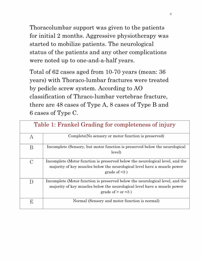
Table 1: Frankel Grading for completeness of injury
A Complete(No sensory or motor function is preserved)
B Incomplete (Sensory, but motor function is preserved below the neurological
level)
C Incomplete (Motor function is preserved below the neurological level, and the
majority of key muscles below the neurological level have a muscle power
grade of <3 )
D Incomplete (Motor function is preserved below the neurological level, and the
majority of key muscles below the neurological level have a muscle power
grade of > or =3 )
E Normal (Sensory and motor function is normal)

7
Table 2: White and Panjabi criteria for spinal
instability (Quantitation of acute instability in sub-
axial, cervical, thoracic, and lumbar injuries)
CONDITION POINTS
ASSIGNED
Loss of integrity of anterior (and middle) column 2
Loss of integrity of posterior column(s) 2
Acute resting translational deformity 2
Acute resting angulation deformity 2
Acute dynamic translation deformity exaggeration 2
Acute dynamic translation deformity exaggeration 2
Neural element injury 3
Acute disk narrowing at level of suspected pathology 1
Dangerous loading anticipated 1
A score of 5 points or more implies the presence of instability
8
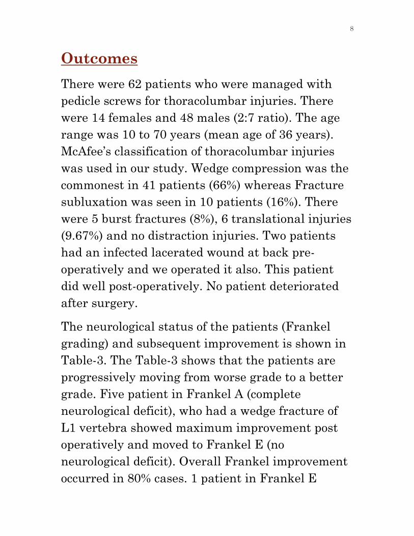
Outcomes
There were 62 patients who were managed with
pedicle screws for thoracolumbar injuries. There
were 14 females and 48 males (2:7 ratio). The age
range was 10 to 70 years (mean age of 36 years).
McAfee’s classification of thoracolumbar injuries
was used in our study. Wedge compression was the
commonest in 41 patients (66%) whereas Fracture
subluxation was seen in 10 patients (16%). There
were 5 burst fractures (8%), 6 translational injuries
(9.67%) and no distraction injuries. Two patients
had an infected lacerated wound at back pre-
operatively and we operated it also. This patient
did well post-operatively. No patient deteriorated
after surgery.
The neurological status of the patients (Frankel
grading) and subsequent improvement is shown in
Table-3. The Table-3 shows that the patients are
progressively moving from worse grade to a better
grade. Five patient in Frankel A (complete
neurological deficit), who had a wedge fracture of
L1 vertebra showed maximum improvement post
operatively and moved to Frankel E (no
neurological deficit). Overall Frankel improvement
occurred in 80% cases. 1 patient in Frankel E

9
remained in the same grade on subsequent follow-
ups. Almost complete removal of vertebral body
was done to get satisfactory alignment of spine in
one case. There were no patients with wound
infection and implant failure. One patient
developed DVT but improved with management of
DVT. One patient became severely depressed and
required long term antidepressants. One patient
developed bedsores. No other complications of
recumbency were found. All the paraplegics could
mobilize with frame independently and could pass
urine using crede manoeuvre.
Table 3: Frankel Grading of the patients
A B C D E
As Presented 51 4 2 2 3
At Follow-up 8 14 18 16 6

10

11

12
13

14

15

16

17
Analysis And Discussion
The management of fractures in the thoracolumbar
region is a controversial subject. Disadvantages of
conservative treatment include deterioration in
neurological status in 17% of the patients,
progressive kyphotic deformity in 20%, persistent
backache, decubitus ulcer and deep venous
thrombosis. Most of these complications can be
avoided by early mobilization and decreased
hospital stay by early surgery. The pedicle offers a
strong point of attachment of the posterior
elements to the vertebral body and pedicle screw
instrumentation has revolutionized spine surgery.
Pedicle screw fixation is considered
biomechanically superior to other stabilization
constructs or parapedicular screws and are
exceptionally rigid.
It has rapidly become one of the most popular
strategies for achieving solid fusion. So
instrumentation with pedicle screws is a commonly
used procedure for correcting deformity and
stabilizing the spine until bony fusion occurs. These
instrumentation systems may be divided into those
using rods and those using plates. Now-a-days,
pedicle screw system using rods is more acceptable

18
and it provides better stability than other implants.
Operative stabilization consists of segmental
distraction with pedicle screw fixation one level
above and one level below the injured segment. By
applying distraction, annulotaxis is exploited to aid
in reduction of retro-pulsed bone and disc
fragments. Similarly, pedicle screws have been
shown to be superior to hooks and Hartshell
fixation in spine.
Intra-operative rod contouring using a French
bender reduces the fatigue life of spinal constructs.
Tapping may decrease the pullout resistance of
screws in osteoporotic spine but not in normal
spine. We utilized rod contouring and tapping for
screw placement in all our cases. Bony fusion was
achieved in 56/62 (90%) of our cases whereas
Sasso30 has reported a 95.6% arthodesis rate with
dynamic compression plates and pedicle screws in
23 patients. Sengupta et al showed similar fusion
rates with iliac crest or local bone in a single level
fusion but less morbidity in case of local
bone. We also used autogenous local bone as graft
in all cases. Zeiller SC et al utilized intraoperative
neurophysiological monitoring in addition to
anatomical landmarks and intraoperative imaging

19
including neuronavigation. Vougioukas VI et al
suggested that the computer-aided navigation may
be beneficial but does not appear to be mandatory.
We localized the pedicles using detailed anatomical
landmarks and use of intraoperative fluoroscopy.
None of our patient required re-exploration for
correction of screws. Yilmaz C et al utilized
percutaneous methyl methacrylate injection around
a loosened screw but it was not required in our
cases. Powers CJ et al found a screw breaching the
spinal canal in one out of 287 percutaneouly placed
pedicle screws but none of our screws breached the
spinal canal. Knop C et al also found 139 out of
2264 screws (6.1%) to be misplaced with open
technique but only 0.6% required revision. White
reported a 23% screw fractures in a series of 76
patients.
In our series, no misplacement or breakage of
screws occurred. Olumide35 showed 0.4, 1.09 and
0.66 Frankel grade improvements with
anterior, posterior and anteroposterior approaches
respectively. Nadeem M et al showed 0.9 Frankel
improvement with one year follow-up while in our
study the average improvement was 1.11 Frankel
grade with similar one year follow-up. It is

20
important to note that Olumide did not study
paraplegic (Frankel A) patients whereas in our
series, 51 out of 62 patients (82.2%) were in
Frankel A. Three patients were neurologically
intact (Frankel E) preoperatively as well as on
follow ups and were excluded while calculating
improvement as by Olumide. Shafiq6 did not show
the neurological improvement whereas significant
neurological improvement was shown in our series.
Two to six percent incidence of postoperative
wound infection has been reported. In our series, no
infection occurred. It was recommended by Sasso
that infection can be managed without the removal
of hardware. No life-threatening complication
occurred in our series. Shafiq6 as well as Olumide
used external orthosis for three months. It was
used for two months only in our cases because the
fixation was strong enough to bear axial loading
without external bracing.

21
Conclusions and Recommendations
Thoracolumbar injury is a common neurosurgical
problem in road traffic accidents and fall from
height. Surgical treatment is a better option for
early ambulation and faster recovery. Pedicle screw
fixation is a useful choice, which achieves reduction
and stability in both anterior and posterior column
injuries, does not require anterior decompression
and does not affect extra motion segments.
The vertebral pedicle screw internal fixation was
technologically applicable, which can efficiently
reposition and stabilize the fractured vertebrae,
indirectly decompress spinal canal, maintain spine
stability, scatter stress of screw system, reduce the
risk of loosening or breakage of screw and loss of
vertebral height, and prevent the formation of
posterior convex after operation.
An early/immediate surgery in the form of pedicle
screw fixation with decompression provides better
relief in neurological recovery also.

22
References
1. Yue JJ, Sossan A, Selgrath C, Deutsch LS,
Wilkens K, Testaiuti M, et al. The treatment of
unstable thoracic spine fractures with
transpedicular screw instrumentation: a 3-year
consecutive series. Spine. 2002;27(24):2782-7
2. Shafiq K, Iqbal M, Hameed A, Mian JM. Role
of transpedicular fixation in thoracolumbar spinal
injuries. Neurol Surg 1998;1:21-7.
3. Sar C, Bilen FE. Flexion was more painful
than extension. Thoracolumbar flexion-distraction
injuries combined with vertebral body fractures.
Am J Orthop 2002;31:147-51.
4. Biomechanical evaluation of pedicle screws
versus pedicle and laminar hooks in the thoracic
spine. Spine J. 2006;6(4):444-9.

23
5. Lindsey C, Deviren V, Xu Z, Yeh RF, Puttlitz
CM. The effects of rod contouring on spinal
construct fatigue strength. Spine 2006;31(15):
1680-87.
6. Carmouche JJ, Molinari RW, Gerlinger T,
Devine J, Patience T. Effects of pilot hole
preparation technique on pedicle screw fixation in
different regions of the osteoporotic thoracic and
lumbar spine. J Neurosurg Spine 2005;3(5):364-70.
7. Lavaste F. Etude des implants rachidiens.
Thesis, Nationale Superieure des Arts et Metiers,
Paris, 1977.
8. Louis R. (ed.) Chirurgie du Rachis, Berlin,
Springer Verlag, 1982.
9. Roy-Camille R, Barcat E, Demeulenbaere C. et
al. Chirurgie par abord posterior du rachis

24
dorsal et lombbaire. Techniques Chirurgicales,
1982; 44; 78-6
10. Roy-Camille R, Judet T, and Badelon O, Rachis
dorsolombaire traumatique non neurologique. In
Roy-Camille R. : Deuxiemes Journees de la Pitie,
Masson, Paris, 1980; pp 3-152.
11. Roy-Camille R, Judet T, Vila T. et al.
Traumatismes recents du rachis avec signes
neurologiques, In Roy-Camille R. :
12. Troisiemes Journees de la Pittie, Paris,
Masson, 1983; pp 151-205
13. Roy-Camille R, Saillant G, Berteaux P. et al.
Early treatment of Spinal injuries. In MacKibbin,
B. (ed.): Recent Advances in Orthopaedics Number
Three. Dublin, Churchill Livingston, 1979; pp 57-
87.

25
14. Roy-Camille R, Saillant G, Bisserie M. et al.
Surgical Treatment of Spinal Metastatic tumors by
posterior plating and laminectomy. 51st Meeting of
the American Academy of Orthopaedic Surgeons,
Atlanta Georgia, February, 1984.
15. Roy-Camille R, Saillant G, Coulon J P. et al.
Spondylolisthesis L4-L5 et L5-S1. In Roy Camille
R.: Troismes Journees de la Pitie, Paris, Masson,
1983, p 91.
16. Roy-Camille R, Saillant G, Lapresle P, et al. A
secret in Spine Surgery: The Pedicle, 51st Meeting
of the American Academy of Orthopaedic Surgeons,
Atlanta, Georgia, February 1984.
17. Roy-Camille R, Saillant G, Sagnet P.
Traumatologie du rachis. In Detrie P. (ed.):
Chirurgie d’Urgence. Paris, Masson et Cie, 1976; pp
690-719.

26
18. Saillant G. Etude anatomique des pedicules
vertebraux, applications chirurgicales, Rev. Chir.
Orthop. Traumatol. 1976; 2:151